Embed Size (px)
Citation preview
International Atomic Energy Agency
Image Quality in Cardiac Image Quality in Cardiac AngiographyAngiography
L 8.1

Lecture 8.1: Image quality in cardiac angiography 2Radiation Protection in Cardiology
Educational ObjectivesEducational Objectives
1. How can image quality of cardiac angiographic images be assessed?
2. How useful can the quality criteria be?

Lecture 8.1: Image quality in cardiac angiography 3Radiation Protection in Cardiology
Rotter, EHJ 2003
+112%
+204%
+75%
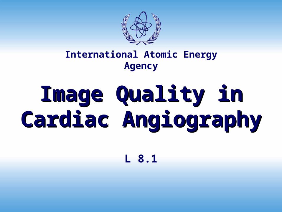
Interventional cardiology in Europe 1992-1999
Lecture 8.1: Image quality in cardiac angiography 4Radiation Protection in Cardiology
EHJ 2001, 2003
PCI in some European Countries(1994-1999)
0
20000
40000
60000
80000
100000
120000
140000
160000
Ger Fra UK Ita Nl Spa
1200 2081
825 1443
242 484
239 763
800 818267 858
1994
1996
1999
per million

Lecture 8.1: Image quality in cardiac angiography 5Radiation Protection in Cardiology
Quality of cardiac imagesQuality of cardiac images
• background
• cardiac cine-angiographic images should allow the cardiologist to evaluate the anatomic (and sometimes functional) details which are relevant for clinical decision making
• variables
• technical performance of the imaging system
• patient cooperation
• angiographic technique

Lecture 8.1: Image quality in cardiac angiography 6Radiation Protection in Cardiology
quality it’s me !!
the interventional cardiologist and quality…

Lecture 8.1: Image quality in cardiac angiography 7Radiation Protection in Cardiology
Scientific societies implemented guidelines to guarantee adequate
level of quality and performance of invasive cardiology
• training of operators
• quantitative standards to maintain the expertise in coronary
angiography or angioplasty
• quality-assurance programme
Pepine, J Am Coll Cardiol 1995;25:14–6Miller, Can J Cardiol 1996;12:470–2Cowley, Cathet Cardiovasc Diagn 1993;30:1–4Heupler, Cathet Cardiovasc Diagn 1993;30:191–
200Scanlon, J Am Coll Cardiol 1999;33:1756–824
Quality in invasive cardiology and Quality in invasive cardiology and scientific societiesscientific societies

Lecture 8.1: Image quality in cardiac angiography 8Radiation Protection in Cardiology
the specific problem of achieving and
maintaining high-quality standards in
angiographic imaging
• responsibility of cardiac catheterization laboratory
directors
• involves periodic cine-angiograms review
• lesion quantification (QCA, calipers)
precise criteria have never been stated for coronary procedures
Quality of cardiac images and scientific Quality of cardiac images and scientific societiessocieties

Lecture 8.1: Image quality in cardiac angiography 9Radiation Protection in Cardiology
do we need a method for do we need a method for image quality assessment image quality assessment in the routine practice of in the routine practice of
diagnostic (and diagnostic (and interventional) cardiologyinterventional) cardiology

Lecture 8.1: Image quality in cardiac angiography 10Radiation Protection in Cardiology
No technical deficiencies 153 49.6%
No reference segment 32 11.4%
Inadequate separation from background 35 11.4%
Inadequate lesion/vessel separation 67 22%
Inadequate opacification flow 48 15.6%
Inadequate opacification technique 68 22%
Inadequate radiographic procedure 10 3.2%
Totally inadequate 7 2.3%
Epicardial vessel not injected 5 1.6%
Types of technical deficiencies in 308 Types of technical deficiencies in 308 cineangiogramscineangiograms (Leape, Am Heart J 2000;139:106-13)(Leape, Am Heart J 2000;139:106-13)
N %
Lecture 8.1: Image quality in cardiac angiography 11Radiation Protection in Cardiology
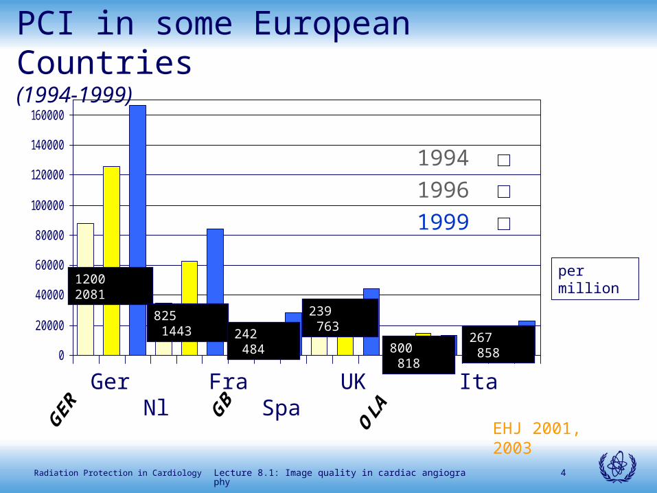
Percentage of inadequate studies by Percentage of inadequate studies by different hospitals different hospitals (Leape, Am Heart J 2000;139:106-13)(Leape, Am Heart J 2000;139:106-13)
In 12/29 hosp. 50% of studies had deficencies
6 of these are teching hosp.
Lecture 8.1: Image quality in cardiac angiography 12Radiation Protection in Cardiology
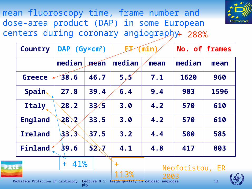
Country DAP (Gy×cm2) FT (min) No. of frames
median mean median mean median mean
Greece 38.6 46.7 5.5 7.1 1620 960
Spain 27.8 39.4 6.4 9.4 903 1596
Italy 28.2 33.5 3.0 4.2 570 610
England 28.2 33.5 3.0 4.2 570 610
Ireland 33.3 37.5 3.2 4.4 580 585
Finland 39.6 52.7 4.1 4.8 417 803
mean fluoroscopy time, frame number and dose-area product (DAP) in some European centers during coronary angiography
Neofotistou, ER 2003+ 41%
+ 113%
+ 288%

Lecture 8.1: Image quality in cardiac angiography 13Radiation Protection in Cardiology
0
5
10
15
20
25
30
35
40
45
50
%
Dublin % Leuven % Athens % Madrid %
LEFT-CR (+,+)
LEFT-CAU (+,-)
RIGHT-CR (-,+)
RIGHT-CAU (-,-)
projections’ distribution
11,5
9,2
7,5
15,413,8
12,4
0,0
2,0
4,0
6,0
8,0
10,0
12,0
14,0
16,0
18,0
Udine Dublin Leuven Greece Treviso Spain
Se
rie
s
mean number of series DIMOND 3 data
1000,4
1045,1
982,4
950,0
960,0
970,0
980,0
990,0
1000,0
1010,0
1020,0
1030,0
1040,0
1050,0
Dublin Greece Spain
SII
D (
cm)
focus-detector mean distances

Lecture 8.1: Image quality in cardiac angiography 14Radiation Protection in Cardiology
based on measurement of some physical parameters
• system transfer factor K
• spatial resolution (MTF, modulation transfer function)
• detective quantum efficiency (DQE)
• noise
they are rather complex and rarely applied to daily practice
quality evaluation of angiographic imagesquality evaluation of angiographic images objective methodsobjective methods

Lecture 8.1: Image quality in cardiac angiography 15Radiation Protection in Cardiology
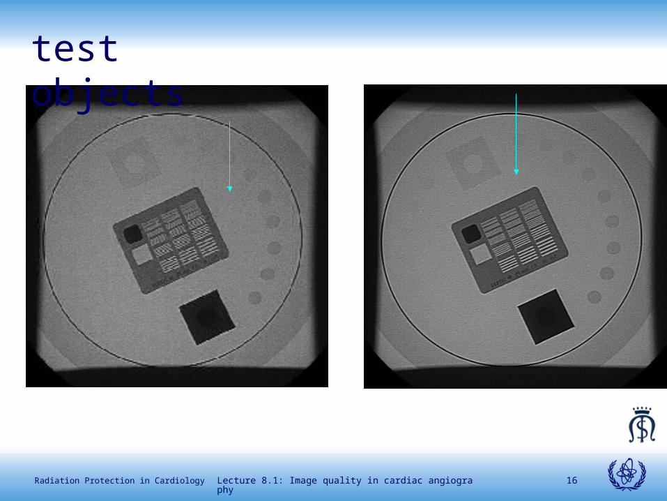
test objects or phantoms
• they are able to simulate the same radiation conditions as the part of the body
• they describe behaviour of radiology equipment in specific operating condition
evaluation of clinical images
• allow evaluation of the overall performance including patient’s collaboration and technique
quality evaluation of angiographic imagesquality evaluation of angiographic images subjective methodssubjective methods
Lecture 8.1: Image quality in cardiac angiography 16Radiation Protection in Cardiology
test objects

Lecture 8.1: Image quality in cardiac angiography 17Radiation Protection in Cardiology
binary classification
• pre-defined feature identification, normal vs. abnormal (this is typically used with test objects )
• correct answer must be known
• borderline visibility
progressive judgement in terms of quality
• variable level quality (clarity of thoracic calcification, arrange images in order of preference)
• strength of agreement by different observers gives indications on superiority
quality evaluation of angiographic images quality evaluation of angiographic images clinical images produced in clinical images produced in different conditionsdifferent conditions
Lecture 8.1: Image quality in cardiac angiography 18Radiation Protection in Cardiology
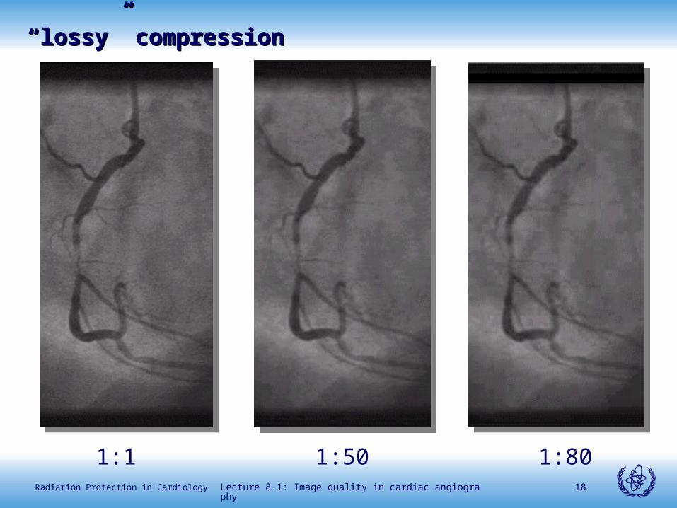
1:801:501:1
““lossy” compressionlossy” compression

Lecture 8.1: Image quality in cardiac angiography 19Radiation Protection in Cardiology

Lecture 8.1: Image quality in cardiac angiography 20Radiation Protection in Cardiology
proper filtering
improper filtering

Lecture 8.1: Image quality in cardiac angiography 21Radiation Protection in Cardiology
set of reference images difficult to obtain
use limited settings where perceptibility of abnormal feature is under experimenter’s control
quality measurement is only relative
clinical adequacy not evaluated
quality evaluation of angiographic imagesquality evaluation of angiographic images limitationslimitations
Lecture 8.1: Image quality in cardiac angiography 22Radiation Protection in Cardiology
quality of images is assessed in comparison to pre-
specified criteria to comply with
effective and relevant in clinical practice
• radiographic images (Maccia, Radiat Protect Dosim
1995; Vañò, Br J Radiol 1995, Radiat Prot Dosim 1998;
Perlmutter, Radiat Prot Dosim 1998)
• CT scan (Calzado, Radiat Prot Dosim 1998)
quality evaluation of angiographic imagesquality evaluation of angiographic images
method of quality criteriamethod of quality criteria

Lecture 8.1: Image quality in cardiac angiography 23Radiation Protection in Cardiology
1995-1996: GISE Società Italiana di Cardiologia
Invasiva and AIFM Associazione Italiana di
Fisica Biomedica
1996–2003: European Concerted Action DIMOND
Cardiology Group (Digital Imaging: Measures for
Optimizing Radiological INformation Content and Dose)
contracts FI 4P-0042DG12-WSMN, FIGM-CT-2000-00061-DIMOND
http://www.dimond3.org/
development of Quality Criteriadevelopment of Quality Criteria

Lecture 8.1: Image quality in cardiac angiography 24Radiation Protection in Cardiology
Diagnostic requirementsDiagnostic requirementsadapted from EUR 16260 ENadapted from EUR 16260 EN
Image criteria
In most cases specify important anatomical structures that should be visible on an image to aid accurate diagnosis. Some of these criteria depend fundamentally on correct positioning and cooperation of the patient or good angiographic technique, whereas others reflect technical performance of the imaging system
Important image detailsProvide quantitative information on the minimum sizes at which important anatomical details should become visible on the image. Some of these anatomical details may be pathological and therefore may not be present (ex. mitral insufficiency)

Lecture 8.1: Image quality in cardiac angiography 25Radiation Protection in Cardiology
Objectivesto set guidelines and give methods for the
evaluation of image quality in–Left Ventriculography
–Left Coronary Angiography
–Right Coronary Angiography
–Angiography of Venous Graft or Arterial Free Graft
–Angiography of Left Mammary Artery ‘In Situ’
ModelEuropean guidelines on quality criteria for
diagnostic radiographic images (EUR 16260 EN) where the diagnostic requirements and image criteria are settled

Lecture 8.1: Image quality in cardiac angiography 26Radiation Protection in Cardiology
What was not intendedto repeat what has already been included in
the manuals of Coronary Angiography, but to give some guidelines about how an angiogram should appear provided that good equipment and a correct angiographic technique are used
Warningsunder no circumstances should an image
which fulfils all clinical requirements but does not meet all image criteria ever be rejected*
*EUR 16260 EN

Lecture 8.1: Image quality in cardiac angiography 27Radiation Protection in Cardiology
Clinical criteria are defined as important anatomical features that should be visible; the level of visualisation is as follows
visualization: characteristic features are detectable, but details are not fully reproduced (features just visible)
reproduction: details of anatomical structures are visible, but not necessarily clearly defined (details emerging)
visually sharp reproduction: anatomical details are clearly defined (details clear)
Technical criteria
help to asses the technical quality of the procedure
features not necessarily impair the clinical information content (panning, arms position, etc.)
Aspects of an optimised angiographic technique
set of technical information
aimed to an optimised radiological technique
not mandatory
definition of termsdefinition of terms

Lecture 8.1: Image quality in cardiac angiography 28Radiation Protection in Cardiology
visualization: characteristic features are detectable, but details are not fully reproduced (features just visible)

Lecture 8.1: Image quality in cardiac angiography 29Radiation Protection in Cardiology
reproduction: details of anatomical structures are visible, but not necessarily clearly defined (details emerging)

Lecture 8.1: Image quality in cardiac angiography 30Radiation Protection in Cardiology
visually sharp reproduction: anatomical details are clearly defined (details clear)

Lecture 8.1: Image quality in cardiac angiography 31Radiation Protection in Cardiology
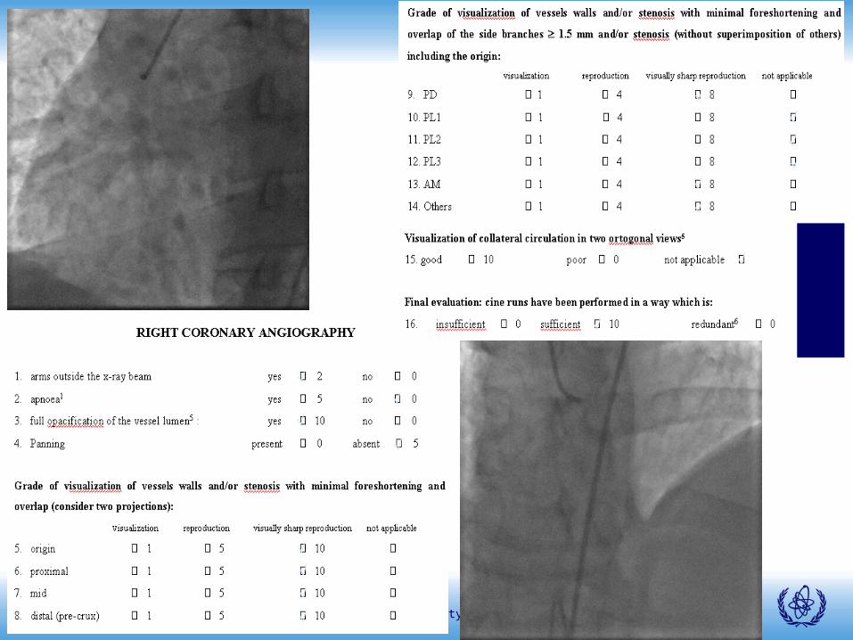
1) Visually sharp reproduction of the origin, proximal, mid (especially the crux region) and distal portion in at least two orthogonal views, with minimal foreshortening and overlap
2) Visually sharp reproduction of side branches 1.5 mm in at least two orthogonal views, with minimal foreshortening and overlap. The origin should be seen in at least one projection
3) Visually sharp reproduction of lesions in vessels 1.5 mm in at least two orthogonal views, with minimal foreshortening and overlap
4) Visualization of collateral circulation when present
clinical criteria for RCA projections based on operator’s choice
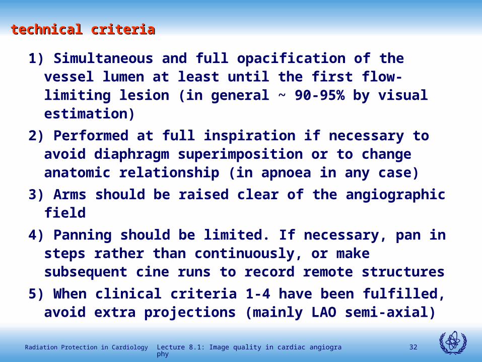
Lecture 8.1: Image quality in cardiac angiography 32Radiation Protection in Cardiology
1) Simultaneous and full opacification of the vessel lumen at least until the first flow-limiting lesion (in general ~ 90-95% by visual estimation)
2) Performed at full inspiration if necessary to avoid diaphragm superimposition or to change anatomic relationship (in apnoea in any case)
3) Arms should be raised clear of the angiographic field
4) Panning should be limited. If necessary, pan in steps rather than continuously, or make subsequent cine runs to record remote structures
5) When clinical criteria 1-4 have been fulfilled, avoid extra projections (mainly LAO semi-axial)
technical criteriatechnical criteria

Lecture 8.1: Image quality in cardiac angiography 33Radiation Protection in Cardiology
aspects of an optimised angiographic techniqueaspects of an optimised angiographic technique
1) Use of the wedge filter on bright peripheral areas
2) 2-3 sequences (except for difficult anatomic details)
3) 12.5-15 frames/s (25-30 only if heart rate exceeds 90-
100 bpm or in paediatric patients)
4) 60 images per sequence at average (12.5-15 fr/s)
except if collaterals have to be imaged or in case of
slow flow
Lecture 8.1: Image quality in cardiac angiography 34Radiation Protection in Cardiology
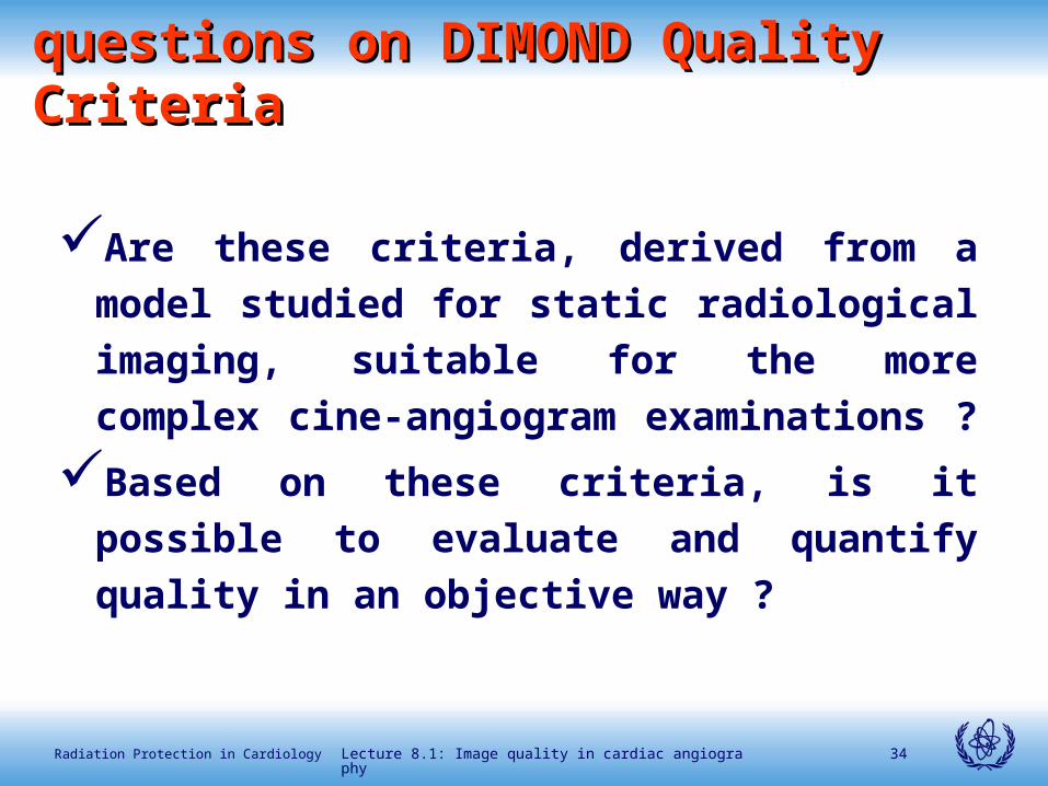
Are these criteria, derived from a model
studied for static radiological imaging,
suitable for the more complex cine-
angiogram examinations ?
Based on these criteria, is it possible to
evaluate and quantify quality in an objective
way ?
questions on DIMOND Quality Criteriaquestions on DIMOND Quality Criteria

Lecture 8.1: Image quality in cardiac angiography 35Radiation Protection in Cardiology
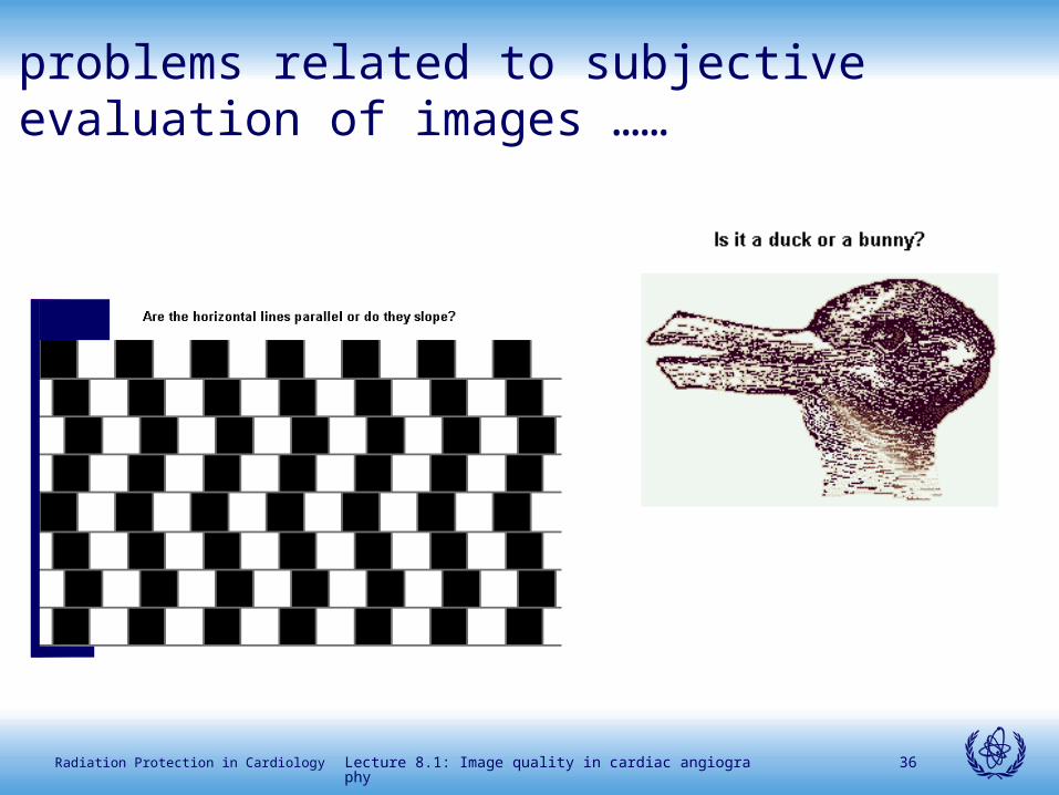
problems related to subjective evaluation of images ……
Lecture 8.1: Image quality in cardiac angiography 36Radiation Protection in Cardiology
problems related to subjective evaluation of images ……

Lecture 8.1: Image quality in cardiac angiography 37Radiation Protection in Cardiology
the method of image quality evaluation based on DIMOND Quality Criteria

Lecture 8.1: Image quality in cardiac angiography 38Radiation Protection in Cardiology
the method of image quality evaluation based on DIMOND Quality Criteria

Lecture 8.1: Image quality in cardiac angiography 39Radiation Protection in Cardiology
the method of image quality evaluation based on DIMOND Quality Criteria
Lecture 8.1: Image quality in cardiac angiography 40Radiation Protection in Cardiology
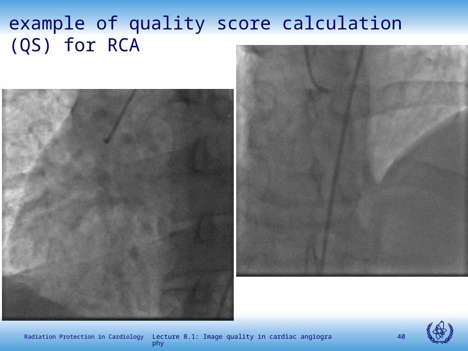
example of quality score calculation (QS) for RCA
Lecture 8.1: Image quality in cardiac angiography 41Radiation Protection in Cardiology

Lecture 8.1: Image quality in cardiac angiography 42Radiation Protection in Cardiology
sum of scores = 91 (actual score)
maximum theoretical score = 96
QS = actual score/theoretical score %
= 65/88x100 = 94%
example of QS calculation for RCA

Lecture 8.1: Image quality in cardiac angiography 43Radiation Protection in Cardiology
total score (mean and std dev.)total score (mean and std dev.)15 angio, 65 readings, 3 european centers15 angio, 65 readings, 3 european centers
75
80
85
90
95
100
Italy
4
Italy
3
Italy
1
Gre 4
Italy
5
Spa 2
Spa 5
Spa 1
Gre 1
Italy
2
Gre 2
Gre 3
Gre 5
Spa 4
Spa 3
0
2
4
6
8
10
12
14
16
18
20
med
std dev
Linear (med)
% within pts variability = 0.08
Lin’s coeff = .76 (CI .67-.84)
AJC, 1999 (abs)

Lecture 8.1: Image quality in cardiac angiography 44Radiation Protection in Cardiology
total score (mean and std dev.)total score (mean and std dev.)30 angio, 160 readings, 6 european centers30 angio, 160 readings, 6 european centers
0
10
20
30
40
50
60
70
80
90
100
1 3 5 7 9 11 13 15 17 19 21 23 25 27 29
-4
1
6
11
16
21
med
std dev
Linear (med)
%
Lecture 8.1: Image quality in cardiac angiography 45Radiation Protection in Cardiology
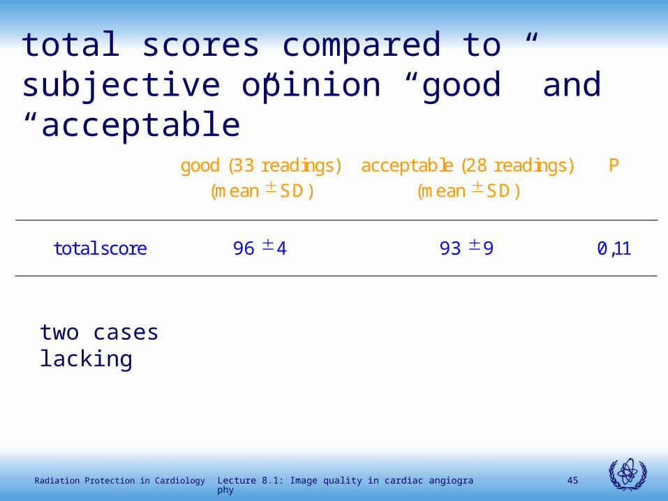
good (33 readings) (mean SD)
acceptable (28 readings) (mean SD)
P
total score 96 4 93 9 0,11
total scores compared to subjective opinion “good” and “acceptable”
two cases lacking

Lecture 8.1: Image quality in cardiac angiography 46Radiation Protection in Cardiology
what is ?what is ?
good
I get all the information needed to treat the patient and I like this examination
acceptable
I get all the information needed to treat the patient but I don’t like very much this examination
unacceptable
I don’t get all the information needed to treat the patient and I don’t like this examination at all

Lecture 8.1: Image quality in cardiac angiography 47Radiation Protection in Cardiology
RemarksRemarks
the method based on Quality Criteria applies to cardiac angiography
reproducibility is goodmeasure of clinical acceptability seems
improved in comparison to subjective opinion
the method “forces” to a systematic and
standardized analysis of the imagesspecific training not requested (but it may
improve agreement)

Lecture 8.1: Image quality in cardiac angiography 48Radiation Protection in Cardiology
Criteri di Qualità dell’Immagine Cineangiografica (documento
preliminare). Emodinamica 1997; 10 (suppl.): 9-11
Quality criteria of imaging in diagnostic and interventional
cardiology. TCT-196: Am J Cardiol, 1999:84(6A):73P-74P
A method based on DIMOND Quality Criteria to evaluate imaging
in diagnostic and interventional cardiology. Radiat Prot Dosim
2001;94:167-172
Quality Criteria for cardiac images in diagnostic and
interventional cardiology. Br J Radiol 2001; 74:852-855
Quality CriteriaQuality Criteria published paperspublished papers

Lecture 8.1: Image quality in cardiac angiography 49Radiation Protection in Cardiology
closing remarksclosing remarks
image quality is not warranted in coronary angiography
a great variability is found in common practice among different
operators and radiological exposure varies considerably
image quality assessment plays a pivotal role in the optimisation
of angiographic procedures
optimisation implies a continuous process of research and audit
which should involve
Scientific Societies
single operators
cooperation of all professionals in the Cath. Lab.