Embed Size (px)
Citation preview

lable at ScienceDirect
International Biodeterioration & Biodegradation 116 (2017) 64e72
Contents lists avai
International Biodeterioration & Biodegradation
journal homepage: www.elsevier .com/locate/ ibiod
Biosorption of copper from aqueous environments by Micrococcusluteus in cell suspension and when encapsulated
Ilya Letnik a, *, Ron Avrahami b, Rafi Port a, Andreas Greiner c, Eyal Zussman b,J. Stefan Rokem a, Charles Greenblatt a
a Department of Microbiology and Molecular Genetics, IMRIC, Hebrew University e Hadassah Medical School, Jerusalem 9112102, Israelb Faculty of Mechanical Engineering, Technion e Israel Institute of Technology, Haifa 32000, Israelc Macromolecular Chemistry II and Bayreuth Center for Colloids and Interfaces, University of Bayreuth, Universit€atsstrasse 30, 95440, Germany
a r t i c l e i n f o
Article history:Received 29 May 2016Received in revised form20 September 2016Accepted 27 September 2016
Keywords:Copper uptakeMicrococcus luteusEncapsulation in electrospun polymer
Abbreviations: MLF, Fibers containing live M. lutetaining liveM. luteus cells; IMAC, Immobilized-metal aEnergy-dispersive X-ray spectroscopy.* Corresponding author.
E-mail address: [email protected] (I. Letnik
http://dx.doi.org/10.1016/j.ibiod.2016.09.0290964-8305/© 2016 Elsevier Ltd. All rights reserved.
a b s t r a c t
The ability to sequester or tolerate copper has been described for a number of bacteria. It was shown thatMicrococcus luteus tolerates copper, and possible mechanisms of uptake for this bacterium have beenproposed. Using a count live of M. luteus (CFU), 10% survived a copper concentration of 370 ppm.Maximum sorption capacity was 59 mg of Cu2þ/g of dry cells at pH 6. Several proteins of M. luteus withcopper affinity were identified by enrichment on a metal-chelating resin followed by LC-MS/MS toidentify the metallome. Use of SEM/EDX showed how copper concentrated on the bacterial surface,whereas TEM indicated that copper was found also inside the cells. The enhanced capability of M. luteusto bind copper was tested using three configurations: free cells on an agar surface and cells encapsulatedin alginate or in electrospun polymer composites. The latter showed the highest capability to bind copper(~76 mg Cu2þ/g dry cells). Such polymer composites may potentially be used in various water-basedapplications such as treatment of wastewater with a high concentration of copper or other heavy metals.
© 2016 Elsevier Ltd. All rights reserved.
1. Introduction
Copper is an essential trace element, like many other heavymetals, and it is also toxic in high concentrations. The interaction ofheavy metals, specifically copper, has been described for manydifferent species of microorganisms. For example, the use of cop-per/nickel alloys in marine environments was considered as ameans to avoid corrosion; however, it was found that sulfate-reducing bacteria could attach and grow on an alloy having ahigh copper concentration as long as the bacteria possess transientoxygen tolerance (Chamberlain et al., 1988). The ability of bacteriato withstand high concentrations of copper was also reported forcopper plumbing, where assimilable organic carbon was requiredfor microbial-induced corrosion (Walker et al., 1991) and highconcentrations of copper in drinking water was found to be relatedto the biofilms of Variovorax sp in copper plumbing systems (Reyes
us cells; MLA, Alginate con-ffinity chromatography; EDX,
).
et al., 2008). In another study, the exopolymer accumulation wasfound to allow for bacterial corrosion, and a mixed culture ofPseudomonas bacteria conferred high resistance to copper to one ofthe bacteria under study, Pseudomonas paucimobilis (Angell andChamberlain, 1991). Copper has also been shown to increasepolymer formation by the marine bacterium Oceanospirillum,allowing for neutralization of copper toxicity permitting otherspecies to become part of a biofilm on copper containing surfaces(Wagner et al., 1996). A similar response with the formation of anextracellular emulsifying agent was described when copper con-centration was increased so that the fungus Curvularia lunata dis-played an increase in total lipid saturation (Paraszkiewicz et al.,2009).
The possible environmental applications of metal uptake byusing microorganisms for wastewater treatment have been re-ported (Wong et al., 2001b). A review of recent studies showedmany microorganisms are capable of metal biosorption, in thestudy of Kiran et al. The cyanobacterium Lyngbya putealis has beenshown to take up copper in the amount of 7.8 mg/g dry cells, with40e45% taken up after just 15min. Adsortptionwas optimizedwithrespect to pH and metal concentration (Kiran and Thanasekaran,2011). Sulfate-reducing bacteria have been shown to have high
I. Letnik et al. / International Biodeterioration & Biodegradation 116 (2017) 64e72 65
metal-reducing capabilities in the study of Chen et al. (2000)although biosorption was limited to Cu(II) 16 mg/g dry cells. Eventhe dead biomass of Fusarium Flocciferum has been shown to be apromissing agent for heavy metal uptake, and equilibrium wasachieved within seconds (Delgado et al., 1996).
One group of cocci, the genus Micrococcus, has repeatedly beenfound under variuos extreme environments such as wood amber(Greenblatt et al., 2004) and hot springs (Liu et al., 2016). Micro-coccus also can be found in grey water from treatment facilities(Keely et al., 2015). Its apparent tolerance to harsh conditionsattracted research in field of bioremediation, for example, boronremoval from wastewater (Lacin et al., 2015). Metal uptake abilitywas shown by Wong et al. (2001b) who described the isolation ofMicrococcus sp, from sludge from a local wastewater treatmentfacility and the bacterium was shown to survive several cycles (atleast five) of biosorption and desorption of copper (Wong et al.,2001a). Micrococcus luteus can also take up strontium (Faisonet al., 1990) and, to a lesser degree lead, nickel and zinc. In addi-tion, it was shown that it is possible to retrieve the metals whilemaintaining bacterial cell viability.
The possible role of M. luteus in bioremediation was the incen-tive for its full genomic sequencing, initiated by our laboratory(Young et al., 2010). There are different strategies of microbial metal(copper) resistance or tolerance; among them are bioaccumulationor sequestration. Others include exclusion, compartmentalizationand complexation by binding proteins such as metallothioneins orphytochelatins (Mejare and Bulow, 2001). In the genomic sequencea repertoire of functions was annotated that deal with metals. Bothchromosomal and plasmid genes are involved in this resistance. InM. luteus the genes of the MerR/ArsR proteins, which are known toact in metal resistance (Young et al., 2010), are present. Further-more, some eight chromosomal genes related to copper transportor function are present in theM. luteus genome, including the 2 Cop(A and C) resistant genes and 3 chaperones. Metal sequestrationcould be by a different means than tolerance, and be related to thethird mechanism – complexation by cell components. Nakajimaet al. (2001) treated M. luteus with a series of chemical extrac-tions in order to remove polysaccharides, proteins, small molecules,lipids, and lastly the more tightly bound proteins and poly-saccharides. Finally, the 17% of what was left after these extractionshad the greatest ability to uptake copper. Although we possesssome understanding of the mechanism governing microbial heavymetal uptake, further studies are required.
The use of immobilized cells for enhanced metal adsorption isgaining interest and several studies showed promising results. Inthe work of Ahmad et al. (2013) biosorption of high amounts zincions from aqueous solution by immobilized Candida utilis andCandida tropicalis cells was shown. Sinha et al. (2012) showed thatalginate immobilized Bacillus cereus cells are able to absorb 104 mgmercury per g dry cells. Since many studies use simple hydrogelssuch as alginate for the encapsultion process, polymers which willdegrade in water based applications, further studies are requiredfor improvement of polymeric formulations.
Recently, Gensheimer et al. (2011); Klein et al. (2012, 2009) andKnierim et al. (2014) have described a system of microbial encap-sulation which entails incorporating bacteria within microtubes ofpolymer on a nano scale. The process, called electrospinning, hasbeen used with M. luteus, Pseudomonas ADP, Nitrobacter winog-radskyi and genetically modified E. coli. Modifications of the orignalprocess (Gensheimer et al., 2007) entail the creation of a two-layered fiber having a water-insoluble outer membrane (shell)and an inner core of a hydrogel (co-electrospinning) (Dror, 2008).This method allows for the creation of a natural environment forthe immobilized cells, mainly due to the viscosity of the water-soluble core polymer. In addition, adjustment of the properties of
the shell polymers allows for tuning the mechanical properties, cellattachment and cell division.
In order to create active polymer-bacteria composites anadequate candidate bacterium should be used. For this work wechose a species that has high affinity for the targeted metals, and iscapable of surviving high concentrations of the metals andaccompanying toxic organic substances. It is preferable that themicrobes be easily separable from their substrate and either themetals can be disposed of, or if they have economic value, retrieved.It is also desirable that organisms be recyclable so the remediativeprocess can be repeated.
Our goal in this study was to shed some light on the copper-sequestering mechanisms of planktonic and encapsulatedM. luteus and, in addition, to create metal-absorbing bacteria/polymer water-insoluble composites.
2. Materials and methods
2.1. Organism and culture media
M. luteus DSM20030T was purchased from DSMZ and wascultured in LB medium in Erlenmeyer flasks at 30 �C, with shakingat 150 rpm. Its identity was confirmed by 16S rDNA sequencing.
2.2. Simple screening methods
Cultures were applied to a copper oxide impregnated cloth (1.0%w/v, at the surface ~30% copper was present). The cloth consists of70% cotton and 30% polyester and is known to inactivate copper-sensitive organisms (Borkow and Gabbay, 2004). Small, 1.0 cmsquares of the fabric were placed on LB culture plates and a drop(10 mL) of cell culture was inoculated on their surface. Growth wasobserved on the surface. Alternatively, a carpet of organisms wasspread on the culture plates in semisolid medium and either thefabric or filter paper discs containing variable concentrations ofCuSO4 or copper gluconate were applied to the surface. The growthcurve was measured at OD600 in a FLUOstar Omega instrumentwith agitation. Readings were done every hour in triplicate.
2.3. Copper sequestration
To measure copper sequestration by planktonic cells, 1010
M. luteus cells/mL were suspended in 10 mL of a 10 mM CuSO4solution and incubated in a shaker at 30 �C. Samples were takenover a 3-h period. At the end of the test period, the bacteria werecentrifuged and the pellet was extracted with 7.5% TCA.
To measure copper sequestration by polymers, calcium alginatebeads or polymer composite were suspended in 10 mL of 10 mMCuSO4 and incubated at 30 �C. After 30 min the supernatant wasremoved by aspiration. The beads were washed twice with waterand then rinsed with a solution of 7.5% TCA. The copper contents ofthe supernatant and the TCA-extracted beads were determined bythe bicinchoninic acid method (Brenner and Harris, 1995).
To measure copper sequestration of the pure polymers, thepolyvinyl alcohol and polyvinyl pyrrolidone were prepared as10 mg per mL solutions and incubated in 40 mM CuSO4 for 30 min.The suspension was filtered through a Microcon filter with anexclusion value of 3 kDa. The copper concentration of the initialfiltrate and a second filtrate of 1.0 mL of 7.5% TCA solution weremeasured. The second filtrate contains the extraction of copperfrom the polymer.
I. Letnik et al. / International Biodeterioration & Biodegradation 116 (2017) 64e7266
2.4. Cell immobilization
2.4.1. AlginateA standard method was applied (Smidsrod and Skjakbraek,
1990). A 4% alginate solution was mixed 1:1 with the M. luteusculture. This mixture was added as a slow drip from a syringe into asolution of 0.1 M calcium chloride, the solidified droplets were leftto harden for 5 min andwashed with PBS, the resulting beads had amean size of 4 mm. Alginate beads without bacteria were preparedas a control.
2.4.2. ElectrospinningA core-shell electrospinning technique was used for immobili-
zation of the bacteria. The system allows for fabrication of micro-tubes in an electrostatic field. Two separate syringes continuouslyejected solutions from a spinneret with two coaxial capillariesunder high voltage. The internal capillary of this spinneret facili-tated the flow of the bacterial cells in an aqueous solution, whilebeing enveloped by the external capillary containing a non-biodegradable polymer solution. The core consisted of 20 vol%cell mass in 15 wt% polyvinylpyrrolidone (PVP) Mw¼ 1300 k in de-ionized water. The shell copolymer consisted of 15% polyvinylidenefluoride-co-hexafluoropropylene (PVDF-HFP) Mw¼ 400 k and 2wt% PEG (Mw ¼ 6 k) in a solvent mixture of tetrahydrofuran (THF)/dimethylformamide (DMF) in a weight ratio of 6.5:3.5, as describedearlier (Nardi et al., 2012). The electrospun fibers were collected onthe surface of an earthed bath of PBS solution and wound ontoplastic carriers. The carriers were stored in PBS solution; the fiberswere not allowed to dry out at any time.
2.5. Copper determination
Copper sulfate anhydrous (CuSO4) was purchased from J.T.BakerChemicals (#32949), and copper (II) D-gluconate from Sigma-Aldrich (#344419). Copper was measured either by flame spec-trophotometry using a Varian Spectra AA Zeeman graphite tubeatomizer or by the bicinchoninic acid chemical method where thecopper-bicinchoninic acid chelated product is oxidized by ascorbicacid to yield a colored substance (Brenner and Harris, 1995). Twomethods were used to measure the amount of copper the organ-isms were able to sequester. CuSO4 was added to a suspension of~0.9 A600/mL of the M. luteus, and supernatant samples were takenat different times (up to 3 days) and centrifuged. In addition copperwas also measured in the pelleted organisms and was determinedboth before and after DDW washes.
2.6. TEM and SEM-EDX
For electron microscopy imaging an extra high resolutionscanning electron microscopy (XHR-SEM) MagellanTM 400L andhigh resolution transmission scanning electron microscope (HR-TEM) Tecnai F20 G2 were used. The scanning electron microscopy(SEM) was combined with an Everhart-Thornley Detector (ET) andenergy-dispersive X-ray spectroscopy (EDX). Samples taken fromthe experiment as described in section 2.3 were air dried and goldplated or were applied to silica and air dried.
For the TEM, samples were fixed with Karnovsky's fixative (2%PFA, 2,5% glutaraldehyde in 0.1 M cacodylate buffer, pH ¼ 7.4) for4 h at room temperature, followed by½ diluted Karnovsky's fixativeovernight at 4 �C. For the TEM-EDX, samples were washed in 0.1 Mcacodylate buffer, dehydrated in a graded series of alcohols, fol-lowed by propylene oxide, and embedded in epoxy resin. Sectionsof 60e80 nm thickness were cut on an ultramicrotome (Reichert-Jung), and examined with the high resolution transmission scan-ning electron microscope Tecnai F20 G2 equipped with an EDX
detector (spectral resolution 133 eV).
2.7. Protein purification
In the procedure, done at 4 �C, in 25 mM TrisHCl buffer and100 mM NaCl at pH 8.0, the chelating resin was charged with100 mM CuSO4 and then washed extensively. The resin was equil-ibrated with 250 mL wash buffer (buffer þ 0.02% Triton X100).Protein was extracted by resuspending the cell pellet (from 2 Lculture) with 30 mL lysis buffer (buffer þ 0.2% TritonX100þDNaseþ lysozymeþ 0.5mM PMSF). The cells, kept on ice atall times, were sonicated 6 times for 10 s and centrifuged for15 min at 12,000 rpm at 4 �C. The pellet was discarded, equilibratedresin was mixed with the supernatant and incubated for 60 min4 �C. The mixed resin/supernatant was centrifuged for 4 min at3500 rpm at 4 �C. The unbound supernatant was discarded, and theresin was washed three additional times. Elution was done with~500 mL elution buffer (buffer þ 0.02% Triton X100þ 10 mM EDTA),with incubation for 5 min with buffer, and then centrifuged for4 min at 3500 rpm at 4 �C. 300 mL of the eluate was loaded in aSuperose 12 53 � 1.6 cm (~107 mL) column in buffer þ 0.1% TritonX100.
2.8. Electrophoresis
20 mg of protein was boiled for 5 min with sample buffer (2xBioRad Laemmli Sample Buffer # 1610737) and loaded on a 12%Mini-PROTEAN TGX Stain-Free™ BioRad Precast Gel (#4568043)with 5 mLmarker. Running buffer was TGS� 1 (25mM Tris,192mMglycine, 0.1% SDS, pH 8.3). The biggest bands were excised and usedfor mass-spec analysis.
2.9. In-gel digestion and protein identification by LC-ESI-MS/MS
Protein bands were excised from an SDS gel stained with Coo-massie blue. The protein bands were subsequently reduced, alky-lated and in-gel digested with bovine trypsin (Promega), at aconcentration of 12.5 ng/mL in 50 mM ammonium bicarbonate at37 �C, as described (Shevchenko et al., 1996). The peptide mixtureswere extracted with 80% CH3CN/1% CF3COOH, and the organicsolvent was evaporated in a vacuum centrifuge. The resultingpeptide mixtures were reconstituted in 80% formic acid andimmediately diluted 1:10 with Milli-Q water before analysis. Liquidchromatographyetandem mass spectrometry (LCeMS/MS) wasperformed using a 15 cm reversed-phase fused-silica capillarycolumn (inner diameter, 75 mm) made in-house and packed with3 mmReproSil-Pur C18AQmedia (Dr. Maisch GmbH). The LC system,an UltiMate 3000 (Dionex). was used in conjunction with an LTQOrbitrap XL (Thermo Fisher Scientific) operated in the positive ionmode and equipped with a nanoelectrospray ion source. Peptideswere separated with a two hour gradient from 5 to 65% acetonitrile(buffer A, 5% acetonitrile, 0.1% formic acid and 0.005% TFA; buffer B,90% acetonitrile, 0.2% formic acid, and 0.005%TFA). The voltageapplied to produce an electrospray was 1.2 kV. The mass spec-trometer was operated in the data-dependent mode. Survey massspectrometry scans were acquired in the Orbitrap with the reso-lution set to a value of 60,000. The sevenmost intense ions per scanwere fragmented and analyzed in the linear ion trap. Raw data fileswere searched with MASCOT (Matrix Science) against the NCBI nrdatabase. Search parameters included a fixed modification of57.02146 Da (carboxyamidomethylation) on Cys, and variablemodifications 15.99491 Da (oxidation) on Met, and 0.984016 Da(deamidation) on Asn and Gln. The search parameters alsoincluded: maximum 2 missed cleavages, initial precursor ion masstolerance 10 ppm, fragment ion mass tolerance 0.6 Da. Samples
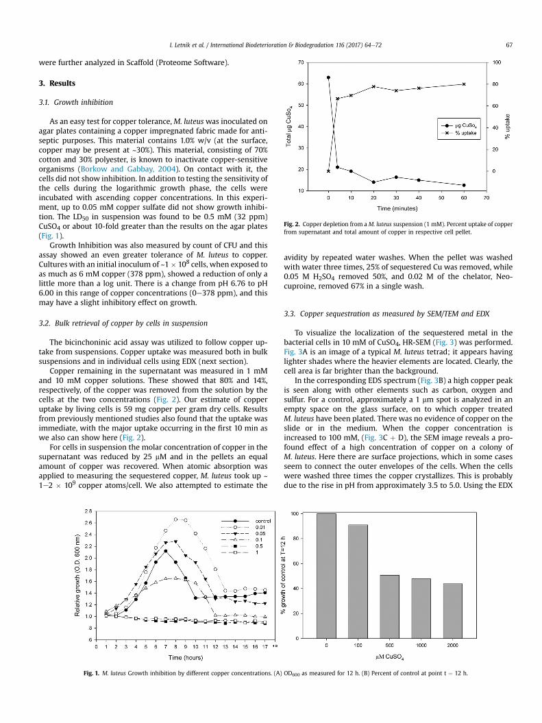
Fig. 2. Copper depletion from a M. luteus suspension (1 mM). Percent uptake of copperfrom supernatant and total amount of copper in respective cell pellet.
I. Letnik et al. / International Biodeterioration & Biodegradation 116 (2017) 64e72 67
were further analyzed in Scaffold (Proteome Software).
3. Results
3.1. Growth inhibition
As an easy test for copper tolerance,M. luteuswas inoculated onagar plates containing a copper impregnated fabric made for anti-septic purposes. This material contains 1.0% w/v (at the surface,copper may be present at ~30%). This material, consisting of 70%cotton and 30% polyester, is known to inactivate copper-sensitiveorganisms (Borkow and Gabbay, 2004). On contact with it, thecells did not show inhibition. In addition to testing the sensitivity ofthe cells during the logarithmic growth phase, the cells wereincubated with ascending copper concentrations. In this experi-ment, up to 0.05 mM copper sulfate did not show growth inhibi-tion. The LD50 in suspension was found to be 0.5 mM (32 ppm)CuSO4 or about 10-fold greater than the results on the agar plates(Fig. 1).
Growth Inhibition was also measured by count of CFU and thisassay showed an even greater tolerance of M. luteus to copper.Cultures with an initial inoculum of ~1� 108 cells, when exposed toas much as 6 mM copper (378 ppm), showed a reduction of only alittle more than a log unit. There is a change from pH 6.76 to pH6.00 in this range of copper concentrations (0e378 ppm), and thismay have a slight inhibitory effect on growth.
3.2. Bulk retrieval of copper by cells in suspension
The bicinchoninic acid assay was utilized to follow copper up-take from suspensions. Copper uptake was measured both in bulksuspensions and in individual cells using EDX (next section).
Copper remaining in the supernatant was measured in 1 mMand 10 mM copper solutions. These showed that 80% and 14%,respectively, of the copper was removed from the solution by thecells at the two concentrations (Fig. 2). Our estimate of copperuptake by living cells is 59 mg copper per gram dry cells. Resultsfrom previously mentioned studies also found that the uptake wasimmediate, with the major uptake occurring in the first 10 min aswe also can show here (Fig. 2).
For cells in suspension the molar concentration of copper in thesupernatant was reduced by 25 mM and in the pellets an equalamount of copper was recovered. When atomic absorption wasapplied to measuring the sequestered copper, M. luteus took up ~1e2 � 109 copper atoms/cell. We also attempted to estimate the
Fig. 1. M. luteus Growth inhibition by different copper concentrations. (A)
avidity by repeated water washes. When the pellet was washedwith water three times, 25% of sequestered Cu was removed, while0.05 M H2SO4 removed 50%, and 0.02 M of the chelator, Neo-cuproine, removed 67% in a single wash.
3.3. Copper sequestration as measured by SEM/TEM and EDX
To visualize the localization of the sequestered metal in thebacterial cells in 10 mM of CuSO4, HR-SEM (Fig. 3) was performed.Fig. 3A is an image of a typical M. luteus tetrad; it appears havinglighter shades where the heavier elements are located. Clearly, thecell area is far brighter than the background.
In the corresponding EDS spectrum (Fig. 3B) a high copper peakis seen along with other elements such as carbon, oxygen andsulfur. For a control, approximately a 1 mm spot is analyzed in anempty space on the glass surface, on to which copper treatedM. luteus have been plated. There was no evidence of copper on theslide or in the medium. When the copper concentration isincreased to 100 mM, (Fig. 3C þ D), the SEM image reveals a pro-found effect of a high concentration of copper on a colony ofM. luteus. Here there are surface projections, which in some casesseem to connect the outer envelopes of the cells. When the cellswere washed three times the copper crystallizes. This is probablydue to the rise in pH from approximately 3.5 to 5.0. Using the EDX
OD600 as measured for 12 h. (B) Percent of control at point t ¼ 12 h.
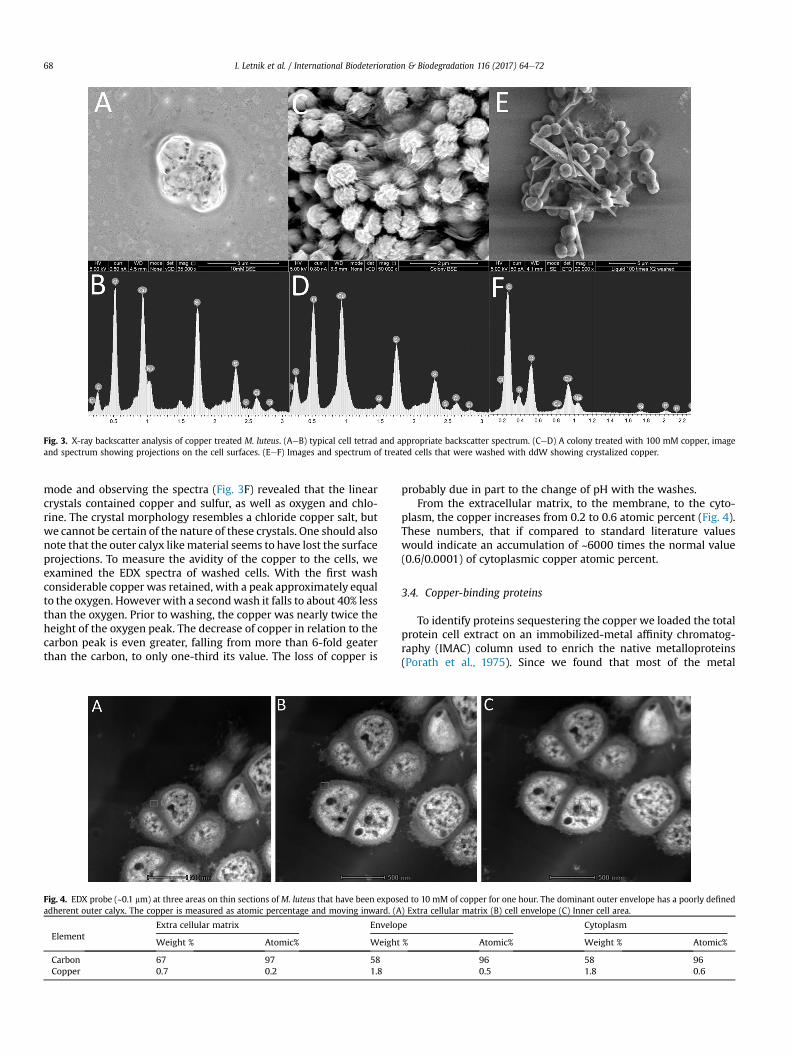
Fig. 3. X-ray backscatter analysis of copper treated M. luteus. (AeB) typical cell tetrad and appropriate backscatter spectrum. (CeD) A colony treated with 100 mM copper, imageand spectrum showing projections on the cell surfaces. (EeF) Images and spectrum of treated cells that were washed with ddW showing crystalized copper.
I. Letnik et al. / International Biodeterioration & Biodegradation 116 (2017) 64e7268
mode and observing the spectra (Fig. 3F) revealed that the linearcrystals contained copper and sulfur, as well as oxygen and chlo-rine. The crystal morphology resembles a chloride copper salt, butwe cannot be certain of the nature of these crystals. One should alsonote that the outer calyx likematerial seems to have lost the surfaceprojections. To measure the avidity of the copper to the cells, weexamined the EDX spectra of washed cells. With the first washconsiderable copper was retained, with a peak approximately equalto the oxygen. However with a secondwash it falls to about 40% lessthan the oxygen. Prior to washing, the copper was nearly twice theheight of the oxygen peak. The decrease of copper in relation to thecarbon peak is even greater, falling from more than 6-fold geaterthan the carbon, to only one-third its value. The loss of copper is
Fig. 4. EDX probe (~0.1 mm) at three areas on thin sections of M. luteus that have been exposadherent outer calyx. The copper is measured as atomic percentage and moving inward. (A
ElementExtra cellular matrix Envelop
Weight % Atomic% Weight
Carbon 67 97 58Copper 0.7 0.2 1.8
probably due in part to the change of pH with the washes.From the extracellular matrix, to the membrane, to the cyto-
plasm, the copper increases from 0.2 to 0.6 atomic percent (Fig. 4).These numbers, that if compared to standard literature valueswould indicate an accumulation of ~6000 times the normal value(0.6/0.0001) of cytoplasmic copper atomic percent.
3.4. Copper-binding proteins
To identify proteins sequestering the copper we loaded the totalprotein cell extract on an immobilized-metal affinity chromatog-raphy (IMAC) column used to enrich the native metalloproteins(Porath et al., 1975). Since we found that most of the metal
ed to 10 mM of copper for one hour. The dominant outer envelope has a poorly defined) Extra cellular matrix (B) cell envelope (C) Inner cell area.
e Cytoplasm
% Atomic% Weight % Atomic%
96 58 960.5 1.8 0.6
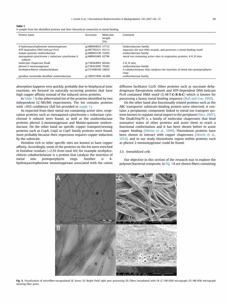
Table 1A sample from the identified proteins and thier theoretical connection to metal binding.
Protein name Accession Molecularweight(Da)
Comment
4-hydroxyacetophenone monooxygenase gij488944925 57712 Oxidoreductase familyATP-dependent DNA helicase PcrA gij497765221 93113 separates the two DNA strands, and possesses a metal binding motifmalate:quinone oxidoreductase gij488943149 53203 oxidoreductase familymenaquinol-cytochrome c reductase cytochrome b
subunitgij488943482 62796 metal ion containing active sites in respiration proteins, 4 H_H sites
molecular chaperone DnaK gij738364993 66564 2 H_H sitesphenol 2-monooxygenase gij738432093 70383 oxidoreductase familySirohydrochlorin cobaltochelatase gij751048160 24834 A cobaltochelatase that catalyzes the insertion of metal into protoporphyrin
ringspyridine nucleotide-disulfide oxidoreductase gi 289557896 44,400 oxidoreductase family
I. Letnik et al. / International Biodeterioration & Biodegradation 116 (2017) 64e72 69
absorption happens very quickly, probably due to biophysical ionicreactions, we focused on naturally occurring proteins that havehigh copper affinity instead of the induced stress proteins.
In Table 1 is the abbreviated list of the proteins identified by twoindependent LC-MS/MS experiments. The list contains proteinswith >95% confidence (full list provided in suppl. 1).
As expected from their metal ion containing active sites, respi-ration proteins such as menaquinol-cytochrome c reductase cyto-chrome b subunit were found, as well as the oxidoreductaseproteins phenol 2-monooxygenase and Malate:quinone oxidore-ductase. On the other hand no specific copper transport/sensingproteins such as CopA, CopZ or CopY family proteins were found,most probably because their expression requires copper inductionby the substrate.
Histidine rich or other specific sites are known to have copperaffinity. Accordingly, some of the proteins on the list were enrichedin histidine residues (<2.5% from total AA) for example sirohydro-chlorin cobaltochelatase is a protein that catalyze the insertion ofmetal into protoporphyrin rings. Another is 4-hydroxyacetophenone monooxygenase associated with the cation
Fig. 5. Visualization of microfiber-encapsulated M. luteus (A) Bright Field right post proceshowing fiber pores.
diffusion facilitator CzcD. Other proteins such as succinate dehy-drogenase flavoprotein subunit and ATP-dependent DNA helicasePcrA contained HMA motif (G-M-T-C-X-X-C) which is known forpossessing a heavy metal binding sequence (Bull and Cox, 1994).
On the other hand also functionally related proteins such as theABC transporter substrate-binding protein were observed, it con-tains a periplasmic component linked to metal ion transport sys-tems known to regulatemetal import to the periplasm (Nies, 2007).The DnaK/Hsp70 is a family of molecular chaperones that bindnonnative states of other proteins and assist them to reach afunctional conformation and it has been shown before to assistcopper binding (Hibino et al., 1999). Thioredoxin proteins havebeen shown to interact with copper chaperones (Abicht et al.,2014), and in our study thioredoxin region within proteins suchas phenol 2-monooxygenase could be found.
3.5. Immobilized cells
Our objective in this section of the research was to explore thepolymer/bacterial composite. In Fig. 5A are shown fibers containing
ssing (B) Fibers Incuabated with LB (C) HR-SEM micrograph (D) HR-SEM micrograph
I. Letnik et al. / International Biodeterioration & Biodegradation 116 (2017) 64e7270
live M. luteus cells (MLF) their uniformity of cell distribution in fi-bers. It should be noted that in subsequent storage cell divisionwasobserved (Fig. 5B). The fiber structure can be seen in Fig. 5C, with anaverage fiber diameter of 5 mm and an average of 40 nmwide pores.Due to the water soluble PVP in the fibers and nano pores on themicro tube surface (Fig. 5D) M. luteus cells remain inside the tubesbut there is no hindrance of liquid flow.
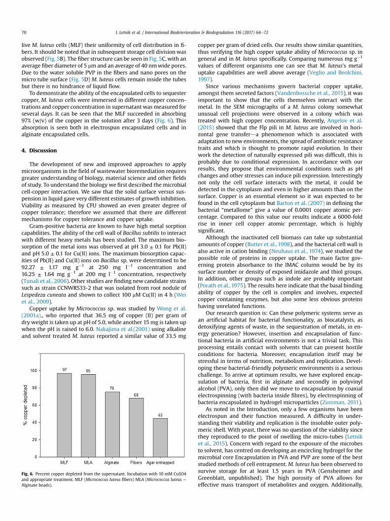
To demonstrate the ability of the encapsulated cells to sequestercopper, M. luteus cells were immersed in different copper concen-trations and copper concentration in supernatant wasmeasured forseveral days. It can be seen that the MLF succeeded in absorbing97% (w/v) of the copper in the solution after 3 days (Fig. 6). Thisabsorption is seen both in electrospun encapsulated cells and inalginate encapsulated cells.
4. Discussion
The development of new and improved approaches to applymicroorganisms in the field of wastewater bioremediation requiresgreater understanding of biology, material science and other fieldsof study. To understand the biology we first described themicrobialcell-copper interaction. We saw that the solid surface versus sus-pension in liquid gave very different estimates of growth inhibition.Viability as measured by CFU showed an even greater degree ofcopper tolerance; therefore we assumed that there are differentmechanisms for copper tolerance and copper uptake.
Gram-positive bacteria are known to have high metal sorptioncapabilities. The ability of the cell wall of Bacillus subtilis to interactwith different heavy metals has been studied. The maximum bio-sorption of the metal ions was observed at pH 3.0 ± 0.1 for Pb(II)and pH 5.0 ± 0.1 for Cu(II) ions. The maximum biosorption capac-ities of Pb(II) and Cu(II) ions on Bacillus sp. were determined to be92.27 ± 1.17 mg g�1 at 250 mg l�1 concentration and16.25 ± 1.64 mg g�1 at 200 mg l�1 concentration, respectively(Tunali et al., 2006). Other studies are finding new candidate strainssuch as strain CCNWRS33-2 that was isolated from root nodule ofLespedeza cuneata and shown to collect 100 mM Cu(II) in 4 h (Weiet al., 2009).
Copper uptake by Micrococcus sp. was studied by Wong et al.(2001a)., who reported that 36.5 mg of copper (II) per gram ofdry weight is taken up at pH of 5.0, while another 15 mg is taken upwhen the pH is raised to 6.0. Nakajima et al.(2001) using alkalineand solvent treated M. luteus reported a similar value of 33.5 mg
Fig. 6. Percent copper depleted from the supernatant. Incubation with 10 mM CuSO4and appropriate treatment. MLF (Micrococcus luteus fibers) MLA (Micrococcus luteus eAlginate beads).
copper per gram of dried cells. Our results show similar quantities,thus verifying the high copper uptake ability of Micrococcus sp. ingeneral and in M. luteus specifically. Comparing numerous mg g�1
values of different organisms one can see that M. luteus's metaluptake capabilities are well above average (Veglio and Beolchini,1997).
Since various mechanisms govern bacterial copper uptake,amongst them secreted factors (Vandenbossche et al., 2015), it wasimportant to show that the cells themselves interact with themetal. In the SEM micrographs of a M. luteus colony somewhatunusual cell projections were observed in a colony which wastreated with high copper concentration. Recently, Angelov et al.(2015) showed that the Flp pili in M. luteus are involved in hori-zontal gene transferda phenomenon which is associated withadaptation to new environments, the spread of antibiotic resistancetraits and which is thought to promote rapid evolution. In theirwork the detection of naturally expressed pili was difficult, this isprobably due to conditional expression. In accordance with ourresults, they propose that environmental conditions such as pHchanges and other stresses can induce pili expression. Interestinglynot only the cell surface interacts with the metal, it could bedetected in the cytoplasm and even in higher amounts than on thesurface. Copper is an essential element so it was expected to befound in the cell cytoplasm but Barton et al. (2007) in defining thebacterial “metallome” give a value of 0.0001 copper atomic per-centage. Compared to this value our results indicate a 6000-foldrise in inner cell copper atomic percentage, which is highlysignificant.
Although the inactivated cell biomass can take up substantialamounts of copper (Butter et al., 1998), and the bacterial cell wall isalso active in cation binding (Neuhaus et al., 1974), we studied thepossible role of proteins in copper uptake. The main factor gov-erning protein absorbance to the IMAC column would be by itssurface number or density of exposed imidazole and thiol groups.In addition, other groups such as indole are probably important(Porath et al., 1975). The results here indicate that the basal bindingability of copper by the cell is complex and involves, expectedcopper containing enzymes, but also some less obvious proteinshaving unrelated functions.
Our research question is: Can these polymeric systems serve asan artificial habitat for bacterial functionality, as biocatalysts, asdetoxifying agents of waste, in the sequestration of metals, in en-ergy generation? However, insertion and encapsulation of func-tional bacteria in artificial environments is not a trivial task. Thisprocessing entails contact with solvents that can present hostileconditions for bacteria. Moreover, encapsulation itself may bestressful in terms of nutrition, metabolism and replication. Devel-oping these bacterial-friendly polymeric environments is a seriouschallenge. To arrive at optimum results, we have explored encap-sulation of bacteria, first in alginate and secondly in polyvinylalcohol (PVA), only then did we move to encapsulation by coaxialelectrospinning (with bacteria inside fibres), by electrospinning ofbacteria encapsulated in hydrogel microparticles (Zussman, 2011).
As noted in the Introduction, only a few organisms have beenelectrospun and their function measured. A difficulty in under-standing their viability and replication is the insoluble outer poly-meric shell. With yeast, there was no question of the viability sincethey reproduced to the point of swelling the micro-tubes (Letniket al., 2015). Concern with regard to the exposure of the microbesto solvent, has centred on developing an encircling hydrogel for themicrobial core Encapsulation in PVA and PVP are some of the beststudied methods of cell entrapment.M. luteus has been observed tosurvive storage for at least 1.5 years in PVA (Gensheimer andGreenblatt, unpublished). The high porosity of PVA allows foreffective mass transport of metabolites and oxygen. Additionally,
I. Letnik et al. / International Biodeterioration & Biodegradation 116 (2017) 64e72 71
they are biocompatible, nontoxic, and have good mechanicaldurability and elasticity. PVAwas found to increase the tolerance ofbiological entities towards changes in pH, high concentrations ofmetabolites and the presence of toxic substances. Finally, it is aneasily available and low cost hydrogel. In an earlier article (Knierimet al., 2014) it was reported on the production of bacteria con-taining PVA particles with a poly (methyl methacrylate) (PMMA)shell and their characterization by ATIR optical microscopy, SEMand TEM. The functionality of the PMMA shell was established byboth the release of fluorescein in buffer and an altered release timeof bacteria on agar plates.
Our encapsulatedM. luteus showed similar results to work doneby Wong et al. (2001a)., with cells in 10% polyacrylamide and 2%calcium alginate. In addition, both of our studies show that the“inert” encapsulating material has significant copper trappingability on its own.
Although electrospun bacteria and those encapsulated in algi-nate showed similar results, actually the fibers have advantages: 1.The shell polymer is water insoluble providing the opportunity touse it in water based application such as a wastewater filter. 2. Thismodel metal sequestering system only hints at how one maybenefit from the hybrid microbe/polymer system, so one may haveto broaden the vision of the bioremediation process. For electro-spun Candida tropicalis when applied for bioremediation ofphenolic compounds in olive water waste, it was found that thewaste water became less toxic for E. coli (Letnik et al., 2015). So inprinciple, the microbial presence cannot only inactivate orsequester but it may also detoxify.
5. Conclusions
We have shown that M. luteus cells were able to absorb a largeamount of copper per cell, probably by passive physical mechanisminstead of an active biological one. This function of the cellsinvolved cell wall as well as inner cell protein mechanisms. Some5% of the un-induced cell proteome can interact with copper cat-ions. We demonstrated that encapsulated Micrococcus luteus inelectrospun fibers and alginate beads are able to absorb largeamounts of copper, although with slower rate.
Acknowledgments
We are indebted to the Trilateral Research Project: Germany-Israel-Palestine of the Deutsche Forschungsgemeinschaft (DFG) forthe funding of this research. We would like to thank Steffen Reichfrom University of Bayreuth for his contribution to this work.
Part of the work was done by the EM Unit of Core ResearchFacility of the Faculty of Medicine and the Hebrew UniversityCenter for Nanoscience & Nanotechnology.
IMAC column experiment was done by Dr. Mario Lebendikerfrom The Protein Purification Facility, Hebrew University. Mass-spec was done by Tevi Mehlman from The Biological ServicesUnit at The Weizmann Institute of Science. We would like to thankAdi Zisman and Rene Gerard Cruz Galera, Jr. for their contribution tothis work.
Appendix A. Supplementary data
Supplementary data related to this article can be found at http://dx.doi.org/10.1016/j.ibiod.2016.09.029
References
Abicht, H.K., Scharer, M.A., Quade, N., Ledermann, R., Mohorko, E., Capitani, G.,Hennecke, H., Glockshuber, R., 2014. How periplasmic thioredoxin TlpA reduces
bacterial copper chaperone ScoI and cytochrome oxidase subunit II (CoxB) priorto metallation. J. Biol. Chem. 289, 32431e32444.
Ahmad, M.F., Haydar, S., Quraishi, T.A., 2013. Enhancement of biosorption of zincions from aqueous solution by immobilized Candida utilis and Candida tropi-calis cells. Int. Biodeterior. Biodegr. 83, 119e128.
Angell, P., Chamberlain, A.H.L., 1991. The role of extracellular products in coppercolonization. Int. Biodeterior. Biodegr. 27, 135e143.
Angelov, A., Bergen, P., Nadler, F., Hornburg, P., Lichev, A., Ubelacker, M., Pachl, F.,Kuster, B., Liebl, W., 2015. Novel Flp pilus biogenesis-dependent natural trans-formation. Front. Microbiol. 6, 84.
Barton, L.L., Goulhen, F., Bruschi, M., Woodards, N.A., Plunkett, R.M., Rietmeijer, F.J.,2007. The bacterial metallome: composition and stability with specific refer-ence to the anaerobic bacterium Desulfovibrio desulfuricans. Biometals Int. J.role metal ions Biol. Biochem. Med. 20, 291e302.
Borkow, G., Gabbay, J., 2004. Putting copper into action: copper-impregnatedproducts with potent biocidal activities. FASEB J. 18, 1728e1730.
Brenner, A., Harris, E.D., 1995. A quantitive test for copper using Bicinchoninic acid.Anal. Biochem. 2261, 80e84.
Bull, P.C., Cox, D.W., 1994. Wilson disease and menkes disease - new handles onheavy-metal transport. Trends Genet. 10, 246e252.
Butter, T.J., Evison, L.M., Hancock, I.C., Holland, F.S., 1998. The kinetics of metaluptake by microbial biomass: implications for the design of a biosorptionreactor. Water Sci. Technol. 38, 279e286.
Chamberlain, A.H.L., Simmonds, S.E., Garner, B.J., 1988. Marine copper-tolerantsulfate reducing bacteria and their effects on 90/10 copper-nickel (Ca 706).Int. Biodeterior. Biodegr. 24, 213e219.
Delgado, A., Barreiros, M.A., J.M., N., 1996. Heavy metal biosorption by the myceliumof Fusarium flocciferum. Int. Biodeterior. Biodegr. 37, 239.
Dror, Y.K.J., Avrahami, R., Zussman, E., 2008. Encapsulation of enzymes in biode-gradable tubular structures. Macromolecules 41, 4187e4192.
Faison, B.D., Cancel, C.A., Lewis, S.N., Adler, H.I., 1990. Binding of dissolved strontiumby micrococcus-luteus. Appl. Environ. Microb. 56, 3649e3656.
Gensheimer, M., Brandis-Heep, A., Agarwal, S., Thauer, R.K., Greiner, A., 2011.Polymer/Bacteria composite nanofiber nonwovens by electrospinning of livingbacteria protected by hydrogel microparticles. Macromol. Biosci. 11, 333e337.
Gensheimer, M.B., Brandis-Heep, A., Wendorff, J.H., Thauer, R.K., Greiner, A., 2007.Novel biohybrid materials by electrospinning: nanofibers of poly(ethylene ox-ide) and living bacteria. Adv. Mater 19, 2480e2482.
Greenblatt, C.L., Baum, J., Klein, B.Y., Nachshon, S., Koltunov, V., Cano, R.J., 2004.Micrococcus luteus e survival in amber. Microb. Ecol. 48, 120e127.
Hibino, T., Kaku, N., Yoshikawa, H., Takabe, T., Takabe, T., 1999. Molecular charac-terization of DnaK from the halotolerant cyanobacterium Aphanothece hal-ophytica for ATPase, protein folding, and copper binding under various salinityconditions. Plant. Mol. Biol. 40, 409e418.
Keely, S.P., Brinkman, N.E., Zimmerman, B.D., Wendell, D., Ekeren, K.M., DeLong, S.K., Sharvelle, S., Garland, J.L., 2015. Characterization of the relativeimportance of human- and infrastructure-associated bacteria in grey water: acase study. J. Appl. Microbiol. 119, 289e301.
Kiran, B., Thanasekaran, K., 2011. Copper biosorption on Lyngbya putealis: appli-cation of response surface methodology (RSM). Int. Biodeterior. Biodegr. 65,840e845.
Klein, S., Avrahami, R., Zussman, E., Beliavski, M., Tarre, S., Green, M., 2012.Encapsulation of Pseudomonas sp. ADP cells in electrospun microtubes foratrazine bioremediation. J. Ind. Microbiol. Biotechnol. 39, 1605e1613.
Klein, S., Kuhn, J., Avrahami, R., Tarre, S., Beliavski, M., Green, M., Zussman, E., 2009.Encapsulation of bacterial cells in electrospun microtubes. Biomacromolecules10, 1751e1756.
Knierim, C., Greenblatt, C.L., Agarwal, S., Greiner, A., 2014. Blocked bacteria escapeby ATRP grafting of a PMMA shell on PVA microparticles. Macromol. Biosci. 14,537e545.
Lacin, B., Tastan, B.E., Donmez, G., 2015. Detection of boron removal capacities ofdifferent microorganisms in wastewater and effective removal process. WaterSci. Technol. 72, 1832e1839.
Letnik, I., Avrahami, R., Rokem, J.S., Greiner, A., Zussman, E., Greenblatt, C., 2015.Living composites of electrospun yeast cells for bioremediation and ethanolproduction. Biomacromolecules 16, 3322e3328.
Liu, L., Salam, N., Jiao, J.Y., Jiang, H.C., Zhou, E.M., Yin, Y.R., Ming, H., Li, W.J., 2016.Diversity of culturable thermophilic actinobacteria in hot springs in tengchong,China and studies of their biosynthetic gene profiles. Microb. Ecol. 72, 150e162.
Mejare, M., Bulow, L., 2001. Metal-binding proteins and peptides in bioremediationand phytoremediation of heavy metals. Trends Biotechnol. 19, 67e73.
Nakajima, A., Yasuda, M., Yokoyama, H., Ohya-Nishiguchi, H., Kamada, H., 2001.Copper biosorption by chemically treated Micrococcus luteus cells. World J.Microb. Biot. 17, 343e347.
Nardi, A., Avrahami, R., Zussman, E., Rokem, S., Greenblatt, C., 2012. Phenolbiodegradation by corynebacterium glutamicum encapsulated in electrospunfibers. J. Environ. Prot. 3, 164e168.
Neuhaus, F.C., Linzer, R., Reusch Jr., V.M., 1974. Biosynthesis of membrane teichoicacid: role of the D-alanine-activating enzyme and D-alanine: membraneacceptor ligase. Ann. N. Y. Acad. Sci. 235, 502e518.
Nies, D.H., Silver, Simon, 2007. Molecular Microbiology of Heavy Metals. Springer.Paraszkiewicz, K., Bernat, P., Dlugonski, J., 2009. Effect of nickel, copper, and zinc on
emulsifier production and saturation of cellular fatty acids in the filamentousfungus Curvularia lunata. Int. Biodeterior. Biodegr. 63, 100e105.
Porath, J., Carlsson, J., Olsson, I., Belfrage, G., 1975. Metal chelate affinity
I. Letnik et al. / International Biodeterioration & Biodegradation 116 (2017) 64e7272
chromatography, a new approach to protein fractionation. Nature 258,598e599.
Reyes, A., Letelier, M.V., De la Iglesia, R., Gonzalez, B., Lagos, G., 2008. Biologicallyinduced corrosion of copper pipes in low-pH water. Int. Biodeterior. Biodegrad.61, 135e141.
Shevchenko, A., Wilm, M., Vorm, O., Mann, M., 1996. Mass spectrometricsequencing of proteins silver-stained polyacrylamide gels. Anal. Chem. 68,850e858.
Sinha, A., Pant, K.K., Khare, S.K., 2012. Studies on mercury bioremediation by algi-nate immobilized mercury tolerant Bacillus cereus cells. Int. Biodeterior. Bio-degr. 71, 1e8.
Smidsrod, O., Skjakbraek, G., 1990. Alginate as immobilization matrix for cells.Trends Biotechnol. 8, 71e78.
Tunali, S., Cabuk, A., Akar, T., 2006. Removal of lead and copper ions from aqueoussolutions by bacterial strain isolated from soil. Chem. Eng. J. 115, 203e211.
Vandenbossche, M., Jimenez, M., Casetta, M., Traisnel, M., 2015. Remediation ofheavy metals by biomolecules: a review. Crit. Rev. Environ. Sci. Technol. 45,1644e1704.
Veglio, F., Beolchini, F., 1997. Removal of metals by biosorption: a review. Hydro-metallurgy 44, 301e316.
Wagner, P., Little, B., Hart, K., Ray, R., Thomas, D., TrzaskomaPaulette, P., Lucas, K.,1996. Environmental fate of sacrificial zinc anodes and influence of a biofilm.
Int. Biodeterior. Biodegr. 37, 151e157.Walker, J.T., Dowsett, A.B., Dennis, P.J.L., Keevil, C.W., 1991. Continuous culture
studies of biofilm associated with copper corrosion. Int. Biodeterior. Biodegr. 27,121e134.
Wei, G.H., Fan, L.M., Zhu, W.F., Fu, Y.Y., Yu, J.F., Tang, M., 2009. Isolation and char-acterization of the heavy metal resistant bacteria CCNWRS33-2 isolated fromroot nodule of Lespedeza cuneata in gold mine tailings in China. J. Hazard.Mater. 162, 50e56.
Wong, M.F., Chua, H., Lo, W., Leung, C.K., Yu, P.H., 2001a. Removal and recovery ofcopper (II) ions by bacterial biosorption. Appl. Biochem. Biotechnol. 91e93,447e457.
Wong, M.F., Chua, H., Lo, W.H., Leung, C.K., Yu, P.H.F., 2001b. Removal and recoveryof copper (II) ions by bacterial biosorption. Appl. Biochem. Biotech. 91e3,447e457.
Young, M., Artsatbanov, V., Beller, H.R., Chandra, G., Chater, K.F., Dover, L.G.,Goh, E.B., Kahan, T., Kaprelyants, A.S., Kyrpides, N., Lapidus, A., Lowry, S.R.,Lykidis, A., Mahillon, J., Markowitz, V., Mavromatis, K., Mukamolova, G.V.,Oren, A., Rokem, J.S., Smith, M.C.M., Young, D.I., Greenblatt, C.L., 2010. Genomesequence of the fleming strain of Micrococcus luteus, a simple free-livingactinobacterium. J. Bacteriol. 192, 841e860.
Zussman, E., 2011. Encapsulation of cells within electropsun fibers. Polym. Adv.Technol. 22, 366e371.