Embed Size (px)
Citation preview
E-JournalOf Science, Medicine And Education
INTERNATIONAL
NOVEMBER 2018 | IeJSME 2018 Vol 12 (3) ISSN 2231-8194
i
Editorial Board
EDITORIAL INTERNATIONAL E-JOURNAL OF SCIENCE, MEDICINE AND EDUCATION (IEJSME)
INTERNATIONAL ADVISORS PROFESSOR JAMES A DICKINSON Professor of Family Medicine and Community Health Sciences University of Calgary, Canada
PROFESSOR RON HARDEN Director of Education The International Virtual Medical School, United Kingdom
PROFESSOR DAVID WILKINSON Head School of Medicine, University of Queensland
EDITOR-IN-CHIEF PROFESSOR DR PATRICIA LIM KIM CHOOI
DEPUTY EDITOR-IN-CHIEF DR MAI CHUN WAI
EDITORIAL BOARD PROFESSOR MAK JOON WAH (IMU)
PROFESSOR DATO ’ JAI MOHAN (IMU)
PROFESSOR DATO ’ KANDASAMI PALAYAN (IMU)
PROFESSOR DATO ’ SIVALINGAM NALLIAH (IMU)
PROFESSOR TENG CHEONG LIENG (IMU)
ASSOCIATE PROFESSOR DR LIM KEAN GHEE (IMU)
PROFESSOR PAUL CHEN (MALAYSIA)
PROFESSOR DATUK DR LOKMAN HAKIM BIN SULAIMAN (IMU)
PROFESSOR DATO’ DR MAIMUNAH BT A HAMID (IMU)
DR GOH PIK PIN (MINISTRY OF HEALTH MALAYSIA)
DR ROZITA HALINA TUN HUSSEIN (MINISTRY OF HEALTH MALAYSIA)
PROFESSOR RAY WILKS (AUSTRALIA)
EDITORIAL ADDRESS PROFESSOR DR PATRICIA LIM KIM CHOOI Editor-in-chief International e-Journal of Science, Medicine and Education 126, Jln Jalil Perkasa 19, Bukit Jalil, 57000 Kuala Lumpur, MALAYSIA
ii
Contents
EDITORIAL
The status of gut microbiota, metagenome and microbiome research in Malaysia 1Chun Wie Chong
REVIEW ARTICLE
Tropical diseases in Malaysia: Past, present and the future 4Lokman Hakim S
ORIGINAL ARTICLE
A retrospective cohort study on unscheduled admissions among patients with 12end stage renal disease (ESRD) receiving maintenance renal replacement therapy(RRT) and its mortality outcomeZher Lin Go, Hon Shen P’ng, Wai Seng Cheong
Perception towards role in psychosocial care among the registered nurses 21in a private hospital in Kuala Lumpur, MalaysiaPei Khim Lee, Wei Fern Siew, Wai Mun Tang
CASE REPORT
Limb-threatening compartment syndrome: A rare complication of dengue fever 30Saiful Azlan Bin Kamisan, Ren Yi Kow, Ed Simor Khan Mor Japar Khan, Sa’adon Bin Ibrahim
1
1Department of Life Science, School of Pharmacy, International University, 126, Jalan Jalil Perkasa 19, Bukit Jalil, 57000 Kuala Lumpur, MALAYSIA2Centre for Translational Research, Institute for Research, Development and Innovation, International Medical University,126, Jalan Jalil Perkasa 19, Bukit Jalil, 57000 Kuala Lumpur, MALAYSIA
Address for Correspondence:Dr. Chong Chun Wie, Department of Life Science, School of Pharmacy, International University,126, Jalan Jalil Perkasa 19, Bukit Jalil, 57000 Kuala Lumpur, MALAYSIAE-mail: [email protected]
The status of gut microbiota, metagenome and microbiome research in MalaysiaChun-Wie Chong1,2
Editorial IeJSME 2018 12(3): 1-3
Human gut microbiota is defined as the community of microorganisms that reside in the human gastrointestinal (GI) tract.1 The GI tract presents one of the biggest interfaces within the human body (250 – 400 m2) where host cells, microorganisms and antigens interact and regulate the functioning of the human host.2 Previous estimates suggested that the number of gut microbiota outnumber human cells by 10:1,3 however, this has been updated to approximately 1:1 in the recent calculation.4 Notwithstanding the revision, the estimated number of gut microbiota in different parts of the GI tract is significant (103-1011).4 Bacteria community in the gut provides essential functions and services, ranging from immunity, digestion to enzymatic regulation.5,6 Due to its importance, the human microbiome project was established by the United States National Institutes of Health (NIH) about a decade ago to improve the understanding of the human microbiota in health and diseases.7 With coordinated efforts and improvement in sequencing techniques and bioinformatics tools, the linkages of gut microbial dysbiosis and various diseases have been established.8,9 For instance, obesity was found to be linked with the imbalance in the taxa affiliated with Firmicutes (i.e. Christensenella) and Bacteriodetes.6,10 Specifically, lower prevalence of Firmicutes is associated with low butyrate-production and higher risk for obesity, colorectal cancer, irritable bowel disease and Crohn’s disease.9,11,12 In addition, low microbial diversity was found to be a consistent pattern for medical conditions such as irritable bowel syndrome, psoriatic arthritis, and diabetes.8 Further, the changes in the gut microbial composition may also affect brain functions and behaviour.13
Cataloguing of the gut microbial taxonomic signatures is commonly carried out based on next generation sequencing of 16S rRNA genes. As such, it is also known as 16S rRNA gene microbiota analysis.1 As the target gene is amplified using universal primers before sequencing, the method is sensitive with low DNA concentration requirement. Nevertheless, it is noteworthy that there are no truly “universal” 16S rDNA primers.14,15 The sequences will then be
aligned, filtered, binned, and classified based on their taxonomic assignment before statistical comparison and interpretation.16 On the other hand, metagenome and microbiome refers to the “collection of genes and genomes from the members of microbiota” and “entire habitat including all the genes and genomes of the residing microorganisms (i.e. virus, fungus, bacteria etc) and their environment” respectively.1 The former can be assessed using whole genome sequencing technology while the latter provides a systems biology view of the gut through the integration of omics such as metagenomics, metabonomics and metaproteomics.
Malaysia as an ideal laboratory for gut microbial research
High inter-individual variation is a common feature of gut microbiome.17,18 Indeed, little overlap in microbiome was observed even in twins19 and the majority of the heritable taxa originated from a single phylum, Firmicutes.20 This is further complicated by confounding factors such as age, diet, genetics and health status. For instance, the progression from infant to elderly is associated with the increase in Bacteroides and Eubacterium but reduction in Bifidobacterium in the gut. Separately, differences in the abundance of gut bacterial taxa such as Prevotella and Bacteriodes were found when comparing the gut microbial composition between subjects from different geographical locations (i.e. Amazonas of Venezuela, rural Malawi and US metropolitan).22 Such differences are likely to be attributed to the variation in the lifestyle and diet.9,23,24. While the contribution of genetics, geographical locations and diets to the development of gut microbiota is well recognised, the current view of “core microbiome” and its association to health status are skewed towards the western populations.25-27
Malaysia is a developing country with a population size of approximately 32 million. The demographics are made up of multiple ethnic groups including Malay, Chinese, Indian, and aborigines. This provides a diverse genetic pool that represents at least three of the most populous countries in the world (i.e. China, India and
2
Editorial - The status of gut microbiota, metagenome and microbiome research in Malaysia IeJSME 2018 12(3): 1-3
Indonesia). Further, the ethnics groups are distinct culturally with each practicing different lifestyles and diets. A microbiome study in Malaysia will therefore cover a wide range of confounders and determinants for the development of microbiota in the GI tract; at the same time, increase the coverage of the Asian cohort in the GenBank database.
Malaysia is also home to tropical diseases such as malaria, dengue, leishmaniasis, schistosomiasis, and soil transmitted helminths (STHs) which are prevalent among the lower income populations such as the aborigines.28 Apart from selected STHs, the interplay between these diseases and gut microbiome modulation is largely unknown. With a relatively more modernised and comprehensive healthcare system (Malaysia ranked 49 based on 2010 WHO Health Care System Performance Rankings) than the majority of the tropical diseases endemic countries, Malaysia possesses the clinical capacity and infrastructure to research the role of gut microbiome in the prevention and prognosis of tropical diseases.
Current status of gut microbiome research in Malaysia and challenges
Gut microbiota and metagenome research in Malaysia are still in their infancy despite the promise. A non-exhaustive search using GenBank with Boolean Search String [(Malaysia) and ((Human Gut Microbiota) or (Human Gut Microbiome))] returned only 6 hits for Bioproject. Among them, 50% are related to colorectal cancer. Using the same search string at PubMed, 50 research results were returned and only approximately 18% (9 hits) are original studies on human gut microbiota/metagenome. Further, >90% of the papers were published within the past five years. The main subjects of the papers are colorectal cancer, helminthic infections and Helicobacter pylori infection.29-31 The surprising lack of studies allude to a great opportunity for gut microbiota/microbiome related research in Malaysia.
It is noteworthy that one of the main hurdles for gut microbial research in Malaysia is the lack of public interest to provide stool samples. Stool is generally
regarded as unhygienic and personal. The hassle of transferring stool samples into sterile containers, and the need to store stools in refrigerated conditions before sample submission further deter potential volunteers. In addition, despite the slight reduction in sequencing cost over the last few years, large scale commercial sequencing cost remains high at RM450 to RM3000 per sample depending on the sequencing platform (Illumina HiSeq, MiSeq or PacBio, personal experience). The cost is prohibitive for medium to large scale sequencing study (e.g. n >500) as the ceiling of “service” budget for most of the research grants in Malaysia is generally around RM60k – RM100k (estimated based on total funding quantum at RM150k – RM250k). Finally, bioinformatics researchers who specialise in gut microbiome related sequencing analyses are rare in Malaysia.
The way forward for gut microbiota, metagenomics and microbiome studies in Malaysia
With the limited resources available, there is a need for a more coordinated and concerted effort in gut microbial research. The establishment of working groups such as “The Malaysia Working Group on Gastrointestinal Health (MYGiH)”32 is a good start to provide consultation for the standardisation of methodology and to coordinate multi-centre-based systematic sampling. These will facilitate better sampling coverage and the ease of inter-laboratories comparison.
Currently, understanding of the workings of gut microbiome is highly skewed to bacteria and relatively little is known about the taxonomy and functions of archaea, viruses and fungi in the gut. This is especially true for the Asian cohort. A comprehensive research into these biotas in the gut is therefore warranted.
In addition, the utilisation of other omics such as metaproteomics, metatranscriptomics and metabonomics is essential to provide a systems biology view of the host-gut microbiome interaction.33 The integration of different biological aspects is expected to provide insight into the complex dynamics of the body systems and to facilitate the modelling of these complex relationships.34
Keywords: Malaysia, gut microbiota, gut microbiome
3
1. Marchesi JR, Ravel J. The vocabulary of microbiome research: a proposal. Microbiome 2015; 3: 31.
2. Thursby E, Juge N. Introduction to the human gut microbiota. Biochemical Journal 2017; 474: 1823.
3. Guarner F, Malagelada J-R. Gut flora in health and disease. The Lancet 2003; 361(9356): 512-9.
4. Sender R, Fuchs S, Milo R. Revised Estimates for the Number of Human and Bacteria Cells in the Body. PLOS Biology 2016; 14(8): e1002533.
5. Kau AL, Ahern PP, Griffin NW, Goodman AL, Gordon JI. Human nutrition, the gut microbiome and the immune system. Nature 2011; 474: 327.
6. Valdes AM, Walter J, Segal E, Spector TD. Role of the gut microbiota in nutrition and health. British Medical Journal 2018; 361.
7. Turnbaugh PJ, Ley RE, Hamady M, Fraser-Liggett CM, Knight R, Gordon JI. The Human Microbiome Project. Nature 2007; 449: 804.
8. Bäckhed F, Fraser Claire M, Ringel Y, Sanders Mary E, Sartor RB, Sherman Philip M, et al. Defining a Healthy Human Gut Microbiome: Current Concepts, Future Directions, and Clinical Applications. Cell Host & Microbe 2012; 12: 611-22.
9. Marchesi JR, Adams DH, Fava F, Hermes GDA, Hirschfield GM, Hold G, et al. The gut microbiota and host health: a new clinical frontier. Gut 2016; 65: 330.
10. Turnbaugh PJ, Ley RE, Mahowald MA, Magrini V, Mardis ER, Gordon JI. An obesity-associated gut microbiome with increased capacity for energy harvest. Nature 2006; 444: 1027.
11. Le Chatelier E, Nielsen T, Qin J, Prifti E, Hildebrand F, Falony G, et al. Richness of human gut microbiome correlates with metabolic markers. Nature 2013; 500: 541.
12. Wang T, Cai G, Qiu Y, Fei N, Zhang M, Pang X, et al. Structural segregation of gut microbiota between colorectal cancer patients and healthy volunteers. The Isme Journal 2011; 6: 320.
13. Mohajeri MH, La Fata G, Steinert RE, Weber P. Relationship between the gut microbiome and brain function. Nutrition Reviews 2018; 76: 481-96.
14. Wang Y, Qian P-Y. Conservative Fragments in Bacterial 16S rRNA Genes and Primer Design for 16S Ribosomal DNA Amplicons in Metagenomic Studies. PLOS ONE 2009; 4: e7401.
15. Sim K, Cox MJ, Wopereis H, Martin R, Knol J, Li M-S, et al. Improved Detection of Bifidobacteria with Optimised 16S rRNA-Gene Based Pyrosequencing. PLOS ONE 2012; 7: e32543.
16. Goodrich Julia K, Di Rienzi Sara C, Poole Angela C, Koren O, Walters William A, Caporaso JG, et al. Conducting a Microbiome Study. Cell. 2014; 158: 250-62.
17. Caporaso JG, Lauber CL, Costello EK, Berg-Lyons D, Gonzalez A, Stombaugh J, et al. Moving pictures of the human microbiome. Genome Biology 2011; 12: R50.
18. Schloissnig S, Arumugam M, Sunagawa S, Mitreva M, Tap J, Zhu A, et al. Genomic variation landscape of the human gut microbiome. Nature 2012; 493: 45.
19. Turnbaugh PJ, Quince C, Faith JJ, McHardy AC, Yatsunenko T, Niazi F, et al. Organismal, genetic, and transcriptional variation in the deeply sequenced gut microbiomes of identical twins. Proceedings
of the National Academy of Sciences. 2010; 107: 7503.20. Goodrich Julia K, Davenport Emily R, Beaumont M, Jackson
Matthew A, Knight R, Ober C, et al. Genetic Determinants of the Gut Microbiome in UK Twins. Cell Host & Microbe 2016; 19: 731-43.
21. Odamaki T, Kato K, Sugahara H, Hashikura N, Takahashi S, Xiao J-Z, et al. Age-related changes in gut microbiota composition from newborn to centenarian: a cross-sectional study. BMC Microbiology 2016; 16: 90.
22. Yatsunenko T, Rey FE, Manary MJ, Trehan I, Dominguez-Bello MG, Contreras M, et al. Human gut microbiome viewed across age and geography. Nature 2012; 486: 222.
23. David LA, Maurice CF, Carmody RN, Gootenberg DB, Button JE, Wolfe BE, et al. Diet rapidly and reproducibly alters the human gut microbiome. Nature 2013; 505: 559.
24. Bressa C, Bailén-Andrino M, Pérez-Santiago J, González-Soltero R, Pérez M, Montalvo-Lominchar MG, et al. Differences in gut microbiota profile between women with active lifestyle and sedentary women. PLOS ONE 2017; 12: e0171352.
25. Arumugam M, Raes J, Pelletier E, Le Paslier D, Yamada T, Mende DR, et al. Enterotypes of the human gut microbiome. Nature. 2011; 473: 174.
26. Shade A, Handelsman J. Beyond the Venn diagram: the hunt for a core microbiome. Environmental Microbiology. 2011; 14: 4-12.
27. Falony G, Joossens M, Vieira-Silva S, Wang J, Darzi Y, Faust K, et al. Population-level analysis of gut microbiome variation. Science 2016; 352(6285): 560.
28. Hotez PJ, Bottazzi ME, Strych U, Chang L-Y, Lim YAL, Goodenow MM, et al. Neglected Tropical Diseases among the Association of Southeast Asian Nations (ASEAN): Overview and Update. PLOS Neglected Tropical Diseases. 2015; 9: e0003575.
29. Chin SF, Megat Mohd Azlan PIH, Mazlan L, Neoh HM. Identification of Schizosaccharomyces pombe in the guts of healthy individuals and patients with colorectal cancer: preliminary evidence from a gut microbiome secretome study. Gut Pathogens 2018; 10: 29.
30. Yap TW, Gan HM, Lee YP, Leow AH, Azmi AN, Francois F, et al. Helicobacter pylori Eradication Causes Perturbation of the Human Gut Microbiome in Young Adults. PLoS One 2016; 11: e0151893.
31. Lee SC, Tang MS, Lim YA, Choy SH, Kurtz ZD, Cox LM, et al. Helminth colonization is associated with increased diversity of the gut microbiota. PLoS Neglected Tropical Diseases 2014; 8: e2880.
32. Lee YY, Hassan SA, Ismail IH, Chong SY, Raja Ali RA, Amin Nordin S, et al. Gut microbiota in early life and its influence on health and disease: A position paper by the Malaysian Working Group on Gastrointestinal Health. Journal of Paediatrics and Child Health 2017 53: 1152-8.
33. Nicholson JK, Holmes E, Kinross J, Burcelin R, Gibson G, Jia W, et al. Host-Gut Microbiota Metabolic Interactions. Science 2012; 336(6086): 1262.
34. Karlsson FH, Nookaew I, Petranovic D, Nielsen J. Prospects for systems biology and modeling of the gut microbiome. Trends in Biotechnology 2011; 29: 251-8.
Editorial - The status of gut microbiota, metagenome and microbiome research in Malaysia IeJSME 2018 12(3): 1-3
REFERENCES
4
Institute for Research, Development and Innovation, International Medical University, 57000 Kuala Lumpur, MALAYSIA.
Address for Correspondence:
Prof Datuk Lokman Hakim Sulaiman, Director, Institute for Research, Development and Innovation, International Medical University,No. 126, Jalan Jalil Perkasa 19, Bukit Jalil, 57000 Kuala Lumpur, MALAYSIAE-mail: [email protected] Tel: 603-27317691
Tropical diseases in Malaysia: Past, present and the futureLokman Hakim S.
The Past
In reminiscing tropical medicine research in Malaysia, it will be a great injustice if the history of the Institute for Medical Research (IMR) is not mentioned. The history began when Sir Patrick Mansor, the father of Tropical Medicine, then the medical adviser to the British Secretary of State for the colonies, pledged the aid of Colonial Office for scientific research into the causes of tropical diseases and announced the intention to establish schools of tropical medicine in the United Kingdom in 18981. Within months, in 1899, the Liverpool and the London Schools of Tropical Medicine were open. The following year in 1900, the first research outpost of the London School of Tropical Medicine was established in Kuala Lumpur, the Kuala Lumpur Pathological Institute, which became the foundation and renamed as it stands now, the Institute for Medical Research (IMR) in 1901.
In the words of Sir Frank Swettenham in his congratulatory message, the IMR work will be devoted towards a scientific and sustained research into the causes and the means of preventing and curing scourges such as beri-beri and all forms of malaria fever. Indeed, the establishment of the IMR was the landmark occasion that propelled the IMR to glory in tropical disease research. Many early studies before independence, contributed to our understanding of the biology, ecology, transmission dynamics, diagnosis and treatment of many tropical diseases such as malaria, lymphatic filariasis, beri-beri and scrub typhus.
Dr Hamilton Wright was the first Director who wasted no time in studying the twin scourges of beri-beri and malaria. Within two years, the Institute’s first two Study Series were published. The first was on the malaria fevers of British Malaya and the second was on the aetiology and pathology of beri-beri published in 1902.2,3 The Institute is also the training ground of the early local legends in various disciplines of tropical medicine in the likes of Dr Lim Boo Liat (zoology) and Dr Nadchatram (acarology) and the recent past outstanding researchers
such as Prof Emeritus CP Ramachandran and Prof Emeritus Mak Joon Wah.
With regards to Malaysian Society for Parasitology and Tropical Medicine (MSPTM) and Southeast Asian Ministers of Education Organisation Network on Tropical Medicine (SEAMEO-TROPMED), two names were legendary. Prof AA Sandosham, was the co-author of the book “Malariology: With special reference to Malaya” which remains a relevant reference text till today.4 MSPTM was officially established in 1964 and Prof Sandosham was elected as its first President. Prof Sandosham was with the University of Singapore and they were part of Malaysia then. The address of the Society has not changed since then, which is the IMR. Prof. Dato’ Dr. Ungku Omar was the first Malay to become a Director of the IMR, an eminent pathologist with public health close to his heart, highlighting the close link between poverty and health. He was instrumental in getting the IMR into the Regional grouping of SEAMEO-TROPMED and enhancing collaboration with international bodies such as the World Health Organisation (WHO). However, the nation had a tragic loss with his passing at a young age of 38 years old in 1969. Nevertheless, he laid the foundation and the IMR was the WPRO Regional Centre for Training and Research in Tropical Medicine and Nutrition for many years and has remained a SEAMEO-TROPMED Centre for Microbiology, Parasitology and Entomology. IMR is also currently a WHO Collaborating Centre for Vectors of Malaria, Filariasis and Dengue.
In the more recent past, we also have two great names in tropical medicines who were closely associated with IMR. Prof Emeritus CP Ramachandran, who started his career in the IMR, is an accomplished scientist and scientist diplomat. As Chief Scientist of WHO Filariasis Programme, he was instrumental in getting research carried out globally on ivermectin as a safer alternative to treat and control onchocerciasis, the major cause of blindness in Africa. The discoverers of ivermectin, Prof Toshi Imura and Prof William Campbell were awarded the Nobel Prize in 2015. He is a scientist diplomat
Review Article IeJSME 2018 12(3): 4-11

Review Article – Lokman Hakim S IeJSME 2018 12(3): 4-11
5
par excellence because he could put together a Global Programme to Eliminate Lymphatic Filariasis (GPELF) through a World Health Assembly Resolution in 1997.5 On top of that, he was able to galvanise the support of the global community, pharmaceutical industries and philanthropists in supporting the implementation of this strategy. The biggest component of GPELF is the donated medicines by the pharmaceutical industries for the mass drug administration (MDA) strategy. The impact is simply remarkable. After 13 years in 55 countries, the global prevalence of lymphatic filariasis (LF) was calculated to have fallen by 59% from 3.55% to 1.47%. In the same study, it is estimated that 96.71 million LF cases, 18.73 million hydrocele cases and a minimum of 5.49 million lymphedema cases have been prevented or cured during this period.6 The prospect of interrupting LF transmission looks bright. LF has been eliminated in Peninsular Malaysia with remaining as isolated foci in Sabah and Sarawak. Ramachandran was awarded the Marry Kingsley Medal by the London School of Tropical Medicine in 1999, placing him among the list of international distinguished leaders of tropical medicine and public health.
Prof Emeritus Mak Joon Wah is also a great son of the nation in tropical medicine particularly in malaria and filariasis. He was the first in the world to culture the larval stage of Brugia malayi to immature adult stage in-vitro,7 opening the floodgates to the study of the life cycle, biology, immunology and pathology of the infection. He is a world recognised expert, having served as a WHO consultant to 17 countries. His contribution to the science and body of knowledge on parasitology and tropical medicine is exemplary, having published more than 350 scientific papers. He co-authored an Atlas of Medically Important Parasites with Dr Choong Mooi Fai, his wife. The book is now in its 3rd Edition with more than 5,000 copies sold worldwide. He was the joint recipient of the National Science Award in 1985 and the winner of the Merdeka Award in 2011 for his outstanding scholastic achievement.
The IMR has continued to do well in some areas of tropical medicine research and recently during the World Health assembly in May 2018, an IMR team of young scientists won a prestigious WHO award, the Dr Lee Joon Wook Memorial Prize for Public Health, for their home grown innovation of maggot therapy for difficult skin ulcers.
Current Situation
Although Malaysia is experiencing a clear shift in the health burden from acute infectious diseases to chronic non-communicable diseases, the challenges of the unfinished agenda and the unexpected of tropical diseases are considerable and cannot be ignored. We are very vulnerable to damaging zoonotic infections, which can cause severe pandemic and tremendous socio-economic loss. Since 1940, 60% of emerging infections were zoonotic in nature.8 Predicting zoonotic infections as what is next to come, is difficult and almost impossible. While the source of the infection is the animals and humans are at the receiving end, lack of ownership and shifting of responsibility between and among various stakeholders, is a major issue undermining effective and efficient management of the risk and control of emerging zoonotic infections. What more, Malaysia and the South and Southeast Asian countries are in the hotspots of emerging zoonotic infection, where the known drivers include human population density, change in human population density and wildlife diversity.9,10
While we are at risk of facing newly emerging pathogens, we are still grappling with the existing endemic zoonotic infections of public health importance in Malaysia. In order to get a better picture of the situation, the Ministry of Health Malaysia (MOH) has decided to make leptospirosis a notifiable disease under the Prevention and Control of Infectious Diseases Act in 2010. Since then the incidence of leptospirosis continues to rise peaking in 2015 with an incidence rate of 27.2 per 100,000 population. Comparison between countries is difficult because of differences in notification
Review Article – Lokman Hakim S IeJSME 2018 12(3): 4-11
6
practices and diagnostic criteria used. It is nevertheless much higher than the reported incidences in Philippines (4.8) and Singapore (2.0) but lower than Thailand (48) and Seychelles (432).11 The incidence is also highly fluctuating on a weekly basis but it generally peaks at the end and the beginning of the year, coinciding with the monsoon season and the risk of flooding which is a common contributing factor. In contrast to the popular belief that the problem is mainly in natural recreational areas such as waterfalls, it is interesting to note that reported outbreaks mostly occurred in urban residential areas as well as in rural community settlements such as the long houses, detention centres and the Orang Asli villages (MOH and Personal Communication).
This drives the important point that leptospirosis in Malaysia and everywhere else, has gone beyond the occupational hazard to become a living hazard. It boils around the issue of environmental cleanliness and hygiene, as well as issues of food wastage and waste management. While individual habits of food wastage are deplorable, the management of waste and urban sanitation is also much to be desired. No city in the world is free from rats. In developed nation cities, rats are confined to the sewage system to source for food and thus have minimal contact with humans, therefore minimising the risk of leptospirosis. But in Malaysia especially in the wet markets and popular outdoor eating outlets, the rats are bigger than the cats and environmental samples tested are heavily contaminated with Leptospira.12 Management of environmental sanitation in Malaysia is under the local government and unless they take the ownership of leptospirosis problem as part of the overall environmental sanitation and waste management performance indicator, we will continue to live with this risk.
Malaysia has a fair share in contributing to the list of newly emerging infections in the form of Nipah virus encephalitis, which some of us here had the opportunity of experiencing first-hand the excitement, turbulence, drama, chaos, initial sense of helplessness and final relief in managing this severe devastating outbreak. Looking back, the emergence of Nipah virus is a classic example
of how anthropogenic factors drive the emergence and how unprepared we were in facing such as a severe health threat.13
But do we learn from this terrible incidence? Based on the author’s 8 years helming the disease control and public health programme of the Ministry of Health Malaysia, he personally felt that many have forgotten the lesson learnt. The level of inter-sectoral cooperation and information sharing is much more to be desired. Malaysia’s resources with regards to laboratory support are still fragmented and the Centre for Disease Control project mooted by the then Prime Minister in 2002 during the ASEAN Summit has yet to take off because of competing priorities.
Interestingly, it will be much more challenging now to do what was thought was the right thing to do then, with regards to managing the affected animals. In a recently published article, it is becoming much more difficult now to justify for the culling of animals in the light of another Nipah event.14 The same challenge was faced by the Malaysian authority when trying to manage stray dogs in responding to an outbreak of rabies in the northern states of Penang, Perlis and Kedah last year in 2017.
Malaysia had a crude shock with regards to rabies, a fatal infection if not treated correctly and early. The official statistics of the Disease Control Division, MOH showed that there is no rabies case for the last 10 years until 2016. In fact, the Department of Veterinary Services (DVS) had submitted an official declaration in 2012 that Malaysia is free from rabies among its animal population. Barely 3 years from that rabies-free declaration, Malaysians were caught by surprise in 2015 of an outbreak of suspected rabies in the northern states of Perlis, Penang and Kedah. Clinical cases conforming to rabies with history of dog bites were admitted to hospitals but no human confirmed cases were notified and registered with MOH. MOH was frantically looking for the information of the infection in the dogs from the DVS counterpart, to provide the epidemiological link to the suspected human cases, but it was not forthcoming,
Review Article – Lokman Hakim S IeJSME 2018 12(3): 4-11
7
despite rumours that dogs were found to be positive. The problem created a huge policy decision gap on the part of MOH in managing the suspected human cases outbreak and in carrying out risk communication to the public.
As mentioned earlier, it is almost impossible to predict a new emerging zoonotic pathogen but for endemic diseases such as rabies, there are tell-tale signs that we need to pay attention to. Rabies is still largely endemic in neighbouring countries of Malaysia that share the land border, namely Thailand to north of Peninsular Malaysia and Indonesia to the south of north Borneo states of Sabah and Sarawak. Cross border control of dog movement and rabies virus surveillance programme in dogs are supposed to prevent the re-introduction of rabies to Malaysia but how much does the border patrol appreciate the importance of such border control and how extensive and comprehensive is the virus surveillance, remains a question.
And two years later, last year 2017, the stark reality faced the Malaysian public, when the first confirmed rabies death after 20 years was reported in Sarawak.15
The war against rabies still continues to this day and Malaysia will continue to record unnecessary casualties; the eighth death was reported on 3 May 2018. While vaccinating all dogs is the main strategy, the question still remains as to what is the actual implementation plan that the state government is implementing, especially in dealing with stray dogs.
While public assurance and risk communications are critical in managing the outbreak, a question remains as to why MOH must continue to take the ownership and leadership in this issue, as apparent by the various press releases made by the Ministry. Licensing the dogs and requirement for vaccination is under the purview of the Local Authority and maybe the Department of Veterinary Sciences (DVS). Managing stray dogs, which is a public nuisance, is also the responsibility of the Local Authority. As with leptospirosis, unless the root problem is resolved, which is the issue with the Local Authority, our efficiency and effectiveness in managing
such an outbreak and our ability to maintain the state of control, will be compromised.
Malaysia has done well in managing malaria as a public health problem. There is a strong sense of optimism among the global community too that Malaysia can now talk about eliminating the local transmission of malaria. WHO announced the global agenda during the World Malaria Day of 2016. The optimism stems from the fact that we already have all the effective tools to tackle the four components of the malaria transmission dynamics namely, protecting the host, treating the infecting agents and thus removing the source of infection to the mosquito vector, managing the vectors and manipulating the environment to minimise vector breeding. Indeed, we are progressing well too in the local context. Human malaria incidence continues to decline steadily in Malaysia and many states are now malaria free. We can even eliminate Plasmodium vivax from many areas without any resurgence.
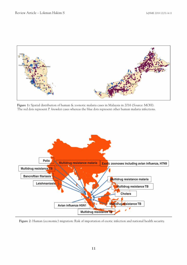
But our concern is the increasing trend of P. knowlesi infection. It is now the most dominant species infecting humans in Malaysia. The proportion is higher in Sarawak and Sabah where it constitutes to about two thirds of the total reported malaria cases annually. As such, an important question was raised, will simian malaria impact on the global programme to eliminate malaria? The infection can impact on global elimination if human-to-human transmission occurs, and so far we are lucky as there is no evidence of human-to-human transmission of P. knowlesi. Indeed, the infection foci for human and simian malaria are quite distinct even in Sabah and Sarawak where most of the cases are being reported (Figure 1).
With the restricted distribution of the natural host and the absence of compelling evidence of human-to-human transmission, the impact on the global elimination programme is considered as not significant at this stage. Furthermore, the distribution of P. knowlesi follows closely the distribution of it natural hosts which are the two macaques, Macaca nemestrina and M. fascicularis,
Review Article – Lokman Hakim S IeJSME 2018 12(3): 4-11
8
which are practically restricted to the Southeast Asian region.16 Despite the wide distribution of the natural host in the region, P. knowlesi in humans is practically reported only from Malaysia. There are imported cases from Indonesia and Thailand although the one imported from Papua New Guinea is a misnomer since the country is outside the normal distribution of the natural hosts. An imported case from Indonesia was also reported by Australia.17 This contention that simian malaria will not impact on the elimination of human malaria is further supported by the work of Imai et al., 2014. They described a comprehensive modelling study and it indicated that simian malaria should not be a major problem to local programme managers because existing strategies like the use of bed-net and early diagnosis and early treatment should be able to provide positive impact on the prevalence of the infection.18
However, the problem of simian malaria brought about a bigger challenge in managing the eco-system, in promoting the balance between socio-economic needs through commodity crop plantation and the impact on natural eco-system, which borders the overall national policy decision-making process. Nevertheless, at the population level, better understanding of the behaviour of the natural primate hosts as well as the mosquito vectors and the interaction with human behaviour in disease transmission, would provide better solutions to the problem, and this would entail a multi-disciplinary approach in the study design.
Currently the biggest challenge is with dengue. It is the most rapidly increasing infectious disease burden in world and also in Malaysia, rising exponentially over the years. In Malaysia, dengue outbreak is cyclical with the major outbreak occurring every 4-5 years. What is most obvious is that not only are the cycles getting closer, the peak of each outbreak cycle is getting higher and higher exponentially too, with the last highest peak in 2015 with more than 120,000 confirmed cases. And this cyclical pattern of dengue is occurring on a weekly basis and within a year, it is quite common to see two or more peaks of dengue outbreak. Selangor, the greater
Klang Valley for that matter, contributed about 60% of the total reported cases. Considering the limitation in access to diagnosis, the estimated minimum number of dengue cases is 2000 cases a week. We seem to be not able to suppress it any further, to such an extent that the MOH has been questioned for not doing the right thing.
The Future
With the current persistent challenges of tropical diseases of public health importance, elements of uncertainty and the unknown risk of emerging infections in the future and threat of cross border health security, it is reckoned that tropical medicine is still an important medical discipline which warrants continued investment in human resource development, research and development. Unlike malaria, dengue is still largely an unfinished agenda not only for Malaysia but also regionally and globally. With climate change and global warming, the risk of geographical spread into the temperate countries is real. In fact, sporadic local outbreaks in some of these countries are already occurring.
The real problem with dengue, unlike malaria, lies in the fact that we do not have enough effective tools to attack all the four components of the transmission dynamics, compounded by the super-efficient Aedes vector and the lackadaisical attitude of the community that contribute to the breeding of the mosquito. We do not have an effective vaccine to protect the host and the use of bed-net is impractical and ineffective. There is no drug to clear the infecting virus and the environment is difficult to manage because of the Aedes cryptic breeding behaviour and human behaviour in littering. A systemic review and meta-analysis evaluated the evidence of the effectiveness of vector control intervention and found that among others, there is strong evidence that community-based campaign can impact on vector abundance, moderate evidence that house screening can reduce vector abundance and no robust studies on the impact of fogging on dengue transmission.19
Review Article – Lokman Hakim S IeJSME 2018 12(3): 4-11
9
MOH continues to explore new technologies and methods in trying to find sustainable solutions to the dengue menace. Malaysia participated in the multi-centre Phase II/III clinical trial of candidate vaccine. Malaysia also experimented on transgenic mosquito but had to abandon it half-way through for various reasons. Currently the IMR with its international partner is conducting field trial on Wolbachia infected Aedes in the control of dengue. At the same time, the IMR is exploring the use of Sterile Insect Technique for the same purpose. In adopting any new intervention technology, it is important to be guided by the principle that it must be scientifically sound, feasible, practical and affordable to ensure sustainability over time.
The risk of another Nipah virus-like outbreak is real. The natural reservoir flight range, the fruit bats of the Pteropodidae family, is huge and expanding across continents. Recent studies showed that repeated introduction of the virus, prime the persistence and emergence of Nipah virus, refuting earlier hypothesis that the El-Nino phenomena drives the emergence of the virus. The seroprevalence rates of Hepanivirus infection among sampled fruit bats were also relatively high.20 In Bangladesh, the risk of human infection has been persistent and human cases and mortality have been reported almost every year for the last 15 years since it first introduction in 2001, two years after the only outbreak in Malaysia in 1999.21
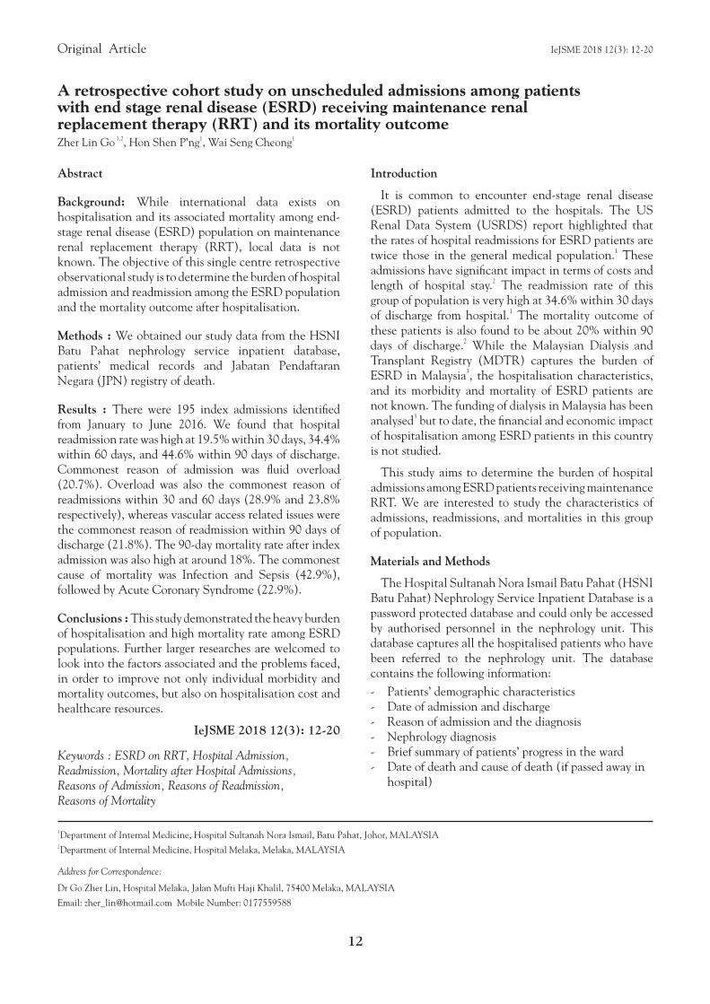
Malaysia being centrally located and a popular destination for economic migrants, both legal and illegal, is at high risk of importing exotic and severe tropical infections thus compromising its national health security (Figure 2). With an estimated number of 4 million illegal migrants, the risk is real. For example, we do have the vector for bancroftian filariasis which is Culex quinquefasciatus which is abundant in most urban areas and lymphatic filariasis is not in the list of infections screened under the Foreign Workers Medical Screening programme. MOH surveillance activities showed that microfilaraemia is prevalent among migrant workers from South Asia. Visceral leishmaniasis has
been diagnosed in an aboriginal patient several years ago in Malaysia. Importation of multi-drug resistant malaria and tuberculosis may cause a huge economic burden and compromise their elimination effort as well as may threaten national health security.
Conclusion
In conclusion, Malaysian scientists are still very actively engaging in tropical diseases research with significant impact globally as evidenced by some of the recent awards received. For her outstanding work on intestinal helminthiases, another much neglected tropical disease area, Prof Yvonne Lim, Deputy Dean (Research), Medical Faculty, University of Malaya and an active member of MSPTM, was listed by Nature as one of the Science Stars of East Asia in the recent News Feature. With two international accolades received by Malaysian researchers within the month by Dr Nazni and Prof Yvonne, it proves that Tropical Medicine research is still very much alive in Malaysia. Interestingly, the areas that brought fame to Malaysia recently are the two probably most unpopular fields – maggot and intestinal worms!
During her inaugural lecture as Fellow of the Academy of Science titled “Debunking the myth about gut worms by unlocking the secrets of gut microbiota”, Prof Yvonne has described a new frontier in understanding the relationship between our human co-existence which the much frowned creature called the gut worms. Who knows one day because of her team’s effort, we might be swallowing capsules of Ascaris lumbricoides and Trichuris trichuria eggs to treat inflammatory bowel diseases.
Review Article – Lokman Hakim S IeJSME 2018 12(3): 4-11
10
1. The Institute for Medical Research 1900-1950. Studies from the Institute for Medical Research, Federation of Malaya. Jubilee Volume No. 25. Field JW, Green R & Byron FE (ed).
2. Wright H. The malaria fevers of British Malaya. Studies of the Institute for Medical Research, Federated Malay State, No. 1, 1901.
3. Wright H. An enquiry into the aetiology and pathology of beri-beri. Studies of the Institute for Medical Research, Federated Malay State, No. 2., 1902
4. Sandosham AA and Thomas V. Malariology: With special reference to Malaya. National of Singapore University Press, 1983.
5. World Health Assembly. Resolution No. 50.29: Elimination of lymphatic filariasis as a public health problem. World Health Organisation, 1997.
6. Ramaiah KD & Ottesen EA. Progress and Impact of 13 Years of the Global Programme to Eliminate Lymphatic Filariasis on Reducing the Burden of Filarial Disease. PLoS Negl Trop Dis 2014; 8(11): e3319. DOI: 10.1371/journal.pntd.0003319.
7. Mak JW, PKC, Sim BKL and Liew LM. Brugia malayi and B. pahangi: Cultivation in vitro of infective larvae to the fourth and fifth stages. Exp Parasitol 1983; 55(2), 243-8.
8. Taylor LH, Latham SM, Woolhouse ME (2001). Risk factors for human disease emergence. Philos Trans R Soc Lond B: Biol Sci 2001; 356: 983–9.
9. Morse SS, Mazet JAK, Woolhouse M, Parrish CR, Carroll D, Karesh WB, Zambrana-Torrelio C, Lipkin WI and Daszak P. Prediction and prevention of the next pandemic zoonosis. Lancet 2012; 380: 1956–65.
10. Coker RJ, Hunter BM, Rudge JW, Liverani M and Hanvoravongchai P. Emerging infectious diseases in southeast Asia: regional challenges to control. Lancet 2011; 377: 599–609.
11. Pappas G, Papadimitriou P, Siozopoulou V, Christou L and Akritidis N. The globalization of leptospirosis: worldwide incidence and trends. Int J Infect Dis 2008;12: 351-7.
12. Ali MRM, Safiee AWM, Yusof NY, Fauzi MH, Yean CY and Ismail N. Isolation of Leptospira kmetyi from residential areas of patients with leptospirosis in Kelantan, Malaysia. J Infect Pub Hlth 2017; 11: 578-580.
13. Chua KB. Nipah virus outbreak in Malaysia. J Clin Virol 2002; 26: 265-75.
14. Lysaght T, Capps B, Bailey M, Bickford D, Coker R, Lederman Z, Watson S and Tambyah PA. Justice is the missing link in One Health: Results of a mixed methods study in an Urban City State. PLoS One 2017; 12(1): e0170967. doi: 10.1371/journal.pone.0170967.
15. New Straight Times On-line news portal. First rabies deaths in Malaysia in 20 years. Published 6 July, 2017.
16. World Health Organisation. Meeting Report: Expert consultation on Plasmodium knowlesi malaria to guide malaria elimination strategies. RS/2017/GE/05/(MYS). World Health Organization Regional Office for the Western Pacific Manila, Philippines, 2017.
17. Figtree M, Lee R, Bain L, Kennedy T, Mackertich S, Urban M, Cheng Q and Hudson BJ. Plasmodium knowlesi in human, Indonesian Borneo. Emerging Infect Dis 2010; 16: 672-4.
18. Imai N, White MT, Ghani AC and Drakeley CJ. Transmission and Control of Plasmodium knowlesi: A Mathematical Modelling Study. PLOS Neglected Trop Dis 2014; 8 (7): e2978
19. Olliaro P, Fouque F, Kroeger A, Bowman L, Velayudhan R, Santelli AC, Garcia D, Ramm RS, Sulaiman LH, et al. Improved tools and strategies for the prevention and control of arboviral diseases: A research-to-policy forum. PLOS Neglected Trop Dis 2018; https://doi.org/ 10.1371/journal.pntd.0005967
20. Pullium JRC, Epstein JH, Dushoff J, Rahman SA, Bunning M, Jamaluddin AA, Hyatt AD, Field HE, Dobson AP, Daszak P and Hepanivirus Ecology Research Group. Agricultural intensification, priming for persistence, and the emergence of Nipah virus: a lethal bat-borne zoonosis. J R Soc Interface 2011; doi:10.1098/rsif.2011.0223.
21. Hegde ST, Sazzad HSS, Hossain MJ, Alam MUI, Kenah E, Daszak P, Rollin P, Rahman M, Luby SP and Gurley ES. Investigating Rare Risk Factors for Nipah Virus in Bangladesh: 2001–2012. Ecohealth 2016; 13(4): 720–8. doi:10.1007/s10393-016-1166-0.
REFERENCES
Review Article – Lokman Hakim S IeJSME 2018 12(3): 4-11
11
Figure 1: Spatial distribution of human & zoonotic malaria cases in Malaysia in 2016 (Source: MOH). The red dots represent P. knowlesi cases whereas the blue dots represent other human malaria infections.
Figure 2: Human (economic) migration: Risk of importation of exotic infection and national health security.
PolioMultidrug resistance malaria
Multidrug resistance malaria
Multidrug resistance TB
Multidrug resistance TB
Multidrug resistance TB
Multidrug resistance TB
Avian influenza H5N1
Leishmaniasis
Bancroftian filariasis
Cholera
Exotic zoonoses including avian influenza, H7N9
12
1Department of Internal Medicine, Hospital Sultanah Nora Ismail, Batu Pahat, Johor, MALAYSIA2Department of Internal Medicine, Hospital Melaka, Melaka, MALAYSIA
Address for Correspondence:
Dr Go Zher Lin, Hospital Melaka, Jalan Mufti Haji Khalil, 75400 Melaka, MALAYSIA
Email: [email protected] Mobile Number: 0177559588
A retrospective cohort study on unscheduled admissions among patientswith end stage renal disease (ESRD) receiving maintenance renalreplacement therapy (RRT) and its mortality outcomeZher Lin Go 1,2, Hon Shen P’ng1, Wai Seng Cheong1
Original Article IeJSME 2018 12(3): 12-20
Abstract
Background: While international data exists on hospitalisation and its associated mortality among end-stage renal disease (ESRD) population on maintenance renal replacement therapy (RRT), local data is not known. The objective of this single centre retrospective observational study is to determine the burden of hospital admission and readmission among the ESRD population and the mortality outcome after hospitalisation.
Methods : We obtained our study data from the HSNI Batu Pahat nephrology service inpatient database, patients’ medical records and Jabatan Pendaftaran Negara (JPN) registry of death.
Results : There were 195 index admissions identified from January to June 2016. We found that hospital readmission rate was high at 19.5% within 30 days, 34.4% within 60 days, and 44.6% within 90 days of discharge. Commonest reason of admission was fluid overload (20.7%). Overload was also the commonest reason of readmissions within 30 and 60 days (28.9% and 23.8% respectively), whereas vascular access related issues were the commonest reason of readmission within 90 days of discharge (21.8%). The 90-day mortality rate after index admission was also high at around 18%. The commonest cause of mortality was Infection and Sepsis (42.9%), followed by Acute Coronary Syndrome (22.9%).
Conclusions : This study demonstrated the heavy burden of hospitalisation and high mortality rate among ESRD populations. Further larger researches are welcomed to look into the factors associated and the problems faced, in order to improve not only individual morbidity and mortality outcomes, but also on hospitalisation cost and healthcare resources.
IeJSME 2018 12(3): 12-20
Keywords : ESRD on RRT, Hospital Admission, Readmission, Mortality after Hospital Admissions, Reasons of Admission, Reasons of Readmission,Reasons of Mortality
Introduction
It is common to encounter end-stage renal disease (ESRD) patients admitted to the hospitals. The US Renal Data System (USRDS) report highlighted that the rates of hospital readmissions for ESRD patients are twice those in the general medical population.1 These admissions have significant impact in terms of costs and length of hospital stay.2 The readmission rate of this group of population is very high at 34.6% within 30 days of discharge from hospital.1 The mortality outcome of these patients is also found to be about 20% within 90 days of discharge.2 While the Malaysian Dialysis and Transplant Registry (MDTR) captures the burden of ESRD in Malaysia3, the hospitalisation characteristics, and its morbidity and mortality of ESRD patients are not known. The funding of dialysis in Malaysia has been analysed3 but to date, the financial and economic impact of hospitalisation among ESRD patients in this country is not studied.
This study aims to determine the burden of hospital admissions among ESRD patients receiving maintenance RRT. We are interested to study the characteristics of admissions, readmissions, and mortalities in this group of population.
Materials and Methods
The Hospital Sultanah Nora Ismail Batu Pahat (HSNI Batu Pahat) Nephrology Service Inpatient Database is a password protected database and could only be accessed by authorised personnel in the nephrology unit. This database captures all the hospitalised patients who have been referred to the nephrology unit. The database contains the following information:- Patients’ demographic characteristics- Date of admission and discharge- Reason of admission and the diagnosis- Nephrology diagnosis- Brief summary of patients’ progress in the ward- Date of death and cause of death (if passed away in hospital)
Original Article - Zher Lin Go, Hon Shen P’ng, Wai Seng Cheong IeJSME 2018 12(3): 12-20
13
We identified patients in the above database who were admitted to HSNI Batu Pahat during January to June 2016, with follow up period of 90 days after index admission. Thus, we analysed the database until the end of September 2016, ninety days after the last patient was recruited at the end of June 2016. We included ESRD patients with unscheduled admissions and who were on maintenance RRT. We excluded non-ESRD patients, patients who were not on maintenance RRT, and patients with scheduled admissions (e.g. admission for an elective imaging procedure).
Demographic characteristics (age, gender, race), traceable co-morbidities, date of admissions and discharges, length of stay (LOS), reasons of admission, reasons of readmissions, dates and causes of death (if passed away in hospital) were extracted from the database. When further information was required, patients’ medical notes were traced from the HSNI Batu Pahat Medical Record Unit, reviewed, and returned.
While the HSNI Batu Pahat Nephrology Service Inpatient Database captured inpatient mortality, there were patients who died at home or in other hospitals. Thus, we officially contacted Jabatan Pendaftaran Negeri Johor (JPN of Johor state), and through the Registry of Death, we obtained our studied populations’ survival status, their dates of death and causes of death.
Data collected were categorised. Numerators and denominators were determined, followed by calculation of percentages of individual causes of admissions, readmissions and mortality. Readmission rates and mortalities rates were calculated by dividing the numbers of readmissions (and mortalities), by the number of index admissions. Cumulative event rates for 30-day, 60-day and 90-day were analysed and plotted.
Mean values of “days to readmission” and “days to mortality” were calculated by dividing the total number of “days to readmissions” (and “days to mortality”), by the total number of readmissions (and mortalities).
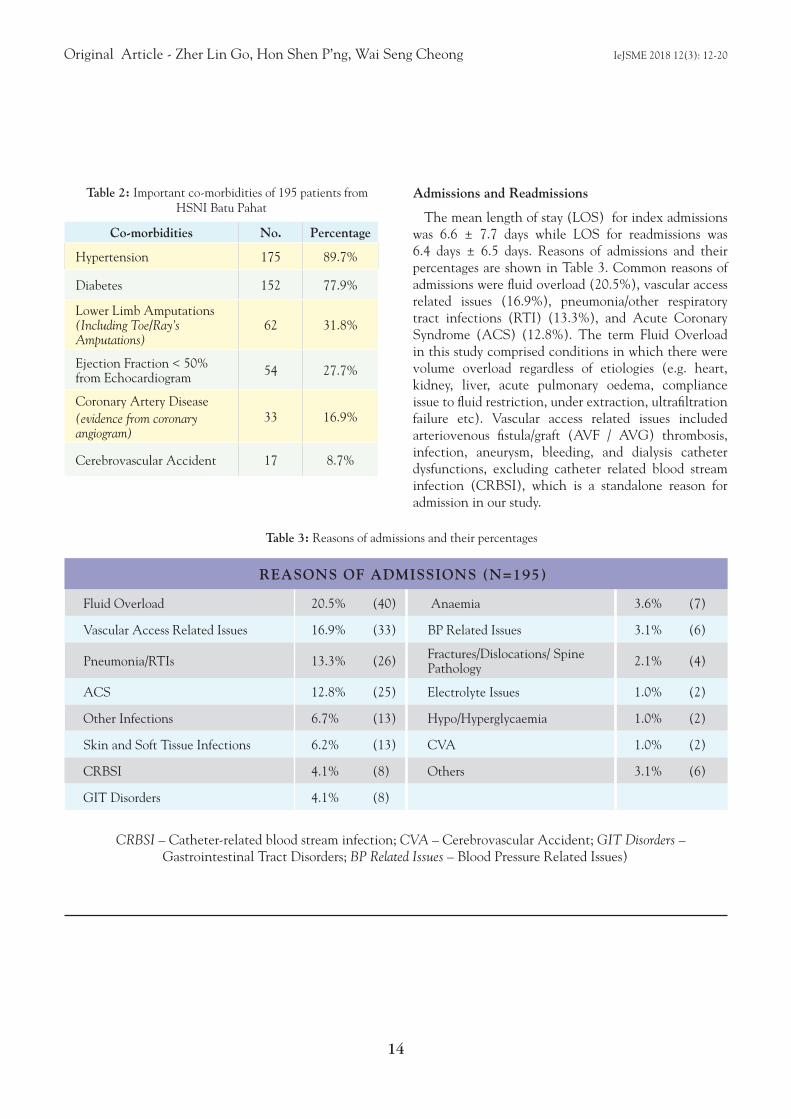
Results
There were 195 index admissions (195 patients) identified from January to June 2016 which met our study criteria. The demographic data is shown in Table 1. Mean age was 58.5 ± 13.3 yrs. The majority of these patients were on maintenance haemodialysis (89.2%) as the mode of RRT. Table 2 shows some of the traceable important co-morbidities of these patients.
Table 1: Demographic Data of 195 patients from HSNI Batu Pahat
N = 195No. Percentage
SEX
Male 98 50.3%
Female 97 49.7%ETHNICITY
Malay 148 75.9%
Chinese 42 21.5%
Indian 3 1.5%
Others 2 1.0%MODE OF RRT
Haemodialysis 174 89.2%
Peritoneal Dialysis 18 9.2%
Transplant 3 1.5%AGE GROUP
19 or below 1 0.5%
20 – 29 9 4.6%
30 – 39 10 5.1%
40 – 49 28 14.4%
50 – 59 47 24.1%
60 – 69 65 33.3%
70 – 79 31 15.9%
80 or above 4 2.1%
Mean Age 58.5 ± SD 13.3
Original Article - Zher Lin Go, Hon Shen P’ng, Wai Seng Cheong IeJSME 2018 12(3): 12-20
14
Table 2: Important co-morbidities of 195 patients from HSNI Batu Pahat
Admissions and Readmissions
The mean length of stay (LOS) for index admissions was 6.6 ± 7.7 days while LOS for readmissions was 6.4 days ± 6.5 days. Reasons of admissions and their percentages are shown in Table 3. Common reasons of admissions were fluid overload (20.5%), vascular access related issues (16.9%), pneumonia/other respiratory tract infections (RTI) (13.3%), and Acute Coronary Syndrome (ACS) (12.8%). The term Fluid Overload in this study comprised conditions in which there were volume overload regardless of etiologies (e.g. heart, kidney, liver, acute pulmonary oedema, compliance issue to fluid restriction, under extraction, ultrafiltration failure etc). Vascular access related issues included arteriovenous fistula/graft (AVF / AVG) thrombosis, infection, aneurysm, bleeding, and dialysis catheter dysfunctions, excluding catheter related blood stream infection (CRBSI), which is a standalone reason for admission in our study.
Co-morbidities No. Percentage
Hypertension 175 89.7%
Diabetes 152 77.9%
Lower Limb Amputations(Including Toe/Ray’s Amputations)
62 31.8%
Ejection Fraction < 50% from Echocardiogram 54 27.7%
Coronary Artery Disease(evidence from coronary angiogram)
33 16.9%
Cerebrovascular Accident 17 8.7%
REASONS OF ADMISSIONS (N=195)
Fluid Overload 20.5% (40) Anaemia 3.6% (7)
Vascular Access Related Issues 16.9% (33) BP Related Issues 3.1% (6)
Pneumonia/RTIs 13.3% (26) Fractures/Dislocations/ Spine Pathology 2.1% (4)
ACS 12.8% (25) Electrolyte Issues 1.0% (2)
Other Infections 6.7% (13) Hypo/Hyperglycaemia 1.0% (2)
Skin and Soft Tissue Infections 6.2% (13) CVA 1.0% (2)
CRBSI 4.1% (8) Others 3.1% (6)
GIT Disorders 4.1% (8)
Table 3: Reasons of admissions and their percentages
CRBSI – Catheter-related blood stream infection; CVA – Cerebrovascular Accident; GIT Disorders – Gastrointestinal Tract Disorders; BP Related Issues – Blood Pressure Related Issues)
Original Article - Zher Lin Go, Hon Shen P’ng, Wai Seng Cheong IeJSME 2018 12(3): 12-20
15
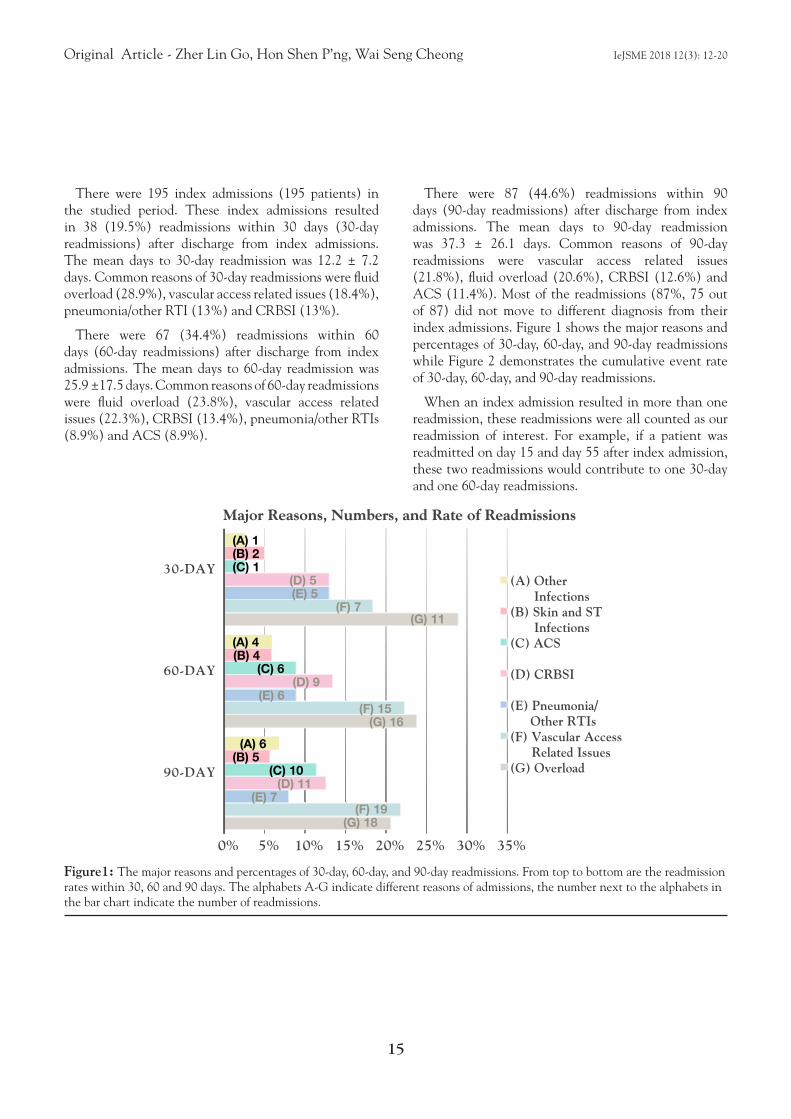
There were 195 index admissions (195 patients) in the studied period. These index admissions resulted in 38 (19.5%) readmissions within 30 days (30-day readmissions) after discharge from index admissions. The mean days to 30-day readmission was 12.2 ± 7.2 days. Common reasons of 30-day readmissions were fluid overload (28.9%), vascular access related issues (18.4%), pneumonia/other RTI (13%) and CRBSI (13%).
There were 67 (34.4%) readmissions within 60 days (60-day readmissions) after discharge from index admissions. The mean days to 60-day readmission was 25.9 ±17.5 days. Common reasons of 60-day readmissions were fluid overload (23.8%), vascular access related issues (22.3%), CRBSI (13.4%), pneumonia/other RTIs (8.9%) and ACS (8.9%).
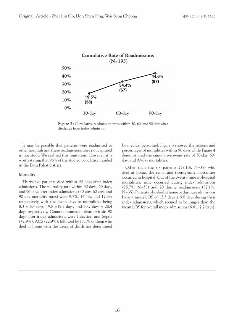
There were 87 (44.6%) readmissions within 90 days (90-day readmissions) after discharge from index admissions. The mean days to 90-day readmission was 37.3 ± 26.1 days. Common reasons of 90-day readmissions were vascular access related issues (21.8%), fluid overload (20.6%), CRBSI (12.6%) and ACS (11.4%). Most of the readmissions (87%, 75 out of 87) did not move to different diagnosis from their index admissions. Figure 1 shows the major reasons and percentages of 30-day, 60-day, and 90-day readmissions while Figure 2 demonstrates the cumulative event rate of 30-day, 60-day, and 90-day readmissions.
When an index admission resulted in more than one readmission, these readmissions were all counted as our readmission of interest. For example, if a patient was readmitted on day 15 and day 55 after index admission, these two readmissions would contribute to one 30-day and one 60-day readmissions.
(G) 18
(G) 16
(G) 11
(F) 19
(F) 15
(F) 7
(E) 7
(E) 6
(E) 5
(D) 11
(D) 9
(D) 5
(C) 10
(C) 6
(C) 1
(B) 5
(B) 4
(B) 2
(A) 6
(A) 4
(A) 1
(A) Other Infections
(B) Skin and ST Infections
(C) ACS
(D) CRBSI
(E) Pneumonia/Other RTIs
(F) Vascular Access Related Issues
(G) Overload
Major Reasons, Numbers, and Rate of Readmissions
0% 5% 10% 15% 20% 25% 30% 35%
90-DAY
60-DAY
30-DAY
Figure1: The major reasons and percentages of 30-day, 60-day, and 90-day readmissions. From top to bottom are the readmission rates within 30, 60 and 90 days. The alphabets A-G indicate different reasons of admissions, the number next to the alphabets in the bar chart indicate the number of readmissions.
Original Article - Zher Lin Go, Hon Shen P’ng, Wai Seng Cheong IeJSME 2018 12(3): 12-20
16
It may be possible that patients were readmitted to other hospitals and these readmissions were not captured in our study. We realised this limitation. However, it is worth stating that 96% of the studied population resided in the Batu Pahat district.
Mortality
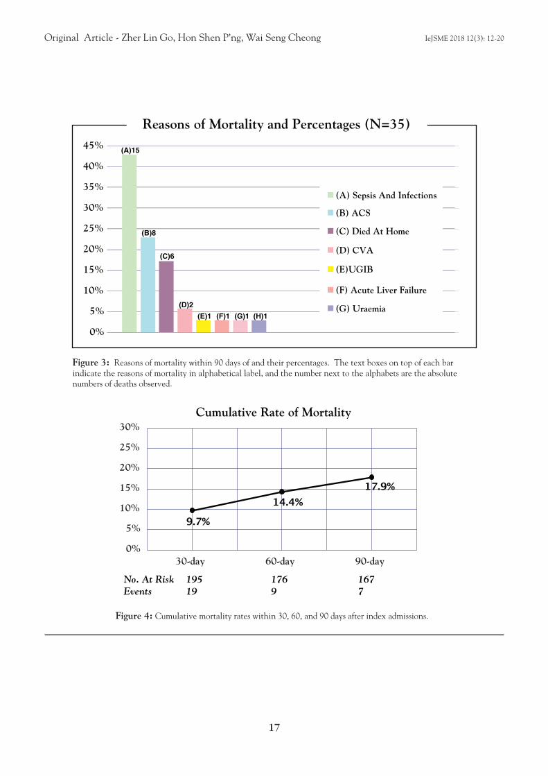
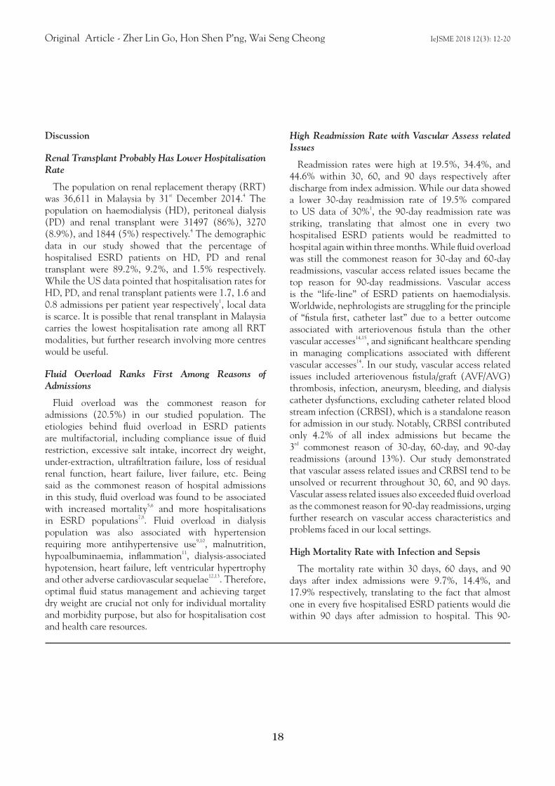
Thirty-five patients died within 90 days after index admissions. The mortality rate within 30 days, 60 days, and 90 days after index admissions (30-day, 60-day, and 90-day mortality rates) were 9.7%, 14.4%, and 17.9% respectively with the mean days to mortalities being 8.3 ± 6.4 days, 19.8 ±19.2 days, and 30.7 days ± 28.4 days respectively. Common causes of death within 90 days after index admissions were Infection and Sepsis (42.9%), ACS (22.9%), followed by 17.1% of them who died at home with the cause of death not determined
by medical personnel. Figure 3 showed the reasons and percentages of mortalities within 90 days while Figure 4 demonstrated the cumulative event rate of 30-day, 60-day, and 90-day mortalities.
Other than the six patients (17.1%, N=35) who died at home, the remaining twenty-nine mortalities occurred in hospital. Out of the twenty-nine in-hospital mortalities, nine occurred during index admissions (25.7%, N=35) and 20 during readmissions (57.1%, N=35). Patients who died at home or during readmissions have a mean LOS of 12.3 days ± 9.8 days during their index admissions, which seemed to be longer than the mean LOS for overall index admissions (6.6 ± 7.7 days).
19.5%(38)
34.4%(67)
44.6%(87)
Cumulative Rate of Readmissions (N=195)
0%
10%
20%
30%
40%
50%
30-day 60-day 90-day
Figure 2: Cumulative readmission rates within 30, 60, and 90 days after discharge from index admission.
Original Article - Zher Lin Go, Hon Shen P’ng, Wai Seng Cheong IeJSME 2018 12(3): 12-20
17
0%
5%
10%
15%
20%
25%
30%
35%
40%
45%
(H)1(F)1 (G)1(E)1(D)2
(C)6
(B)8
(A)15
(A) Sepsis And Infections
(B) ACS
(C) Died At Home
(D) CVA
(E)UGIB
(F) Acute Liver Failure
(G) Uraemia
Reasons of Mortality and Percentages (N=35)
Figure 3: Reasons of mortality within 90 days of and their percentages. The text boxes on top of each bar indicate the reasons of mortality in alphabetical label, and the number next to the alphabets are the absolute numbers of deaths observed.
Figure 4: Cumulative mortality rates within 30, 60, and 90 days after index admissions.
9.7%
14.4%17.9%
No. At Risk 195 176 167Events 19 9 7
0%
5%
10%
15%
20%
25%
30%
30-day 60-day 90-day
Cumulative Rate of Mortality
Original Article - Zher Lin Go, Hon Shen P’ng, Wai Seng Cheong IeJSME 2018 12(3): 12-20
18
Discussion
Renal Transplant Probably Has Lower Hospitalisation Rate
The population on renal replacement therapy (RRT) was 36,611 in Malaysia by 31st December 2014.4 The population on haemodialysis (HD), peritoneal dialysis (PD) and renal transplant were 31497 (86%), 3270 (8.9%), and 1844 (5%) respectively.4 The demographic data in our study showed that the percentage of hospitalised ESRD patients on HD, PD and renal transplant were 89.2%, 9.2%, and 1.5% respectively. While the US data pointed that hospitalisation rates for HD, PD, and renal transplant patients were 1.7, 1.6 and 0.8 admissions per patient year respectively1, local data is scarce. It is possible that renal transplant in Malaysia carries the lowest hospitalisation rate among all RRT modalities, but further research involving more centres would be useful.
Fluid Overload Ranks First Among Reasons of Admissions
Fluid overload was the commonest reason for admissions (20.5%) in our studied population. The etiologies behind fluid overload in ESRD patients are multifactorial, including compliance issue of fluid restriction, excessive salt intake, incorrect dry weight, under-extraction, ultrafiltration failure, loss of residual renal function, heart failure, liver failure, etc. Being said as the commonest reason of hospital admissions in this study, fluid overload was found to be associated with increased mortality5,6 and more hospitalisations in ESRD populations7,8. Fluid overload in dialysis population was also associated with hypertension requiring more antihypertensive use9,10, malnutrition, hypoalbuminaemia, inflammation11, dialysis-associated hypotension, heart failure, left ventricular hypertrophy and other adverse cardiovascular sequelae12,13. Therefore, optimal fluid status management and achieving target dry weight are crucial not only for individual mortality and morbidity purpose, but also for hospitalisation cost and health care resources.
High Readmission Rate with Vascular Assess related Issues
Readmission rates were high at 19.5%, 34.4%, and 44.6% within 30, 60, and 90 days respectively after discharge from index admission. While our data showed a lower 30-day readmission rate of 19.5% compared to US data of 30%1, the 90-day readmission rate was striking, translating that almost one in every two hospitalised ESRD patients would be readmitted to hospital again within three months. While fluid overload was still the commonest reason for 30-day and 60-day readmissions, vascular access related issues became the top reason for 90-day readmissions. Vascular access is the “life-line” of ESRD patients on haemodialysis. Worldwide, nephrologists are struggling for the principle of “fistula first, catheter last” due to a better outcome associated with arteriovenous fistula than the other vascular accesses14,15, and significant healthcare spending in managing complications associated with different vascular accesses14. In our study, vascular access related issues included arteriovenous fistula/graft (AVF/AVG) thrombosis, infection, aneurysm, bleeding, and dialysis catheter dysfunctions, excluding catheter related blood stream infection (CRBSI), which is a standalone reason for admission in our study. Notably, CRBSI contributed only 4.2% of all index admissions but became the 3rd commonest reason of 30-day, 60-day, and 90-day readmissions (around 13%). Our study demonstrated that vascular assess related issues and CRBSI tend to be unsolved or recurrent throughout 30, 60, and 90 days. Vascular assess related issues also exceeded fluid overload as the commonest reason for 90-day readmissions, urging further research on vascular access characteristics and problems faced in our local settings.
High Mortality Rate with Infection and Sepsis
The mortality rate within 30 days, 60 days, and 90 days after index admissions were 9.7%, 14.4%, and 17.9% respectively, translating to the fact that almost one in every five hospitalised ESRD patients would die within 90 days after admission to hospital. This 90-
Original Article - Zher Lin Go, Hon Shen P’ng, Wai Seng Cheong IeJSME 2018 12(3): 12-20
19
day mortality rate was similar to US data of 20%2. The commonest causes of mortality within 90 days in our study were Infection and Sepsis (42.9%), ACS (22.9%), followed by 17.1% of them who died at home with the cause of death not determined by medical personnel. This result mirrored that of Malaysian Dialysis and Transplant Registry (MDTR) data on causes of mortality among ESRD patients, except that CV deaths ranked first in MDTR, followed by Sepsis, and Died at Home16. Sepsis in ESRD population carries a 50-fold higher risk of mortality compared to the general population17. A single-centre study found a 25.6% mortality rate at 28 days after discharge from hospital for an admission of sepsis among ESRD patients18. In addition to high mortality rate, the standard protocol for management of sepsis has its limitations in ESRD populations and has to be applied cautiously19, making the management of sepsis in this group of population challenging. The probable explanation for Infection and Sepsis as the number one cause of death in our studied population was that ESRD patients are less immunocompetent and prone to infection and sepsis after hospital admissions. Exposure to pathogens in hospitals and dialysis centres, dialyser reuse, dialysis catheter use, invasive procedures in hospitals, requirement of central venous cannulation during hospital stay, underlying diabetes, age factor, and deterioration of nutritional status after hospital admission are the other probable explanations20,21,22. The mortality data in our study highlighted the need to look into factors in minimising infection risks in ESRD population, and in another way emphasises the importance of strict adherence to infection control strategies.
Conclusion
Our study found that fluid overload, vascular access related issues, respiratory tract infections and coronary events are the commonest reasons of admissions among ESRD patients receiving maintenance RRT. Rate of readmission is high at almost one in every two patients within 90 days of discharge. Vascular access related issues tends to be unsolved and becomes the commonest reason of readmission at 90 days.
Rate of mortality within 90 days of admission to hospital among this group of population is high at almost 18%. Common causes of death within 90 days of discharge are Infection and Sepsis, ACS, and “Died at Home”. The results of this single centre study has its limitations if we generalise the data in Malaysia. Nevertheless, certain results on admissions, readmissions, and mortality prompt future larger research in this country to look into the problems faced, as they carry significance not only on patients’ outcome, but also on health care cost and resources.
Acknowledgement
We would like to express our gratitude to Jabatan Pendaftaran Negeri Johor for their kind assistance.
REFERENCES
1. Saran R, Robinson B, Abbott KC, et al. US Renal Data System 2016 Annual Data Report: epidemiology of kidney disease in the United States. Am J Kidney Dis. 2017; 69(3)(suppl 1): S1-S688.
2. Kshirsagar AV, Hogan SL, Mandelkehr L and Falk RJ. Length of stay and costs for hospitalized hemodialysis patients: nephrologists versus internists. J Am Soc Nephrol 2000; 11: 1526–33.
3. Chapter 2, Dialysis in Malaysia, 22nd Report of the Malaysian Dialysis and Transplant Registry 2014.
4. Chapter 1, All Renal Replacement Therapy in Malaysia, 22nd Report of the Malaysian Dialysis and Transplant Registry 2014.
5. Zoccali C, Moissl U, Chazot C, Mallamaci F, Tripepi G, Arkossy O, Wabel P, Stuard S. Chronic Fluid Overload and Mortality in ESRD. J Am Soc Nephrol. 2017; 28(8): 2491-7.
6. Wizemann V, Wabel P, Chamney P, Zaluska W, Moissl U, Rode C, Malecka-Masalska T, Marcelli D. The mortality risk of overhydration in haemodialysis patients. Nephrol Dial Transplant. 2009; 24: 1574-9.
7. Goldstein SL, Smith CM, Currier H. Noninvasive interventions to decrease hospitalization and associated costs for paediatric patients receiving hemodialysis. J Am Soc Nephrol. 2003; 14: 2127-31.
8. Rodriguez HJ, Domenici R, Diroll A, Goykhman I. Assessment of dry weight by monitoring changes in blood volume during hemodialysis using Crit-Line. Kidney Int. 2005; 68: 854-61.
9. Wabel P, Moissl U, Chamney P, Jirka T, Machek P, Ponce P, Taborsky P, Tetta C, Velasco N, Vlasak J, Zaluska W, Wizemann V. Towards improved cardiovascular management: the necessity of combining blood pressure and fluid overload. Nephrol Dial Transplant. 2008; 23: 2965-71.
Original Article - Zher Lin Go, Hon Shen P’ng, Wai Seng Cheong IeJSME 2018 12(3): 12-20
20
10. Agarwal R, Alborzi P, Satyan S, Light RP. Dry-weight reduction in hypertensive hemodialysis patients (DRIP): a randomized, controlled trial. Hypertension. 2009; 53: 500-7.
11. Demirci MS, et al. Relation between malnutrition, inflammation, atherosclerosis and volume status: the usefulness of bioimpedence in peritoneal dialysis patients. Nephrol Dial Transplant. 2011; 2626: 1708-16.
12. Wizemann V, Schilling M. Dilemma of assessing volume state-the use and limitations of a clinical score. Nephrol Dial transplant, 1995; 10: 2114-7.
13. Wizemann V, Leibinger A, Mueller K, et al. Influence of hydration state on plasma volume changes during ultrafiltration, Artif Organs 1995; 19: 416-9.
14. Charmaine E. Lok, Robert Foley. Vascular Access Morbidity and Mortality: Trends of the Last Decade. Clin J Am Soc Nephrol. 2013; 8:1213-9. doi: 10.2215/CJN.01690213.
15. Toshikazu Ozeki, Hideaki Shimizu, Yoshiro Fujita, Daijo Inaguma, Shoichi Maruyama, Yukako Ohyama, Shun Minatoguchi, Yukari Murai, Maho Terashita, and Tomoki Tagaya. The Type of Vascular Access and the Incidence of Mortality in Japanese Dialysis Patients. Intern Med. 2017; 56: 481–5.
16. Chapter 3, Death and Survival on Dialysis, 22nd Report of the Malaysian Dialysis and Transplant Registry Year 2014.
17. Sarnak MJ, Jaber BL. Mortality caused by sepsis in patients with end-stage renal disease caompared to general population. Kidney Int. 2000; 58: 1758-64.
18. Gilbert Abou Dagher, Elie Harmouche, Elsy Jabbour, Rana Bachir, Dina Zebian, and Ralphe Bou Chebl. Sepsis in hemodialysis patients. BMC Emerg Med. 2015; 15: 30.
19. Jason J. Lee, Sandra L. Taylor, Jason Y. Adams. Fluid Resuscitation and Mortality in Sepsis with End-Stage Renal Disease. American Journal of Respiratory and Critical Care Medicine 2017;195: A5020.
20. Wang HE, Gamboa C, Warnock DG, Muntner P. Chronic kidney disease and risk of death from infection. Am J Nephrol. 2011;34: 330-6.
21. Powe NR, Jaar B, Furth SL, Hermann J, Briggs W. Septicemia in dialysis patients: incidence, risk factors, and prognosis. Kidney Int. 1999; 55:1081-90.
22. Hoen B, Paul-Dauphin A, Hestin D, Kessler M. EPIBACDIAL: a multicenter prospective study of risk factors for bacteremia in chronic hemodialysis patients. J Am Soc Nephrol. 1998; 9: 869-76.
21
1Division of Nursing, School of Health Sciences, International Medical University,No. 126, Jalan Jalil Perkasa 19, Bukit Jalil, 57000 Kuala Lumpur, MALAYSIA2School of Medicine, International Medical University, No. 126, Jalan Jalil Perkasa 19, Bukit Jalil, 57000 Kuala Lumpur, MALAYSIA
Address for Correspondence:
Dr Wei Fern Siew, School of Medicine, International Medical University, No. 126, Jalan Jalil Perkasa 19, Bukit Jalil, 57000, Kuala Lumpur, MALAYSIA E-mail: [email protected]
Perception towards role in psychosocial care among theregistered nurses in a private hospital in Kuala Lumpur, MalaysiaPei Khim Lee1, Wei Fern Siew2, Wai Mun Tang1
Original Article IeJSME 2018 12(3): 21-29
Abstract
Background: Psychosocial care remains an important component in holistic care nursing and is crucial for patients’ recovery outcomes.
Objective: The purpose of the study was to determine nurses’ perception towards their role in psychosocial care.
Methodology: The research design was descriptive and cross-sectional. Nurses’ Role in Psychosocial Care Questionnaire (NRPCQ) was used for data collection and approximately 110 registered nurses (response rate = 52.38%) participated in the study via convenience sampling. Descriptive and inferential statistics, Mann-Whitney U test were used for data analysis.
Results: In general, nurses’ perception towards their role in providing psychosocial care was positive (M = 73.71, SD ± 12.20). Items on “demonstrating warmth and friendliness by smiling” (M = 3.92, SD ± 0.28); and “explaining nursing procedures or interventions to the patient” (M = 3.88, SD ± 0.32) were rated most positive. Nevertheless, items on “referring patients to other health care team members” (M = 3.32, SD ± 0.83), and “discussing with patient and patient’s family regarding planned care” (M = 3.44, SD ± 0.69) were rated the least positive. The Mann-Whitney U test analysis revealed significant association between nurses’ age and perception towards their role in psychosocial care (p = 0.025), in which the older nurses have a more positive perception towards their role in psychosocial care than the younger nurses.
Conclusion: The findings highlighted some important gaps in the practice of psychosocial care among the registered nurses. The information serves as a baseline for the planning and implementing of relevant strategies in enhancing nurses’ role in psychosocial care provision.
IeJSME 2018 12(3): 21-29
Keywords: Perception, role, psychosocial care, registered nurses, hospital
Introduction
Psychosocial care involves the provision of psychological, social and spiritual care to patients and their family members. As every individual enters the hospital, he or she will respond to the stress of illness in a unique way. Henceforth, providing emotional and social support helped to protect the hospitalised patient from undesirable emotional breakdown due to physical condition in relation to the intimidating environment (Chivukula, Hariharan, Rana, Thomas & Swain, 2014). Furthermore, the psychosocial support is crucial in boosting patients’ confidence, and thus, reducing the stress of illness, giving the patient time to think through and decide the treatment options (Chivukula et al., 2014).
Legg (2010) also reinforced that effective psychosocial care comes down to good communication skills, both verbal and non-verbal. Some examples which include listening to patients’ problems, providing explanation, and giving appropriate advice. In addition, supporting individuals going through illnesses through one-to-one interaction, and being empathetic were deemed to be the utmost basic support (Legg, 2010). On another note, qualitative findings by Attree (2001) revealed that nurses who were friendly, sociable, approachable, and demonstrate kindness and sensitivity were highly appreciated by both patients and caregivers/ families. A smile is a simple action but this warm personality was rated the most important among the patients and their family members (Attree, 2001).
In general, past studies have affirmed the importance of psychosocial care provision among the nurses. Ausserhofer et al. (2014) had conducted a large scale study among the European nurses across twelve countries. The study found that the most frequent nursing care activities which “left undone” were “providing emotional care/ talking with patients” (53%) and “‘educating patients and families” (41%) which clearly reflected the lesser priority of providing psychosocial care among the nurses. Instead, nurses placed high priority in technical
Original Article - Pei Khim Lee, Wei Fern Siew, Wai Mun Tang IeJSME 2018 12(3): 21-29
22
roles particularly, in the aspects of administrating treatment and procedure (Ausserhofer et al., 2014; Ball et al., 2013). Similarly, a separate study conducted by Pavlish and Ceronsky (2009) revealed that the oncology nurses perceived that provision of emotional and palliative care should only be rendered to end-of-life patients. In addition, 89.5% of the Chinese nurses reported that the provision of emotional care was the role of the family caregivers instead of nurses (Jiang, Ma, Li & Gu, 2015). Nonetheless, information pertaining to Malaysian nurses’ perceptions towards their role in providing psychosocial care remained scarce. Thus, the purpose of the study was to determine Malaysian nurses’ perception towards their role in psychosocial care. The findings of the study can be used as baseline information to facilitate interventions in strengthening the provision of psychosocial care among nurses.
Methodology
Research design
The research design was quantitative, cross-sectional and descriptive. The study was conducted in a private hospital in Kuala Lumpur, Malaysia. The hospital provides treatments for patients with emergencies and referral for medical specialties. The study duration was between November and December 2016.
Study participants
Participants of the study were full-time Malaysian registered nurses who were working in the in-patient departments. The entire population of the study (N = 270) was invited to participate in the study. Nevertheless, 110 (response rate 52%) participants responded to the questionnaire. The slightly low response rate was due to the data collection period in which relatively high proportion of registered nurses took leave for festive and year-end holidays. Registered nurses who held managerial and supervisory positions such as ward managers, deputy ward managers, and clinical instructors were excluded from the study in view of the differences in the nature of their job scope.
In general, the average age of the participants was 27.08 years old (SD ± 5.51). As shown in Table 1, 52.70% of nurses were single while 47.30% of nurses were married. Majority of the nurses were diploma holders (71.80%) while the remaining possessed qualifications higher than diploma level (28.20%). In terms of working areas, almost half of the participants were working in the medical surgical wards (42.70%), whereas the remaining of the nurses (53.70%) were working in the specialised areas such as intensive care unit (ICU), coronary care unit (CCU), high dependency ward (HDU), pediatric ward, emergency department, orthopedic ward and labour ward. The average number of years of working experience for the nurses was 4.78 (SD ± 4.14). Furthermore, at least half of the recruited study participants rated moderate for their workload (52.70%), while the other nurses rated their workload as satisfactory (4.50%), heavy (36.40%), extremely heavy (6.40%). On another note, the average double shift days in a month for the staff nurses was 4.62 (SD = 1.77).
Instrument
The instrument used for the study was the Nurses’ Role in Psychosocial Care Questionnaire (NRPCQ). NRPCQ is a self-developed questionnaire based on few literature (Lu, While & Barriball, 2008; Jacob, McKenna & D’Amore, 2015). In addition, the items of the questionnaire were aligned with the competency standards stipulated in the code of professional conduct for nurses (Nursing Board Malaysia, 1998). The response format was rated using a four-points Likert scale: 1 = sometimes/year, 2 = sometimes/month, 3 = sometimes/week, and 4 = almost every day. Nurses’ perceptions towards their role in psychosocial care was reflected by the aggregated score on the questionnaire. The score ranged between 20 and 80. A higher score is suggestive of more positive perception.
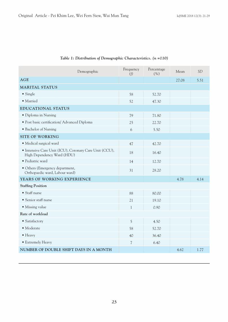
Original Article - Pei Khim Lee, Wei Fern Siew, Wai Mun Tang IeJSME 2018 12(3): 21-29
23
DemographicFrequency
(f)Percentage
(%)Mean SD
AGE 27.08 5.51
MARITAL STATUS
• Single 58 52.70
• Married 52 47.30
EDUCATIONAL STATUS
• Diploma in Nursing 79 71.80
• Post basic certification/ Advanced Diploma 25 22.70
• Bachelor of Nursing 6 5.50
SITE OF WORKING
• Medical surgical ward 47 42.70
• Intensive Care Unit (ICU), Coronary Care Unit (CCU), High Dependency Ward (HDU)
18 16.40
• Pediatric ward 14 12.70
• Others (Emergency department, Orthopaedic ward, Labour ward)
31 28.20
YEARS OF WORKING EXPERIENCE 4.78 4.14
Staffing Position
• Staff nurse 88 80.00
• Senior staff nurse 21 19.10
• Missing value 1 0.90
Rate of workload
• Satisfactory 5 4.50
• Moderate 58 52.70
• Heavy 40 36.40
• Extremely Heavy 7 6.40
NUMBER OF DOUBLE SHIFT DAYS IN A MONTH 4.62 1.77
Table 1: Distribution of Demographic Characteristics. (n =110)
Original Article - Pei Khim Lee, Wei Fern Siew, Wai Mun Tang IeJSME 2018 12(3): 21-29
24
Validity and reliability
The content validity of the instrument had been verified by a panel of experts comprising of nursing educator, hospital counselor, and a clinical instructor of the study site, a private hospital in Kuala Lumpur. Content validity was validated using the content validity index (CVI). The average scale content validity index (S-CVI) of NRPCQ was 0.83 which is an acceptable index (Polit & Beck, 2014). In terms of reliability, a pilot study involving 35 registered nurses was conducted in another private hospital. The Cronbach’s alpha coefficient was 0.95, which indicated acceptable internal reliability. Thus, NRPCQ was deemed to be valid and reliable to be used on the study population.
Data collection procedure
Ethical approval was obtained from the International Medical University Joint-Committee on Research and Ethics. Institutional permission was also obtained from the chief executive officer of the private hospital. Furthermore, participants provided written consent prior to participating in the study. Privacy and anonymity of each participant were maintained. Participation of the study was on a voluntary basis.
As for data collection, eligible registered nurses were approached after their shift duties from Monday to Friday. They were briefed about the study and provided with the questionnaire along with a consent form. In total, there were 110 (52% response rate) registered nurses who participated in the study. They were given approximately five to ten minutes to complete the questionnaire which was then returned directly to the researcher on the same day or the day after.
Data analysis
Data analysis of the study was done using Statistical Package for the Social Science (SPSS) version 24. Descriptive statistics were used to analyse the demographic characteristics of the study sample and research variables. The overall and composite mean scores were calculated to determine the nurses’ perception towards their role in psychosocial care. The data was also analysed in terms standard deviation, frequencies and percentages of each item on the questionnaire. Mann Whitney U test was used to determine the association between the demographic variable and nurses’ perception towards their role in psychosocial care.
Results
Nurses’ perception towards their role in psychosocial care
The results in Table 2 revealed that in general, the nurses’ perception towards their role in psychosocial aspect of care was positive (M = 73.71, SD ± 12.20). According to the analysis, nurses showed the most positive perception towards their roles in demonstrating warmth and friendliness by smiling (M = 3.92, SD ± 0.28), followed by explaining nursing procedures or interventions to the patient (M = 3.88, SD ± 0.32) and maintaining patients’ confidentiality, privacy and dignity (M = 3.84, SD ± 0.44). The high frequency in demonstrating such roles could be due to the study hospital which is a private sector healthcare setting in which branding is prioritised and customer service is very highly emphasised among the staff.
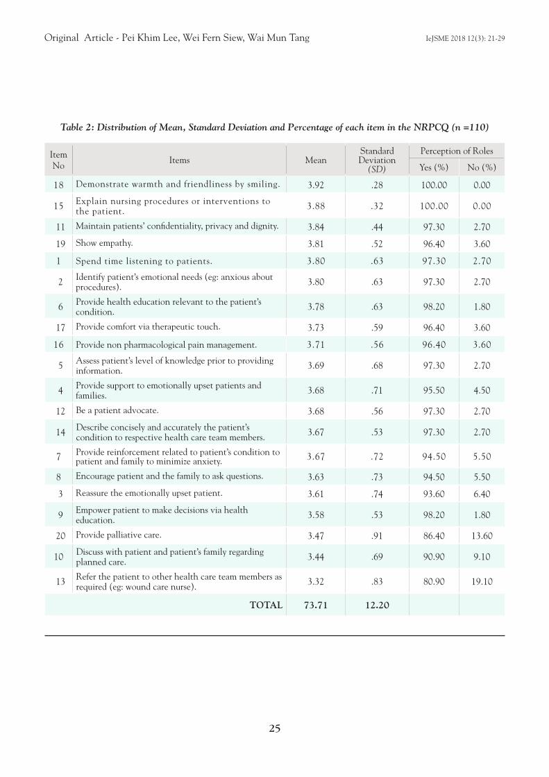
Original Article - Pei Khim Lee, Wei Fern Siew, Wai Mun Tang IeJSME 2018 12(3): 21-29
25
Item No
Items MeanStandard Deviation
(SD)
Perception of Roles
Yes (%) No (%)
18 Demonstrate warmth and friendliness by smiling. 3.92 .28 100.00 0.00
15 Explain nursing procedures or interventions to the patient. 3.88 .32 100.00 0.00
11 Maintain patients’ confidentiality, privacy and dignity. 3.84 .44 97.30 2.70
19 Show empathy. 3.81 .52 96.40 3.60
1 Spend time listening to patients. 3.80 .63 97.30 2.70
2 Identify patient’s emotional needs (eg: anxious about procedures). 3.80 .63 97.30 2.70
6 Provide health education relevant to the patient’s condition. 3.78 .63 98.20 1.80
17 Provide comfort via therapeutic touch. 3.73 .59 96.40 3.60
16 Provide non pharmacological pain management. 3.71 .56 96.40 3.60
5 Assess patient’s level of knowledge prior to providing information. 3.69 .68 97.30 2.70
4 Provide support to emotionally upset patients and families. 3.68 .71 95.50 4.50
12 Be a patient advocate. 3.68 .56 97.30 2.70
14 Describe concisely and accurately the patient’s condition to respective health care team members. 3.67 .53 97.30 2.70
7 Provide reinforcement related to patient’s condition to patient and family to minimize anxiety. 3.67 .72 94.50 5.50
8 Encourage patient and the family to ask questions. 3.63 .73 94.50 5.50
3 Reassure the emotionally upset patient. 3.61 .74 93.60 6.40
9 Empower patient to make decisions via health education. 3.58 .53 98.20 1.80
20 Provide palliative care. 3.47 .91 86.40 13.60
10 Discuss with patient and patient’s family regarding planned care. 3.44 .69 90.90 9.10
13 Refer the patient to other health care team members as required (eg: wound care nurse). 3.32 .83 80.90 19.10
TOTAL 73.71 12.20
Table 2: Distribution of Mean, Standard Deviation and Percentage of each item in the NRPCQ (n =110)
Original Article - Pei Khim Lee, Wei Fern Siew, Wai Mun Tang IeJSME 2018 12(3): 21-29
26
Contrariwise, nurses showed the least positive perception towards their roles in referring the patient to other health care team members as required (M = 3.32, SD ± 0.83), discussing with patient and patient’s family regarding planned care (M = 3.44, SD ± 0.69), as well as providing palliative care (M = 3.47, SD ± 0.91). Based on the findings, why nurses perceived these roles less positively could be due to a few reasons. Referring the patient to other health care team members has always been the doctor’s role in the local context. Nurses, on the other hand are less empowered to refer the patient to the respective health care worker because most of them do not have the autonomy to do so. Discussing with patient and patient’s family members regarding planned care may be difficult for the nurses to perform as not all family members are by the patients’ side in the hospital. Instead, caregivers such as maids and private nurses were the ones accompanying the patient in most
of procedures. That being said, therefore it was slightly difficult for nurses to carry out these responsibilities fully. Besides, in the nursing curriculum, palliative care is not included for all education programmes except for specialisation in specific courses, such as in the oncology field. Hence this can be the reason nurses are not competent enough or not well equipped to provide palliative care to patients.
Association between nurses’ demographic characteristics and perception towards their role in psychosocial care
Mann-Whitney U test was used to determine whether there is any significant association between nurses’ demographic characteristics and perception towards their role in psychosocial care. The analysis results are presented in Table 3.
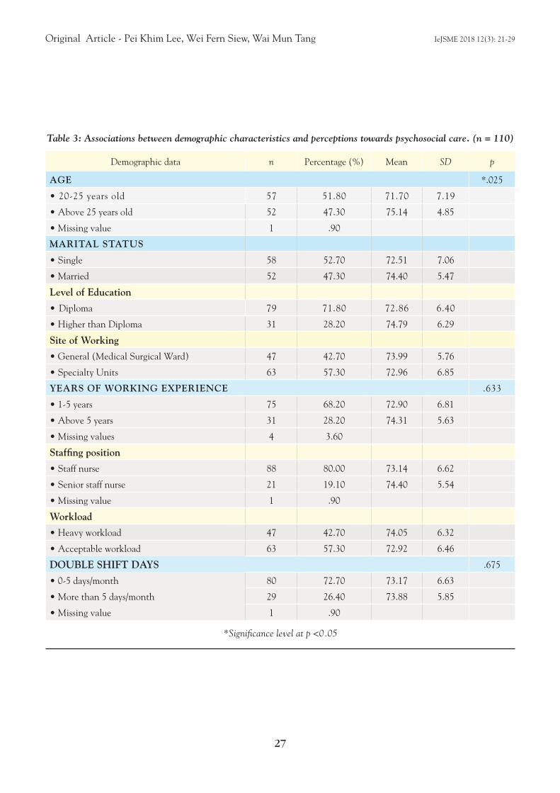
Original Article - Pei Khim Lee, Wei Fern Siew, Wai Mun Tang IeJSME 2018 12(3): 21-29
27
Demographic data n Percentage (%) Mean SD p
AGE *.025
• 20-25 years old 57 51.80 71.70 7.19
• Above 25 years old 52 47.30 75.14 4.85
• Missing value 1 .90
MARITAL STATUS
• Single 58 52.70 72.51 7.06
• Married 52 47.30 74.40 5.47
Level of Education
• Diploma 79 71.80 72.86 6.40
• Higher than Diploma 31 28.20 74.79 6.29
Site of Working
• General (Medical Surgical Ward) 47 42.70 73.99 5.76
• Specialty Units 63 57.30 72.96 6.85
YEARS OF WORKING EXPERIENCE .633
• 1-5 years 75 68.20 72.90 6.81
• Above 5 years 31 28.20 74.31 5.63
• Missing values 4 3.60
Staffing position
• Staff nurse 88 80.00 73.14 6.62
• Senior staff nurse 21 19.10 74.40 5.54
• Missing value 1 .90
Workload
• Heavy workload 47 42.70 74.05 6.32
• Acceptable workload 63 57.30 72.92 6.46
DOUBLE SHIFT DAYS .675
• 0-5 days/month 80 72.70 73.17 6.63
• More than 5 days/month 29 26.40 73.88 5.85
• Missing value 1 .90
*Significance level at p <0.05
Table 3: Associations between demographic characteristics and perceptions towards psychosocial care. (n = 110)
Original Article - Pei Khim Lee, Wei Fern Siew, Wai Mun Tang IeJSME 2018 12(3): 21-29
28
The analysis revealed there was no significant association between nurses’ demographic characteristics (i.e., marital status, level of education, working areas, years of working experience, staff position, workload, and number of double shift days in a month) and perception towards their role in psychosocial care (p > 0.05). Nonetheless, significant association was found between nurses’ age and perception towards their role in psychosocial aspect of care (p = 0.025). Nurses who were older (above 25 years old) were found to have a more positive perception towards their role in psychosocial care (M = 75.14, SD ± 4.85) as compared to younger nurses (M = 71.70, SD ± 7.19).
Furthermore, the findings revealed that nurses who were married were more positive towards their psychosocial roles (M = 74.40, SD ± 5.47) compared to nurses who were single (M = 72.51 SD ± 7.06). In addition, nurses who had higher than diploma educational qualification were found to be more positive towards their role in psychosocial care (M = 74.79, SD ± 6.29). Besides, nurses who had more than five years of working experience (M = 74.31, SD ± 5.63) and those who held senior staff nurse positions (M = 74.4, SD ± 5.54) generally had more positive perception towards their responsibilities in psychosocial care. Nevertheless, it was found that nurses who worked in general medical surgical wards had more positive perception towards their psychosocial role (M = 73.99, SD ± 5.76) compared to nurses who worked in specialty units (M = 72.96, SD ± 6.85). It was also shown that nurses who experienced heavy workload (M = 74.05, SD ± 6.32) and had more than five double shift days in a month (M = 73.88, SD ± 5.85) had more positive perception on their role in psychosocial care.
Discussion and implications
Psychosocial care remains as an integral part of holistic nursing care today. The findings of the study showed that registered nurses working in a private hospital had a positive perception towards their role in psychosocial care. These basic psychosocial nursing roles such as demonstrating kindness, care and concern should be maintained by the current nurses as these are
good qualities of care that can bring a positive effect to patient’s health outcome. Nurses should therefore place high emphasis on to these roles in order to establish a long term patient care quality outcome.
Furthermore, the study found that nurses’ age was significantly associated with nurses’ perception towards their role in psychosocial care. This finding suggested that the older nurses were more positive towards their role in psychosocial care. The findings were congruent with a past study conducted by Lange, Thom and Kline (2008), in which age was considered as one of the strongest indicators of exhibiting a positive attitude towards providing psychosocial care. Furthermore, the current study found that nurses who had more than five years of working experience and those who held senior staff nurse position generally had more positive perception towards their responsibilities in psychosocial care. Similarly, the findings supported previous studies which revealed that nurses with more years of practice experience were found to be more comfortable in providing psychosocial care, which includes caring for the dying children and their families as well as providing palliative care (Lange et al., 2008; Chen & Raingruber, 2014).
In addition, the study found that nurses who had higher than diploma educational qualification had better perception towards their role in psychosocial aspects of care. In terms of providing palliative care to the patients, 86.4% of nurses in this study had positive perception towards this role. Past study done by Jacob et al., (2015) revealed that students with a degree background are expected to perform this role more than nurses who have a diploma educational background. Thus, the findings implicated the need to review and refine the existing nursing curriculum whereby psychosocial nursing role such as palliative care should be included as a core module in the basic and/ or advanced nursing academic programmes.
It was also shown that nurses who had heavy workload had more positive perception on their role in psychosocial care. The findings in this study are in contrast with the results obtained from a paper by Pehlivan and Küçük (2016), in which the nurses reported that workload was
Original Article - Pei Khim Lee, Wei Fern Siew, Wai Mun Tang IeJSME 2018 12(3): 21-29
29
ranked as the first reason that nurses did not have extra time to fulfill patients’ psychosocial needs. The additional tasks that nurses carried on their shoulder increased the work pressure which hindered them from carrying out psychosocial care. The additional pressure at work could cause nurses to avoid in-depth communication and long conversation with clients (Kenny & Allenby, 2013; Pehlivan & Küçük, 2016).
Limitations and recommendations
The study had yielded important results on the private hospital nurses’ perception towards their role in psychosocial care. However, a few limitations were observed. The sample size of the study was relatively small (n = 110) and the data was collected from one private hospital, therefore the generalisability is restricted. Henceforth, a larger randomised sample of registered nurses across the city or country from both healthcare sectors (i.e., public and private) is recommended for future studies.
Conclusion
Providing psychosocial care to hospitalised patients is extremely important in ensuring positive patients’ outcomes. Comparison of research findings is helpful in identifying the potential gaps in providing the optimum psychosocial care to patients. Planning and implementation of relevant interventions will be appropriate to encourage nurses to provide each elements of psychosocial care.
Acknowledgements
The authors would like to acknowledge and express their deepest gratitude to the International Medical University for funding this study (Ref No: 4.13/JCM-125/2016) and to the IMU Joint-Committee on Research and Ethics for granting the ethics approval for the study. Furthermore, the kind assistance and cooperation from the chief executive office, chief nursing officer, nurse managers and registered nurses are highly appreciated and acknowledged.
REFERENCES
Attree, M. (2001). Patients’ and relatives’ experiences and perspectives of ‘good ‘and ‘not so good’quality care. Journal of Advanced Nursing, 33(4), 456-466.
Ausserhofer, D., Zander, B., Busse, R., Schubert, M., De Geest, S., Rafferty, A. M., ... & Sjetne, I. S. (2014). Prevalence, patterns and predictors of nursing care left undone in European hospitals: results from the multicountry cross-sectional RN4CAST study. BMJ Quality & Safety, 23(2), 126-135.
Ball, J. E., Murrells, T., Rafferty, A. M., Morrow, E., & Griffiths, P. (2013). ‘Care left undone’during nursing shifts: associations with workload and perceived quality of care. BMJ Quality & Safety, 1-10. http://dx.doi.org/10.1136/bmjqs2012-001767
Chen, C. H., & Raingruber, B. (2014). Educational needs of inpatient oncology nurses in providing psychosocial care. Clinical Journal of Oncology Nursing, 18(1), 1-5.
Chivukula, U., Hariharan, M., Rana, S., Thomas, M., & Swain, S. (2014). Role of psychosocial care on ICU trauma. Indian Journal of Psychological Medicine, 36(3), 312-316.
Nursing Board Malaysia. (1998). Code of professional conduct for nurses. Retrieved from http://nursing.moh.gov.my/wp-content/uploads/2015/04/nursing_board_malaysia-code_of_professional_conduct_1998.pdf
Jacob, E. R., McKenna, L., & D’amore, A. (2015). The changing skill mix in nursing: considerations for and against different levels of nurse. Journal of Nursing Management, 23(4), 421-426.
Jiang, H., Li, H., Ma, L., & Gu, Y. (2015). Nurses’ roles in direct nursing care delivery in China. Applied Nursing Research, 28(2), 132-136.
Kenny, A., & Allenby, A. (2013). Barriers to nurses providing psychosocial care in the Australian rural context. Nursing & Health Sciences, 15(2), 194-200.
Lange, M., Thom, B., & Kline, N. E. (2008). Assessing nurses’ attitudes toward death and caring for dying patients in a comprehensive cancer center. Oncology Nursing Forum, 35(6), 955-959.
Legg, M. J. (2011). What is psychosocial care and how can nurses better provide it to adult oncology patients. Australian Journal of Advanced Nursing, 28(3), 61-67.
Lu, H., While, A. E., & Barriball, K. L. (2008). Role perceptions and reported actual role content of hospital nurses in Mainland China. Journal of Clinical Nursing, 17(8), 1011-1022.
Pavlish, C., & Ceronsky, L. (2009). Oncology nurses’ perceptions of nursing roles and professional attributes in palliative care. Clinical Journal of Oncology Nursing, 13(4), 404-412.
Pehlivan, T., & Küçük, L. (2016). Skills of oncology nurses in diagnosing the psychosocial needs of the patients. International Journal of Caring Sciences, 9(1), 658-668.
Polit, F.D., & Beck, C.T. (2014). Essentials of nursing research: Appraising evidence for nursing practice (8th ed.). Philadelphia: Lippincott Williams & Wilkins.
30
1Orthopaedic Department, Hospital Sultan Ismail, Johor Bahru, Johor, MALAYSIA2Orthopaedic Department, International Islamic University Malaysia, Kuantan, Pahang, MALAYSIA.
Address for Correspondence: Dr Ren Yi Kow, Orthopaedic Department, International Islamic University Malaysia, Kuantan, Pahang, MALAYSIAE-mail: [email protected]
Limb-threatening compartment syndrome: A rare complication of dengue feverSaiful Azlan Bin Kamisan1, Ren Yi Kow2, Ed Simor Khan Mor Japar Khan2, Sa’adon Bin Ibrahim1
Case Report IeJSME 2018 12(3): 30-32
Abstract
Dengue fever, a common mosquito-borne disease in Malaysia, has a wide range of clinical presentations. Dengue shock syndrome, a potentially fatal complication of dengue, is associated with derangement of numerous haematologic markers. A compromised coagulation profile, coupled with a traumatic incident may lead to a serious haemorrhagic complication. We present a rare complication of acute compartment syndrome of the right upper limb in a 17- year-old gentleman who was admitted for decompensated dengue shock syndrome after an unsuccessful attempt of venous cannulation. An emergency fasciotomy was performed and the right upper limb was salvaged albeit with ulnar nerve and radial nerve palsy.
Introduction
Dengue fever is common in Malaysia, with 83,849 cases reported in year 2017 alone.1 Dengue fever is a single stranded RNA virus which is transmitted by mosquitoes, specifically Aedes aegypti and Ae. albopictus. Dengue fever is an acute febrile illness as defined by the presence of fever and two or more features: rash, headache, arthralgia, leucopenia, retro-orbital pain, a hemorrhagic manifestation with a history of exposure to a dengue endemic area.2 Complication of dengue fever can manifest in multiple systems in the human body, including the central nervous system (encephalopathy), gastrointestinal tract (hepatic dysfunction), cardiovascular system (myocarditis), respiratory system (pleural effusion) and coagulation profile (thrombocytopenia). We present a rare complication of acute compartment syndrome of the right upper limb in a patient who was admitted for decompensated dengue shock syndrome.
Case Report
Mr MA, a 17-year-old gentleman, presented to the emergency department with a 4-day history of fever, myalgia, arthralgia, reduced oral intake and epigastric
pain. Upon examination, he seemed lethargic with hypotension, with evidence of plasma leakage. His full blood count showed neutropenia (3.4 x 10⁹/L) and thrombocytopenia (42 x 109/L), consistent with dengue infection. Dengue serology for non-structural antigen (NS-1), dengue IgM and IgG were all positive. His renal and liver profiles were normal. He was admitted and treated for dengue shock syndrome.
He remained hemodynamically stable while in the ward until day 6 of illness, where he complained of pain and swelling over the right arm after a failure of venous cannulation. The pain was increasing in nature, associated with numbness over the median, ulnar and radial nerve distribution and he had reduced range of motion. The vascular circulation of the right arm remained intact with good Doppler signal for all arteries.
He was diagnosed to have acute compartment syndrome of the right upper limb and an emergency fasciotomy was performed to salvage the right upper limb. A long incision was made over the anterolateral aspect of the forearm and distal aspect of the arm, releasing all the compartments together with carpal tunnel release and cubital tunnel release. Intra-operatively there was hematoma formation over the medial aspect of the cubitus region extending towards the elbow region and along the medial aspect of the arm towards the axillary region. The biceps brachii muscle appeared contuse and swollen.
Post-operatively he was admitted to the intensive care unit as he developed massive plural effusion. He remained stable and subsequently recovered. A secondary suturing and split skin graft was performed for wound closure and he was discharged well. Although the upper limb was salvaged, it was associated with ulnar and radial nerve palsy as evidenced by ulnar clawing and wrist drop of the right hand.
Discussion
Compartment syndrome is defined as a clinical presentation that occurs as the result of increase in tissue
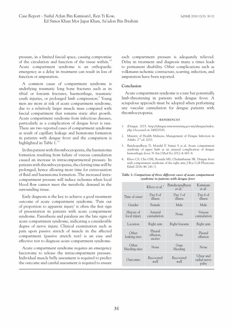
Case Report - Saiful Azlan Bin Kamisan1, Ren Yi Kow, IeJSME 2018 12(3): 30-32
Ed Simor Khan Mor Japar Khan, Sa’adon Bin Ibrahim
31
pressure, in a limited fascial space, causing compromise of the circulation and function of the tissue within.3,4 Acute compartment syndrome is an orthopaedic emergency as a delay in treatment can result in loss of function or amputation.
A common cause of compartment syndrome is underlying traumatic long bone fractures such as in tibial or forearm fractures, haemorrhage, traumatic crush injuries, or prolonged limb compression.4 Young men are more at risk of acute compartment syndrome, due to a relatively larger muscle mass compared with fascial compartment that remains static after growth. Acute compartment syndrome from infectious diseases, particularly as a complication of dengue fever is rare. There are two reported cases of compartment syndrome as result of capillary leakage and hematoma formation in patients with dengue fever and the comparison is highlighted in Table 1.3,4
In this patient with thrombocytopenia, the haematoma formation resulting from failure of venous cannulation caused an increase in intracompartmental pressure. In patients with thrombocytopenia, the clotting time will be prolonged, hence allowing more time for extravasation of fluid and haematoma formation. The increased intra-compartment pressure will induce ischemia when local blood flow cannot meet the metabolic demand in the surrounding tissue.
Early diagnosis is the key to achieve a good treatment outcome of acute compartment syndrome. ‘Pain out of proportion to apparent injury’ is often the first sign of presentation in patients with acute compartment syndrome. Paresthesia and paralysis are the late signs of acute compartment syndrome, indicating a considerable degree of nerve injury. Clinical examination such as pain upon passive stretch of muscle in the affected compartment (passive stretch test) is an easy and effective test to diagnose acute compartment syndrome.
Acute compartment syndrome requires an emergency fasciotomy to release the intracompartment pressure. Individual muscle belly assessment is required to predict the outcome and careful assessment is required to ensure
each compartment pressure is adequately relieved. Delay in treatment and diagnosis many a times leads to permanent disability. Other complications such as volkmann ischemic contracture, scarring, infection, and amputation have been reported.
Conclusion
Acute compartment syndrome is a rare but potentially limb-threatening in patients with dengue fever. A scrupulous approach must be adopted when performing any vascular cannulation for dengue patients with thrombocytopenia.
REFERENCES
1. iDengue. 2015. http://idengue.remotesensing.gov.my/idengue/index.php (Accessed on 14/8/2018).
2. Ministry of Health Malaysia. Management of Dengue Infection in Adults. 2nd ed. 2010.
3. Bandyopadhyay D, Mondal P, Samui S et al. Acute compartment syndrome of upper limb as an unusual complication of dengue hemorrhagic fever. N Am J Med Sci 2012; 4: 667–8.
4. Khoo CS, Chu GSE, Rosaida MS, Chidambaram SK. Dengue fever with compartment syndrome of the right arm; J Roy Coll Physicians Edinb 2016; 46: 241–3.
Khoo et al.4 Bandyopadhyay et al.3
Kamisan et al.
Time of onset Day 8 of illness
Day 3 ofillness
Day 6 of illness
Gender Female Male Male
History of local injury
Arterial cannulation None Venous
cannulation
Location Right arm Right forearm Right arm
Otherleaking sites
Pleural effusion, ascites
None Pleural effusion
Other bleeding sites None Gum
bleeding None
Outcomes Recovered well
Recoveredwell
Ulnar and radial nerve
palsy
Table 1: Comparison of three different cases of acute compartment syndrome in patients with dengue fever
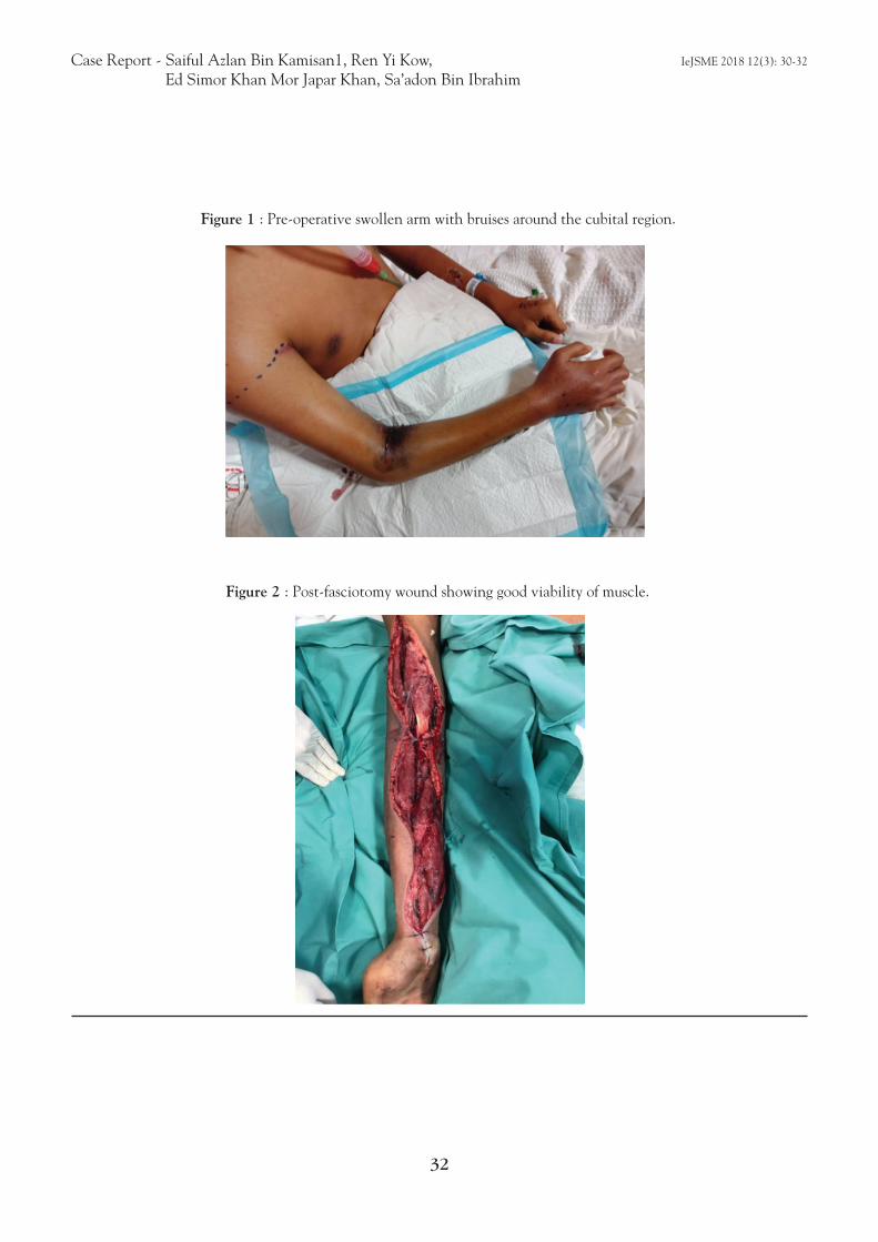
Case Report - Saiful Azlan Bin Kamisan1, Ren Yi Kow, IeJSME 2018 12(3): 30-32
Ed Simor Khan Mor Japar Khan, Sa’adon Bin Ibrahim
32
Figure 1 : Pre-operative swollen arm with bruises around the cubital region.
Figure 2 : Post-fasciotomy wound showing good viability of muscle.
33
List of Reviewers for IeJSME 2018 (Vol 1-Vol 3)
Assoc Prof Srikumar Chakravathi
Dr Nicole Chen
Dr Chen Seong Ting
Dr Jon Chua Yul Chen
Prof Esha Das Gupta
Ms Norul Hidayah
Assoc Prof Mohammed Arshad Ikram
Assoc Prof Lo Yoke Lin
Prof Lim Pek Hong
Prof Victor Lim
Prof Lum Siew Keong
Prof Mak Joon Wah
Ms Patricia Matizha
Assoc Prof Nilesh Kumar Mitra
Prof Dato’ Sivalingam Nalliah
Assistant Prof Mohd Said Bin Nurumal
Assoc Prof Joachim Perera
Dr Sangeetha Shyam
Dr Tang Wai Mun
Dr Tong Chin Voon