Embed Size (px)
Citation preview

Keywords:
Maxillectomy, obturator, speech, formant.
Corresponding Author:
Dr. Mariko Hattori
Department of Maxillofacial Prosthetics, Graduate School, Tokyo Medical and Dental University, 1-5-45 Yushima Bunkyo-ku Tokyo 113-8549, Japan
Phone number: +81-358-035556Email: [email protected]
Received: 12 October 2020 Received in revised form: 08 December 2020Accepted: 13 December 2020
This report describes the prosthetic rehabilitation of a patient with a large maxillary defect. It is often risky and difficult to take an impression of a large defect in a single step procedure. In this study, we present a clinical technique where the maxillary obturator prosthesis was fabricated by combining two methods: conventional processing using an impression followed by relining of the obturator part to improve retention and support of the prosthesis. This report also describes how digital speech analysis using formants of five vowels can be utilized during the relining and adjustment step, resulting in improved patient satisfaction and comfort. (Int J Maxillofac Prosthetics 2020;3:32-36)
International Journal of Maxillofacial Prosthetics (iJMP)
CASE REPORT
Two-step fabrication of an obturator prosthesis for a large maxillary defect with digital speech analysis: A case reportYujia Wang, BDS1 , Mariko Hattori, DDS, PhD1* , Pooja Garg, BDS, Dclindent2 , Christian Wesemann, DDS3 , Tengku Fazrina Tengku Mohd Ariff, DDS, MClinDent4 , Cristian Lagos Fredes, DDS5 , Yuka I. Sumita, DDS, PhD1 1Department of Maxillofacial Prosthetics, Graduate School, Tokyo Medical and Dental University, Tokyo, Japan.2Department of Oral Restorative Sciences, Westmead Centre for Oral Health, Sydney, Australia.3Medical Center – University of Freiburg, Center for Dental Medicine, Department of Prosthetic Dentistry, Faculty of Medicine, University of Freiburg, Freiburg, Germany.4Centre for Restorative Dentistry Studies, Faculty of Dentistry, Universiti Teknologi MARA, Sungai Buloh Campus, Selangor, Malaysia.5Center Of Advance Prosthodontics and Implant Dentistry, School of Dentistry, Univesty of Concepción, Chile.
ABSTRACTARTICLE INFO
INTRODUCTIONAcquired maxillary defects are caused by the
surgical removal of benign or malignant tumor, trauma or infection. In patients with maxillary defects, the oral defect is often connected to the nasal cavity1 which impairs functional ability especially speech.2-4 Maxillofacial prostheses play an important role in the rehabilitation of these patients.5 However, the fabrication and adjustment of the prosthesis is complicated because of the varied anatomy of the defect and the unique function of the orofacial region. For large maxillary defects in particular, the prosthesis fabrication can be a challenging and tedious for both the clinician and the patient.6 For example, taking an impression of a large defect has the risk of impaction, aspiration, or swallowing of the impression material, as well as mucosal injury. Thus, a complex fabrication
procedure with multiple adjustments is needed. Functional evaluation at various stages can help find a suitable approach.
Digital speech analysis involving formant frequency analysis of vowels has been used for the functional evaluation of patients requiring a maxillofacial prosthesis.2-4,7 For the five vowels in Japanese, the formant frequency and its range are significantly different between patients with a maxillofacial defect and healthy individuals.2 It has also been reported that the formant frequency and its range in five vowels spoken by maxillectomy patients is different between patients with an obturator prosthesis and those not wearing one.3 The speech of maxillectomy patients has characteristic formant frequencies according to the type of defect under the Aramany classification system.4
Articles published in Int J Maxillofac Prosthetics are licensed under a Creative Commons Attribution-NonCommercial 4.0 International License.32 Int J Maxillofac Prosthetics 2020;3:32-36. DOI: 10.26629/ijmp.2020.07

During treatment planning, the design of the prosthesis was discussed with the patient, and treatment options were explained. Considering the size of defect, an acrylic obturator with cast clasps was selected for ease of base adjustment and better retention. An open-bulb design was selected in order to reduce the weight of the obturator. The use of the undercuts was planned to ensure suitable retention.9
Fabrication of the obturator prosthesis A special tray was made from the preliminary impression. The final impression was made with impression compound and hydrophilic vinyl polysiloxane impression material using the tray. The new prosthesis was processed with denture acrylic resin with acrylic artificial teeth and cobalt chromium cast clasps on teeth #16, #12, and #23.
The prosthesis was completed and tried in the patient’s mouth in June 2019 (Figure 2a). The fit to the mucosal surface was adjusted using pressure-indicating paste (Mizzy Pressure Indicating Paste Pump, Keystone Industries, Oss, Netherlands).
Digital speech analysis and adjustment of the prosthesis Informed consent was obtained from the patient to perform digital speech analysis, which was approved by the Ethics Committee of the Faculty of Dentistry, Tokyo Medical and Dental University (approval number D2017-080). A speech analysis system (CSL 4400, KayPentax, Lincoln Park, NJ, USA) was used with a high-quality dynamic microphone positioned at a constant distance of 20 cm from the patient’s lips. Five Japanese vowels /a/, /e/, /i/, /o/, and /u/ were recorded. A 1-s segment was extracted from the amplitude-by-time waveform on the computer monitor. Linear predictive coding was used to reveal the formant data for each vowel. The frequencies of the first to third formant (F1 to F3) of each vowel were extracted.
To evaluate the fit of the obturator, a fit-checking silicone material (Fit Checker 1-1 package, GC Corp., Tokyo, Japan) was applied on the intaglio surface of the defect area (Figure 2b), and the vowels were recorded again to evaluate the utterance with the fit checking material on the prosthesis. Based on the results of voice analysis and fit checking, the prosthesis was relined with acrylic denture reline material (Functional Denture Rebase, Kamemizu Chem. Ind. Co., Osaka, Japan). After a week, a visible light irradiator was used to cure the acrylic material (Figure 2c) and his speech was re-analysed. Four sets of formant data were obtained for comparison: without the prosthesis, with the prosthesis tried for the first time, with the prosthesis after application of fit-checking material, and with the prosthesis after relining with acrylic material.
Wang et al.
Since the formant range of vowels is related to speech ability,2 digital speech analysis using the five Japanese vowels can be expected to be useful for evaluating the effect of adjusting maxillofacial prostheses.
This report describes the prosthetic rehabilitation of a patient with a large maxillary defect. Since it was risky and difficult to make an impression of the large and complicated defect in a single step, the obturator prosthesis was fabricated by combining conventional procedures of impression making followed by relining of the obturator part of the prosthesis to improve retention and support for the prosthesis and reduce leakage. This case report also describes the process of relining and adjustment of the prosthesis, performed as per the results of speech analysis using formants of five vowels.
CASE REPORT A 68 year old male patient visited the Clinics for Maxillofacial Prosthetics, Tokyo Medical and Dental University (TMDU) after being referred by the Department of Oral and Maxillofacial Surgery for prosthetic rehabilitation following maxillectomy. In 2016, he developed hoarseness and dysphagia. An adenoid cystic carcinoma of the left maxilla with N0 cervical lymph node was identified two years later. Prior to surgery, he was referred for the fabrication of an immediate surgical obturator. An Aramany Class II maxillectomy with full-thickness skin graft from the inguinal region was performed. The surgical obturator was fitted after surgery.
The surgeon referred the patient for a definitive obturator prosthesis one year after the surgery because of favorable wound healing and post-operative recovery. Figure 1 shows the intraoral condition of the patient after surgery. He had an Aramany Class II defect8 of the left maxilla which also extended to the soft palate. The teeth #16 and #12 - #23 remained.
Fig 1. Intraoral view of the maxilla after maxillectomy resulting in an Aramany Class II defect.
Int J Maxillofac Prosthetics 2020;3:32-36. DOI: 10.26629/ijmp.2020.07 33

Wang et al.
RESULTS
The patient’s speech improved significantly when the prosthesis was first worn, but some hypernasality was noted. The prosthesis was retained well, and the patient felt it was comfortable to wear. After relining, the patient observed that his speech improved, and was satisfied with the prosthesis.
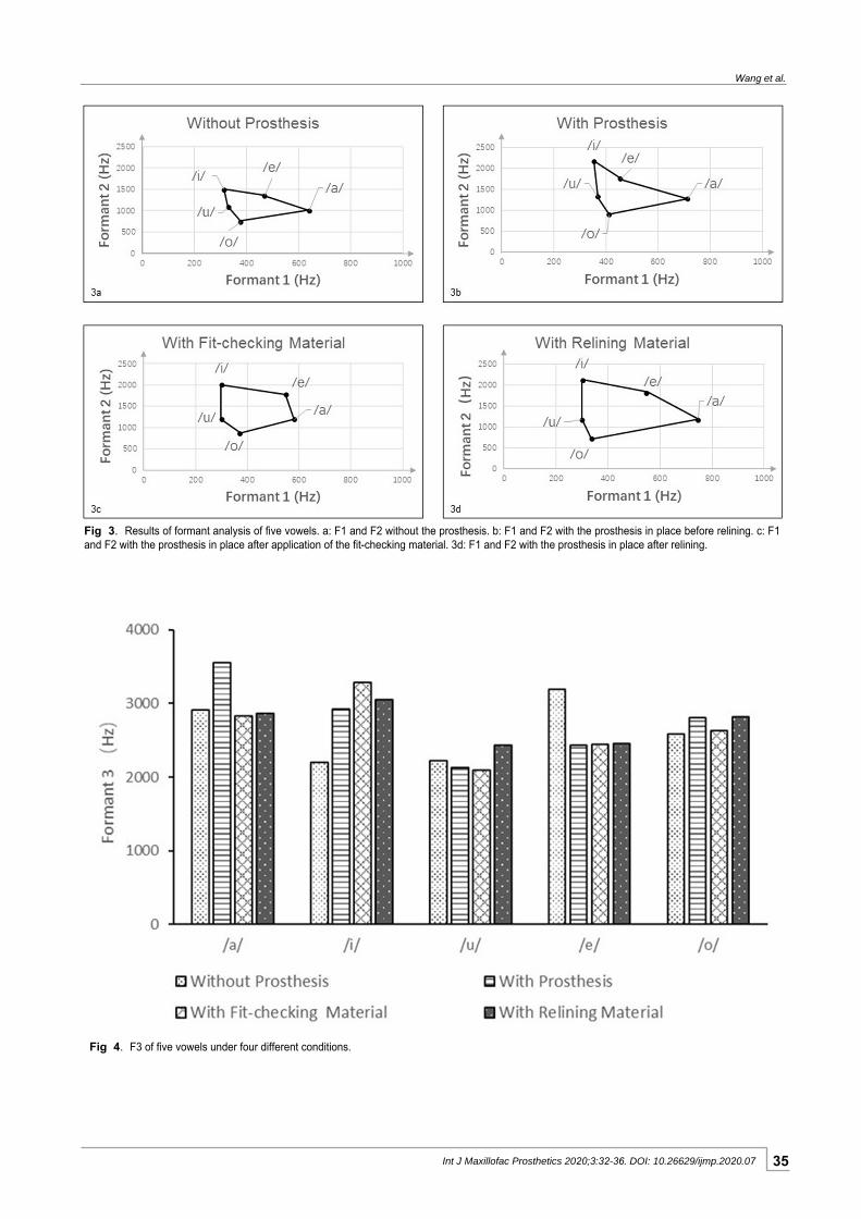
The results of formant analysis are shown in Figure 3. The x- and y-axes indicate the F1 and F2 frequencies, respectively. Both F1 and F2 range of the five vowels were larger with the prosthesis in place than without it. The F2 range when wearing the relined prosthesis was the largest among the four data sets. The F3 frequencies of the five vowels under the four different conditions are shown in Figure 4. The F3 frequency was largest for /i/ with the fit-checking material.
Fig 2. The prosthesis in three different states. a: Before adjustment. b: With fit-checking silicone material. c: After relining with acrylic.
DISCUSSION In this case, undercuts of the maxillary defect area
were not impressed in a single step to avoid iatrogenic complications like aspiration of impression material. Instead, the prosthesis was relined after the initial processing. The relining and adjustment were carefully performed according to the results of digital speech analysis.
The F1 and F2 frequencies of vowels are related to tongue position,7,10,11 and characterize the sound of each vowel. The results showed that the formant range was significantly enlarged by obturator wearing. This is consistent with the findings of a previous study.3 The speech analysis scores demonstrated that the prosthesis restored proper resonance and that the five vowels were more clearly differentiated from each other after relining the prosthesis, resulting an improvement in speech function.
34 Int J Maxillofac Prosthetics 2020;3:32-36. DOI: 10.26629/ijmp.2020.07
Wang et al.
Fig 3. Results of formant analysis of five vowels. a: F1 and F2 without the prosthesis. b: F1 and F2 with the prosthesis in place before relining. c: F1 and F2 with the prosthesis in place after application of the fit-checking material. 3d: F1 and F2 with the prosthesis in place after relining.
Fig 4. F3 of five vowels under four different conditions.
Int J Maxillofac Prosthetics 2020;3:32-36. DOI: 10.26629/ijmp.2020.07 35

Wang et al.
The third formant is related to the space compression
of the throat cavity.12, 13 The F3 frequency of /i/ was
lower in this patient without wearing the prosthesis
than in healthy individuals14and became higher when
the prosthesis was worn. This could indicate that
nasal leakage was blocked by the obturator. The F3
frequency of /i/ became even higher with the fit-
checking material, reaching the normal range.
Therefore, the obturator was relined with acrylic to
achieve a better fit and further improve the patient’s
speech. Further studies of the F3 frequency in
maxillectomy patients are needed since it may be
closely related to hypernasal speech.
In this case, the results of formant analysis with
the relined prosthesis in place were similar to the
results with the fit-checking material. This indicates
that digital speech analysis with a temporary material
gave useful information for predicting the results of
relining the obturator. Due to the difficulty in making
impression of the undercuts of the large defect area,
the obturator part of the prosthesis did not fit after the
initial processing step. Two-step fabrication of the
prosthesis including relining of obturator part based
on the results of digital speech analysis was effective
for this patient with a large maxillary defect.
CONCLUSION
An obturator prosthesis was fabricated in two
steps for a patient with a large maxillary defect. The
results of digital speech analysis can be used for
planning the fabrication procedure and predicting the
treatment outcome.
ACKNOWLEDGMENTS
This clinical case report was a part of clinical
observation activities for a group of educators that
attended the 1st biennial international workshop of
young maxillofacial prosthetic educators (yMPE) which
was held March 2019 at Tokyo Medical and Dental
University (TMDU). This work was supported by
project for Promoting Leading Edge Research in Oral
Science at Tokyo Medical and Dental University.
REFERENCES
1. Beumer J III, Marunick MT, Esposito SJ. Maxillofacial rehabilitation Prosthodontic and surgical management of cancer-related, acquired and congenital defects of the head and neck. 3rd ed. Hanover Park: Quintessence Publishing 2011;155-156.
2. Sumita YI, Ozawa S, Mukohyama H, Ueno T, Ohyama T, Taniguchi H. Digital acoustic analysis of five vowels in maxillectomy patients. J Oral Rehabil 2002;29:649-656.
3. Sumita YI, Hattori M, Murase M, Elbashti ME, Taniguchi H. Digitised evaluation of speech intelligibility using vowels in maxillectomy patients. J Oral Rehabil 2018;45:216-221.
4. Elbashti ME, Sumita YI, Hattori M, Aswehlee AM, Taniguchi H. Digitized Speech Characteristics in Patients with Maxillectomy Defects. J Prosthodont 2019;28:649-655.
5. Mukohyama H, Kadota C, Ohyama T, Taniguchi H. Lip plumper prosthesis for a patient with a marginal mandibulectomy: a clinical report. J Prosthet Dent 2004;92:23-26.
6. Taylor TD. Clinical maxillofacial prosthetics. Quintessence Publishing 2000;85-102.
7. Yoshida H, Furuya Y, Shimodaira K, Kanazawa T, Kataoka R, Takahashi K. Spectral characteristics of hypernasality in maxillectomy patients. J Oral Rehabil 2000;27:723-730.
8. Aramany MA: Basic principles of obturator design for partially edentulous patients. Part I: classification. J Prosthet Dent 1978;40:554-557.
9. Wu YL, Schaaf NG. Comparison of weight reduction in different designs of solid and hollow obturator prostheses. J Prosthet Dent 1989;62:214-217.
10. Fox RA. Perceptual structure of monophthongs and diphthongs in English. Language and Speech 1983;26:21-60.
11. Rakerd B. Linguistic and acoustic correlates of the perceptual structure found in an individual differences scaling study of vowels. J Acoust Soc Am 1985;77:296-301.
12. Raphael LJ, Borden GJ, Harris KS Speech Science
Primer:Physiology, Acoustics, and Perception of Speech Sixth Edition. Williams & Wilkins 2011;88-113.
13. Keith J. Acoustic and auditory phonetics. 2nd ed. Black Publishing 2003;102-119.
14. Deterding D. The formants of monophthong vowels in standard Southern British English pronunciation. J Int Phon Assoc 2007;27:47-55.
36 Int J Maxillofac Prosthetics 2020;3:32-36. DOI: 10.26629/ijmp.2020.07