Embed Size (px)
Citation preview
C
Csm
Da
b
c
a
ARRA
KHMLS
1
cfL2(2dt
ppiT
n
(
h1
International Journal of Paleopathology 12 (2016) 11–16
Contents lists available at ScienceDirect
International Journal of Paleopathology
j ourna l ho mepage: www.elsev ier .com/ locate / i jpp
ase report
haracterization of hypertrophic osteoarthropathy in an identifiedkeleton from Évora, Portugal, using combined and comparativeorphology and microscopy
aniela Anselmo a, Fernando Capela e Silva a,b, Teresa Fernandes a,c,∗
Departamento de Biologia, Escola de Ciências e Tecnologia, Universidade de Évora, Évora 7002-554, PortugalInstituto de Ciências Agrárias e Ambientais Mediterrânicas (ICAAM), Universidade de Évora, Évora 7002-554, PortugalCentro de Investigac ão em Antropologia e Saúde (CIAS), Universidade de Coimbra, Apartado 3046, Coimbra 3001-401, Portugal
r t i c l e i n f o
rticle history:eceived 24 July 2015eceived in revised form 28 October 2015ccepted 4 November 2015
eywords:ypertrophic osteoarthropathyorphology
a b s t r a c t
Hypertrophic osteoarthropathy (HOA) is a bone pathology that can be associated with a number of focalor systemic diseases, particularly those affecting the respiratory system. HOA is characterized by pro-liferative periostosis of the long bones, especially in their distal and periarticular regions. This reportpresents a probable case of HOA in the skeleton of an elderly Portuguese male from Évora, Portugal, whodied in 1970.
The skeleton was evaluated by morphological, radiological, and histological methods. We found bilat-eral periosteal proliferation of the tubular bones, remodeling of the phalanges of the feet, and new bone
ight microscopycanning electron microscopy
formation on rib visceral surfaces. Bone alterations are more severe on the left tibia and fibula, wherelesions display a ‘tree bark’ appearance, leading to severe thickening of the diaphysis.
Microscopically, we observed deposition of porous woven bone. Our evaluation also considers a dif-ferential diagnosis of the lesions, which includes melorheostosis, hypervitaminosis A, fluorosis, thyroidacropachy, tuberculosis, and treponematosis.
© 2015 Elsevier Inc. All rights reserved.
. Introduction
Hypertrophic osteoarthropathy (HOA) is a bone pathology thatan be associated with a number of disease conditions. HOA isound mainly in dogs (Headley et al., 2005; Cetinkaya et al., 2011;awler et al., 2015) and humans (Golder and Wolf, 2001; Utine et al.,008), but has been observed occasionally in various other speciesLenehan and Fetter, 1985; Golder and Wolf, 2001; Mair and Tucker,004; Ferguson et al., 2008; Guyot et al., 2011). Although HOA wasescribed in 1868 by Friedenreich (as cited in Kuhn et al., 2007),he exact aetiology remains unknown.
Clinically, there are two forms of HOA, the primary, or idio-athic form, and the secondary, formerly called hyperthrophic
ulmonary osteoarthropathy. Only the secondary form is describedn dogs. The primary form, also called pachyodermoperiostosis orouraine-Solente-Golé syndrome, is a rare genetic disease that is
∗ Corresponding author at: Departamento de Biologia, Escola de Ciências e Tec-ologia, Universidade de Évora, Évora 7002-554, Portugal.
E-mail addresses: daniela [email protected] (D. Anselmo), [email protected]. Capela e Silva), [email protected] (T. Fernandes).
ttp://dx.doi.org/10.1016/j.ijpp.2015.11.001879-9817/© 2015 Elsevier Inc. All rights reserved.
inherited in an autosomal fashion (Latos-Bielenska et al., 2007),recently mapped to human chromosome 4q33–q34 and to genemutations encoding 15-hydroxyprostangladin degradation (Uppalet al., 2008; Yao et al., 2009). This form, usually severe, affects pri-marily males (Aufderheide and Rodriguez-Martín, 1998), appearsaround the time of puberty, and its progression is limited to thepuberal growth period (Christensen et al., 2013).
Secondary HOA is also called Marie-Bamberger syndrome orhyperthrophic pulmonary osteoarthropathy, emphasizing the pul-monary diseases that represent the major human associates ofHOA. Among pulmonary diseases carcinomas are the major causeof HOA. In fact, bronchial carcinoma accounts for 80% of humanHOA, pleural tumors for 10%, other intrathoracic tumors for 5%,and, rarely, Hodgkin’s lymphoma is observed (Atkinson et al., 1976;Aufderheide and Rodriguez-Martín, 1998; Resnick and Kransdorf,2005). Several studies suggest an association between HOA andtuberculosis (Mays and Taylor, 2002; Assis et al., 2011). Otherextra-pulmonary conditions that may be associated with HOA
include various gastrointestinal, cardiovascular, hepatobiliary, andendocrine disorders (Bazar et al., 2004; Shih, 2004; Vandemergelet al., 2004; Yao et al., 2009).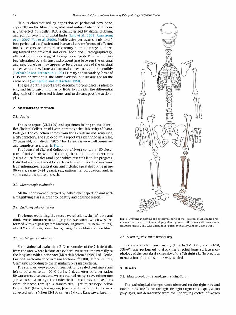
1 rnal of Paleopathology 12 (2016) 11–16
eiaefbiatac(Hs
idg
2
2
fiPa7a
t(Df6s
2
a
2
fifa
2
ftEG
l8(wEc
Fig. 1. Drawing indicating the preserved parts of the skeleton. Black shading rep-
2 D. Anselmo et al. / International Jou
HOA is characterized by deposition of periosteal new bone,specially on the tibia, fibula, ulna, and radius. Subchondral bones unaffected. Clinically, HOA is characterized by digital clubbingnd painful swelling of distal limbs (Jajic et al., 2001; Armstrongt al., 2007; Yao et al., 2009). Proliferative periostosis leads to dif-use periosteal ossification and increased circumference of affectedones. Lesions occur more frequently at mid-diaphysis, taper-
ng toward the proximal and distal bone ends. Radiographically,ffected bone may suggest having been “pasted” onto the cor-ex (identified by a distinct radiolucent line between the originalnd new bone), or may appear to be a dense part of the originalortex where new bone and normal cortex merge imperceptiblyRothschild and Rothschild, 1998). Primary and secondary forms ofOA can be present in the same skeleton, but usually not on the
ame bone (Rothschild and Rothschild, 1998).The goals of this report are to describe morphological, radiolog-
cal, and histological findings of HOA, to consider the differentialiagnosis of the observed lesions, and to discuss possible aetiolo-ies.
. Materials and methods
.1. Subject
The case report (CEIE109) and specimen belong to the Identi-ed Skeletal Collection of Évora, curated at the University of Évora,ortugal. The collection comes from the Cemitério dos Remédios,
city cemetery. The subject of this report was identified as a male,3 years old, who died in 1970. The skeleton is very well preservednd complete, as shown in Fig. 1.
The Identified Skeletal Collection of Évora contains 160 skele-ons of individuals who died during the 19th and 20th centuries90 males, 70 females) and upon which research is still in progress.ata that are maintained for each skeleton of this collection come
rom inhumation registrations and include: age at death (mean age0 years, range 3–91 years), sex, nationality, occupation, and, inome cases, the cause of death.
.2. Macroscopic evaluation
All the bones were surveyed by naked eye inspection and with magnifying glass in order to identify and describe lesions.
.3. Radiological evaluation
The bones exhibiting the most severe lesions, the left tibia andbula, were submitted to radiographic assessment which was per-
ormed with a digital system Mammo Diagnost UC system (Philips),t 28 kV and 25 mA, coarse focus, using Kodak Min-R screen film.
.4. Histological evaluation
For histological evaluation, 2–3 cm samples of the 7th right rib,rom the area where lesions are evident, were cut transversally tohe long axis with a bone saw [Materials Science (NW) Ltd., Settle,ngland] and embedded in resin (Technovit® 9100, Heraeus Kulzer,ermany) according to the manufacturer’s instructions.
The samples were placed in hermetically sealed containers andeft to polymerise at −20 ◦C during 5 days. After polymerization0 �m transverse sections were obtained using a saw microtome
Leica 1600, Germany). The undecalcified and unstained sectionsere observed through a transmitted light microscope Nikonclipse 600 (Nikon, Kanagawa, Japan), and digital pictures wereollected with a Nikon DN100 camera (Nikon, Kanagawa, Japan).
resents more severe lesions and grey shading more mild lesions. All bones weresurveyed visually and with a magnifying glass to identify and describe lesions.
2.5. Scanning electronic microscopy
Scanning electron microscopy (Hitachi TM 3000, and SU-70,30 keV) was performed to study the affected bone surface mor-phology of the vertebral extremity of the 7th right rib. No previouspreparation of the rib sample was needed.
3. Results
3.1. Macroscopic and radiological evaluations
The pathological changes were observed on the right ribs andlower limbs. The fourth through the eighth right ribs display a thingray layer, not demarcated from the underlying cortex, of woven
D. Anselmo et al. / International Journal o
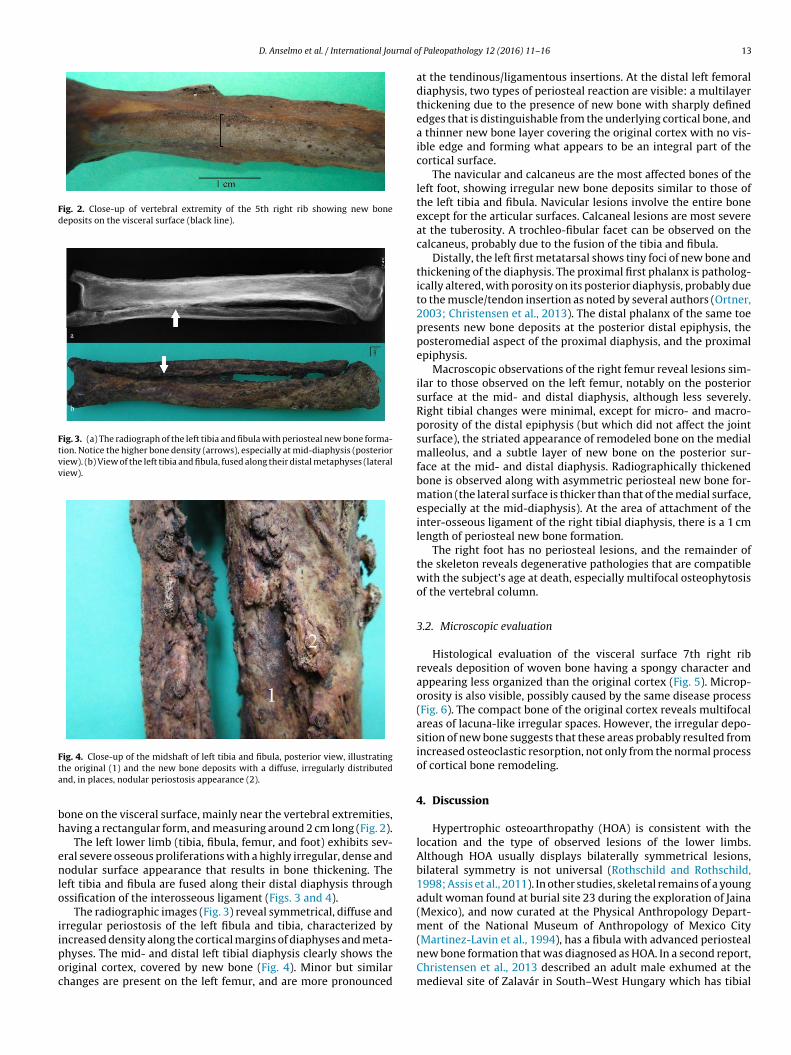
Fig. 2. Close-up of vertebral extremity of the 5th right rib showing new bonedeposits on the visceral surface (black line).
Fig. 3. (a) The radiograph of the left tibia and fibula with periosteal new bone forma-tion. Notice the higher bone density (arrows), especially at mid-diaphysis (posteriorview). (b) View of the left tibia and fibula, fused along their distal metaphyses (lateralview).
Fta
bh
enlo
iipoc
(Martinez-Lavin et al., 1994), has a fibula with advanced periosteal
ig. 4. Close-up of the midshaft of left tibia and fibula, posterior view, illustratinghe original (1) and the new bone deposits with a diffuse, irregularly distributednd, in places, nodular periostosis appearance (2).
one on the visceral surface, mainly near the vertebral extremities,aving a rectangular form, and measuring around 2 cm long (Fig. 2).
The left lower limb (tibia, fibula, femur, and foot) exhibits sev-ral severe osseous proliferations with a highly irregular, dense andodular surface appearance that results in bone thickening. The
eft tibia and fibula are fused along their distal diaphysis throughssification of the interosseous ligament (Figs. 3 and 4).
The radiographic images (Fig. 3) reveal symmetrical, diffuse andrregular periostosis of the left fibula and tibia, characterized byncreased density along the cortical margins of diaphyses and meta-
hyses. The mid- and distal left tibial diaphysis clearly shows theriginal cortex, covered by new bone (Fig. 4). Minor but similarhanges are present on the left femur, and are more pronouncedf Paleopathology 12 (2016) 11–16 13
at the tendinous/ligamentous insertions. At the distal left femoraldiaphysis, two types of periosteal reaction are visible: a multilayerthickening due to the presence of new bone with sharply definededges that is distinguishable from the underlying cortical bone, anda thinner new bone layer covering the original cortex with no vis-ible edge and forming what appears to be an integral part of thecortical surface.
The navicular and calcaneus are the most affected bones of theleft foot, showing irregular new bone deposits similar to those ofthe left tibia and fibula. Navicular lesions involve the entire boneexcept for the articular surfaces. Calcaneal lesions are most severeat the tuberosity. A trochleo-fibular facet can be observed on thecalcaneus, probably due to the fusion of the tibia and fibula.
Distally, the left first metatarsal shows tiny foci of new bone andthickening of the diaphysis. The proximal first phalanx is patholog-ically altered, with porosity on its posterior diaphysis, probably dueto the muscle/tendon insertion as noted by several authors (Ortner,2003; Christensen et al., 2013). The distal phalanx of the same toepresents new bone deposits at the posterior distal epiphysis, theposteromedial aspect of the proximal diaphysis, and the proximalepiphysis.
Macroscopic observations of the right femur reveal lesions sim-ilar to those observed on the left femur, notably on the posteriorsurface at the mid- and distal diaphysis, although less severely.Right tibial changes were minimal, except for micro- and macro-porosity of the distal epiphysis (but which did not affect the jointsurface), the striated appearance of remodeled bone on the medialmalleolus, and a subtle layer of new bone on the posterior sur-face at the mid- and distal diaphysis. Radiographically thickenedbone is observed along with asymmetric periosteal new bone for-mation (the lateral surface is thicker than that of the medial surface,especially at the mid-diaphysis). At the area of attachment of theinter-osseous ligament of the right tibial diaphysis, there is a 1 cmlength of periosteal new bone formation.
The right foot has no periosteal lesions, and the remainder ofthe skeleton reveals degenerative pathologies that are compatiblewith the subject’s age at death, especially multifocal osteophytosisof the vertebral column.
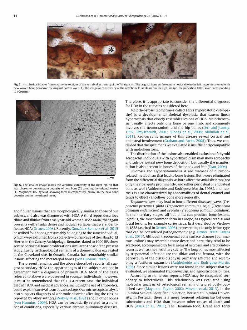
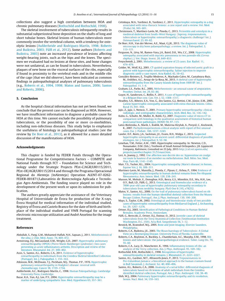
3.2. Microscopic evaluation
Histological evaluation of the visceral surface 7th right ribreveals deposition of woven bone having a spongy character andappearing less organized than the original cortex (Fig. 5). Microp-orosity is also visible, possibly caused by the same disease process(Fig. 6). The compact bone of the original cortex reveals multifocalareas of lacuna-like irregular spaces. However, the irregular depo-sition of new bone suggests that these areas probably resulted fromincreased osteoclastic resorption, not only from the normal processof cortical bone remodeling.
4. Discussion
Hypertrophic osteoarthropathy (HOA) is consistent with thelocation and the type of observed lesions of the lower limbs.Although HOA usually displays bilaterally symmetrical lesions,bilateral symmetry is not universal (Rothschild and Rothschild,1998; Assis et al., 2011). In other studies, skeletal remains of a youngadult woman found at burial site 23 during the exploration of Jaina(Mexico), and now curated at the Physical Anthropology Depart-ment of the National Museum of Anthropology of Mexico City
new bone formation that was diagnosed as HOA. In a second report,Christensen et al., 2013 described an adult male exhumed at themedieval site of Zalavár in South–West Hungary which has tibial
14 D. Anselmo et al. / International Journal of Paleopathology 12 (2016) 11–16
Fig. 5. Histological images from transverse sections of the vertebral extremity of the 7th rinew woven bone (2) above the original cortex layer (1). The irregular consistency of the nto 100 �m).
Fig. 6. The smaller image shows the vertebral extremity of the right 7th rib thatwas chosen to demonstrate deposits of new bone (2) covering the original cortex(1). Magnified 30× by SEM, showing focal microporosity (arrow) in the new boned
astpfidwHssal
garidcar(b
Coimbra Skeletal Identified Collection, housed at Coimbra Univer-sity, in Portugal, there is a more frequent relationship between
eposits and in the original layer.
nd fibular lesions that are morphologically similar to those of ourubject, and also was diagnosed with HOA. A third report describesibiae and fibulae from a 58-year-old woman, IPAZ 6646, that againresents with similar dense and nodular surfaces that were identi-ed as HOA (Ortner, 2003). Recently, González-Reimers et al., 2015escribed four bones, presumably belonging to the same individual,hich were exhumed from a collective burial cave of the island of Elierro, in the Canary Archipelago. Remains, dated to 1000 BP, show
evere periosteal bone proliferations similar to those of the presenttudy. Lastly, archaeological remains of a domestic dog excavatedt the Cleveland site, in Ontario, Canada, has remarkably similaresions affecting the metacarpal bones (von Hunnius, 2009).
The present remains, and the above-described reports, all sug-est secondary HOA; the apparent ages of the subjects are not ingreement with a diagnosis of primary HOA. Most of the caseseferred to above were observed in younger individuals; however,t must be remembered that this is a recent case, the individualied in 1970, and medical advances, including the use of antibiotics,ould explain survival to an advanced age. Our microscopic analysislso supports diagnosis of a chronic disorder affecting the ribs, as
eported by other authors (Wakely et al., 1991) and in other bonesvon Hunnius, 2009). HOA can be secondarily related to a num-er of conditions, especially various chronic pulmonary diseases.ght rib. The original bone surface (more noticeable in the left image) is covered withew bone (*) is clearer in the right image (magnification 100X, scale corresponding
Therefore, it is appropriate to consider the differential diagnosesfor HOA in the remains considered here.
Melorheostosis (sometimes called Leri’s hyperostotic osteopa-thy) is a developmental skeletal dysplasia that causes linearhyperostosis that closely resembles lesions of HOA. Melorheosto-sis usually affects only one bone or one limb, and commonlyinvolves the neurocranium and the hip bones (Leri and Joanny,1992; Freyschmidt, 2001; Subhas et al., 2008; Abdullah et al.,2011). Radiographic images of this disease reveal cortical andendosteal involvement (Graham and Parke, 2005). Thus, we con-cluded that the specimens we evaluated is insufficiently compatiblewith melorheostosis.
The distribution of the lesions also enabled exclusion of thyroidacropachy. Individuals with hyperthyroidism may show acropachyand sub-periosteal new bone deposition, but usually the manifes-tation is also present in bones of the hands and feet (Tran, 2004).
Fluorosis and Hypervitaminosis A are diseases of nutrition-related metabolism that lead to bone lesions. Both were eliminatedfrom the differential diagnosis, as both affect the axial skeleton (notonly the ribs) quite prominently, and either periosteal or endostealbone as well (Aufderheide and Rodríguez-Martín, 1998), and fluo-rosis is also characterized by abnormalities of dental enamel andtends to effect cancellous bone more generally.
Treponemal spp. may lead to four different diseases: yaws (Tre-ponema pertenue), pinta (Treponema carateum), bejel (Treponemapallidum endemicum) and syphilis (Treponema pallidum pallidum).In their tertiary stages, all but pinta can produce bone lesions.Syphilis, the most common form in Europe, has typical cranial andfacial lesions, for example caries sicca, first described by Virchowin 1858 (as cited in Ortner, 2003), representing the only lesion typethat can be considered pathognomonic (e.g. Ortner, 2003; Santoset al., 2013). Although treponemal bone surface changes (gumma-tous lesions) may resemble those described here, they tend to bescattered, accompanied by focal areas of necrosis, and affect endos-teum as well as the medullary cavity. The long bones most affectedby treponemal infection are the tibiae and the femora, with theperiosteum of the distal diaphysis primarily affected and resem-bling a fusiform expansion (Aufderheide and Rodriguez-Martín,1998). Since similar lesions were not found in the subject that weevaluated, we eliminated Treponema ssp. as diagnostic possibilities.
According to numerous reports, HOA may be recognized sec-ondary to tuberculosis. This relationship was evaluated usingmolecular analysis of osteological remains of a previously pub-lished case (Mays and Taylor, 2002; Masson et al., 2013). In the
tuberculosis and HOA than between other causes of death andHOA (Assis et al., 2011). The Hamman-Todd, Grant and Terry
rnal o
cc
sscoaBwmwpiofi(a
5
cwHtitrd
A
tNnPRFgdl
HÉRpet
R
A
A
A
A
A
B
D. Anselmo et al. / International Jou
ollections also suggest a high correlation between HOA andhronic pulmonary diseases (Rothschild and Rothschild, 1998).
The skeletal involvement of tuberculosis infrequently results inubstantial subperiosteal bone deposition on the shafts of long andhort tubular bones. Skeletal lesions of human tuberculosis moreommonly involve the vertebral column, with a tendency for oste-lytic lesions (Aufderheide and Rodriguez-Martín, 1998; Robertsnd Buikstra, 2003; Pálfi et al., 2012). Some authors (Roberts anduikstra, 2003) note an increased prevalence of lesions affectingeight-bearing joints, such as the hips and the knees. The speci-en we evaluated had no lesions at these sites, and bone changesere not unilateral, as can be found in tuberculosis. Nevertheless,
laques of new bone on the visceral surfaces of the ribs, especiallyf found in proximity to the vertebral ends and in the middle ribsf the cage (that we did observe), have been indicated as commonndings in paleopathological analyses of pulmonary tuberculosis
e.g., Roberts et al., 1994, 1998; Matos and Santos, 2006; Santosnd Roberts, 2006).
. Conclusion
As the hospital clinical information has not yet been found, weonclude that the present case can be diagnosed as HOA. However,e have insufficient information to diagnose a probable cause forOA at this time. We cannot exclude the possibility of pulmonary
uberculosis, or the possibility of one or more co-morbidities,ncluding non-infectious diseases. This study helps to emphasizehe usefulness of histology in paleopathological studies (see theeview by De Boer et al., 2013), as it allowed for a more detailediscussion of the manifestations of HOA.
cknowledgments
This chapter is funded by FEDER Funds through the Opera-ional Programme for Competitiveness Factors – COMPETE andational Funds through FCT – Foundation for Science and Tech-ology under the Strategic Projects PEst-C/AGR/UI0115/2011,Est-OE/AGR/UI0115/2014 and through the Programa Operacionalegional do Alentejo (InAlentejo) Operation ALENT-07-0262-EDER-001871/Laboratório de Biotecnologia Aplicada e Tecnolo-ias Agro-Ambientais. The funding source played no role in theevelopment of the present work or upon its submission for pub-
ication.The authors greatly appreciate the assistance of: the Veterinary
ospital of Universidade de Évora for production of the X-rays,vora Hospital for medical information of the individual studied,egistry of Évora and Castelo Branco for the date of birth and birth-lace of the individual studied and VWR Portugal for scanninglectronic microscope utilization and André Anselmo for the imagereatment.
eferences
bdullah, S., Pang, G.M., Mohamed-Haflah, N.H., Sapuan, J., 2011. Melorheostosis ofthe ulna. J. Chin. Med. Assoc. 74, 469–472.
rmstrong, D.J., McCausland, E.M., Wright, G.D., 2007. Hypertrophic pulmonaryosteoarthropathy (HPOA) (Pierre Marie-Bamberger syndrome): two casespresenting as acute inflammatory arthritis. Description and review of theliterature. Rheumatol. Int. 27, 399–402.
ssis, S., Santos, A.L., Roberts, C.A., 2011. Evidence of hyperotrophicostearthropathy in individuals from the Coimbra Skeletal Identified Collection(Portugal). Int. J. Paleopathol. 1, 155–163.
tkinson, M.K., McElwain, T.J., Peckham, M.J., Thomas, P.P., 1976. Hypertrophicpulmonary osteoarthropathy in Hodgkin’s disease: reversal with
chemotherapy. Cancer 38, 1729–1734.ufderheide, A.C., Rodriguez-Martín, C., 1998. Human Paleopathology. CambridgeUniversity Press, Cambridge.
azar, K.A., Yun, A.J., Lee, P.Y., 2004. Hypertrophic osteoarthropathy may be amarker of underlying sympathetic bias. Med. Hypotheses 63, 357–361.
f Paleopathology 12 (2016) 11–16 15
Cetinkaya, M.A., Yardimci, B., Yardimci, C., 2011. Hypertrophic osteopathy in a dogassociated with intra-thoracic lesions: a case report and a review. Vet. Med.(Praha) 56, 595–601.
Christensen, T., Martínez-Lavín, M., Pineda, C., 2013. Periostitis and osteolysis in amedieval skeleton from South–West Hungary: (leprosy, treponematosis,tuberculosis or hypertrophic osteoarthropathy) a diagnostic challenge! Int. J.Osteoarchaeol. 23, 69–82.
De Boer, H.H., Van der Merwe, A.E., Maat, G.J.R., 2013. The diagnostic value ofmicroscopy in dry bone palaeopathology: a review. Int. J. Paleopathol. 3,113–121.
Ferguson, N., Lévy, M., Ramos-Vara, J.A., Baird, D.K., Wu, C.C., 2008. Hypertrophicosteopathy associated with mycotic pneumonia in two juvenile elk (Cervuselaphus). J. Vet. Diagn. Invest. 20, 849–853.
Freyschmidt, J., 2001. Melorheostosis: a review of 23 cases. Eur. Radiol. 11,474–479.
Golder, W., Wolf, K.J., 2001. CT-guided aspiration biopsy of infected aortic graft in apatient with hypertrophic osteoarthropathy. Saline injection to improvediagnostic yield-a case report. Acta Radiol. 42, 59–62.
González-Reimers, E., Trujillo-Mederos, A., Machado-Calvo, M., Castaneyra-Ruiz,M., Ordónez, A.C., Arnay-de-la-Rosa, M., 2015. A skeletal case of hypertrophicosteoarthropathy from the Canary Islands dating from 1000 BP. Int. J.Paleopathol. 11, 1–6.
Graham, L.E., Parke, R.C., 2005. Melorheostosis—an unusual cause of amputation.Prosthet. Orthot. Int. 29, 83–86.
Guyot, H., Sandersen, C., Rollin, F., 2011. A case of hypertrophic osteoarthropathyin a Belgian blue cow. Can. Vet. J. 52, 1308–1311.
Headley, S.A., Ribeiro, E.A., Von, G., dos Santos, G.J., Bettini, C.M., Júnior, E.M., 2005.Canine hypertrophic osteopathy associated with extra-thoracic lesions. Ciênc.Rural 35, 941–944.
Jajic, Z., Jajic, I., Nemcic, T., 2001. Primary hypertrophic osteoarthropathy: clinical,radiologic, and scintigraphic characteristics. Arch. Med. Res. 32, 136–142.
Kuhn, G., Schultz, M., Muller, R., Ruhli, F.J., 2007. Diagnostic value of micro-CT incomparison with histology in the qualitative assessment of historical humanpostcranial bone pathologies. HOMO 58, 97–115.
Latos-Bielenska, A., Marik, I., Kuklik, M., Materna-Kiryluk, A., Povysil, C., Kozlowsk,K., 2007. Pachydermoperiostosis-critical analysis with report of five unusualcases. Eur. J. Pediatr. 166, 1237–1243.
Lawler, D.F., Reetz, J.A., Sackman, J.E., Evans, R.H., Widga, C., 2015. Suspectedhypertrophic osteopathy in an ancient canid: differential diagnosis of possibleetiologies. Int. J. Paleopathol. 9, 52–58.
Lenehan, T.M., Fetter, A.W., 1985. Hypertrophic osteopathy. In: Newton, C.D.,Nunamaker, D.M. (Eds.), Textbook of Small Animal Orthopedics. J.B. LippincottCompany, Baltimore, Consulted on 23 July, 2015 http://cal.vet.upenn.edu/projects/saortho/chapter 51/51mast.htm.
Leri, A., Joanny, J., 1992. Une affection non decrite des os: hyperostose en coulgesur toute la hauteur d’un member ou mélorhéostose. Bull. Mém. Soc. Med.Hop. Paris 46, 1141–1145.
Mair, T.S., Tucker, R.L., 2004. Hypertrophic osteopathy (Marie’s disease) in horses.Equine Vet. J. 16, 308–311.
Martinez-Lavin, M., Mansilla, J., Pineda, C., Pijoan, C., Ochoa, P., 1994. Evidence ofhypertrophic osteoarthropathy in human skeletal remains from Pre-HispanicMesoamerica. Ann. Intern. Med. 120, 238–241.
Masson, M., Molnár, E., Donoghue, H.D., Besra, G.S., Minnikin, D.E., Wu, H.H., Lee,O.Y., Bull, I.D., Pálfi, G., 2013. Osteological and biomolecular evidence of a7000-year-old case of hypertrophic pulmonary osteopathy secondary totuberculosis from neolithic hungary. PLoS One 8 (10), e78252.
Matos, V., Santos, A.L., 2006. On the trail of pulmonary tuberculosis based on riblesions: results from the human identified skeletal collection from the MuseuBocage (Lisbon, Portugal). Am. J. Phys. Anthropol. 130, 190–200.
Mays, S., Taylor, G.M., 2002. Osteological and biomolecular study of two possiblecases of hypertrophic osteoarthropathy from Mediaeval England. J. Archaeolog.Sci. 29, 1267–1276.
Ortner, D.J., 2003. Identification of Pathological Conditions in Human SkeletalRemains. Academic Press, Amsterdam.
Pálfi, G., Bereczki, Z., Ortner, D.J., Dutour, O., 2012. Juvenile cases of skeletaltuberculosis from the Terry Anatomical Collection (Smithsonian InstitutionWashington, D.C., USA). Acta Biol. Szegediensis 56, 1–12.
Resnick, M., Kransdorf, M.J., 2005. Bone and Joint Imaging. Elsevier Saunders,Philadelphia.
Roberts, C.A., Buikstra, J.E., 2003. The Bioarchaeology of Tuberculosis: A GlobalView on a Reemerging Disease. University Press of Florida, Gainesville.
Roberts, C.A., Boylston, A., Buckley, L., Chamberlain, A.C., Murphy, E.M., 1998. Riblesions and tuberculosis: the palaeopathological evidence. Tuber. Lung Dis. 79,55–60.
Roberts, C.A., Lucy, D., Manchester, K., 1994. Inflammatory lesions of ribs: ananalysis of the Terry collection. Am. J. Phys. Anthropol. 95, 169–182.
Rothschild, B.M., Rothschild, C., 1998. Recognition of hypertrophicosteoarthropathy in skeletal remains. J. Rheumatol. 21, 2221–2227.
Santos, A.L., Gardner, M.T., Allsworth-Jones, P., 2013. Treponematosis inPre-Columbian Jamaica: a biocultural approach to the human cranium foundin Bull Savannah. J. Archaeol. Sci. 40, 490–496.
Santos, A.L., Roberts, C.A., 2006. Anatomy of a serial killer: differential diagnosis oftuberculosis based on rib lesions of adult individuals from the Coimbraidentified skeletal collection, Portugal. Am. J. Phys. Anthropol. 130, 38–49.
Shih, W.J., 2004. Pulmonary hypertrophic osteoarthropathy and its resolution.Semin. Nucl. Med. 34, 159–163.
1 rnal o
S
TU
U
Wakely, J., Manchester, K., Roberts, C., 1991. Scanning electron microscopy of rib
6 D. Anselmo et al. / International Jou
ubhas, N., Sundaram, M., Bauer, T.W., Seitz, W.H., Recht, M.P., 2008. Glenoidlabrum ossification and mechanical restriction of joint motion: extraosseousmanifestations of melorheostosis. Skeletal Radiol. 37, 177–181.
ran, H.A., 2004. Thyroid acropachy. Intern. Med. J. 34, 513–514.ppal, S., Diggle, C.P., Carr, I.M., Fishwick, C.W., Ahmed, M., Ibrahim, G.H., Helliwell,
P.S., Latos-Bielenska, A., Phillips, S.E., Markham, A.F., Bennett, C.P., Bonthron,D.T., 2008. Mutations in 15-hydroxyprostaglandin dehydrogenase cause
primary hypertrophic osteoarthropathy. Nat. Genet. 40, 789–793.tine, E.G., Yalc in, B., Karnak, I., Kale, G., Yalc in, E., Dogru, D., Kiper, N., Akyüz, C.,Büyükpamukcu, M., 2008. Childhood intrathoracic Hodgkin lymphoma withhypertrophic pulmonary osteoarthropathy: a case report and review of theliterature. Eur. J. Pediatr. 167, 419–423.
f Paleopathology 12 (2016) 11–16
Vandemergel, X., Blocklet, D., Decaux, G., 2004. Periostitis and hypertrophicosteoarthropathy: etiologies and bone scan patterns in 115 cases. Eur. J. Intern.Med. 15, 375–380.
von Hunnius, T., 2009. Using microscopy to improve a diagnosis: an isolated caseof tuberculosis-induced hypertrophic osteopathy in archaeological dogremains. Int. J. Osteoarchaeol. 19, 397–405.
lesions. Int. J Osteoarchaeol. 1, 185–189.Yao, Q., Altman, R.D., Brahn, E., 2009. Periostitis and hypertrophic pulmonary
osteoarthropathy: report of 2 cases and review of the literature. Semin.Arthritis Rheum. 38, 458–466.