Embed Size (px)
Citation preview
Paleopathology: Nutritional Stress
As a Measure of Environmental Stress
By
Yelda Raheen
A THESIS
Presented to the Department of General Science and the Robert D. Clark Honors College
in partial fulfillment of the requirements for the Degree of Bachelor of Science
NOVEMBER 2016
brought to you by COREView metadata, citation and similar papers at core.ac.uk
provided by University of Oregon Scholars' Bank
ii
Abstract of the Thesis of
Yelda Raheen for the degree of Bachelor of Science in the Department of General Science to be taken November 2016
Title: Paleopathology: Nutritional Stress As a Measure of Environmental Stress
Approved: _______________________________________ Terry L. Hunt
The purpose of this thesis is to understand what kinds of societies were
experiencing stresses and see what that implies about their lifestyle, environment,
health, and diseases of the time. Paleopathology can be defined as the study of the
evidence of trauma, disease, and congenital defects in human remains. Utilizing
paleopathology can illuminate how healthy and stable a civilization may have been in
comparison to other regions and may either confirm or disapprove previous notions. I
will be analyzing 3-4 societies in depth, while utilizing the rest of my data for a broad
comparison of stress indicators globally. With more research from other regions
experiencing similar stresses, we can compare and contrast their lifestyles and hopefully
gain some insight into possibly preventing such epidemics from reoccurring.
iii
Acknowledgement
Thank you Dr. Terry Hunt for agreeing to be my Primary Thesis Adviser. Thank
you to the rest of my Thesis Committee, Professors Elliot Berkman and Ulrick Casimir,
for their support and input on this document as well as Academic and Thesis Programs
Manager Miriam Alexis Jordan for assisting me with the formatting of my thesis
document.
iv
Table of Contents
Research Question…………………………………………………………………………………1 Methods of Measuring Nutritional Stress………………………………………………….....................................................3
Case Studies……………………………………………………………………………….......9 Implications & Conclusions……………………………………………………………….…………....44 Bibliography…………………………………………………………………………....48
Research Question
The aim for this thesis is to analyze combination of similar studies from several
historic and prehistoric skeletal samples worldwide to provide context and discussion
about stress related morbidity and mortality from throughout the globe. This thesis
examined the relationship between developmental stress and environmental stresses
including climate, foreign invasion, diet, SES, & urbanization. The use of multiple
stress indicators is the key to assessing the true health status of populations, as there is
no universal view to health between societies. The hope for this thesis is to provide an
unbiased review of techniques, implications, and interpretations across multiple regions
around the world for a better understanding of nutritional standards across ancient
populations. These studies illuminate the bigger picture whereas infectious diseases and
malnutrition still affect more of the world than chronic disease. Epidemiologists,
biologists, archaeologists, & osteoarchaeologists working together to add to the
bioarchaeological record lead to mitigation and aid in areas where medical technology
and SES may not be able to buffer the effects. Future research can illuminate the
contextual factors affecting the relationship between nutritional health and
environmental stresses in order to gain a better understanding of human health overall.
Throughout my thesis I hope to emphasize perfect examples of combing
multiple facets of science to achieve a better understanding of disease and the
transmission of disease. There are two basic problems in epidemiology, hence the
history of medicine, which have been the identification of the places where infectious
diseases originated and discovery of the routes by which they travelled. For too long
researchers have been relying on written documents that are supported by a small extent
2
of anatomical examination. This process is simply inadequate, which is why I’m
arguing for a more cooperative approach towards studying diseases. The authors in
previous studies and propose numerous techniques for examining human remains that
will allow people to obtain reliable data. These techniques range from simply reviewing
untapped parish records to utilizing immunocytochemistry to observe material stored
for years. Our focus should not just be on how a disease is travelling and progressing in
modern times, but also what disease loads and stressors were already active in
aboriginal communities before contact. Research initiatives should include
archaeological, physical anthropological, and ethno historic evidence in specific
regional sequences to evaluate pre-contact disease loads.
Paleopathology can also teach us about how we perceive “violence” and what
hidden biases may have written history rather than actual research. In the case of Rapa
Nui, not only did paleopathology illuminate the progress of certain diseases, it also
illustrated how non-violent the people of Rapa Nui actually were in contrast to what
previous explorers and scientists have claimed. The “violence” of a society is still
relevant to the overall health of a society and can offer deep insight into ancient
populations.
3
Methods of Measuring Nutritional Stress
Paleopathology focuses on the health stress indicators left behind on human
remains. A specimen’s health can be gauged through the recording of certain infectious
diseases and stress indicators. Even if only skulls are obtained for observation, “some
diseases can cause specific modifications of
the skeleton and thus be identified in the
archaeological record” (Polet 2011). Abstract
Hypoplastic defects of tooth enamel, Harris
lines in the long bones, and Cribra Orbitalia
have been heralded as potentially useful
indicators of health conditions in
prehistoric populations. In order to have stressors applicable to multiple regions
and time periods, I will review the credibility of such indicators.
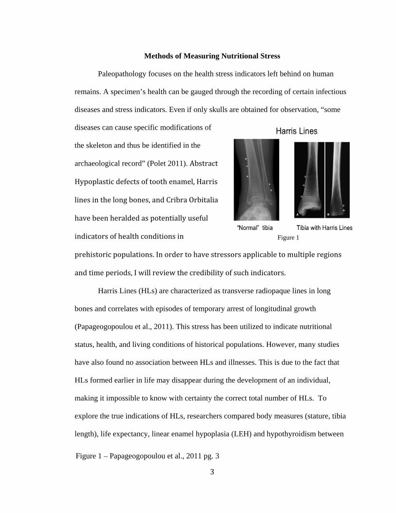
Harris Lines (HLs) are characterized as transverse radiopaque lines in long
bones and correlates with episodes of temporary arrest of longitudinal growth
(Papageogopoulou et al., 2011). This stress has been utilized to indicate nutritional
status, health, and living conditions of historical populations. However, many studies
have also found no association between HLs and illnesses. This is due to the fact that
HLs formed earlier in life may disappear during the development of an individual,
making it impossible to know with certainty the correct total number of HLs. To
explore the true indications of HLs, researchers compared body measures (stature, tibia
length), life expectancy, linear enamel hypoplasia (LEH) and hypothyroidism between
Figure 1
Figure 1 – Papageogopoulou et al., 2011 pg. 3
4
individuals with and without HLs (Papageogopoulou et al., 2011). This is key because
in most studies, the researchers failed to correlate the presence or absence of HLs with
specific stress markers and consider HLs as bone reactions to pathological or nutritional
stress. Results indicate that peaks of high HL frequency were found to parallel normal
growth spurts and growth hormone secretions, rather than a pure outcome of nutritional
or pathologic stress (Papageogopoulou et al., 2011). In accordance with the results, it
seems appropriate to discard Harris Lines as a health indicator for the purposes of this
review (Papageogopoulou et al., 2011). In the same study, linear enamel hypoplasia,
also referred to as dental enamel hypoplasia, was one of the variables measured in
accordance with number of HLs. Teeth remain the most useful ancient skeletal
specimens for detecting markers of stress, as other bone pathologies acquired during
childhood are generally difficult to diagnose in adult samples, due to bone modeling and
remodeling and thus the disappearance of lesions during bone growth (McHenry &
Schulz, 1976; Roberts & Manchester, 2005). LEH itself is seen as a dental analog of
HLs. However, there was no correlation found between the number of HLs and the
percentage of LEH (Papageogopoulou et al., 2011). Many prior LEH publications
utilize a variety of sources for LEH age determination, such as tooth mineralization
stages by Bass (1995) (e.g. Boldsen, 2007) or more conventional methods by Massler et
al. (1941) (e.g Palubeckaite et al. 2002, -Niedbala & Kozlowski, 2011).1 However, not
all projects implement consistent methodological controls when collecting LEH, which
ultimately restricts addressing potential stress. When examining stress indicators in
1 Miszkiewicz, Justyna Jolanta. "Linear Enamel Hypoplasia and Age-at-Death at Medieval (2012)
5
teeth, controls need to account for “age-related tooth loss and dental wear, and also
identify systemic stress by comparing more than one tooth type, particularly when two
or more groups are contrasted (Miszkiewicz and Jolanta, 2012).” Factors that
predispose enamel defects include low socioeconomic status, low birth weight, poor
maternal response to stress, young maternal age, and conditions that disrupt calcium and
phosphate metabolism such as vitamin C deficiency and hyposphatemia. The presence
LEH not only indicates the occurrence of stress episodes and physiological disruption
but also of recovery and continued growth (Merret et al, 2016). Many
osteoarchaeologists assign weaning related significance to ‘peak’ LEH records (e.g.
Lanphear, 1990, Iregren, 1992, Ubelaker, 1992, Moggi-Cecchi et al., 1994, Wright,
1997), while some studies have shown that such a link is weak or possibly non-existent
(e.g Blakey et al., 1994, Corruccini et al., 1985, Santos & Coimbra, 1999, Saunders &
Keenleyside, 1999, Wood, 1996).2 Weaning can be defined as the cessation of breast-
feeding and inclusion of solid non-maternal food sources into juvenile diet.3 However,
the age at which non-maternal food sources are introduced to weaned children is highly
variable among human societies as a consequence of traditional practices and cultural
influences. All of these are valid concerns, however, “the degree to which such aspects
would influence population-based health profiles is relatively low, as high frequency
and severity of LEH would still reflect a continuum of stressful events in a child’s life”
(Miszkiewicz and Jolanta, 2012).
2 Miszkiewicz and Jolanta, 2012 3 World Health Organization Website, 2002; Griffiths et al., 2007
6
Early child life is important to emphasize because there are studies of skeletal
stress indicators that lend further support to the hypothesis that early childhood stress
results in a reduced age at death. One study analyzing tooth crown size and age at death
in prehistoric Native Americans from Auerbuch, found that permanent teeth of juveniles
were significantly smaller than those of adults.4 It is suggested that environmental
stressors prevent the development of teeth
to their maximum size. Another study
shows that smaller vertebral neural canals
are associated with an earlier age at
death.5
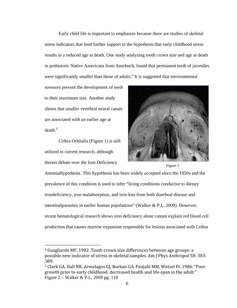
Cribra Orbitalia (Figure 1) is still
utilized in current research, although
thereis debate over the Iron Deficiency
AnemiaHypothesis. This hypothesis has been widely accepted since the 1950s and the
prevalence of this condition is used to infer “living conditions conducive to dietary
irondeficiency, iron malabsorption, and iron loss from both diarrheal disease and
intestinalparasites in earlier human populations” (Walker & P.L, 2009). However,
recent hematological research shows iron deficiency alone cannot explain red blood cell
production that causes marrow expansion responsible for lesions associated with Cribra
4 Guagliardo MF. 1982. Tooth crown size differences between age groups: a possible new indicator of stress in skeletal samples. Am J Phys Anthropol 58: 383-389. 5 Clark GA, Hall NR, Armelagos GJ, Borkan GA, Panjabi MM, Wetzel Ft. 1986. “Poor growth prior to early childhood: decreased health and life-span in the adult.” Figure 2 – Walker & P.L, 2009 pg. 110
Figure 2
7
Orbitalia and Porotic Hyperostosis. In general, “Age-related changes in red blood cell
production and cortical plasticity mean that cribra orbitalia probably forms between six
months and four years of age in response to nutritional stress” (Watts 2013). Thus the
presence of cibra orbitalia in an adult skeleton represents health issues that occurred
before the age of four. One study claims that anemia-induced marrow hypertrophy is
probably a common cause of cribra orbitalia, “other pathological processes such as
those associated with scurvy, rickets, hemangiomas and traumatic injuries can produce
sub periosteal hematomas that can lead to orbital roof lesions” (Walker & P.L, 2009). One of the alternative explanations offered was that many cribra orbitalia lesions may
results from “megaloblastic anemia acquired by nursing infants through the synergistic
effects of depleted maternal vitamin B12 reserves and unsanitary living conditions that
are conducive to additional nutrient losses from gastrointestinal infections around the
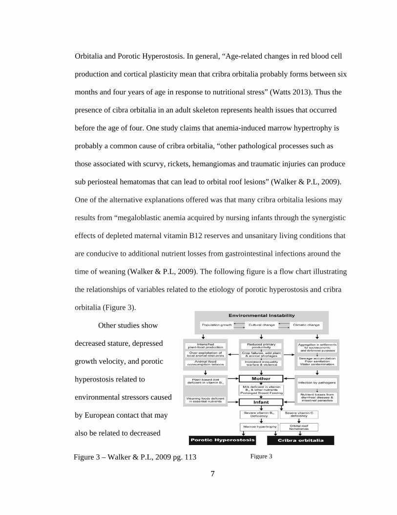
time of weaning (Walker & P.L, 2009). The following figure is a flow chart illustrating
the relationships of variables related to the etiology of porotic hyperostosis and cribra
orbitalia (Figure 3).
Other studies show
decreased stature, depressed
growth velocity, and porotic
hyperostosis related to
environmental stressors caused
by European contact that may
also be related to decreased
Figure 3 Figure 3 – Walker & P.L, 2009 pg. 113

8
longevity in adulthood (Klaus and Tam, 2009; Littleton, 2005). So if we are to measure
health via mortality and longevity of a population, there are more factors involved.
Cribra Orbitalia can still be utilized as a stress indicator as its etiology may be elusive
but still interpreted as a sign of stress. Depending on the research team and location of
certain studies, different indicators or techniques may be utilized, but cribra orbitalia
and dental enamel hypoplasia will be the over arching variables for broad comparison.
Both techniques essentially look at the process of development as a measure of health of
a society. It seems plausible to address the overarching health of a society by examining
the health of children and if they can recover from childhood stress episodes later in
life. I must also pay close attention to the age differences between individuals in each
sample and how each study reaches these dates of origin. This is because a 150+ years
difference between individuals essentially indicates different environments. In other
words, you wouldn’t compare my teeth to my great great grandmother’s teeth because
the environment/health care was completely different. Therefore, when I have a sample
ranging hundreds of years I need to treat that data with a bit of caution and perspective.
9
Case Studies
Among archaeologists to this day, Rapa Nui is portrayed as a cautionary tale of
ecocide and carelessly consuming resources. There is no doubt that such a message
should not be passed on, but this story can hardly be justified by actual biological
evidence. It is the most isolated inhabited island of the Pacific, which probably factors
into why it is so popular amongst archaeologists (Hamlin 1939). Unfortunately, “the
disappearance of the majority of the ancestral culture holders during the slave raids and
the epidemics led to the loss of most of the information relating to the history of the
island and its traditions” (Polet 2011). Caroline Polet wrote a paper gauging the relative
health and diet of ancient Easter Islanders based on human skeletons discovered
between 1934 and 2009. The percentage of adults and children on Rapa Nui with
enamel hypoplasia and the percentage with Cribra Orbitalia are in the range of variation
of other histories and prehistoric samples. However, “these are much lower than
European medieval populations from the 6th to the 15th century AD” (Polet 2011). This
suggests that the people of Rapa Nui may have actually been healthier than people in
the middle ages, disproving the notion that the islanders collapsed due to malnutrition as
some previous scientists have claimed. Polet also concludes “infantile malnutrition was
far from being severe as opposed to the catastrophist theories proposed by Diamond”
and “the palaeopathologic study reveals, nevertheless, new cases of infectious diseases
probably resulting from contact with Europeans or Americans” (Polet 2011).
A dissertation submitted to the graduate division of the University of Hawaii by
John Vede Dudgeon also supports the idea that Rapa Nui was a stable environment
prior to European contact by studying a total of 88 dental specimens from the island.
10
They utilized laser ablation inductively coupled plasma mass spectrometry of the
subsurface dental enamel to test for the presence of minor and trace elements in the
structure of enamel during formation. These elements in the enamel were then
compared to the “elemental signature from soils and sediments that were pedogenically
derived from the different volcanic flow events on the island” (Dudgeon 2008).
Statistical comparisons between these two datasets were used to confirm residential
stability because “the signature of the trace elements incorporated in the dental enamel
during permanent tooth formation in early life [was] similar to the trace element
compositional variation in the sediments of the location of recovery of the skeleton, and
the skeletons [could] be chemically grouped by their location of recovery” (Dudgeon
2008).
However, with all this wealth of information available, there are scientists who
have built up an exciting yet false story about the island. In the early stages of discovery
and digging on Easter Island it is clear that there are some biases between the native
Islander’s and the Eurocentric perspective on its people. However, even with those
biases put aside, there is not enough reliable evidence to conclude that Easter Island’s
ecosystem was fragile enough to destroy the once thriving native civilization.
Diamond’s claims that the natives “had the misfortune [of] living in one of the most
fragile environments, at the highest risk for deforestation, of any Pacific people” is
entirely an assumption, which leads to the cautionary tale that “of a society that
destroyed itself by overexploiting its own resources” and therefore, a cautionary tale to
every modern society (Diamond 2005). Even though there is a positive message in this
11
fabricated story, in reality, deforestation is blamed for the population collapse that
occurred as a result of Old World diseases being introduced to the native population.
In other words, Rapa Nui is not a tale of ecocide, cannibalism, or warfare, but
really a story of brilliant engineering and survival. Perhaps Mr. Hunt said it best when
he claimed “They did exactly the right thing for that island...they avoided collapse, on a
small poor, and isolated island. They are the story of remarkable success. The only
collapse came with the diseases introduced from the outside world after 1722 and as a
result of it”.6 Paleopathology, in this case, has added to the archaeological record of the
region.
The sweet potato connects Hawaii and Easter Island. This incredible human link
between lands 4000 miles apart is a fair enough reason to compare these regions. We
can now examine Hawaii, as Polynesians were the first people to arrive around the
fourth and fifth centuries A.D (Pietrusewsky 2014). Pietrusewsky and M.T. Douglas
studied the skeletal and dental biology of twenty-four sets of human remains disinterred
in 1988 from a late historic Hawaiian cemetery in Kailua-Kona. This is an ideal sample
to compare to Rapa Nui because the specimens are generally from the same time period
as Polet’s sample. The ideal situation is that a “cemetery reflects the demographic
profile of the community which contributes to it”, but this seems to be a unique sample
because of the large number of fetal remains (Pietrusewsky 2014). Paleopathological
observations of the remains include “a low dental caries rate and slight periodontal
disease reflecting the young age-at-death of the sample” (Pietrusewsky 2014). They did
find evidence of metabolic and nutritional stress, more specifically, Cribra Orbitalia.
6 YouTube National Geographic Live https://youtu.be/rut16-AfoyA
12
However, this was only observed in the remains of a single middle-aged female.
Enamel hypoplasia was also present in 15% of the permanent teeth (Pietrusewsky
2014). Already we can see similar diseases present in two different environments that
contained the same origin of people. The similarities also support Polet’s claims that the
percentage of people in Rapa Nui who had hypoplasia and Cribra Orbitalia were not
severe in comparison to other regions, such as Hawaii in this instance.
Paralleling Hawaii with Rapa Nui, its not an unfair suggestion that Europeans
may have simply introduced a variety of infectious diseases that could easily devastate a
population of isolated island inhabitants. They discovered that enamel hypoplasia is
more common in teeth from historic skeletons than in those from pre-contact times. The
comparative data suggests “these indicators of general stress occurring at early ages
were more common in contact times than in prehistory” (Pietrusewsky 2014). However,
for Cribra Orbitalia, no significant difference in pre-contact and post contact adults was
observed. They also found an individual female that has vertebral lesions consistent
with tuberculosis. This is important because no signs of skeletal tuberculosis were
recorded before, which may reflect “the arrival of new diseases associated with contact
prior to the noticeable changes in mortuary practices” (Pietrusewsky 2014). There is
only one published case of possible treponematosis from Hawaii prior to Captain
Cook’s arrival, and DNA from this skeleton reveals “the nine base-pair deletion seen
frequently in Asians and Polynesians” (Pietrusewsky 2014). The chapter reveals
significant differences between pre-contact and post contact skeletal remains for a
variety of health indicators. Overall, the investigation illustrates deterioration in health.
The study also states that certain infectious diseases, “such as tuberculosis and
13
treponematosis (yaws and syphilis), traditionally assumed to have been introduced to
Hawaii with Western contact are now either confirmed or challenged by the
osteological record” (Pietrusewsky 2014).
As a stand-alone study, this paper follows the typical pattern of a
paleopathologic study. It ultimately points to the underlying issues facing this particular
sample, which was the introduction of new infectious diseases coupled with lower
fertility. Researchers also mark the transmission of certain diseases, making this study
valuable to the medical community. One small detail that may easily be overlooked is
the fact that native scholars were referenced and utilized in this study. This is not
always the case, and “outsiders” digging up graves of native people’s may be
troublesome for researchers who need permission for such research. It works in
everyone’s best interest to recruit the guidance and input of local scholars as they
provide insight into history with all the cultural nuances being addressed as well. This
case also challenges the historical perception of these early European explorers, as they
are usually revered in their homelands. However, when looking closely at the natives, a
serious decline in overall health due to new infectious diseases is most commonly the
case. Although reliability may be in question, comparing the skeletal sample to
surrounding samples is the ideal method when researching a region, as even settlements
that are near each other may have a number of factors and/or lifestyle habits that
contribute to completely different health profiles.
The researchers attributed the slight increase found in the post-contact to more
stress during childhood rather than survival, once again pointing to the significance of
childhood stress .In most cases, methodological differences in recording and reporting
14
frequencies limit the use of other researchers' data. However, this study emphasizes how
in Hawai'i “we are fortunate that workers are increasingly adopting a common standard
for collecting osteological information (Pietrusewsky 2014).” Oddly after stating this
fact, the researchers state that they will only emphasize the data in this particular study
and not others due to “incompatibility issues”. The inclusion of an infectious diseases
section provides health-related insight that can be helpful towards modern medicine and
should be included in the normal agenda of paleopathology.
Another comparable case study to Hawaii and Rapa Nui would be to look at the
health of the ancient Mariana Islanders. I include this study because it focuses more on
the environmental factors that may have influenced the islander’s health in prehistoric
conditions and re-examines the question of inter-island variability in health and lifestyle
of the natives. The native people called the Chamarro were hypothesized to have higher
stress levels on smaller islands, as smaller lands are more susceptible to “environmental
perturbations which would affect human health because of smaller land mass,
remoteness, lower rainfall, freshwater lens, slightly cooler temperatures, narrow and
non-continuous fringing reefs, earthquakes, volcanic eruptions, and sea-level
fluctuations” (Pietrusewsky 2014). The four islands in question include Tinian, Saipan,
Rota, and Guam. These islands are located in the southern area of the archipelago, and
are also the largest islands, which indicates why people preferred to live there in the
past and presently. As far as the people go, it has been suggested that the northern
Philippines is a possible source of the earliest human settlements. However, recent
DNA evidence suggests that the Mariana Islands were initially settled approximately
4000 years BP by a small group of people from Southeast Asia. This initial population
15
remained genetically isolated until approximately 1000 years BP when another wave of
migrants arrived. Pre-contact agriculture and economies included reliance on
“cultivated tree and root crops typical of tropical island environment such as coconut,
banana, breadfruit, taro, yam, and possibly rice – all foods high in carbohydrates”
(Pietrusewsky 2014). The diet was also rich in marine sources such as fish and shellfish.
Along with agriculture, the Chamarro relied on hunting and gathering tactics to catch
birds, fruit bats, monitor lizards, turtles, and crabs. Taking the natives geographic
landscape, diet, and social background into account, I can now analyze the skeletons
with a broad perspective in mind. The main stress indicators that were found on this
island suggest cribra orbitalia, dental enamel hypoplasia, stature, long bone fractures,
spondylolysis, tuberculosis, and leprosy (Pietrusewsky 2014).
Previous research suggested that the precontact inhabitants of the smaller
islands, such as Rota, Tinian and Saipan, were less healthy than those from Guam, the
largest Island. However, tests of statistical significance in the frequencies of 13
indicators between the four largest islands revealed, “the majority are not statistically
different” (Pietrusewsky 2014). The results suggest greater similarities in health and
lifestyle in skeletons from Rota and Guam in contrast to those from Tinian and Saipan.
Unexpectedly, several frequencies of indicators from Rota, the smallest island, were
similar to those from Guam, the largest island. The proximity of the islands may explain
this finding, more specifically, a formation of large island/small island partnership.
This case is intriguing as it goes against what previous research indicates, which
is that smaller islands struggling more than larger islands. The study really focuses on
possible sex differences, a variable that may be influential in discerning the health
16
profiles of a society as men and different are biologically different and quite possibly
may react differently to stress. Women’s health is a hot topic in the U.S right now, as
most of the current treatments available for modern illnesses are based on research
conducted only on men, as it was perceived to be too harmful for women. This
ultimately resulted in treatments/procedures that are less efficient or unhelpful to
women. Utilizing paleopathology, the research side of medicine could benefit greatly
from sexual differences indicated when observing health insults on the body. By doing
so, they can better strategize how to account for these differences when progressing the
field of women’s health research. As far as this particular paper goes, sex differences
were discovered but they were not statistically significant. However, that does not mean
that these differences should not be observed in other studies, which may look at
different indicators/environments. The paper utilizes 13 different health indicators,
which seems to be a bit too broad for the purposes of gauging a society’s health. All 13
indicators must have previously established credibility, and there must be a logical basis
for why these are chosen. The researchers present fair interpretations of their results
across all indicators yet spend little time establishing the reliability of each one.
Although much more research is needed, this study offers some answers to
questions regarding intra- and interisland variability in skeletal indicators of health,
temporal changes in health, and variability by sex and age. This study is also
comparable to the Pietrusewsky’s Hawaii analysis of pre-contact and post-contact as the
natives of the Mariana Islands faced a similar stressor. However, the researchers only
focused on signs for treponemal infection instead of including others. Uneven and small
sample sizes make it difficult to reliably view this skeletal series as representative of the
17
population. Any stress indicators involving dental health is difficult to interpret, as
differences seem to be related to the use of the betel nut. The influence of the chewing
of betel is debatable as some believe it protects from dental caries while other research
illustrate how periodontal disease and dental calculus may be greater in chewers
(Chatrchaiwiwatana 2006). A WHO study analyzing the affects of betel chewing found
that there are multiple confounding variables such as “level of oral hygiene, dietary
factors, general health, and dental status”.7 Thus, results regarding dental
pathology/indicators in this paper must be approached cautiously, but will not affect
LEH results as these lesions form during early childhood and the chewing of the betel
nuts as adults would have no effect. However, the methods chosen by these researchers
are taken from Pietrusewsky’s 2002 paper, which provides a reliable baseline for
comparison, at least to Pietrusewsky’s and Douglas’ work.
From the islands we move to examine a particularly interesting source of
skeletal remains dating from 17-19th century Caribbean slave population in Barbados.
After slavery ended in the British Empire from the middle of the 17th century to 1834,
the wealth of Barbados depended on the agricultural economy of the tens of thousands
of slaves of African birth or descent. Thus, by the early 19th century, a much greater
percentage of the slave population was native-born. This contrasts to earlier periods in
which a greater percentage of the slaves were African-born and thus African cultural
patterns were also followed to a much greater extent. According to researchers, “This
7 International Agency for Research on Cancer (IARC). 2004. Betel-quid and Areca- nut Chewing and Some Areca-nut-derived Nitrosamines, Volume 85. Lyon, France: Monographs on the Evaluation of Carcinogenic Risks to Humans.
18
population is probably unique in terms of environment, nutritional deficiency and other
severe environmental stresses, and (especially) association with historical resources that
might allow more specific correlation of stresses with hypoplasia chronology” (Robert
S 699). The specific site chosen was a slave cemetery at Newton plantation because of
the availability of historical documents as well as the permission of excavations in
relatively undisturbed contexts. Researchers concluded that “Newton typified medium-
to large-scale Barbados sugar plantations of the slave era; thus the analysis of Newton’s
slaves may be generalized to other plantation slave populations on the island”
(Corruccini 1985).
Of the 103 individual specimens observed with teeth, 56 were hypoplastic
(54.5%). This is the earliest and largest excavated group of African and African-
descended slaves reported from the Caribbean and North America. The historical
evidence indicates Caribbean slaves experienced nutritional deprivation, which would
cause severe developmental disruption. From this data, researchers concluded that
hypoplasia was most likely to occur around age three for any given individual slave
surviving infancy. The tendency for more numerous and serious hypoplasia’s to occur
late in calcifying teeth may possibly be related to a late weaning period in Caribbean
slaves. Documentation illustrates how two Barbados-born medical doctors who
practiced among plantation slaves alluded specifically to the risks of the weaning
period. One doctor in particular attributed high infant mortality to “the highly improper
food mothers gave their children” as well as “the period of nursing the child being
protracted to an age when it is both injurious to the mother and infant” while the other
doctor also assigned the cause for high infant mortality to housing conditions

19
(Corruccini 1985). The tendency for hypoplasia to form 3-4 years in postnatal surviving
Barbados slaves is late in comparison with other groups studied in this paper such as
Chicago Whites and medieval Swedes. With the exception of medieval swedes, the
Barbados slave total hypoplasia distribution appears less pronounced peaks or
leptokurtic.
The strength of this paper lies within the fact that it is a snapshot of a slave
population. This ultimately represents a “negative control” in the sense that it should be
one of the poorest if the not the worst health profiles on the spectrum. This is also an
interesting case in regards to LEH, as typically the high rate of LEH would be attributed
to weaning children too early rather than taking too long as in the case of the Barbados
mothers. For a deeper understanding of the health implications of LEH alone, this study
provides an interesting counter perspective to the weaning hypothesis most commonly
accepted in current literature. This case also provides insight as to how a colonial
economic system affected the health of the oppressed that carried on the same line of
work, even when the colonizers are gone. The deeper embrace of African culture by the
newborns of the previous enslaved generation did not adequately buffer the health
insults, and may in fact, have exacerbated it instead. The testimonies of two medical
doctors who lived alongside the sample being studied gives this paper a unique window
into the not only the underlying issues facing the people, but also the medical
perspective embraced by local physicians at the time. It would be a fascinating
comparison to compare the medial diagnoses of these two physicians to two modern
day physicians who practice in the same area today. This case is a perfect example of
20
how Paleopathology can open up new avenues of further study that can add value to
multiple scientific and medical fields.
The methods of this paper towards LEH is interesting as they hone in on the
central tendency of 3-4 years for the formation of hypoplasia’s. They analyze the
percent concentration between 3-4 years and the residual occurrences at 4+ years
compared to other reported populations. The assumption here is that hypoplasia peak is
related to the post-natal time of maximum environmental/metabolic stress on children in
non-industrial societies. The age of hypoplasia formation in Barbados slaves is late
whereas “Chicago Whites peak at 0-1 year, composite prehistoric Amerinds and
Missouri Mississippian Amerinds peak at 2-3 years, and Dickson Mound combined pre-
agricultural and agricultural Amerinds and medieval Westerhus Swedes peak at 2.5-3
years” (Corrucini 1985). One downfall of this study is the lack of demographic
information regarding the skeletal sample, to which the researchers admit is
“unavoidably subjective” (Corruccini 1985). The classification of enamel hypoplasia is
based on modifications of earlier sources, which include three different forms of
hypoplasia. Although this particular method is somewhat based on previous work, the
modifications make the data difficult to compare to others. However, the true value of
this study is not seen through comparison, but more so as an individual unique study
analyzing factors specific to these people in this time period. It is important to note that
the weaning hypothesis regarding LEH is supported, as this is the first study observing a
specific population wherein hypoplasia concentration is shown to occur the year
following a historically documented time of weaning.
21
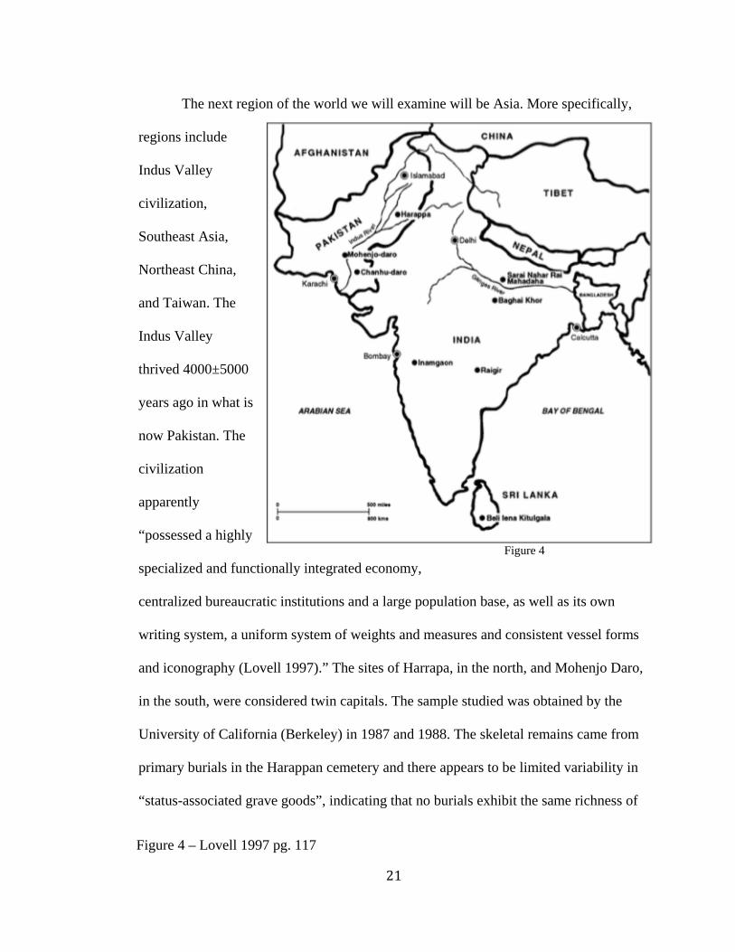
The next region of the world we will examine will be Asia. More specifically,
regions include
Indus Valley
civilization,
Southeast Asia,
Northeast China,
and Taiwan. The
Indus Valley
thrived 4000±5000
years ago in what is
now Pakistan. The
civilization
apparently
“possessed a highly
specialized and functionally integrated economy,
centralized bureaucratic institutions and a large population base, as well as its own
writing system, a uniform system of weights and measures and consistent vessel forms
and iconography (Lovell 1997).” The sites of Harrapa, in the north, and Mohenjo Daro,
in the south, were considered twin capitals. The sample studied was obtained by the
University of California (Berkeley) in 1987 and 1988. The skeletal remains came from
primary burials in the Harappan cemetery and there appears to be limited variability in
“status-associated grave goods”, indicating that no burials exhibit the same richness of
Figure 4
Figure 4 – Lovell 1997 pg. 117
22
elite burials such as in ancient Egyptian or Mayan civilizations (Lovell 1997). The site
contained 92 individuals, however, only 29 crania were preserved for assessment due to
poor preservation. Only two of the 29 crania recovered displayed cranial lesions of
porotic hypersostosis, suggestive of chronic anemia. However, in a sample of 40
individuals obtained from Mohenjo-Daro, seven individuals (18%) displayed porotic
hyperostosis, and both children and adults were affected. It was also reported that 25%
of several hundred individuals that comprise a total human sample from various sites of
the Harappan civilization contain vault lesions. Although nearly one third of the 92
individuals at Harappa displayed pathological lesions, enamel hypoplasia was exhibited
in 72% of permanent dentitions. However, enamel hypoplasia was not observed in any
skeletons from Mohenjo-Daro. Researchers conclude that the etiology of anemia, which
was already debated as the main source of cribra orbitalia, may be best explained by
multiple factors in each physical and social context. The data indicates that anemia was
more prevalent in Mohenjo-Daro. This location can be categorized as a highly
urbanized center which often “exhibits high frequencies of acquired iron deficiency due
to the prevalence of infectious diseases caused by contaminated water supplies and
close contact of inhabitants” (Lovell 1997). The lower prevalence in Harappan may be
attributed to a good nutritional base in a diverse ecological setting, and few
gastrointestinal and other infections due to high standards of personal and community
hygiene (Lovell 1997). The iron deficiency anemia may alternatively result from dietary
inadequacies of intake or absorption or genetic inheritance. Nutritional deficiencies are
an unlikely explanation as conditions such as Rickets and Scurvy are rare in South Asia.
In this sample in particular, the Harappan skeletons indicate episodic morbidity, but
23
reflect acute rather than chronic stress because this sample is not associated with high
frequencies of porotic hyperostosis (Cribra Orbitalia) or periostitis.
This paper hones in on the affects of transitioning from a primarily nomadic
lifestyle to a more sedentary, urbanized society. One would assume that the
technological advances implemented would reduce stress, but this does not seem to be
the case. However, the fact that the sample indicated acute rather than chronic stress
implies that it was not necessarily sedentism that deteriorates health in this scenario.
There is not solid obvious factor in this study that explains the results, but the standards
of hygiene is a logical conclusion for the researchers to make. This parallels medieval
Europe, which struggled with hygiene, creating the perfect breeding grounds for
diseases to flourish. In this literature review, this is perhaps the most advanced
civilization included in the analysis, and it would not be unreasonable to compare to
modern day civilizations as well. The established society, thriving on new technologies
and systems of order, acts as a basis of comparison to either other greatly advanced
civilizations or any societies today. This allows researchers to really narrow and focus
their efforts on the confounding variables interacting with the health of peoples who, in
theory, should be healthier than before such advances.
This paper heavily focuses on the probable causes of anemia through physical,
sociocultural, and environmental evidence. However, they come to the conclusion that
many factors are involved, hardly a surprise. This case in particular may be difficult to
compare to others simply because of all the possible explanations offered. There would
have to be some similarity between regions, such as an endemic or environmental
disaster, to provide a basis of comparison. In regards to Mohenjo-Daro, environmental
24
differences related to Indus flooding and malaria may explain the relatively greater
frequency of porotic hyperostosis. Going with this theory, researchers claim that there
may be a genetic anemia present at Mohenjo-Daro. The reasoning behind this theory is
based off the geographic distribution of inherited abnormal hemoglobins and
widespread malaria in South Asia. However, the scarcity of skeletal remains provides
no evidence to confirm this interpretation. The Pakistani state of Sind, where Mohenjo-
Daro is located, harbors mosquito vector A. culcifacies, which feeds primarily on cattle
but also humans if cattle are scarce. It seems humans are the primary food source in
urban areas where the mosquito breeds, but it is still a stretch to assume that this
endemic was in full swing in the past because it is presently. This paper does have
medical value as medical historians were previously unconvinced of malaria until
Roman Times89. The discovery of fossilized remains of several types of blood-sucking
mosquito expose their existence in the Old World for over 60 million years. This is
what led researchers to theorize that “irrigation practices and the ponding of water after
river flooding may have helped form breeding places for malaria-bearing mosquitos and
endemic malaria may have followed from the commencement of sedentary subsistence
technologies in South Asia (Lovell 120).” Regardless of the legitimacy of this
interpretation, this discovery adds to the pathological history of malaria in the Old
World and benefits the medical community. Although no definitive answer was
presented, the researchers thoroughly consider all pathogenic, environmental, and
societal factors that could logically contribute to the big picture overall. Ultimately, this
8 Ackernecht, E. H. History and Geography of the Most Important Diseases. New York: Haffner, 1965. 9 Henschen, F. The History of Diseases. London: Longmans, 1966.

25
paper provided more definitive insight into the migration patterns and effects of malaria
than the causative agent(s) postcranial lesions.
We will now direct our attention to Northeast China, honing in on the Dongbai
plain, which acts as the boundary between sedentary agriculturists and nomadic
pastoralists. Examining this region allows us to explore the relationship between stress
exposure, subsistence, and cultural complexity. The human remains of one study were
recovered from the upper level of the Houtaomuga site on the Dongbie plain. These
specimens date back to the Hanshu II cultural period (2250-2050 BP) (Merrett 2016).
Results show overall 72.73% of the individuals examined (40/55) exhibited Linear
Enamel Hypoplasia (LEH). In adults alone the prevalence is 96.8% (30/31). These
prevalence’s are similar to those of Hokkaido Jomon, slightly higher than the two other
Jomon data group, and much higher than those observed in Inui and Neanderthal
foragers. The follow table shows comparison across the regions (Table 1).
Table 1
Table 1 – Merrett et al, 2016 pg. 5
26
Among prehistoric populations, hunter-gatherers live to approximately 21.6 years
(SD =2.1 years), horticulturalists live to approximately 21.2 years (SD=3.9 years),
and agriculturalists live to about 24.9 years (SD=8.5 years) (Wolfe 2014). The
distribution of LEH across age categories by individual varies significantly. High LEH
was also observed in Hokkaido Jomon (73.9%, N=23), both Coastal and Inland Jomon
(61.1%, N = 69) foragers from Japan, and in south Asian Mesolithic foragers from
northern India (83.3%, N=57) (Merrett et al, 2016). This contrasts with past forager and
modern sample in which the proportion of LEH is much lower at between 6-49%. What
is particularly interesting about the Houtamuga site is that almost all who survived to
age 4 years (36/37) had experienced at least one severe stress episode. According to
researchers, if the older women without LEH (1/13) had lost her LEH due to 50% tooth
crown attrition, then everyone at Houtaomuga who survived past the age of four years
had experienced substantial stress episodes in childhood. Therefore, the children of
Houtaomuga represent a highly stressed and vulnerable faction of the society (Merrett et
al, 2016).
Regardless of the high prevalence of LEH, there is high survivorship data that
includes “timing of stress episodes relative to developmentally critical times, pathogen-
specific factors, and archaeological/cultural context. Perhaps stress events did occur in
critical times during development and cultural buffering may have been at least partly
effective. Or perhaps stressful events did occur during critical times but subsided later
in life so that later survivorship was not compromised. Stress duration was not
correlated with subsistence strategy, since “Houtaomuga inland hunger-gatherer-fisher-
millet cultivators exhibit similar mean duration to coastal Hokkaido Jomon foragers,
27
significantly longer duration than inland Jomon foragers, and significantly shorter than
the Coastal LF Jomon foragers, Inuit, and Neanderthal hunter/gatherers (Merrett
2016).” Location relative to coastal resources also does not appear to show any
relationship to stress duration, indicating inland HTMG and coastal Hokkaido as similar
(Merrett et al, 2016).
This high prevalence of LEH is astonishing for a time period that was politically
stable. It is possible that climatic conditions juxtaposed with cultural factors such as
cultural complexity may have an impact on population exposure to stressors. It is
suggested that the facilitation of control of resources may have predisposed the
population with periods of climatic stress, crop failure, and food shortages. Modeling
data using the climate model NCAR CAM2 indicates that the precipitation in Northeast
China has been driven by the East Asian summer monsoon system. Slight changes in
climate would also be predicted to result in crop failure every 3 or 4 years. The fact that
almost all of the individuals of the population managed to survive enough to develop
LEH and recover illustrates strong resilience, which can be argued as a genetic
component but is most likely due to a diversity of subsistence practices, cultural
buffering mechanisms, climate fluctuations, and adaptive phenotypic plasticity. The
broad-spectrum economy may have increased immune competence and reduced stress
episodes, thus compromised survival later in life did not occur. It can also be inferred
that during years of reduced food resources, there may have been more communication
and interactions with other communities, thus increasing potential for pathogen
exchange. Factors relating to economic, social, cultural, biological and pathogen loads
juxtaposed within a setting of climate instability contribute to the high LEH prevalence
28
and variation. This complex milieu of relationships highlights the importance of site-
specific context when evaluating the implications of LEH prevalence and other stress
parameters amongst human remains (Merrett et al, 2016).
Researchers did not choose this region to study in order to prove a certain theory
or the prevalence of certain factors, but because these people played important roles in
the formation of Chinese civilization. To put this in perspective, the previous paper
(Lovell) veered more strongly to the medical side of paleopathology. This paper leans
more towards the archaeological side, with the goals of truly understanding the people
rather than the health of the people. This paper truly embraces all the potentials of
paleopathology by considering all possible factors that could be involved in this case.
Unlike most other papers, this one does not seem to follow a predisposed set of methods
directly aimed to prove a certain theory. Rather, this group of researchers seems to
embrace an open mind whilst conducting research. This makes for a much longer debate
as to what the true causes are in regards to this sample’s health, but it is a more
thorough analysis of the situation which holds more value in the end.
This paper is also similar to Lovell in the sense that Lovell attributed a myriad
of factors to postcranial lesions and Merrett also lists multiple elements to the
development of LEH. Unlike the previous studies, this study interprets the presence of
LEH as not only an indicator of stress episodes and physiological disruption, but also as
recovery and continued growth. This paper aims to narrow down the number of stress
episodes, calculated as ranging from 17 to 28 days at the low end to 98 to 154 days for
the longer duration events, and compare this to other regions. This is a good sign, as
these results will have more credibility due to common data collection techniques.
29
Similar to the Mariana Islands paper, climate is examined as a possible influence of
stress for this time and region. It’s worth noting that this paper also includes adults, and
not just LEH in children. However, the minimum standard for being included in the
study was that each skeleton had to have at least one anterior tooth, with only 4 out of
55 at this minimum level. This means that there are significantly less teeth present per
individual, and may skew the reliability of results. In other words, it may be difficult to
compare this study with a sample of 55 skeletons because not all the teeth are available.
They detect defects utilizing methods presented in Skinner et al., (1995) and Armelagos
et al. (2009), a methodology not referenced in the previous studies. Regardless of what
methodology is quoted, the researchers make numerous compromises and minimum
standards due to the poor condition of the specimens overall. At the same time, these
modifications are needed to obtain meaningful results and counteract all the obstacles
inherent in the sample. This makes this data set extremely difficult to compare, but that
does not mean it offers no insight of value, results will simply remain credible for this
particular region and time. They do estimate duration of episodes, similar to Merrett,
ranging from 16 to 102 days. The sample at Houtaomuga showed the highest prevalence
of LEH observed so far in this review even when taking into consideration and the
political stability and reliability at the time. These factors were not enough to buffer the
stress, which seems odd, as previous studies have linked LEH to nutritional
deficiencies1011. Perhaps the more valuable branch of the paper is in regards to climate.
10 Sweeney, E.A., Saffir, A.J., de Leon, R., 1971. Linear hypoplasia of deciduous incisor teeth in malnourished children. American Journal of Clinical Nutrition 24, 29-31. 11 Masumo, R., Bårdsen, A., Åstrøm, A.N., 2013. Developmental defects of enamel in primary teeth and association with early life course events: a study of 6e36 month old
30
Previous research has shown that climactic changes have preceded changes in human
societies before, sometimes as serious as societal collapse (Mayewski et al., 2004;
Weninger et al., 2009; Hsiang et al., 2013). Previous examples include Mesopotamia,
Thailand, and Peru (de Menocal, 2001; Wu and Liu, 2004). This paper emphasizes the
importance of site-specific context in evaluating the meaning of LEH prevalence, an
approach that should be adopted by all future studies.
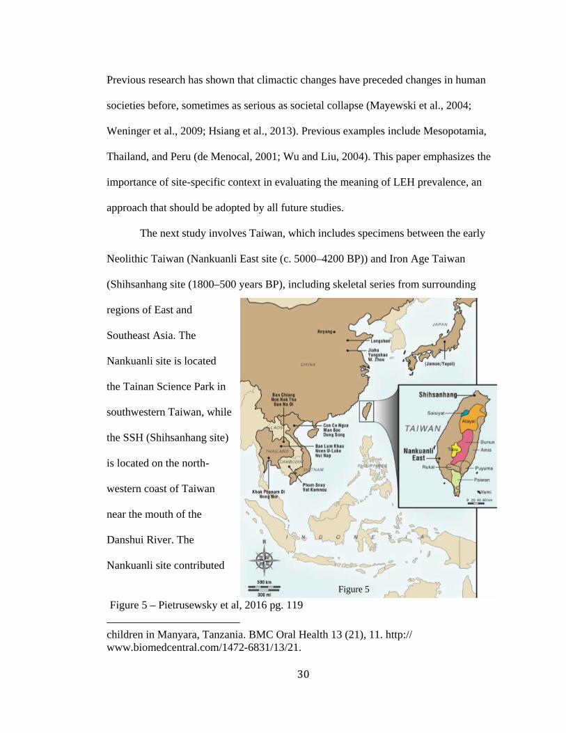
The next study involves Taiwan, which includes specimens between the early
Neolithic Taiwan (Nankuanli East site (c. 5000–4200 BP)) and Iron Age Taiwan
(Shihsanhang site (1800–500 years BP), including skeletal series from surrounding
regions of East and
Southeast Asia. The
Nankuanli site is located
the Tainan Science Park in
southwestern Taiwan, while
the SSH (Shihsanhang site)
is located on the north-
western coast of Taiwan
near the mouth of the
Danshui River. The
Nankuanli site contributed
children in Manyara, Tanzania. BMC Oral Health 13 (21), 11. http:// www.biomedcentral.com/1472-6831/13/21.
Figure 5
Figure 5 – Pietrusewsky et al, 2016 pg. 119
31
twenty-four of the most complete adult burials representing 14 males and 10 females to
this study. Twenty-three of the best-preserved and most complete adult skeletons from
the SSH site (Pietrusewsky and Tsang, 2003) are used in the present study.
Based on previous research (Cohen and Armelagos, 1984; Larsen, 2006; Cohen
and Crane-Kramer, 2007; Temple and Larsen, 2013), it is predicted that the livelihood
of Iron Age Taiwan will be associated with an increase in systematic stress and certain
dental indicators of health when compared to the early Neolithic communities in
Taiwan. Thus, frequencies of childhood stress (CO and LEH) and dental pathology are
expected to increase from early Neolithic to later Iron Age Taiwan. This studies also
observes CO lesions and LEH linked to iron deficiency anemia as a result of nutritional
deficiencies, especially during early childhood, infectious diseases, gastrointestinal
parasitic infections leading to infant diarrheal disease, as well as hereditary hemolytic
anemia’s.
Contrary to expectations, results show the frequency of LEH significantly lower
in the Iron Age skeletons (37.1%) (Pietrusewsky et al., 2013) and the expected increase
in dental pathology (AMTL, caries, and alveolar defects) in the Iron Age skeletons was
not confirmed. Few differences of systemic stress indicators between the Neolithic and
Iron Age Taiwan series were found, suggesting there is no increased reliance on cereals
or change in emphasis between millet and rice. Instead, researchers propose that the
subsistence base remained very broad, including marine, riverine and terrestrial re-
sources. They also suggest a decline in childhood stress over time that may be linked to
improvements in food- processing techniques, better weaning foods, and less
contamination of food/water in later Iron Age Taiwan (Pietrusewsky et al., 2013).

32
The researchers run into an issue comparing the prevalence of CO among the
locations, suggesting differences in the methods of recording this stress indicator. This
is due to a lack of standardization in reporting data in the studies conducted by different
researchers. Regardless, their numbers show some of the highest frequencies of CO in
the region were collected from early skeletal series from Vietnam and the Khok
Phanom Di series from Thailand. While on the other hand, some of the lowest
frequencies of this indicator are skeletons from sites in Northeast Thailand, and early
and middle Yangshao skeletons from China. The frequencies of CO in the NKLE
skeletons are most similar to the skeletal series from China and Thailand. As reported in
Pietrusewsky et al. (2013), LEH is moderately elevated in the NKLE series, suggesting
that the early Neolithic inhabitants of Taiwan experienced relatively high levels of
physiological stress during childhood.
This paper also compares a more active lifestyle to a more advanced, yet
sedentary lifestyle. However, this is unlike Northeast China in the sense that it is the
same location over a span of time rather than two different societies coexisting during
the same era. The researchers ultimately conclude that the lack of difference is due to a
broad diet, ultimately pointing to the fact that the natives basically satisfied their
nutritional needs early on and didn’t feel the need to change core elements, regardless
of the new technologies at hand. However, the way foods were processed and prepared
were improved which would have a slight impact on results, but not significant enough.
The paper seems to have tunnel vision as it focuses on the nutritional profile of the
natives and not delving too deep into the cultural, climatic, or political atmosphere. It
does reference other close areas for comparison, which should be included in every
33
paper as it better represents the population.
Unfortunately, it is hard to consider these comparisons due to the lack of
standardization of recording techniques, a reoccurring problem in these studies. On the
other hand, research methods are based off of Pietrusewsky et al. (2013), which
provides a basis for comparison although it is no surprise that the researchers are using
the same methods they’ve already established themselves. Hopefully this is a sign of
confidence in techniques and not blind obedience to what seems to work without taking
into consideration other research techniques. This study found very little differences in
contrast to the people of Northeast, who also went through a similar transition in
lifestyle. Similar to the natives of the Mariana Islands, chewing the seed of the areca
palm (Areca catechu L.), which happens to actually be incorrectly referred to as the
betel nut. It’s amazing to find this cultural practice spanning from SouthEast Asia to the
Mariana Islands. This nut is a confounding variable regarding dental defects, however,
because both peoples engage in this practice, it would not be unreasonable to compare
these two data sets. However, this study looks at cribra orbitalia and not LEH in
particular. This study is meant to be observed as a unique, isolated case, as the paper
even states “ there are limited comparative data for skeletal series outside of Taiwan
that overlap temporally and spatially with those from NKLE and SSH” (Pietrusewsky
2016). This study also seems to focus on the differences between males and females, a
category of analysis not emphasized in the previous studies except for the Mariana
Islands paper.
The next region of examination will be Europe, in England and the UK. Linear
Enamel Hypoplasia and Age-at-Death was recorded from medieval human skeletal
34
remains recovered at the St. Gregory’s Priory and nearby Cemetery in the Northgate are
of Canterbury (Kent, UK). This monastery was established in the 11th century and
functioned until early 16th century (Miszkiewicz & Jolanta, 2012). This structure was
established for clergy to aid the sick and provide burial services for financially
disadvantaged members of society. According to historical and archaeological textual
records, the individuals buried at the priory (Priory n=19) were of a higher
socioeconomic status (SES) than those buried in the cemetery (Cemetery n=30)
(Miszkiewicz & Jolanta, 2012). This presents a much more controlled and limited
comparison between socioeconomic status rather than a sample that is representative of
the entire region, providing a controlled view of how SES can affect health.
Researchers chose to adopt Ried and Dean’s (2000, 2006), work on tooth
formation times based on histological examinations that take into account hidden
(appositional) enamel (King et al. 2002) in order to obtain reliable data regarding LEH
formation age (Miszkiewicz & Jolanta, 2012). Results indicated that hypoplasia was
recorded in the majority of individuals, with only two samples from the cemetery and
four samples from the Priory displaying unaffected dentition. Both groups were almost
completely affected by LEH, but Cemetery individuals displayed significantly higher
LEH frequencies, implying that they experienced heavily disrupted health during
childhood. Age-at-death estimates differed significantly between Cemetery (mean =
39.8 years) and Priory (mean = 44.1 years) samples (Miszkiewicz & Jolanta, 2012). The
study supports the notion that a low SES can negatively affect an individual’s
wellbeing. In this case in particular, low SES disrupted enamel formation during
childhood, and consequently a shortened longevity. SES and its affect on health and
35
development in children in particular is a current hot topic for current psychology
research. By utilizing paleopathology and examining this ancient sample, researchers
have proved SES is a reliable indicator of stress in a time period before psychology was
even an organized, recognized, and formulaic practice.
This paper takes some time to mention previous studies of LEH in population
and how not all projects implement consistent methodological controls. The example
provided is a study that increases sample size by choosing only one tooth, a process we
have already seen in the Haotaomuga paper. Miszkiewicz provides a standard of
comparison in the following sentence: “When examining stress markers registered in
teeth, it is indeed of importance to assign as many methodological controls as possible
to account for age-related tooth loss and dental wear, and also identify systemic stress
by comparing more than one tooth type (e.g. King et al., 2005), particularly when two
or more groups are contrasted (Miszkiewicz et al., 2012).” However, this study is the
first, to my knowledge, that considers the possibility that childhood stress is linked to
social status. When interpreting LEH results, the paper addresses that many
osteoarchaeologists assign weaning related significance to peak LEH records while
other studies show that this link is weak or non-existent. With the literature providing
no definitive interpretation, researchers opt to attribute differences in LEH formation
ages to “non-specific physiological disruptions specific to social status” (Miszkiewicz
en et al 2012). The study does not include the skeletal remains of sub-adult and
juveniles, which could’ve offered a more holistic picture. The paper concludes that the
etiology of LEH and definition of stress are influenced by many factors, but ultimately
wrap up by emphasizing that a high frequency and severity of LEH would still reflect a
36
continuum of stressful events in a child’s life (Miszkiewicz 2012). This is a refreshing
perspective as the writers do not ignore the multi-faced nature of LEH, but instead,
emphasize how it can still be meaningful regardless.
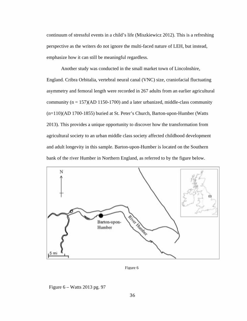
Another study was conducted in the small market town of Lincolnshire,
England. Cribra Orbitalia, vertebral neural canal (VNC) size, craniofacial fluctuating
asymmetry and femoral length were recorded in 267 adults from an earlier agricultural
community (n = 157)(AD 1150-1700) and a later urbanized, middle-class community
(n=110)(AD 1700-1855) buried at St. Peter’s Church, Barton-upon-Humber (Watts
2013). This provides a unique opportunity to discover how the transformation from
agricultural society to an urban middle class society affected childhood development
and adult longevity in this sample. Barton-upon-Humber is located on the Southern
bank of the river Humber in Northern England, as referred to by the figure below.
Figure 6
Figure 6 – Watts 2013 pg. 97

37
Barton’s growth was limited by poor land access and by increasing urbanization and
development of other port towns in the region during the early 14th century. Conditions
remained relatively stable for most of its history, maintaining a population of around
1000 individuals. By the 15th century Barton-upon-Humber was described as a “poor
rural town” (Clapson, 2005:17) and children were active members of the workforce and
began receiving wages at the age of ten (Watts 2013). Significant changes occurred in
the late 17th century when factories were being built. By the 19th century, the population
had risen to nearly 4000 individuals, prompting problems with overcrowding, waste
disposal, and contributed to the risk of contracting infectious and gastrointestinal
diseases (Watts 2013). A total of 2750 skeletons were dated to clearly defined periods
of cemetery use, and the individuals from St. Peter’s Church are considered to represent
a genetically homogenous population. Thus, results obtained are likely to reflect change
in environmental conditions and not variations in genotype frequencies (Watts 2013).
The sample was split into two groups: Group 1 (n = 157) and excavated from
cemeteries used between AD 1150-1700, & Group 2 (n = 110) were excavated from
cemeteries in use between AD 1700-1855. Cribra Orbitalia frequencies and LEH
frequencies are displayed.
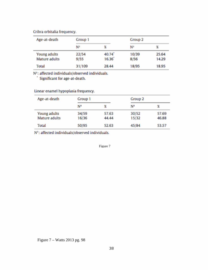
38
Figure 7
Figure 7 – Watts 2013 pg. 98
39
LEH data also demonstrates similar frequencies between groups, with 50
individuals (53%) in Group 1 and 45 individuals in Group 2 (54%). This indicates that
just over half of the individuals from both periods who survived into adulthood
experienced health insults which disrupted amelogenesis between one and six years of
age (Watts 2013). Surprisingly, despite the transition from rural to an urban middle-
class living environment, prevalence of stress indicators after AD 1700 did not change
significantly between cemetery periods. This is high in comparison to rural medieval
population of Wharram Percy, where 32% of adults displayed LEH (Mays 2007). These
high levels may be attributed to an increase of levels of pollution and hygiene in a small
town vs. a rural village. Regardless of improvements in housing and economic status,
such improvements were not enough to protect individuals from non-specific stressors,
including fevers, infections, and nutritional stress (Watts 2013).
Cribra Orbitalia was observed in 31 individuals (28%) from Group 1, and 18
individuals (19%) from Group 2. These prevalence rates are lower than in urban
populations from medieval York, where 66% of adult individuals from low status
population and 37% of individuals with higher status displayed orbital lesions.12 It is
hypothesized that these high rates can be attributed to overcrowding and poor
sanitation. Skeletal remains from Christ Church Spitalfields in London (AD 1700-1850)
showed that 34% of the total sample displayed Cribra Orbitalia.13 For this sample,
12 Grauer, A.L., 1993. Patterns of anemia and infection from medieval York, England.; Sullivan, A., 2005. Prevalence and etiology of acquired anemia in medieval York, England. 13 Molleson, T., Cox, M., 1993. The Spitalfields Project. Volume 2.; The Middling Sort. Cox, M., 1996. Life and Death in Spitalfields, 1700–1850.

40
Cribra Orbitalia is hypothesized results from the use of fashionable infant feeding
practices, which caused malnutrition and gastrointestinal diseases.
This is the only paper so far that has points distinctly to gastrointestinal disease,
as opposed to multiple causative agents, and the severe nutrient losses associated with
these may have been a factor in the development of cribra orbitalia in children under
four years of age in both cemetery periods at St. Peter’s Church. One fascinating claim
is that age-at-death individuals who experienced health insults that caused cribra
orbitalia no longer experienced a reduced adult lifespan. The explanation given here is
that the changing environment and economic conditions seemed to have benefited the
population in the long term. This is the first study to show a positive affect as a result of
societal movement towards a more sedentary, yet secure and advanced lifestyle.
However, it is unclear whether these lesions have the ability to remodel completely by
adulthood. Some studies contradict this notion as higher rates of healed orbital lesions
were observed in mature adults, illustrating that cribra orbitalia lesions can be retained
well into adulthood.14 Until more information is known about the remodeling abilities
of such lesions, it is important to approach these findings with caution.
The paper is inherently similar to Taiwan and Northeast China studies as it
examines a transition from hunter-gatherer to a more technologically advanced, yet
sedentary lifestyle. The results seem to follow the same pattern, as improvements in
economics and food reliability were not strong enough buffers to counteract stress. This
paper really dives into the history of the location as they have information spanning
14 Walker, P.L., 1986. Porotic hyperostosis in a marine-dependent Californian Indian population.; Grauer, 1993.
41
centuries for this region. This allows for in depth analysis of urbanization and how
society’s improvements affected general health. It’s worth noting that children during
this time were active members of the workforce. This could significantly impact
childhood stress, whereas the previous studies have not provided insight as to what the
children were actually doing during that time. Similar to the Haotaomuga paper,
individuals were required at least three permanent anterior teeth to be included in the
data set for LEH.
The last region of examination is West Virginia, America. In this study, linear
enamel hypoplasia (LEH), tests the hypothesis that early life stressors contribute to
decreased adult longevity in a human skeletal sample from the Late Fort Ancient site of
Neale’s Landing, West Virginia. Neale’s Landing represents an extremely stressed
population indicative of prehistoric agricultural societies. Researchers claim the most
likely cause for stress at Neale’s Landing is seasonal resource scarcity exacerbated by
the ongoing Little Ice Age. Unlike other contemporary year round settlements in the
area, Neale’s landing never exceeded two acres and contained only a few families with
a population at maximum of 40-50 individuals. It is important to note that there were
preservation issues due to human modification of the land, bank erosion, agriculture,
and bioturbation (root growth and woodchuck burrows), which contributed to this small
sample size (Hemmings, 1977). Radiocarbon dating suggests initial occupation between
A.D 1500 & 1550 and trade goods indicate occupation lasted until early 17th century.
Evidence also shows that the population was preoccupied warfare and hunting relative
to farming. Maize agriculture was prominent along with the hunting and gathering of
local species such as nuts, large and small mammals, birds, and aquatic resources. This
42
region allows the researcher the ability to analyze “the effect of stress on Native
American populations present in the Ohio Valley just prior to European contact.”
(Wolfe 2014)
Researchers uncovered 34 human burials in 27 graves at Neale’s Landing. The
assumption is that there are three extended families within the site that persisted for
about a century. This study examined 7 sub adults and 11 adults. Four of the adults are
male, three are female, and four are of indeterminate sex. Researchers looked at all teeth
for each individual and simply marked if the signs of linear enamel hypoplasia were
present or not. If an LEH existed, we measured the position of each defect from the
cement-enamel junction (CEJ) using digital sliding calipers accurate within 1/100 of a
millimeter to assess chronological age at insult. Two other sites are from Illinois (A.D.
1000 – 1350) and Ohio (A.D. 800-1100) is included to provide material to contextualize
the stress experienced at Neale’s Landing (Wolfe 2014).
Thirteen individuals (72%) in this study show signs of linear enamel
hypoplasia. This includes four adult males, two adult females, five juveniles, and
three adults of indeterminate sex. These individuals are as young as five and
perhaps as old as 50 years of age. Based on the average age of the sample, individuals
with linear enamel hypoplasia (n=13) died on average at the age of 24.23 and
individuals without linear enamel hypoplasia (n=5) died on average at the age of 25.10.
The data are not significant at the α=0.05 level (p=0.905)(Wolfe 2014).
The paper does an impressive job of utilizing the right variance tests to still
gather meaningful information from the data and observe differences between each
subsistence group. The comparison of many locations did show a significantly lower
43
mean age at death for individuals with enamel defects compared to individuals with
normal teeth, at least supporting the notion of LEH being related to mortality. The
comparison of this sample to other nearby samples once again reaffirms the
representativeness of skeletal remains of the population. If post-contact remains could
be recovered, this would be a great paper to study the affects of European contact. This
paper is very thorough in terms of methodology, but the focus of the paper is narrow
and not all possible factors are included. There could be legitimate reasons as to why
the researchers don’t explore other avenues, but such explanations are not offered when
they should be.
This paper has a small sample size so the results may not be representative of
the population at the time. The sample is also a relatively young population and cannot
represent the full life span of the group. Researchers also state “the fragmentary nature
of the sample prevents complete accuracy when ageing these specimens.” (Wolfe 2014)
Regardless of the downfalls of this sample, the data set from Neale’s Landing is
compared with several other sites from various geographic locations. The biggest fault
in this analysis is revealed by the writer himself when he states, “Although compelling,
the significant difference present masks the fact that the comparative sample includes
samples from numerous populations that vary in subsistence practice, settlement
structure, settlement size, among other variables”. (Wolfe 2014)
44
Implications & Conclusions
Although there are a plethora of previous studies analyzing enamel hypoplasia
and the relationship between decreased mortality, to date, no study compares these
results cross-culturally. But there are studies that do have some plausible theories to
consider. Steckel and Rose (2002) created a skeletal health index indicating that general
health declines throughout human history as society transitions from simple to complex
and hierarchical. This indicates that people who lived in large settlements were
generally less healthy than hunter-gatherers. This paints a stark picture chronic disease
replaces infectious disease and nutritional deficiency as the prominent driver of
morbidity and mortality due to the modern developing world. However, LEH is present
in a modernized society as evidenced by the samples of Lisbon, Portugal and Cleveland,
United States, thus giving a broad factor of comparison across time periods (Wolfe
2014). Another theory called the osteological paradox questions the true implications
of LEH. This suggests that those who survived long after the episode were the healthiest
while those individuals without lesions may have died before the chance to manifest
themselves. This questions if the presence of LEH really equates to less healthy. There
is also the factor of sample size to consider. How can one decipher how many skeletal
samples are really needed in order to consider the collection representative of the
region. And if they are a reliable sample, how large of a region can they truly compare
to?
One common problem became clear the more I sifted through the literature.
There is a lack of standardized methods when analyzing for specific markers of stress.
This makes it extremely difficult to compare data sets to one another and broaden the
45
context with which we are viewing the results. Some papers reference their methods
from previous studies, which is a step in the right direction, but there is not one
accepted practice. It doesn’t have to be the most flawless methods, but a foundation of
similarities must be present to be able to provide a global context. For the same certain
fields of research the “scientific method”, which essentially lays out the minimum
standards for gathering proper evidence, paleopathology needs a method of its own.
Only when this is accomplished, can we accept comparative results without doubt.
It is also important to recognize that markers of stress in bones and teeth imply
poor health. Stress is not an objective reaction for everyone, there are more factors
involved. There are lots of research about protective factors (e.g., warm, nurturing
parenting; wealth), homeostatic mechanisms (e.g., metabolic adjustment to caloric
deprivation during the prenatal period), and cultural factors (e.g., familism among
Latino/a people appears to buffer against the effects of stress). The primary hormonal
mediators of the stress response are glucocorticoids and catecholamines, which are
essential for adaptation and maintenance of homeostasis and survival. Over time, they
contribute to an allostatic load, which can accelerate disease processes. This load
centers on the brain as the interpreter and responder to environmental challenges. Early
childhood experiences of abuse and neglect increase allostatic load later in life that can
lead individuals into social isolation, hostility, depression, and even conditions as
serious as extreme obesity and CVD (Mcewen et al., 2000). Animal models support this
claim of lifelong influences of early experience on stress hormone reactivity. Although
the psychological factors of these ancient populations cannot be obtained, it is important
46
to keep this variable in mind as it may play a critical role in understanding results now
and in future studies.
After all cases taken into consideration, Steckel & Rose’s theory seems to be the
most plausible explanation for any paleopathologic study that analyzes the same
population over time, or two neighboring societies with different lifestyles. Some
studies in this review reflect the presence of this paradox, such as in Lovell’s paper. In
the Indus Valley paper, a conclusion was reached that pathogen load increases with
“sendentism and with denser populations because these facilitate both the exchange of
infectious agents between people in close contact and the accumulation of human
wastes and other refuse in and around the settlement” (Lovell 2014). Lincolnshire,
England and Northeast China also reflected a decline in health when moving towards a
more sedentary lifestyle. This provides insight into today’s society as we are more
sedentary than possibly ever before in history. We have outlived diseases and bred new
ones as well, and there is valuable knowledge in paleopathology that can help combat
our ever-changing biosphere.
Paleopathology can be referred to as a gateway study in the sense that
professionals from multiple fields of science can look at the results and add a piece to
the puzzle. In Rapa Nui, anthropologists and historians remedy the archaeological
record and redeem the locals by erasing a history of cannibalism & warfare. Hawaii
further emphasizes the impact of infectious diseases, providing valuable knowledge to
etiologists and pathologists who aim to understand, create, and implement medical
treatments based the historical and medical profile of each disease. The Mariana Islands
challenges environmentalists to truly observe the impact of climate on the health of a
47
population. The Barbados slave population can be utilized as a basis of comparison for
other slave populations globally as well as the represent the less healthy end of the
spectrum. Anthropologists could be the most helpful in this case as the weaning
practices seem to be the main factor and such practices are tied to culture. Lovell’s
study in the Indus Valley adds to the medical history of Malaria and explores all
possible avenues, allowing researchers to easily add to the proposed theories through
their own work. The cases in England may perhaps be of most interest to psychologists
as the affect of SES on childhood stress is a thriving area of research. West Virginia as a
stand alone case would improve if all other possibilities were thoroughly investigated, a
task that could be accomplished via a diverse team of scientists. The applicability of
paleopathology spans to modern times, as these same stress indicators will continue to
appear on skeletal remains. Ultimately these studies reveal that bone lesions are still
relevant to the study of health today and in the past. With the help of biologists,
epidemiologists, archaeologists, anthropologists, and environmental scientists, the
combined data could eventually lead to mitigation and aid in areas where medical
technology and socioeconomic status may not be able to buffer the effects. With this
perspective in mind, future studies should illuminate the unreliable shortcomings
present, and further contextualize and interpret the association between development,
nutritional, and environmental stress, in whatever form they may present themselves.
48
Bibliography
Armelagos, G.J., Goodman, A.H., Harper, K.N., Blakey, M.L., 2009. Enamel hypoplasia and early mortality: bioarchaeological support for the Barker Hypothesis. Evolutionary Anthropology 18, 261e271.
Bass WM. 1995. Human Osteology: A Laboratory and Field Manual. Colombia, MO:
Missouri Archaeological Society. Special Publication No. 2. Boldsen JL. 2007. Early childhood stress and adult age mortality – a study of dental
enamel hypoplasia in the Medieval Danish village of Tirup. American Journal of Physical Anthropology 132: 59–66. DOI: 10.1002/ajpa.20467
Chatrchaiwiwatana, S. 2006. Dental caries and periodontitis associated with betel quid
chewing: Analysis of two data sets. Journal of the Medical Association of Thailand 89:1004–1011.
Corruccini, Robert S., Jerome S. Handler, and Keith P. Jacobi. “Chronological
Distribution of Enamel Hypoplasias and Weaning in a Caribbean Slave Population”. Human Biology 57.4 (1985): 699–711.
Cox, M., 1996. Life and Death in Spitalfields, 1700–1850. Council for British
Archaeology, Halifax. Diamond, Jared. Collapse: How Societies Choose to Fail or Succeed. Viking, 2005. 81-
119. Print. Dudgeon, John. "The Genetic Architecture of the late Prehistoric and Protohistoric Rapa
Nui" U of Hawaii, 2008. Print. Grauer, A.L., 1993. Patterns of anemia and infection from medieval York, England.
American Journal of Physical Anthropology 91, 203–213. Griffiths LJ, Tate AR, Dezateux C, Millenium Cohort Study Child Health Group. 2007.
Do early infant feeding practices vary by maternal ethnic group? Public Health Nutrition 10: 957–964. DOI: 10.1017/S1368980007665513
Hamlin, H. “The Geography of Treponematosis .” The Yale Journal of Biology and
Medicine 12.1 (1939): 29–50. Print. Iregren E. 1992. Scandinavian women during the medieval period; health, childbirth
and childcare. Collegium Anthropologicum 16: 59–82. Klaus HD, Tam ME. 2009. Contact in the Andes: bioarchaeology of systemic stress in
colonial Mórrope, Peru. Am J Phys Anthropol 138: 356-368.
49
Krenz-Niedbała M, Kozłowski T. 2011. Comparing the chronological distribution of enamel hypoplasia in Rogowo, Poland (2nd century AD) using two methods of defect timing estimation. International Journal of Osteoarchaeology DOI: 10.1002/oa.1262
Lanphear KM. 1990. Frequency and distribution of enamel hypoplasia’s in a historic
skeletal sample. American Journal of Physical Anthropology 81: 35–43. DOI: 10.1002/ ajpa.1330810106
Littleton, J. 2005. Invisible impacts but long-term consequences: hypoplasia and contact
in central Australia. Am J Phys Anthropol 126: 295-304. Lovell, Nancy C. "Anemia in the Ancient Indus Valley." International Journal of
Osteoarchaeology Int. J. Osteoarchaeology. 7.2 (1997): 115-23. Web. Massler M, Schour I, Poncher HG. 1941. Developmental pattern of the child as
reflected in the calcification pattern of the teeth. American Journal of Disease of Children 62: 33–67.
Mcewen, B. "Allostasis and Allostatic Load Implications for
Neuropsychopharmacology." Neuropsychopharmacology 22.2 (2000): 108-24. Web.
Merrett, Deborah C., Hua Zhang, Xiaoming Xiao, Quanchao Zhang, Dong Wei, Lixin
Wang, Hong Zhu, and Dongya Y. Yang. "Enamel Hypoplasia in Northeast China: Evidence from Houtaomuga." Quaternary International 405 (2016): 11-21. Web.
Miszkiewicz, Justyna Jolanta. "Linear Enamel Hypoplasia and Age-at-Death at
Medieval (11 Th -16 Th Centuries) St. Gregory's Priory and Cemetery, Canterbury, UK." International Journal of Osteoarchaeology Int. J. Osteoarchaeol. 25.1 (2012): 79-87. Web.
Moggi-Cecchi J, Pacciani E, Pinto-Cisternas J. 1994. Enamel hypoplasia and age at
weaning in 19th century Florence, Italy. American Journal of Physical Anthropology 93: 299–306.
Molleson, T., Cox, M., 1993. The Spitalfields Project. Volume 2. The Middling Sort.
CBA Research Report 86. Council for British Archaeology, York. Palubeckaite Z, Jankauskas R, Boldsen J. 2002. Enamel hypoplasia in Danish and
Lithuanian late Medieval/ early modern samples: a possible reflection of child morbidity and mortality patterns. International Journal of Osteoarchaeology 12: 189–201. DOI: 10.1002/oa.607
50
Papageogopoulou, Christina, Susanne Sutter, Frank Ruhli, and Frank Segmund. "Harris Lines Revisited: Prevalence, Comorbidities, and Possible Etiologies." American Journal of Human Biology (2011)
Pietrusewsky M., Lauer A., Tsang C.-H, Li K.-T., and Douglas M.T. (2013) Dental
indicators of health in early Neolithic and Iron Age Skeletons from Taiwan. Journal of Austronesian Studies, 4: 1–33.
Pietrusewsky, Michael, and Michele Douglas. "An Osteological Assessment of Health
and Disease in Precontact and Historic (1778) Hawai'I." An Osteological Assessment of Health and Disease in Precontact and Historic (1778) Hawai'I. Honolulu: New York: Wiley Liss, 2014. Print.
Pietrusewsky, Michael, Adam Lauer, Michele Toomay Douglas, Cheng-Hwa Tsang,
and Kuang-Ti Li. "Patterns of Health in Early Neolithic and Iron Age Taiwan." AS Anthropological Science 124.2 (2016): 117-33. Web.
Pietrusewsky, Michael, Michele Toomay Douglas, Marilyn K. Swift, Randy A. Harper,
and Michael A. Fleming. "Health in Ancient Mariana Islanders: A Bioarchaeological Perspective." The Journal of Island and Coastal Archaeology (2014): 319-40. Print.
Polet, Caroline. "Health and Diet of Ancient Easter Islanders: Contribution of
Paleopathology, Dental Microwear and Stable Isotopes." 2011. Print. Skinner, M.F., Dupras, T.L., Moya-Sola, S., 1995. Periodicity of enamel hypoplasia among Miocene Dryopithecus from Spain. Journal of Paleopathology 7,
197e222. Sullivan, A., 2005. Prevalence and etiology of acquired anemia in medieval York,
England. American Journal of Physical Anthropology 128, 252–272. Ubelaker DH. 1992. Enamel hypoplasia in ancient Ecuador. In: Recent Contributions to
the Study of Enamel Developmental Defects. Journal of Paleopathology, Monographic Publication, 2, AH Goodman, LL Capasso (eds.). Edigrafital: Teramo; 207–217.
Walker, P.L., 1986. Porotic hyperostosis in a marine-dependent Californian Indian
population. American Journal of Physical Anthropology 69, 345–354. Walker, Phillip L., Rhonda R. Bathurst, Rebecca Richman, Thor Gjerdrum, and
Valerie A. Andrushko. "The Causes of Porotic Hyperostosis and Cribra Orbitalia: A Reappraisal of the Iron-deficiency-anemia Hypothesis." Am. J. Phys. Anthropology. American Journal of Physical Anthropology 139.2 (2009): 109-25. Web.
51
Watts, Rebecca. "Childhood Development and Adult Longevity in an Archaeological Population from Barton-upon-Humber, Lincolnshire, England." International Journal of Paleopathology 3.2 (2013): 95-104. Web.
Wolfe, Christopher. Enamel Hypoplasia and Early Mortality: Interpreting
Developmental Stress at Neale’s Landing, West Virginia (46Wd39). Thesis. Dickinson College, May 2014. N.p.: n.p., n.d. Print.
Wright LE. 1997. Intertooth patterns of hypoplasia expression: implications for
childhood health in the classic Maya collapse. American Journal of Physical Anthropology
Wolfe, Christopher. Enamel Hypoplasia and Early Mortality: Interpreting
Developmental Stress at Neale’s Landing, West Virginia (46Wd39) Christopher Wolfe. Thesis. Dickinson College, 2014.