Embed Size (px)
Citation preview

LKD
May 31 - June 1, 2013
Vilnius, Lithuania
World Colorectal
C O N F E R E N C E
International Society of University Colon
and Rectal Surgeons (ISUCRS) Interim
meeting in Vilnius 2013
May 31 - June 1, 2013, Vilnius, Lithuania
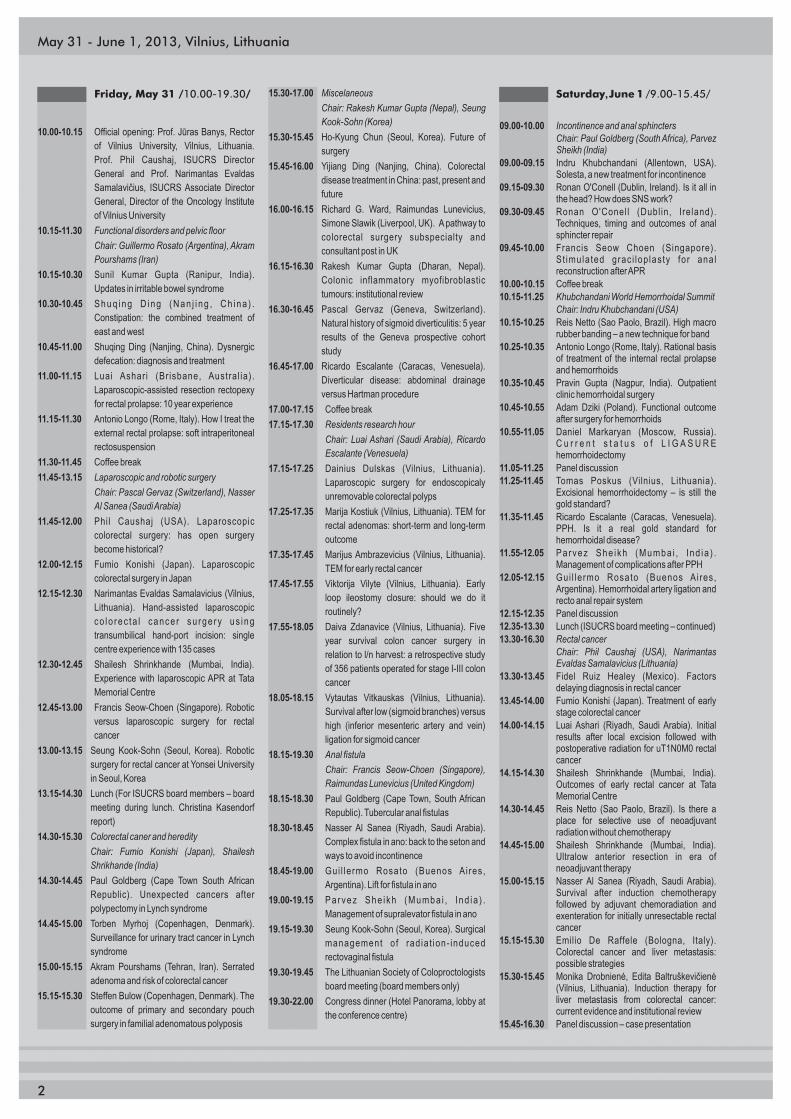
Friday, May 31 /10.00-19.30/
10.15-11.30 Functional disorders and pelvic floor
Chair: Guillermo Rosato (Argentina), Akram
Pourshams (Iran)
10.15-10.30 Sunil Kumar Gupta (Ranipur, India).
Updates in irritable bowel syndrome
10.30-10.45 S h u q i n g D i n g ( N a n j i n g , C h i n a ) .
Constipation: the combined treatment of
east and west
10.45-11.00 Shuqing Ding (Nanjing, China). Dysnergic
defecation: diagnosis and treatment
11.00-11.15 Luai Ashari (Br isbane, Austral ia).
Laparoscopic-assisted resection rectopexy
for rectal prolapse: 10 year experience
11.15-11.30 Antonio Longo (Rome, Italy). How I treat the
external rectal prolapse: soft intraperitoneal
rectosuspension
11.30-11.45 Coffee break
11.45-13.15 Laparoscopic and robotic surgery
Chair: Pascal Gervaz (Switzerland), Nasser
Al Sanea (Saudi Arabia)
11.45-12.00 Phil Caushaj (USA). Laparoscopic
colorectal surgery: has open surgery
become historical?
12.00-12.15 Fumio Konishi (Japan). Laparoscopic
colorectal surgery in Japan
12.15-12.30 Narimantas Evaldas Samalavicius (Vilnius,
Lithuania). Hand-assisted laparoscopic
co lo rec ta l cance r su rge ry us ing
transumbilical hand-port incision: single
centre experience with 135 cases
12.30-12.45 Shailesh Shrinkhande (Mumbai, India).
Experience with laparoscopic APR at Tata
Memorial Centre
12.45-13.00 Francis Seow-Choen (Singapore). Robotic
versus laparoscopic surgery for rectal
cancer
13.00-13.15 Seung Kook-Sohn (Seoul, Korea). Robotic
surgery for rectal cancer at Yonsei University
in Seoul, Korea
13.15-14.30 Lunch (For ISUCRS board members – board
meeting during lunch. Christina Kasendorf
report)
14.30-15.30 Colorectal caner and heredity
Chair: Fumio Konishi (Japan), Shailesh
Shrikhande (India)
14.30-14.45 Paul Goldberg (Cape Town South African
Republic). Unexpected cancers after
polypectomy in Lynch syndrome
14.45-15.00 Torben Myrhoj (Copenhagen, Denmark).
Surveillance for urinary tract cancer in Lynch
syndrome
15.00-15.15 Akram Pourshams (Tehran, Iran). Serrated
adenoma and risk of colorectal cancer
15.15-15.30 Steffen Bulow (Copenhagen, Denmark). The
outcome of primary and secondary pouch
surgery in familial adenomatous polyposis
10.00-10.15 Official opening: Prof. Jūras Banys, Rector
of Vilnius University, Vilnius, Lithuania.
Prof. Phil Caushaj, ISUCRS Director
General and Prof. Narimantas Evaldas
Samalavičius, ISUCRS Associate Director
General, Director of the Oncology Institute
of Vilnius University
Saturday, June 1 /9.00-15.45/
09.00-10.00 Incontinence and anal sphincters Chair: Paul Goldberg (South Africa), Parvez Sheikh (India)
09.00-09.15 Indru Khubchandani (Allentown, USA). Solesta, a new treatment for incontinence
09.15-09.30 Ronan O'Conell (Dublin, Ireland). Is it all in the head? How does SNS work?
09.30-09.45 Ronan O'Conel l (Dubl in, Ireland). Techniques, timing and outcomes of anal sphincter repair
09.45-10.00 Francis Seow Choen (Singapore). St imulated grac i lop lasty for anal reconstruction after APR
10.00-10.15 Coffee break
10.15-11.25 Khubchandani World Hemorrhoidal Summit Chair: Indru Khubchandani (USA)
10.15-10.25 Reis Netto (Sao Paolo, Brazil). High macro rubber banding – a new technique for band
10.25-10.35 Antonio Longo (Rome, Italy). Rational basis of treatment of the internal rectal prolapse and hemorrhoids
10.35-10.45 Pravin Gupta (Nagpur, India). Outpatient clinic hemorrhoidal surgery
10.45-10.55 Adam Dziki (Poland). Functional outcome after surgery for hemorrhoids
10.55-11.05 Daniel Markaryan (Moscow, Russia). C u r r e n t s t a t u s o f L I G A S U R E hemorrhoidectomy
11.05-11.25 Panel discussion
11.25-11.45 Tomas Poskus (Vilnius, Lithuania). Excisional hemorrhoidectomy – is still the gold standard?
11.35-11.45 Ricardo Escalante (Caracas, Venesuela). PPH. Is it a real gold standard for hemorrhoidal disease?
11.55-12.05 Parvez She i kh (Mumba i , I nd ia ) . Management of complications after PPH
12.05-12.15 Gui l lermo Rosato (Buenos Ai res, Argentina). Hemorrhoidal artery ligation and recto anal repair system
12.15-12.35 Panel discussion
12.35-13.30 Lunch (
13.30-16.30 Rectal cancer Chair: Phil Caushaj (USA), Narimantas Evaldas Samalavicius (Lithuania)
13.30-13.45 Fidel Ruiz Healey (Mexico). Factors delaying diagnosis in rectal cancer
13.45-14.00 Fumio Konishi (Japan). Treatment of early stage colorectal cancer
14.00-14.15 Luai Ashari (Riyadh, Saudi Arabia). Initial results after local excision followed with postoperative radiation for uT1N0M0 rectal cancer
14.15-14.30 Shailesh Shrinkhande (Mumbai, India). Outcomes of early rectal cancer at Tata Memorial Centre
14.30-14.45 Reis Netto (Sao Paolo, Brazil). Is there a place for selective use of neoadjuvant radiation without chemotherapy
14.45-15.00 Shailesh Shrinkhande (Mumbai, India). Ultralow anterior resection in era of neoadjuvant therapy
15.00-15.15 Nasser Al Sanea (Riyadh, Saudi Arabia). Survival after induction chemotherapy followed by adjuvant chemoradiation and exenteration for initially unresectable rectal cancer
15.15-15.30 Emilio De Raffele (Bologna, Italy). Colorectal cancer and liver metastasis: possible strategies
15.30-15.45 Monika Drobnienė, Edita Baltruškevičienė (Vilnius, Lithuania). Induction therapy for liver metastasis from colorectal cancer: current evidence and institutional review
15.45-16.30
ISUCRS board meeting – continued)
Panel discussion – case presentation
15.30-17.00 Miscelaneous
Chair: Rakesh Kumar Gupta (Nepal), Seung
Kook-Sohn (Korea)
15.30-15.45 Ho-Kyung Chun (Seoul, Korea). Future of
surgery
15.45-16.00 Yijiang Ding (Nanjing, China). Colorectal
disease treatment in China: past, present and
future
16.00-16.15 Richard G. Ward, Raimundas Lunevicius,
Simone Slawik (Liverpool, UK). A pathway to
colorectal surgery subspecialty and
consultant post in UK
16.15-16.30 Rakesh Kumar Gupta (Dharan, Nepal).
Colonic inflammatory myofibroblastic
tumours: institutional review
16.30-16.45 Pascal Gervaz (Geneva, Switzerland).
Natural history of sigmoid diverticulitis: 5 year
results of the Geneva prospective cohort
study
16.45-17.00 Ricardo Escalante (Caracas, Venesuela).
Diverticular disease: abdominal drainage
versus Hartman procedure
17.00-17.15 Coffee break
17.15-17.30 Residents research hour
Chair: Luai Ashari (Saudi Arabia), Ricardo
Escalante (Venesuela)
17.15-17.25 Dainius Dulskas (Vilnius, Lithuania).
Laparoscopic surgery for endoscopicaly
unremovable colorectal polyps
17.25-17.35 Marija Kostiuk (Vilnius, Lithuania). TEM for
rectal adenomas: short-term and long-term
outcome
17.35-17.45 Marijus Ambrazevicius (Vilnius, Lithuania).
TEM for early rectal cancer
17.45-17.55 Viktorija Vilyte (Vilnius, Lithuania). Early
loop ileostomy closure: should we do it
routinely?
17.55-18.05 Daiva Zdanavice (Vilnius, Lithuania). Five
year survival colon cancer surgery in
relation to l/n harvest: a retrospective study
of 356 patients operated for stage I-III colon
cancer
18.05-18.15 Vytautas Vitkauskas (Vilnius, Lithuania).
Survival after low (sigmoid branches) versus
high (inferior mesenteric artery and vein)
ligation for sigmoid cancer
18.15-19.30 Anal fistula
Chair: Francis Seow-Choen (Singapore),
Raimundas Lunevicius (United Kingdom)
18.15-18.30 Paul Goldberg (Cape Town, South African
Republic). Tubercular anal fistulas
18.30-18.45 Nasser Al Sanea (Riyadh, Saudi Arabia).
Complex fistula in ano: back to the seton and
ways to avoid incontinence
18.45-19.00 Gui l lermo Rosato (Buenos Ai res,
Argentina). Lift for fistula in ano
19.00-19.15 Parvez She i kh (Mumba i , I nd ia ) .
Management of supralevator fistula in ano
19.15-19.30 Seung Kook-Sohn (Seoul, Korea). Surgical
management of rad iat ion- induced
rectovaginal fistula
19.30-19.45 The Lithuanian Society of Coloproctologists
board meeting (board members only)
19.30-22.00 Congress dinner (Hotel Panorama, lobby at
the conference centre)
2

International Society of University Colon and Rectal Surgeons (ISUCRS) Interim meeting in Vilnius 2013
I feel delighted to be able to address with this welcoming word to all of you on the occasion of the
International Society of University Colon and Rectal Surgeons Interim meeting in Vilnius 2013, a World
Colorectal Conference, which is held in Vilnius Panorama Hotel, Lithuania, on May 31- June 1, 2013.
The International Society of University Colon and Rectal Surgeons (ISUCRS) was found in Mexico city, th
Mexico, on November 24, 1962, and last year celebrated its 50 anniversary. This society has performed
its role as an outstanding international organization, contributing over these long years enormously to
the progress of the treatment of the diseases of colon, rectum and anus. The Society takes pride in having
held innumerable congresses and meetings throughout the world, providing a platform of exchanging
valuable scientific knowledge on a global level. That is the reason a new initiative to organize an interim
meeting between the usual biennial meetings of this Society is very special. For us it is very special for an
additional reason - it is held for the first time, and it takes place in Lithuania. That not only points out the input of Lithuanian colorectal surgeons to the activities of ISUCRS, but allows us to attract people from
our region (Poland, Belorussia, Latvia and Estonia, and obviously the whole Lithuania), to introduce them
to our Society and of what I am completely sure - significantly increase our Society membership over this
weekend.
The Lithuanian Society of Coloproctologists, hosting this event, is very glad to continue its annual
international colorectal meetings with this one, providing world-class lectures from ISUCRS members
and experts throughout the world, most updated knowledge in our field, as well and exceptional
possibility to learn from our differences and our similarities.
Wishing you the most fruitful, inspiring academic meeting and best memories from tiny, both known
and yet enough undiscovered medieval city called Vilnius,
Dear guests,
members of the International Society
of University Colon and
Rectal Surgeons and
Lithuanian Society of Coloproctologists,
colleagues and friends,
3
Yours -
Prof. Narimantas Evaldas Samalavièius
Associate Director General, International Society of University Colon and Rectal Surgeons
President, Lithuanian Society of Coloproctologists
Director, Oncology Institute of Vilnius University

May 31 - June 1, 2013, Vilnius, Lithuania
Invited speakers:
Assoc. Prof. Akram PourshamsAssociate Professor in MedicineGastroenterologist & HepatologistTehran University of Medical SciencesDigestive Disease Research CenterKargar Ave,Shariati Hospital14117-13135,TehranIran
Prof. Adam DzikiUniversity HospitalUl. Zeromskiego 113, LodzPoland
Dr. Audrius DulskasCenter of OcosurgeryInstitute of Oncology, Vilnius UniversitySantariskiu 1LT-08406 VilniusLithuania
Prof. Antonio LongoClinica Madonna della FiduciaVia Alfredo Baccarini, 5400179 RomaItaly
Dr. Daiva ZdanaviceCenter of OcosurgeryInstitute of Oncology, Vilnius UniversitySantariskiu 1LT-08406 VilniusLithuania
Dr. Daniel MarkaryanDepartment of coloproctology and endoscopic surgeryFaculty of postgraduate professional education of physiciansDepartment of colorectal surgeryUniversity Hospital #2, Clinical CenterFirst Moscow State Medical University n.a. I.M. Sechenov1/1 Pogodinskaya str. 119435 MoscowRUSSIA
Dr. Emilio De Raffele, MD, PhDDepartment of Emergency, General Surgery and TransplantsS.Orsola University Hospital of BolognaVia Massarenti, 940138 BolognaITALY
Dr Francis Seow-Choen Colorectal Surgeon & DirectorSeow-Choen Colorectal Surgery PLCSingapore, Singapore
Dr. Fidel Ruiz Healy, MD, FICS (Hon), Prof. (Hon), Dr. (h.c.) Durango 290-309. Col. Roma, México City, DF. 06700 México
Prof. Fumio KonishiDepartment of SurgeryNerima Hikaragaoka HospitalJapanProf. Guillermo Rosato MD MAAC MSACP FASCRS MISUCRSStaff Colorectal Surgery-Surgical Department-Hospital Universitario Austral-Buenos Aires-Argentina. Profesor in Surgery - Universidad AustralSchool of MedicineBuenos AiresArgentina
Prof. Ho-Kyung Chun, MD, PhDProfessor, Department of SurgeryExecutive Vice PresidentKamgbuk Samsung HospitalSungkyunkwan University School of Medicine29, Saemunan-Ro, Jongno-Gu, Seoul 110-746Korea
Prof. Indru T. Khubchandani MD, MS, FRCS, FACS, FICS (Hon), FASCRSProf. Honoris Causa (Bolivia)Prof. of Surgery, Complutenis University, Madrid, Spain.Lehigh Valley Health Network Allentown, Pa. 18103Professor of SurgeryMorsani College of MedicineUniversity of South FloridaUSA
Prof. Yijang DingNational center of Colorectal disease of Chinese Medicine Nanjing Municipal TCM Hospital,3rd affiliated hospital of Nanjing University of Chinese MedicineNo.1 Jinling Road, Nanjing, 210001P.R.China
Dr. Luai AshariDirector, residency programSection Colon & Rectal SurgeryKing Faisal Specialist Hospital & Research Center-RiyadhSaudi Arabia
Dr. Marijus AmbrazeviciusCenter of OcosurgeryInstitute of Oncology, Vilnius UniversitySantariskiu 1LT-08406 VilniusLithuania
Dr. Marija KostiukCenter of OcosurgeryInstitute of Oncology, Vilnius UniversitySantariskiu 1LT-08406 VilniusLithuania
Prof. Narimantas Evaldas SamalavičiusChief of Clinic of OncosurgeryDirector, Institute of Oncology, Vilnius UniversityPresident of the Lithuanian Society of ColoproctologistsSantariskiu 1LT-08406 VilniusLithuania
Dr. Nasser Al-SaneaHead Section, Colon & Rectal SurgeryKing Faisal Specialist Hospital & Research Center-RiyadhPresident, Saudi Society of Colon & Rectal SurgerySaudi Arabia
Prof. Patrick Ronan O'Connell, M.D.University College DublinElm Park, Dublin, 4 Ireland
Prof. Phil Caushaj, MD, PhDProfessor and Surgeon in ChiefWest Virginia SOMGreenbrier, West Virginia USA
Prof. Pascal Gervaz ChirurgienClinique de La Colline1206 GenèveSwitzerland
Dr. Parvez SheikhHon. Colorectal SurgeonSaifee HospitalMumbai & Nova Specialty SurgeryMumbaiIndia
Prof. Paul GoldbergColorectal UnitUniversity of Cape Town and Groote Schuur HospitalCape TownSouth Africa
Dr. Richard G. Ward, MA, FRCS (Engl)Consultant Surgeon, General Emergency Surgery and Major Trauma Units, Aintree University Hospitals NHS Foundation Trust,Longmoor Lane, Liverpool L9 7AL, UKHead, School of Surgery, MerseyDeanery, Regatta Place, Summers Road, Brunswick Business Park, Liverpool, L3 4BLUK
Dr. Raimundas Lunevicius, MD, FRCS (Engl.)Consultant Surgeon, General Emergency Surgery and Major Trauma Units, Aintree University Hospitals NHS Foundation Trust, Longmoor Lane, Liverpool L9 7AUK
Assoc. prof. Rakesh GuptaDepartment of SurgeryB.P. Koirala Institute of Health Sciences, DharanNepal
Prof. Reis NetoMedical Director at Clinica Reis Neto. Campinasgal osorio 2273 Sao Paulo f 551932525611Surgeon at hospital Casa da Saude Campinas Sao PauloProf of Surgery at PUCCAMP Campinas Sao PauloBrazil
Prof. Ricardo EscalantePresident Venezuelan Society of Parenteral and Enteral NutritionUniversidad Central de VenezuelaLoira Medical CenterCaracasVenesuela
Assoc. Prof. Steffen BülowOverlæge, dr. med.Tjørnekrogen 4DK-2820 GentofteDenmark
Prof. Seung-Kook Sohn Department of SurgeryKangnam Severance HospitalYonsei University College of MedicinePO Box 1217,Seoul 135-720Korea
Prof. Shailesh V. Shrikhande, MS, MDProfessor and Consultant SurgeonGastrointestinal and Hepato-Pancreato-Biliary SurgeryConvener, GI - Disease Management GroupTata Memorial CentreErnest Borges Marg, Parel,Mumbai, India
Dr. Shuqing Ding, MD, PhDNational center of Colorectal disease of Chinese Medicine Nanjing Municipal TCM Hospital,3rd affiliated hospital of Nanjing University of Chinese MedicineNo.1 Jinling Road, Nanjing, 210001P.R.China
Dr. Torben MyrhojCopenhagenDenmark
Dr. Tomas PoškusDepartment of SurgeryVilnius University Hospital Santariskiu KlinikosSantariskiu 1LT-01128 VilniusLithuania
Dr. Vytautas VitkauskasCenter of OcosurgeryInstitute of Oncology, Vilnius UniversitySantariskiu 1LT-08406 VilniusLithuania
Dr. Viktorija VilyteCenter of OcosurgeryInstitute of Oncology, Vilnius UniversitySantariskiu 1LT-08406 VilniusLithuania
Dr. Sunil Kumar GuptaMain Hospital,Bharat Heavy Eletricals Limited,Ranipur, Hardwar-249403, UttarakhandIndia
4

International Society of University Colon and Rectal Surgeons (ISUCRS) Interim meeting in Vilnius 2013
5

May 31 - June 1, 2013, Vilnius, Lithuania
6
UPDATES IN IRRITABLE BOWEL SYNDROME
SUNIL KUMAR GUPTA MS (SURGERY), FAIS, FICS, FACRSI, FACS. HEAD (SURGERY), MAIN HOSPITAL, BHEL, RANIPUR, HARDWAR, UTTARAKHAND, INDIA
Irritable bowel syndrome (IBS) is a common Functional Gastro Intestinal Disorder (FGID) characterized by bouts of recurrent abdominal pain, bloating or discomfort associated with disturbances in defaecation which are not explained by structural abnormalities. Earliest description of
1 2symptoms appeared in literature in 1849 . Irritable bowel syndrome (IBS) term was first given by C.J. DeLor in 1967 . Other historical terms used to describe IBS include mucous colitis, spastic colon, nervous colon, unstable colon, non-specific colitis, neurogenic mucous colitis, spastic bowel syndrome. Overall incidence of IBS is 10-15%, with age range of 30 to 50 years. IBS has clear female predominance in the ratio of 2:1. Its one of common conditions accounting for 3.5 million physician visits in US alone associated with total costs of 19 billion US dollars.
IBS occurs worldwide with similar incidence in US & Europe. Incidence of IBS is increasing in developing countries. Since the etiology is not 3clearly understood, the disease has long been dismissed as psychosomatic condition and incidence/prevalence were not extensively monitored
in the past. A variety of factors are believed to play a role in the development of IBS including altered bowel motility, visceral hypersensitivity, altered brain-
gut interactions, immune activation/low grade inflammation, psychosocial stresses, changes in gut microbiome and genetic factors. Recent research in understanding pathophysiology of the disease has resulted in exciting new insights which suggest IBS may be product of pathogenic mechanisms which include serotonergic disorder, small intestine bacterial overgrowth (SIBO), potential role of mast cells and abnormal pain processing/pain memory. British Society of Gastroenterology guidelines have shown that awareness in understanding IBS has led to more
4effective treatment . To aid diagnosis, treatment and to allow international comparisons, several international consensus meetings have culminated into
5introduction of Rome III diagnostic criteria for IBS . These consensus meetings also highlighted alarm indicators (red flag indicators) which should be excluded before diagnosing IBS i.e. age >50 years, male, short history, documented weight loss, nocturnal symptoms, rectal bleeding, family history of colon cancer and recent antibiotic use.
IBS is often classified into four subtypes viz. constipation predominant (IBS-C), diarrhoea predominant (IBS-D), mixed IBS (IBS-M) and gas/bloating predominant unsubtyped (IBS-U). Diagnosis of IBS is by and large clinical in the absence of any biological markers. Clinicians use a holistic approach to diagnose IBS taking note of features beyond gut such as behavior, previous medical history, pelvic symptoms, backache, headache etc. 50% IBS patients are depressed or anxious having sleep disturbances. Somatization is a feature of many IBS patients which can easily be recognized by personal health questionnaire 15.
All patients consulting physician for IBS related symptoms should be judiciously investigated to exclude organic pathology. There is a lack of sine qua non biomarkers hence the diagnosis remains on symptoms based criteria. Two recent well performed meta-analysis have investigated the performance of Rome III and Manning Criteria.
Management recommendations for IBS are directed towards determination of psychological concerns like anxiety/depression, cognitive behavioral therapy and psychodynamic interpersonal therapy and hypnotherapy. Traditional symptoms based therapies of IBS are directed at the relief of individual symptoms but they are often of limited efficacy in addressing the entire symptoms complex. Combinations of drugs to target bothersome symptoms are suggested as Ist line of treatment which offer significant gain over placebo. Increasing knowledge of pathophysiology
6and molecular mechanisms of IBS have resulted in development of several new therapeutic approaches . Thirteen consensus statements for treatment of IBS have been developed using modified Delphi approach. Exclusion diets have modest efficacy in improving symptoms in IBS patients. Symptoms based therapies with dietery fibre, bulking agents and laxatives are effective in IBS-C. Luboprostine and tegaserod have offered a therapeutic gain of 7-15% over placebo in constipation predominant IBS. Bifidobacter infantis, nonabsorbable antibiotic (rifaximin),
7loperamide and alosetron help relieving symptoms in diarrhoea predominant IBS . Additionally there is persuasive evidence to suggest that selected antispasmodics and tricyclic antidepressants (TCA) and selective
serotonin reuptake inhibitors (SSRI) are effective in improving global IBS symptoms and abdominal pain. Recently mesalamine, pregabalin, gabapentene, clonidine and octreotide have been used to treat IBS with promising results. Complementary and alternative medicine therapies
8such as probiotics, herbal therapies and acupuncture are gaining popularity amongst IBS sufferers although manufacturing standards and safety concerns remain unanswered.
Despite all limitations, this remains quite exciting that IBS represents an area of intense investigations from multiple directions. Therapy that alters the natural history of IBS is yet to be identified and current knowledge of IBS is constantly in evolution. As new insights into pathophysiology of IBS emerge, better therapies will be in vogue.
References:1. Cumming W. Lond Med Gazette 1849, NS9, 969-9732. DeLor C J Am J Gastroenterol May 1967, 47 427–434 3. Maxwell et al Lancet Dec. 1997 350 1691-1695 4. Spiller R, Aziz Q, Creed F, et al. The British Society of Gastroenterology. Guidelines on the irritable bowel syndrome : Mechanisms and
practical management. Gut 2007. 5. Longstreth GF, Thompson WG, Chey WD, Houghton LA, Mearin F, Spiller RC. Functional bowel disorders, Gastroenterology 2006;
130:1480-916. Kwon JG, Park KS, Park JM, Park CH, Lee KJ, Park HJ, Rhee JC: Korean Society of Neurogastroenterology and Motility. Korean J
Gastroenterol. 2011 Feb; 57 (2):82-997. Olden KW Clin Exp Gastroenterol. 2012;5:69-1008. Chey WD, Maneerattaporn M, Saad R. Gut Liver. 2011 Sep;5(3):253-66

International Society of University Colon and Rectal Surgeons (ISUCRS) Interim meeting in Vilnius 2013
7

May 31 - June 1, 2013, Vilnius, Lithuania
8
CONSTIPATION: THE COMBINED TREATMENT OF EAST AND WEST
Ding, S. Q.; Ding, Y. J. Colorectal section, Nanjing TCM hospital, Nanjing, Jiangsu, China
Purpose.
To show the constipation multidisciplinary and integreated treatment in China pelvic floor center practice.
Methods.
According Nanjing pelvic floor center database from June 2008 to June 2011, 1045 constipation patients got integrated treatment
protocol enrolled including diet, psychological and psychiatric treatment, pelvic floor biofeedback therapy, Chinese medicine,
Acupuncture or operation etc. To investigate the symptoms ,treatment protocols, initial results and follow up etc.
Results.
Patients usually abused in cathartic or enema with poor quality of life, routine practice was usually invalid. Multidisciplinary
rehabilitation team work was needed , integrated treatment protocol did according to the diagnosis and treatment flowchart. The
results will be evaluated by Patient Reported Outcome questionnaires and physiology tests respectively.
Conclusions.
Integrated multidisciplinary treatment improved success rate. According to research work, Acupuncture improved intestine motility
in slow treatment constipation, sensation and muscle control in pelvic floor relaxation of outlet obstructed constipation, pelvic floor
biofeedback therapy improved muscle variability, endurance and rectal sensation in dysnergic defecation, Chinese herb's
advantage was in laxative withdraw, motility promotion and body constitution modulation. Operation just worked in very small
percentage of rectocele, slow transit constipation and pelvic floor organ prolapse relevant constipation.

International Society of University Colon and Rectal Surgeons (ISUCRS) Interim meeting in Vilnius 2013
9

May 31 - June 1, 2013, Vilnius, Lithuania
10
DYSSNERGIC DEFECATION: DIAGNOSIS AND TREATMENT
Ding, S. Q.; Ding, Y. J.Colorectal section, Nanjing TCM hospital, Nanjing, Jiangsu, China
Purpose.
Outline dyssnergic defecation diagnosis and treatment in China pelvic floor center practice.
Methods.
According Nanjing pelvic floor center database from June 2008 to June 2012, 1,650 constipation patients enrolled , according to
RomeⅢcriterion, dyssnergic defecation accounts for 45.4%, which divided into 4 subsets according to manometry which provided
more information and painless than defeography, needle EMG etc. Patients characteristic symptoms usually combined upper GI,
psychological , Irritable bowel syndrome etc which should be evaluated before treatment. The management protocols including diet,
laxative, psychological or psychiatric consultation, BTX injection, pelvic floor biofeedback , Chinese medicine, Acupuncture etc
should be investigated and individualized.
Results.
Symptoms with digital examination and manometry are preferred in dyssnergic defecation diagnosis, the success rate is 85% while
40%, 62% and 89% respectively. Biofeedback is in first line recommendation, it is sufficient in friendly compliance patients. The
success rate is 65%, almost 3-4 sessions. BTX injection can shorten treatment course into 1-2 sessions. Chinese medicine is helpful
in laxative quitting , bloating, loss of appetite etc. Acupuncture is practically helpful in motility and anxiety insomnia. Operation has no
requirement.
Conclusion.
Combined and customized treatment protocol is the key.

International Society of University Colon and Rectal Surgeons (ISUCRS) Interim meeting in Vilnius 2013
11

May 31 - June 1, 2013, Vilnius, Lithuania
12
LAPAROSCOPICALLY-ASSISTED RESECTION RECTOPEXY FOR RECTAL PROLAPSE: TEN
YEARS' EXPERIENCE
, Lumley JW, Stevenson AR, Stitz RW. Colorectal Unit, Royal Brisbane Hospital, Brisbane,
Australia
Purpose.
This study has been undertaken to audit a single-center experience with laparoscopically-assisted resection rectopexy for full-
thickness rectal prolapse. The clinical outcomes and long-term results were evaluated.
Methods.
The data were prospectively collected for the duration of the operation, time to passage of flatus postoperatively, hospital stay,
morbidity, and mortality. For follow-up, patients received a questionnaire or were contacted. The data were divided into quartiles over
the study period, and the differences in operating time and length of hospital stay were tested using the Kruskal-Wallis test.
Results.
Between March 1992 and October 2003, a total of 117 patients underwent laparoscopic resection rectopexy for rectal prolapse. The
median operating time during the first quartile (representing the early experience) was 180 minutes compared with 110 minutes for
the fourth quartile (Kruskal-Wallis test for operating time = 35.523, 3 df, P < 0.0001). Overall morbidity was 9 percent (ten patients),
with one death (<1 percent). One patient had a ureteric injury requiring conversion. One minor anastomotic leak occurred,
necessitating laparoscopic evacuation of a pelvic abscess. Altogether, 77 patients were available for follow-up. The median follow-
up was 62 months. Eighty percent of the patients reported alleviation of their symptoms after the operation. Sixty-nine percent of the
constipated patients experienced an improvement in bowel frequency. No patient had new or worsening symptoms of constipation
after surgery. Two (2.5 percent) patients had full-thickness rectal prolapse recurrence. Mucosal prolapse recurred in 14 (18 percent)
patients. Anastomotic dilation was performed for stricture in five (4 percent) patients.
Conclusions.
Laparoscopically-assisted resection rectopexy for rectal prolapse provides a favorable functional outcome and low recurrence rate.
Shorter operating time is achieved with experience. The minimally invasive technique benefits should be considered when offering
rectal prolapse patients a transabdominal approach for repair, and emphasis should now be on advanced training in the
laparoscopic approach.
Ashari LH

International Society of University Colon and Rectal Surgeons (ISUCRS) Interim meeting in Vilnius 2013
13
May 31 - June 1, 2013, Vilnius, Lithuania
14
LAPAROSCOPIC COLORECTAL CANCER SURGERY IN JAPAN: SHORT-TERM CLINICAL
OUTCOMES FROM A LARGE SCALE RANDOMIZED CONTROLLED TRIAL TO EVALUATE
LAPAROSCOPIC AND OPEN TOTAL MESOCOLIC EXCISION FOR STAGE II-III
COLORECTAL CANCER
Konishi F, Yamamoto S, Inomata M, Watanabe M, and Kitano S. ( Japan Clinical Oncology Group: JCOG
0404)
Introduction.
In Japan laparoscopic colectomy for cancer started in 1992, and the standardization of the technique was established in 2002 by the
working group of the technique. The nationwide qualification of laparoscopic colectomy skill started in 2004 (Japan Society for
Endoscopic Surgery: JSES). In the same year we started a large scale randomized controlled trial to confirm the non-inferiority of
LAP to OPEN in terms of overall survival with less frequent post-operative morbidity. The benefits of laparoscopic surgery (LAP) in
comparison with open surgery (OPEN) have been suggested by several RCTs in the world. However, the long-term survival after
LAP for advanced colorectal cancer (stage II & III) requiring complete mesocolic excision is still unclear.
Methods.
The primary analysis is planned in 2014, and short-term outcomes including post-operative complications are presented here.
Inclusion criteria were as follows: Tumor located in cecum, ascending colon, sigmoid colon or rectosigmoid, preoperatively T3 or T4
(without involvement of other organs), N0-2 and M0, and tumor size < 8 cm. In all the cases D3 lymph node dissection (total
mesocolic excision) was performed. Primary endpoint was overall survival. Secondary endpoints were relapse-free survival, short
term clinical outcomes, incidence of adverse events and conversion rate. Sample size was 1050.
Results.
Actual accrual was carried out from October 2004 to Match 2009. 1,057 patients were recruited for the JCOG 0404 study, and short-
term clinical benefits of LAP were demonstrated. Conversion rate was low (5.4%). Postoperative grade 3/4 complication rate was
similar in both groups, the amount of intraoperative bleeding and rate of wound related complication were significantly lower in LAP
than in OPEN, and postoperative hospital stay was significantly shorter in LAP than in OPEN.
Colnclusoins.
Short-term results in this study were superior in laparoscopic surgery than in open surgery. Long-term oncological results will be
available in 2014.

International Society of University Colon and Rectal Surgeons (ISUCRS) Interim meeting in Vilnius 2013
15

May 31 - June 1, 2013, Vilnius, Lithuania
16
HAND-ASSISTED LAPAROSCOPIC COLORECTAL CANCER SURGERY USING TRANSUMBILICAL HAND-PORT INCISION: SINGLE CENTRE EXPERIENCE WITH 135 CASES
Samalavicius Narimantas Evaldas. Center of Ocosurgery, Oncology Institute of Vilnius University, Santariskiu 1, Vilnius, Lithuania
Background.
Since its introduction in 1991, adoption of laparoscopic colectomy (LAC) to date has been slow because of its technical complexity, steep learning curve and prolonged operative times. To overcome the hurdles of LAC, Hand-assisted laparoscopic surgery (HALS) colectomy is introduce as an alternative technique that addresses these problems while preserving the short-term benefits of laparoscopic colectomy.
Methods.
A prospectively maintained database was analysed for all HALS colectomies using transumbilical hand port incision from July 1, 2009 to April 1, 2013. All the patients who underwent HALS colectomy for colorectal cancer were included in this study. The patient's demography, operative details, short term and long term outcome were reviewed.
Results.
Over fourty five months, 135 patients underwent hand-assisted laparoscopic colorectal resections for malignancy. The patients' mean age was 64 years (range 32-89), 60 male and 75 female. Cancer in sigmoid colon in 66 (48,9%) patients, upper rectal in 54 (40%), descending colon in 9 (6,7%), mid rectal in 5 (3,7%) and splenic flexure in 1(0,7%). Stage I colorectal cancer was present in 40 (29,6%) patients, stage II - in 37 (27,4%), stage III - in 47 (34,8%) and stage IV - in 11 (8,1%). Anterior rectal resection with partial mesorectal excision (LAR with PME) was performed on 54 (40%) patients, left hemicolectomy - 50 (37%), sigmoid colectomy – 22 (16,3%0, total mesorectal excision (TME) with covering ileostomy – 5 (3,7%), and subtotal colectomy with ileorectal anastomosis – 3 (2,2%). The mean operating time was 110 minutes. The conversion to open surgery was required in 4 (2.9%) cases (massive adhesions – 2, unexpected tumor localization – 1, oncological reasons – 1). The median postoperative length of hospital stay was 7 (range, 3–31) days. Postoperative complications occurred in 11 (8,1%) cases and mortality was 0.7 % (1 case).
Conclusion.
HALS colorectal resection is a safe and effective procedure. The data in terms of length of hospital stay and operative time compare favorably with published data for LAC. With experience, it is associated with significantly reduced operative times. HALS effectively bridges the complexity divide between minimal access and open procedures.
Keywords.
Hand assisted laparoscopic surgery (HALS); Laparoscopic colectomy (LAC); Outcomes.
Type of operation Complications N=135
1
1
1
1
1
1
1
3
1
Management Outcome
LAR with PME
Left hemicolectomy
LAR with PME
TME
LAR with PME
Subtotal colectomy withileorectal anastomosis
LAR with PME
Left hemicolectomy
LAR with PME(conversion to open)
Suture dehiscence(anastomotic leak)
Perforation above the sutureline with paracolic abscess
Relaparotomy, lavage,covering ileostomy
Relaparotomy, drainage of theabscess, covering ileostomy
Recovered
Recovered
Recovered
Recovered
Recovered
Recovered
Recovered
Recovered
Died
Urinary retention
Urinary retention
Bleeding from theanastomotic line
Stroke
Myocardial infarction
Subacute intestinal obstruction
Septic pneumonia
Epicystostomy
Epicystostomy
Conservative
Conservative
Conservative
Conservative
Conservative

International Society of University Colon and Rectal Surgeons (ISUCRS) Interim meeting in Vilnius 2013
17

May 31 - June 1, 2013, Vilnius, Lithuania
18
LAPAROSCOPIC APR AT TATA MEMORIAL CENTRE
Ashwin Desouza, Mahesh Goel, Shailesh V. Shrikhande. GI and HPB Service, Department of Surgical
Oncology, Tata Memorial Centre, Mumbai, India
Laparoscopy is associated with a number of immediate post operative patient benefits in terms of decreased post operative pain,
faster return of bowel function, lower blood loss etc. all of which contribute to a shorter hospital stay. It is therefore not surprising that
the last decade has seen an increased use of laparoscopy for colorectal cancer.
While the evidence for laparoscopic colon surgery is fairly robust with long term results of multicentre randomized trials being
recently published, the data for laparoscopy for rectal cancer is less mature. The first randomized controlled trial to include patients
with rectal cancer patients was the MRC CLASSIC trial. A higher incidence of circumferential margin positivity in this trial was a
matter of justifiable concern as margin positivity is an accepted independent predictor for local recurrence. However long term data
from the MRC CLASSIC trial did not report a higher incidence of local recurrence or an inferior survival in this group of patients.
Following the MRC trial, two independent groups have reported randomized trials comparing laparoscopic and open surgery for
rectal cancer. These two trials, namely the COLOR II and the COREAN trials have supported laparoscopy for rectal cancer in terms
of oncological adequacy of resection and immediate post operative outcomes.
Since September 2009 we have maintained a prospective database of all patients undergoing laparoscopic colorectal surgery at
the Tata Memorial hospital, Mumbai, India. From this time till February 2013 we have performed 42 laparoscopic abdominoperineal
resections, with 64% of these patients male with a mean age of 47.2 years. Apart from one patient with anal canal melanoma, 2
patients with anal canal squamous cell carcinoma and one patient with a large adenomatous polyp at the anal verge, the remaining
patients had biopsy proven adenocarcinoma. 76.3% of these patients received neoadjuvant chemoradiation. This series of 42 2patients included patients of ASA Class 1 and 2 and with a mean BMI of 23.4Kg/m .
The mean duration of surgery, blood loss and duration of hospital stay were 197.7min, 257.7ml and 6.8 days respectively. One
patient was converted to the open approach (conversion rate 2.4%) because of posterior vaginal wall invasion. Thirty day morbidity
was 11.9% (5 patients). One patient developed ischemia of the sigmoid and descending colon and was re-explored with resection of
the ischemic bowel and formation of a transverse stoma. This patient however went into sepsis and expired on the seventh
postoperative day. This was the only mortality in this series (2.4%). One patient complained of persistent perineal pain which was
effectively managed with oral medication; 2 patients had post operative urinary retention requiring prolonged catheterisation and
one patient developed perineal wound infection which healed by secondary intention.
The mean nodal yield was 12.1 nodes and all but three patients had negative circumferential resection margins (CRM). One
patient with positive CRM underwent a palliative resection for a locally advanced, fungating anal canal melanoma and the other 2
patients had locally advanced disease post neoadjuvant therapy with threatened a CRM on MRI. Both these patients had T4N2
disease with a positive CRM on final histopathology.
The results of this consecutive series of patients are in keeping with reported literature and confirm the oncological adequacy of a
laparoscopic approach for abdominoperineal resection.

International Society of University Colon and Rectal Surgeons (ISUCRS) Interim meeting in Vilnius 2013
19

May 31 - June 1, 2013, Vilnius, Lithuania
20
ROBOTIC VERSUS LAPAROSCOPIC COLORECTAL SURGERY
Francis Seow-Choen MBBS FRCSEd FAMS. Medical Director; Fortis Colorectal Hospital Singapore
Most cases of colonic or rectal cancers can be efficaciously managed by a totally laparoscopic approach. There are many
approaches used by different laparoscopic surgeons. However with the current instrumentation available all the various techniques
needed for any colonic or rectal lesion can be managed by laparoscopy. All my colonic resections including total procto-colectomy
make use of 3 ports only. Recovery is very rapid therefore. Even most cases of upper rectal cancers can be operated with a total
laparoscopic approach. However robotic surgery is one more new tool to help patients who may otherwise need open surgery and a
long painful scar with prolonged post operative recovery. Cases of rectal cancer which are very near to the ano-rectal junction or
following neo-adjuvant chemo-irradiation therapy may require robotic surgery. In cases where the cancer is in the low rectum or the
patient is very fat and the pelvis very narrow as in many male patients, then the use of robotic surgery will be very useful indeed. The
robot is useful as it can imitate the surgeons wrist and fine finger movements and allows for complicated 7 degrees of movement
which traditional laparoscopic surgery cannot. In low resection of the rectum the traditional laparoscopic approach encounters
difficulty with the posterior left anorectal junctional dissection. The robot because of its flexibility makes dissection much easier and is
recommended for these sort of low lesions in the rectum.
One disadvantage of the robot is the field of vision is much narrower than the laparoscope and clinic experience is needed to
make sure that the right planes and surgical dissection direction is adhered to. A further disadvantage may be the fact that the robotic
arms is unable to move backwards and in difficult situations multiple dockings of the robot may be needed. The traditional
laparoscopic approach is more flexible then in this regard.

International Society of University Colon and Rectal Surgeons (ISUCRS) Interim meeting in Vilnius 2013
21

May 31 - June 1, 2013, Vilnius, Lithuania
22
ROBOTIC SURGERY FOR RECTAL CANCER AT YONSEI UNIVERSITY COLLEGE OF
MEDICINE IN KOREA
Seung-Kook Sohn, MD, PhD. Department of surgery, Yonsei University College of Medicine, Seoul,
Korea
After the da Vinci® system was been cleared by the U.S. Food and Drug Administration for intra-abdominal surgery in 2000, this
innovative surgical system has been applied in the management of colorectal disease. Because robotic surgery has several
advantages when compared to laparoscopic surgery, such as its articulate movement, eliminating surgeon's tremor, and magnifies
the structure with a 3-dimensional (3-D) view, it is of great interest whether the technical advantages of the robotic system could
translate to better patient outcomes. Compared to colon surgery, rectal cancer surgery was regarded as a good candidate, which
could be realized the benefit of robotic surgical system.
Current Status of robotic surgery in Severance Hospital.
Since the first da Vinci assisted cholecystectomy was performed in July 13th, 2005 in Severance hospital in Seoul, Korea, more
than 8,700 cases robotic surgeries including over 700 cases of colorectal surgeries were performed so far. As the number of robotic
surgeries for various departments such as general surgery, urology, gynecology and cardiothoracic surgery etc. increased rapidly,
the need for training program also expanded. In 2008 January, Da Vinci Training Center had begun in Severance Hospital for the first
time in South Korea. Since then, 752 medical co-workers visited our training center and participated in various training programs
such as observation course, basic course, and also advanced cadaver dissection course. In addition, the multi-specialty live surgery
symposium, “Severance (Yonsei) da Vinci Live” was begun in 2007. All programs are composed of da Vinci assisted robotic surgery, thand live surgery demonstrations from the experts of each fields. The 7 Symposium will be held in Severance Hospital at 29th-31st
August, 2013.
Evidence of robotic surgery for colorectal disease.
Several comparative studies were performed to prove the potential benefits, such as decreased morbidity, better specimen
quality and the improvement of oncologic outcomes. In most of studies, the overall complication rate, functional outcome, and
oncologic outcome after robotic rectal cancer surgery were comparable to laparoscopic surgery. The safety and feasibility of robotic
rectal surgery were demonstrated but it was hard to find out the significant benefit of robotic surgery, whereas the cost of robotic
surgery was high. For the reason, colorectal surgeons hesitated to adapt the robotic rectal surgery in their daily practice.
Since the first report of the robotic surgery in 2008, about 15 papers on this new innovative field were reported from our center so
far. Recently, our group published several results of robotic rectal surgery, which demonstrated the benefit of robotic rectal surgery.
In the comparative study among open(OS), laparoscopy(LS), and robotic surgery(RS), using propensity scores for adjustment of
several variables, a well-balanced cohort with 165 patients in each group, was created by matching each patient who underwent RS
as the study group with one who underwent OS or LS as the control group. In this study, we could find out the benefit of robotic
surgery, in terms of decreased anastomosis leakage, less involvement of circumferential resection margin, and decreased acute
voiding difficulty. This study was performed with the patients who had rectal cancer within 10 cm from the anal verge. The lesson of
this study was that the benefit of robotic surgery for mid-low rectal cancer, which is quite demanding procedure, could be maximized.
We also performed the study regarding the functional outcome after robotic surgery. This study was a prospective cohort study
and 69 patients who underwent laparoscopic TME (n = 39) or robotic TME (n = 30) were prospectively enrolled. As a result, the total
IIEF (International Index of Erectile Function) score was significantly decreased 1 month after surgery in both laparoscopy and
robotic surgery groups, but the recovery was faster in the robotic surgery group. This study concluded that Robotic TME for rectal
cancer is associated with earlier recovery of voiding and sexual function compared than Laparoscopic TME.
In our experience, the benefit of robotic surgery was maximized in carefully selected patients. Although this new technique
should be validated with more objective data, it is clear that the application of robotic surgical system would be expanded in the
management of colorectal cancer.

International Society of University Colon and Rectal Surgeons (ISUCRS) Interim meeting in Vilnius 2013
23

May 31 - June 1, 2013, Vilnius, Lithuania
24
SURVEILLANCE FOR URINARY TRACT CANCER IN LYNCH SYNDROME
Torben Myrhoj and Inge Thomsen Bernstein. The Danish HNPCC-register,Department of Gastroenterology and Clinical Research Center, Hvidovre Hospital, Copenhagen
Hereditary non-polyposis colorectal cancer (HNPCC) is an inherited multiorgan cancer syndrome, which when caused by a
germ line mutation in the mismatch repair (MMR) genes is known as Lynch Syndrome (LS). Mutation carriers are at risk for
developing cancer primarily in the colon, rectum and endometrium.
Several epidemiological studies have demonstrated that urinary tract cancer (UTC) is integrated in LS with the highest lifetime
risk for male MSH2-mutation carriers. The majority of the studies find the increased risk of urothelial cancer in the upper urinary tract
i.e. the renal pelvis and the ureter, with a lifetime risk varying from a few per cent to 12%. UTC seems to be the third most frequent
cancer (5%) after colon and endometrial cancer in LS.
It has been discussed among researchers and clinicians whether or not screening for urological tumours should be included in
the surveillance programme in LS, and if so what screening procedures are justifiable.
Only a few studies have systematically evaluated the outcome of an established UTC screening programme in HNPCC patients.
Myrhøj et al. analysed the outcome of biennial urine cytology (UC) in persons from LS, Amsterdam I/II and “Amsterdam suspected”
families and found UC useless as screening procedure. The sensitivity of diagnosing asymptomatic UTC was only 29% and only
0.1% of the examinations lead to detection of an asymptomatic UTC. Additionally in 1% of the procedures, there was a false positive
screening result leading to unnecessary invasive diagnostic procedures. Zachhau studied the outcome of screening in a single
urologic department following 20 HNPCC-patients prospectively with a more intensive and invasive program including one initial
contrast CT-scan followed by biannual flexible cystoscopy and UC. In total 26 CTs and 48 flexible cystoscopies with UC were carried
out and two patients with asymptomatic cancers in the ureter were found. The overall conclusions in both studies were that evidence
for systematically screening in LS is lacking.
In conclusion.
There is a moderate increased risk of UTC in LS. Currently there is a lack of knowledge on which screening programme to
establish, if any at all. It is recommended that screening for UTC in LS only should be performed in clinical trials or with a systematic
reporting to a HNPCC-register for future evaluation.

International Society of University Colon and Rectal Surgeons (ISUCRS) Interim meeting in Vilnius 2013
25

May 31 - June 1, 2013, Vilnius, Lithuania
26
SERRATED ADENOMA AND RISK OF COLORECTAL CANCER
Akram Pourshams MD, MPH. Tehran University of Medical Sciences, Digestive Disease Research Center,
Shariati Hospital
Tehran, Iran
Colorectal Cancer (CRC) is the second most common fatal malignancy.
Until recently it is assumed that almost all CRC arise from adenoma formation via the suppressor pathway initiated with a
mutation of the APC gene leading to microsatellite stable carcinomas that has been the target of screening and prevention programs
to date. It is now clear that this pathway accounts for only 60% of CRC.
In the past Serrated polyps (epithelial lesions characterized by saw-toothed in folding of the crypt epithelium) were classified as
hyperplastic polyps and were considered to have no malignant potential. In 1990, Longacre and Fenoglio-Presiser proposed the
term serrated adenoma for polyps showing a mixture of features of hyperplastic and adenomatous polyps. There is evidences that
approximately 35% of CRC arise via the serrated pathway developing from the precursor lesion known as the sessile serrated
adenoma / polyp (SSA/P). SSA/P leads to carcinomas with extensive CpG island promoter methylation (CIMP+) which can be either
microsatellite instable high or microsatellite stable. Now it is recognized that CRC comprises a family of diseases with various
molecular pathways.
CIMP+ microsatellite instable could be a rapidly progressive pathway, much more than carcinogenesis via the conventional APC
pathway, so it sounds that SSA/P will require a different screening strategy from that used for conventional adenomas.
Another concerns regarding SSA/P are:
a- SSA/P is a very subtle lesion and may be difficult to identify on endoscopic examination, especially for who are not familiar
with subtle changes that might indicate the presence of a SSA/P.
b- terminology issues using to describing SSA/P.
In this review the various pathways to CRC with emphasis on the serrated pathway, update on the terminology for SSA/P and the
implications of the serrated pathway for CRC screening programs will be discussed comprehensively.

International Society of University Colon and Rectal Surgeons (ISUCRS) Interim meeting in Vilnius 2013
27

May 31 - June 1, 2013, Vilnius, Lithuania
28
THE OUTCOME OF PRIMARY AND SECONDARY POUCH SURGERY IN FAMILIAL
ADENOMATOUS POLYPOSIS
Steffen Bulow. The Danish Polyposis Register, Dept. of Surgical Gastroenterology, Hvidovre University
Hospital, Copenhagen, Denmark
Aim.
The aim of the study was to evaluate intraoperative difficulties, complications, and long-term bowel function in polyposis patients
operated with conversion of an ileorectal anastomosis into an ileoanal pouch, compared with patients with a primary ileoanal pouch
operation.
Method.
National register based retrospective study with clinical follow-up and a questionnaire concerning long-term bowel function.
Results.
Fifty-nine (71%) patients had a primary pouch operation and a secondary pouch procedure was performed in 24. The median follow-
up was 123 months (range 0-359). Intraoperative difficulties were seen in 0/59 primary operations versus 9/25 secondary (p<0.001).
Postoperative complications within one month occurred in 6/59 primary and in 0/24 secondary operations (p<0.001), and late
surgical complications in 8/55 primary and 8/24 secondary operations (p=0.13). No differences were seen in bowel function between
the two patient groups.
Conclusion.
Reoperation with proctectomy after a previous IRA and conversion to an IPAA is feasible in FAP patients with similar morbidity and
functional results as seen after a primary pouch operation.

International Society of University Colon and Rectal Surgeons (ISUCRS) Interim meeting in Vilnius 2013
29

May 31 - June 1, 2013, Vilnius, Lithuania
30
FUTURE OF SURGERY: EVOLUTION OF MINDS AND MATERIALS
Ho-Kyung Chun, M.D., Ph.D.Department of Surgery, KangbukSamsung HospitalSungkyunkwan University School of Medicine
With the development of science and technology, there have been considerable changes in surgery recently. Since minimally
invasive surgery was first introduced two decades ago, it has been evolved continuously. New surgical procedures such as single
incision laparoscopic surgery and NOTES (Natural Orifice Trans-luminal Endoscopic Surgery) have been developed and lively
studied. Robotics is one of the hottest issues. It still has pros and cons, of course, nevertheless we can't imagine future of surgery
without it.
On the other hands, evidence based medicine changed the paradigm of perioperative care. Critical pathway and ERAS
(Enhanced Recovery After Surgery) program suggested new strategy for surgery patients. Development of the life-support devices
for the critically ill patients gave us the chance to perform more aggressive procedure.
The current state in these related fields with slight expectation of near future will be reviewed. We are expecting profound
changesexisting as an extension of the present, because the future is always based on the past. With this recent progress of minds
and materials, the future surgery will be a both minimally invasive and aggressive one.

International Society of University Colon and Rectal Surgeons (ISUCRS) Interim meeting in Vilnius 2013
31

May 31 - June 1, 2013, Vilnius, Lithuania
32
COLORECTAL DISEASE TREATMENT IN CHINA: PAST, PRESENT AND FUTURE
Ding, Y. J. Colorectal section, Nanjing TCM hospital, Nanjing, Jiangsu, China
Outline the anal benign diseases (including hemorrhoids, anal fissure, perineal abscess, anal fistula) inflammatory bowel disease
and colon rectum cancer diagnosis and treatment past today and future in China.
Hemorrhoids.
From BC770, China has named the name “zhi” and classified 5 subsets according to five main symptoms. In clinical practice, hip
bath, purgation, enema by herb solution were used to relieve the pain edema and constipation etc. More than 1 000 years ago,
vegetable herb has been used in bleeding relieve and wound healing. Nearly same era, arsenic and alum as basic elements have
been used as sclerosing agent from powder nail to injection solution. Ligation therapy, from AD 982, spider silk in internal
hemorrhoids ligation reported. Now this therapy has stead by silk thread, instrument rubber band, Doppler-Guided or by revised M-
M Ferguson procedure, stapler anopexy etc. Future, more research in epidemiology Pathogeny pathology and post operation pain
are needed.
Anal fissure.
Chinese herb medication, hip bath and cream effectively release spasm and pain. External application 0.2% Nitroglycerin cream
and injectable Botulinum toxin used in routine practice.
Perianal abscess.
Take Chinese Medicine theory (dispelling supporting and reinforcing therapy) in different stages. Identify the abscess location and
infection stages by 3D ultrasound before operation.
Anal fistula.
From AD 1556, there was detailed record about seton therapy, it plays a role in drainage, cut, mark and chronic stimulation. Now
although combined with partly incision, partly stitch and tube drainage, complex fistula is still challenging inrecurrence, cure time and
function issues. Fistula plug and LIFT procedure havn't widely used in China.
Rectal prolapse.
Injection “Xiaozhiling” in submucosa and perianal fossa used in several special centers. Most popular procedure is suture rectopexy
or Delome procedure with levator muscle plasty. More concerned in multidisciplinary evaluation before operation in constipation,
fecal incontinence etc.
Colon rectal cancer.
Chinese herb has been used to reduce the side effect of chemoradiotherapy and enhance the immune system as complementary
way. Now fecal occult blood test is the first step in tumor screening. Research in anti-mutation herb and chemoprophylaxis by
customized database are going.
Constipation and fecal incontinence.
Pelvic floor center and multidisciplinary clinical pathway continuously improved the results. Integrated protocol including oral
Chinese herb, acupuncture, biofeedback, psychological instruction etc. Now research in acupuncture shows potential benefits.
IBD.
Herb perfusion by an enema instrument showed symptoms release long term in mild and moderate patients. Mechanism
research showed gut micro bacterial improvement and mediators of inflammation inhibition.
Future.
Pay more attention in practice parameters performance and development. Integrated western and oriental medicine in functional
disorder request, non-operation and microsurgery is the trend.

International Society of University Colon and Rectal Surgeons (ISUCRS) Interim meeting in Vilnius 2013
33
May 31 - June 1, 2013, Vilnius, Lithuania
34
A PATHWAY TO COLORECTAL SURGERY SUBSPECIALTY AND CONSULTANT POST IN
THE UK
Richard G. Ward MA, FRCS (Engl.), Raimundas Lunevicius MD, FRCS (Engl.), Simone Slawik MD, FRCS
(Engl.)
Aintree University Hospitals NHS Foundation Trust, Longmoor Lane, Liverpool L9 7AL, UK
Mersey Deanery School of Surgery, Brunswick Business Park, Liverpool, L3 4BL, UK
The presentation will review a process of training in the specialty of general and colorectal surgery in the UK. Graduates of
University Medical Schools of the UK or other EU country have to complete a two year Foundation Programme covering a range of
subspecialities, not just surgery. Candidates then apply by national selection for entry into a two year Core Surgical Training
programme in one of the Schools of Surgery run by an NHS Deanery (not based on a University). They rotate through themed
programmes of four month posts, two in a chosen subspeciality and two in related surgical disciplines. Achievement of set
Curriculum objectives is expected for satisfactory completion of the Core Surgical Training programme, including The Membership
of the Royal Colleges of Surgeons examination.
The next step is to obtain general surgery trainee status via competitive entry through national selection and complete The
Intercollegiate Surgical Curriculum Programme (ISCP) which has been developed on an intercollegiate basis, involving 13 Royal
Colleges, Associations, and their Specialist Advisory Committees. Both Core Surgical Training programme and ISCP are based on
outcome educational model. That means that within such a model, training is not a matter of hours and years worked in the NHS
Trusts within each Deanery but requires demonstration of achievement of competencies set out in the ISCP for each stage of
training.
Minimum duration of training in general surgery is six years (ST3 – ST8). The aim of the ISCP, 2013, is to train surgeon to be able
to function at an effective level in Emergency General Surgery and at an effective level as a consultant in a team with a special
interest (UGI, HPB, colorectal, etc.). After completion of clinical competencies, all candidates must pass final Intercollegiate
Speciality Examination to be eligible for a Certificate of Completion of Training (CCT). They are then eligible for entry to the General
Medical Council (GMC) Specialist Register which enables surgeons to compete for Consultant Posts.
Unlike most other countries, there is no hierarchy after employment as a Consultant Surgeon. Appointees to new Consultant
posts are therefore expected to perform at a high level from day one. As a result the Specialist Advisory Committees which oversee
training have specified recommended numbers of procedures to be achieved in an attempt to standardise training. We will look at the
numbers and implications for trainers.
Diagnostic and curative flexible sigmoidoscopies and colonoscopies are mandatory components of colorectal surgeon training.
In UK, such training is carried out in conjunction with our gastroenterology colleagues with strict conditions for completion of
endoscopy training to enable independent practise. Training in laparoscopic colorectal surgery is mandatory. This was initially
delivered in post CCT fellowship posts designated for laparoscopic training but as expertise has developed, this training is
increasingly delivered during the six year training programme. A minimum of 20 laparoscopic colorectal resections would be
expected of a CCT trainee. UK training in subspeciality colorectal surgery is now highly developed. However, excessive focus on
colorectal disease to the detriment of general surgical skills could compromise standards of Emergency General Surgery.

International Society of University Colon and Rectal Surgeons (ISUCRS) Interim meeting in Vilnius 2013
33
International Society of University Colon and Rectal Surgeons (ISUCRS) Interim meeting in Vilnius 2013
35
May 31 - June 1, 2013, Vilnius, Lithuania
36
NATURAL HISTORY OF SIGMOID DIVERTICULITIS
Pascal Gervaz. Geneva Medical School, Geneva, Switzerland
Background.
The natural history of sigmoid diverticulitis is poorly investigated. Relevant information is restricted to population-based or
retrospective studies. We prospectively assessed the risks of recurrence and complications in a cohort of patients who had a first
episode of simple diverticulitis.
Methods.
A prospective cohort study of 285 patients who were admitted between January 2007 and December 2011 for a first episode of
simple sigmoid diverticulitis documented with computed tomography (CT) scan. After successful medical management of the first
episode, follow-up was conducted through yearly telephone interviews. A Cox proportional hazard regression was performed to
model the impact of various parameters on eventual recurrences and complications.
Results.
46 patients (16.4%) experienced a second episode of diverticulitis: 1- and 3-year recurrence rates were 20%, and 31% respectively.
6 patients (2.1%) subsequently developed complicated (Hinchey I-IV) recurrent diverticulitis, and 4 patients (1.4%) underwent
emergency surgery for peritonitis (Hinchey III-IV). In multivariate analysis, an elevated serum C-reactive protein (CRP) during the
first attack was correlated with the risk of recurrence: patients with a CRP level >240 mG/L had a 22% recurrence rate, while those
with a CRP <240 mG/L had a recurrence rate of 8% at 6 months (log rank test, p=0.0001).
Conclusion.
This prospective study demonstrates the benign nature of simple sigmoid diverticulitis. Overall recurrence rate in this series is 16%.
Patients with CRP>240 mg/L are three times more likely to recur. The risk to develop a complicated second episode (and for
emergency surgery) is less than 2%. (Trial registration number NCT01015378).

International Society of University Colon and Rectal Surgeons (ISUCRS) Interim meeting in Vilnius 2013
37

May 31 - June 1, 2013, Vilnius, Lithuania
38
LAPAROSCOPIC POLYPECTOMY: SINGLE INSTITUTION EXPERIENCE
Dulskas Audrius, Samalavicius Narimantas Evaldas. Center of Ocosurgery, Oncology Institute of Vilnius
University, Santariskiu 1, Vilnius, Lithuania
Background.
Large colonic polyps or polyps that lie in anatomical locations that are difficult to access at endoscopy may not be suitable for
endoscopic resection without an increased risk for perforation and therefore may require operative procedures. In such
circumstances, laparoscopic resection (colotomy or partial bowel resection) represents a minimally invasive alternative.
Methods.
Between April 2006 and January 2013 total of 273 laparoscopic colorectal operations at the Institute of Oncology, Vilnius University,
Lithuania were performed. 32 of them – laparoscopic polypectomies for polyps which could not be treated by endoscopy due to size,
location, and/or risk of complications. Demographic data, past surgical history, preoperative polyp morphology and histology of the
biopsy, type of surgery, length of postoperative stay, complications, final pathology and stage of cancer (if present) were recorded.
Results.
Thirty-two consecutive patients (15 male and 17 female) with a mean age of 65 ± 9.1 (range from 50 to 83 years) underwent elective
laparoscopic polypectomy. For benign preoperative pathology – 27 (84.4%) patients and 5 (15.6%) – for Ca in situ. 12 (37.5%)
patients had multiple polyps (2 or more), 2 (6.3%) patients had previous abdominal surgery. 10 (31.2%) patients had comorbidities: 8
of them (24.9%) – cardiac, 2 (6.3%) – diabetes. La olotomy and removal of polyp
was performed for 10 (31.2%) polyps. Laparoscopic segmental bowel resection was performed in 22 (68.8%) patients: anterior
rectal resection with – 10 (31.2%), left hemicolectomy – 6 (18.8%), sigmoid resection – 3 (9.4%), ileocaecal resection – 2
(6.3%) and resection of transverse colon in 1 (3.1%).
Mean postoperative hospital stay was 6.1 ± 2.5 days (range from 2 to 14 days). There were three complications (9.4%) – urinary tract
infection in two patients and partial ileus – in one. All There were no deaths or
convertions. Mean polyp size was 3.6 ± 2.2 (range from 1 to 10 cm). Final pathology revealed polyps (juvenile and hyperplastic = 2),
tubular (n = 4), tubulovillous (n = 16), Carcinoma in situ (n = 8) and T1 invasive cancer (n = 2). Invasive carcinoma not identified at
colonoscopy and biopsy was found in 2 polyps (6.3%) – both in tubular adenomas. 2 patients underwent laparoscopic left
hemicolectomies in 14 and 10 days due to T1 cancer after laparoscopic colotomy and polypectomy.
Conclusions.
For the management of endoscopically unresectable polyps, laparoscopic polypectomy is currently the technique of choice. In
addition to the benefits of minimally invasive surgery, in the hands of an experienced surgeon it achieves results comparable with
those of open surgery.
Key words.
Colectomy, colorectal polyp, polyp, laparoscopic surgery.
paroscopic mobilization of colonic segment and c
partial TME
patients recovered after conservative treatment.
p

International Society of University Colon and Rectal Surgeons (ISUCRS) Interim meeting in Vilnius 2013
39
May 31 - June 1, 2013, Vilnius, Lithuania
40
TEM OF RECTAL ADENOMAS: SHORT-TERM AND LONG-TERM OUTCOME
Marija Kostiuk, Narimantas Evaldas Samalavicius. Clinic of of Oncosurgery, Oncology Institute of Vilnius
University, Santariskiu 1, Vilnius
Introduction.
TEM (transanal endoscopic microsurgery) is an alternative method for the treatment of rectal tumors. Since its introduction in 1983,
transanal endoscopic microsurgery (TEM) has emerged as a safe and effective method to treat rectal lesions including benign
tumors, early rectal cancer, strictures and some other conditions.
AIM. The aim of the study was to evaluate short term and long term outcome of TEM for rectal adenomas. Shor-term oucome
evaluation included surgical complications, hospital stay and other parameters, including resection margins and fragmentation of
the specimens. Long-term outcome incuded recurrences, late complications and anal continence (FISI (faecal incontinence severity
index) was used) 1 year after TEM.
Patients and methods.
We included all 65 patients with rectal adenomas treated at the Oncology institute of Vilnius University from IMPLEMENTATION
OF THIS TECHNIQUE 1-09-2009 to 31-12-2012 using TEM. On final histology, 16 adenomas showed Ca in situ (24,6 %). The mean
age of patients was 65 ( std. deviation 8,875, min. 47 , max. 85). 32 patients were males and 33 – females. 14 tumors were located in
the lower third of the rectum, 35 in middle third, 16 in upper third.
Results.
In 63 cases tumor was removed en bloc, in 2 cases specimen was fragmentated. In other 5 cases histological margins of the
specimen were endangered. To evaluate long-term outcome we included patients treated using TEM from 1-09-2009 to 31-12-2011.
43 patients were legible: 9 of them were lost to follow up, 3 were died prior to the study ( prior to 2013-02-07) from the reasons
unrelated to TEM. Overall 31 patients were questioned. 22 patients had no signs of fecal incontinence (FISI 24 points), 5 patients
had mild disorders (23 points), 3 patients suffered of average incontinence (from 21 to 17 points), and only 1 had severe
incontinence (10 points). Only in one case recurrence was recorded (3,23%) from 31 patients followed up for 12 months or more, and
was suvccessfully treated by endoscopic polypectomy.
Conclusion.
TEM is an effective method for the treatment of rectal adenomas with excellent immidiate surgical results and low recurrance rate.
However, the probability of fecal incontinence after this procedue should not be undersetimated.

International Society of University Colon and Rectal Surgeons (ISUCRS) Interim meeting in Vilnius 2013
41

May 31 - June 1, 2013, Vilnius, Lithuania
42
TRANSANAL ENDOSCOPIC MICROSURGERY (TEM) FOR EARLY RECTAL CANCER:
SINGLE CENTER EXPERIENCE
Marijus Ambrazevicus, Narimantas Evaldas Samalavicius. Center of Oncosurgery, Oncology Institute of
Vilnius University, Santariskiu 1, Vilnius, Lithuania
Aim.
To evaluate the initial experience with transanal endoscopic microsurgery (TEM) for early rectal cancer in a single center.
Patients and methods.
From February 2010 to October 2012 a total of 16 patients underwent TEM for early rectal cancer. 7 were women and 9 men, age
range 52 to 88 years (median – 71 years). Postoperative surveillance protocol, which includes rigid proctoscopy, CEA and
endorectal ultrasound every 3 months during first two years, was applied to all patients after TEM.
Results.
Final histology revealed 10 (62,5% ) lesions to be T1 and 6 (37,5%) T2 cancers. There were no postoperative complications. All 6
patients in pT2 group and those in pT1 group with unfavorable histology were offered adjuvant chemoradiotherapy or immediate
radical surgery. Patients were followed up from 1 to 27 months (median – 14 months). There was one local recurrence (6,25%) in a
patient who refused to undergo abdominoperineal excision for T1 low rectal cancer, had unfavorable histology after TEM, for which
reason underwent postoperative chemoradiation. The patient had abdominoperineal resection 7 months after TEM (rpT2N0M0).
One patient was lost to follow-up. The rest of the patients are alive and disease-free.
Conclusions.
In our hands, TEM was an alternative to standard total mesorectal excision in patients with low risk early rectal cancer. Further
follow-up is necessary to evaluate recurrence and survival rates after TEM for patients with invasive rectal cancer.

International Society of University Colon and Rectal Surgeons (ISUCRS) Interim meeting in Vilnius 2013
43

May 31 - June 1, 2013, Vilnius, Lithuania
44
EARLY LOOP ILEOSTOMY CLOSURE: SHOULD WE DO IT ROUTINELLY?
Viktorija Vilyte, Narimantas Evaldas Samalavicius. Center of Oncosurgery, Oncology Institute of Vilnius
University, Santariskiu 1, Vilnius, Lithuania
Background/aim.
Loop ileostomies are frequently used in colorectal surgery after ileoanal or coloanal anastomosis to prevent complications
associated with the anastomosis. These stomas are not without complications. Temporary loop ileostomies constructed to protect
distal anastomoses are generally closed at 8 to 12 weeks, a period long enough to encounter stoma-related complications, which
reduces the quality of life. Early closure may be considered to overcome these adverse effects. The aim of this study was to review
patients after early loop ileostomy closure.
Methods.
Complications and postoperative morbidity after early loop ileostomy closure were assessed retrospectively by reviewing the
medical records.
Results.
Of the 10 patients assessed, 5 were male and 5 were female with the average age 71 years ( from 65 to 85). Ileostoma was
performed: 7 patients with colorectal cancer after rectal resection, one patient with ovarian cancer after colorectal resection, one
patient after anastomotic strictures resection and one patient after bowel reconstruction surgery. Anastomotic integrity was
examined before closure in all patients. The average time from operation to loop ileostomy closure was 11 days. Overall,
complication rate was 30 percent. One patient (10%) had a gas continence, which passed through conservative treatment. One
patient (10%) developed enteric fistula to the ileostomy incision and was one wound infection (10%).
Conclusion.
Due to the small number of patients observed high overall complication rate cannot be considered as scientifically significant.
Nevertheless all complications were managed conservatively without reoperation. According to the literature, loop ileostomies
should be closed as early as medically possible for those patients who undergo adjuvant chemotherapy. This study suggests that
early stoma closure is feasible for selected patients without anastomotic complications.

International Society of University Colon and Rectal Surgeons (ISUCRS) Interim meeting in Vilnius 2013
45
May 31 - June 1, 2013, Vilnius, Lithuania
46
FIVE YEAR SURVIVAL AFTER COLON CANCER SURGERY IN RELATION TO LYMPHNODE
HARVEST: A RETROSPECTIVE STUDY OF 356 PATIENTS OPERATED FOR STAGE I-III
COLON CANCER
Daiva Zdanavice, Narimantas Evaldas Samalavicius. Center of Oncosurgery, Oncology Instiute of
Vilnius University, Santariskiu 1, Vilnius
Background.
Adequate nodal harvest (≥12 lymph nodes) in colorectal cancer has been shown to optimize staging. In a restrospective study, we
invesigated if higher lymph node harvest (≥12 lymph nodes) during surgery had an impact on 5 year survival in patients with stage I-
III colon cancer.
Methods.
A retrospective case study was performed, which included 356 cases ( male 45,8 %, female 54,2%). All patients underwent resection
for primary colorectal cancer, stages I-III, during the period of 5 years (2003.01.01-2007.12.31) at the Oncology Institute of Vilnius
University.
Results.
Among 356 patients, the median lymph node harvest was 11,8 ±6. Higher lymph node harvest (≥ 12 lymph nodes) was performed
in 46,6 % cases (Group 1), and in 53,6% of cases lymph node harvest was less than 12 l/n (group 2). The overall node positivity rate
was 37.6%. Incidence of positive lymph nodes did not differ in two groups: 50,7 % of positive nodes were harvested in group 1 and
49,3% in group 2. Overall 5-year survival was 73,3%: respectively 77,1% in Group 1and 70,0% in Group 2 (p=0,083). The 5- year
survival rate in stage III colon cancer patients was 65,1%: respectively 70% in Group 1 and 59,7% in Group 2 (p=0,124).
Conclusions.
Higher lymph node harvest was associated with a tendency to improved overall survival in colon cancer. However, the differences
between the groups were not statistically reliable.

International Society of University Colon and Rectal Surgeons (ISUCRS) Interim meeting in Vilnius 2013
47

May 31 - June 1, 2013, Vilnius, Lithuania
48
FIVE YEAR SURVIVAL AFTER SIGMOID RESECTION (LOW LIGATION) VERSUS LEFT
HEMICOLECTOMY (HIGH LIGATION) FOR STAGE I-III SIGMOID CARCINOMA: A
RETROSPECTIVE STUDY
Vytautas Vitkauskas, Narimantas Evaldas Samalavicius. Center of Oncosurgery, Oncology Instiute of
Vilnius University, Santariskiu 1, Vilnius
Introduction.
There is still a discussion wheather or not high ligation of inferior mesenteric artery and vein during surgery for sigmoid cancer has
survival benefit compared to low ligation (sigmoid branches). Both operations are used today.
Aim.
The aim of our study was to evaluate 5 year survival after sigmoid resection (low ligation) in comparison with left hemicolectomy
(high ligation) for stage I-III sigmoid cancer retrospectively.
Patients and methods.
We reviewed 127 patients who were operated on for stage I-III sigmoid cancer during the period of 5 years (2003 01 01 - 2007 12 31)
at the Oncology Institute of Vilnius University. Left hemicolectomy was performed in 107 cases (group 1), whereas sigmoid resection
was performed in 20 cases (group 2). In group 1 there were 46 men and 61 women, mean age was 66 (std. dev. 9,964, min. 40, max.
84) and mean duration of the operation was 106 min. (std. dev. 39,916, min. 55, max. 305). In group 2 there were 10 men and 10
women, mean age was 66 (std. dev. 7.816, min. 50, max. 78) and mean duration of the operation was 118 min. (std. dev. 42.855, min.
65, max. 225).
Results.
Hospital stay was 17 days (std. dev. 4,995, min. 7, max. 37) in group 1 and 15 days in group 2 (std. dev. 4,895, min. 10, max. 30).
There were 27 postoperative complications in group 1 (25,2%) and 2 in group 2 (10%) (p<0,005). 5 year survival in group 1 was
72,9% group 2 - 70% (p>0,005).
Conclusions.
There was no difference in operating time and other variables between the groups, except higher postoperative complication rate
and a longer hospital stay after sigmoid resections in comparison with left hemicolectomy. Five year survival rate was not different
between the groups, however.

International Society of University Colon and Rectal Surgeons (ISUCRS) Interim meeting in Vilnius 2013
49

May 31 - June 1, 2013, Vilnius, Lithuania
50
CLINICAL FEATURES OF TUBERCULOUS ANAL FISTULAS
Paul Goldberg. Colorectal Unit, University of Cape Town and Groote Schuur Hospital, Cape Town, South
Africa
Anal sepsis caused by tuberculosis (TB) is rare. We wished to establish the prevalence at a referral hospital in an endemic area.
Patients and methods.
Over a 2 year period from 2004 to 2006, all patients who underwent surgery for anal fistula had specimens collected for
histopathology, Zielh-Neelsen staining and culture for tuberculosis.
Results.
96 patients had 117 operations performed for anal fistula disease. 7 patients had TB demonstrated on histopathology and ZN
staining (in 5) or TB culture (2). None of these 7 patients had constitutional symptoms of TB and only 1 had radiological evidence of
active pulmonary TB. The TB was diagnosed on the median second (range 1-4) biopsy taken.
5 of the 7 patients were HIV –ve and 2 declined testing. The fistulas had multiple external openings in 5 and a single external opening
in 2.
2 fistulas healed after standard 4 drug anti tuberculous therapy, but the rest persisted at a median follow-up 2 years (range 6 to 30
months).
Conclusion.
7.3% of anal fistulas are caused by TB at a referral hospital in an endemic area. The diagnosis is difficult to make clinically and
repeated biopsies and culture are required. Anti-tuberculous treatment seldom heals the fistulas.

International Society of University Colon and Rectal Surgeons (ISUCRS) Interim meeting in Vilnius 2013
51

May 31 - June 1, 2013, Vilnius, Lithuania
52
COMPLEX RECURRENT FISTULA IN ANO: BACK TO THE SETON AND WAYS TO AVOID
INCONTINENCE
Nasser Al-Sanea. Section of Colon & Rectal Surgery, King Faisal Specialist Hospital & Research Center-
Riyadh, Saudi Arabia
Purpose.
A 30-percent gas incontinence rate has been reported after the use of the cutting seton in complex anal fistulas. This study was
undertaken to determine the morbidity and efficacy of the cutting seton in the management of complex anal fistulas at the King Faisal
Specialist Hospital.
Methods.
All patients who had a cutting seton inserted for complex fistua in ano at the colorectal unit at King Faisal Specialist Hospital between
1999 to 2009 were identified from a colorectal data base. The charts of these patients were examined and form the basis of this
report. Setons were inserted and tied under general anesthesia after the fistula tract had been identified. All fistulas were
transsphincteric, and if it seemed that more than 30 percent of the internal sphincter would need to be divided to "lay open" the tract,
a seton was used. Fistulas were designated "high" if the internal opening was above the level of the anal crypts. Setons were
tightened under general anesthesia at intervals of six weeks until cutting was complete. Patients were followed up until wounds had
healed and fistula symptoms had resolved. Patients were questioned about incontinence according to the Waxner's scale.
Results.
Data from 47 patients were analyzed. The mean duration of disease before surgery was 39.1 months. Twenty-five patients had had
previous anorectal abscess drainage. The mean number of previous fistula operations was 2.2. Before seton insertion five patients
were incontinent to gas, two to liquid stool, and none to solid stool. Continence status before seton surgery was unknown in 11
patients. There were 16 "high" fistulas. Diluted Methylene blue dye or Pure saline was used to identify the internal opening in 14
patients when simple probing failed. Setons were tightened on three or more occasions in 12 patients, twice in 19 patients, and once
in 16 patients. Mean perineal wound healing time was six months. The mean length of follow-up was 1.1 years, and during this time
one fistula recurred. After treatment a total of 17 patients (36.2 percent) were incontinent to gas, 4 to liquid feces (8.5 percent), and 1
to solid feces (2.3 percent). Four patients complained of soiling. Of previously continent patients, 7 percent were incontinent to gas.
Conclusion.
The use of the cutting seton resulted in gas incontinence rate of 7 percent after a mean follow-up of 1.1 years in previously continent
patients. Only 1 fistula recurred.

International Society of University Colon and Rectal Surgeons (ISUCRS) Interim meeting in Vilnius 2013
53

May 31 - June 1, 2013, Vilnius, Lithuania
54
LIFT PROCEDURE FOR ANAL FISTULA
Guillermo Rosato MD MAAC MAACP FASCRS, Carina Chwat MD.
Anal fistula is a common cause of chronic irritation to both patients and surgeons.“Probably more surgical reputations have been damaged by unsuccessful treatment of fistula than by excision of the rectum or
gastroenterotomy”(Sir Hugh Lockhart Mummery -1929)Anal fistulas are communications, (hollow tracts lined with granulation tissue) connecting an opening in the anal canal to an
opening in the perineal skin. (figure1-2). Usually associated with a previous anorectal abscesses, due to infection of an anal gland within the intersphincteric space (level III evidence).
They are thought to be a chronic condition after abscess drainage.
Colorectal Surgery-Surgical
Department-Hospital Universitario Austral-Buenos Aires-Argentina
HUA
Figure 2
In the majority of cases, the cause is classified as nonspecific, idiopathic or cryptoglandular in origin. In a small proportion of cases, however, sepsis occurs as a complication of specific diseases such as Crohn's , HIV, tuberculosis or hidradenitis suppurativa .There are other less frequent conditions such as lymphogranuloma venereum,sacrococcygeal teratoma or perianal actinomycosis that might cause anal fistulas.
Secondary tracts can be found in seldom cases and may be multiple, interpreted as complex. Complex fistula are defined under the following circumstances: primary tract crossing 30–50% of the external sphincter i.e.: high
trans-sphincteric, suprasphincteric and extrasphincteric, anterior track in a female, multiple tracks, recurrent fistula, Crohn's disease.
Treating an abscess can be usually straightforward; treating anorectal fistulae can be difficult for the surgeon and frustrating for the patient.
The different techniques described to treat fistula in ano implies that there are no single one yet perfect. Surgical practice varies across centers around the world.
Surgical treatment accomplishes a delicate balance between eradicating the fistula without compromising continence.Quality of life is affected due to post op. pain, healing time, hospital stay, and this must be taken in account when treating patients
with anal fistula.Treatment alternatives evolved through time, and lately, new options are being considered in order to achieve successful results
avoiding compromise of sphincter and adding better quality of life.
Figure1
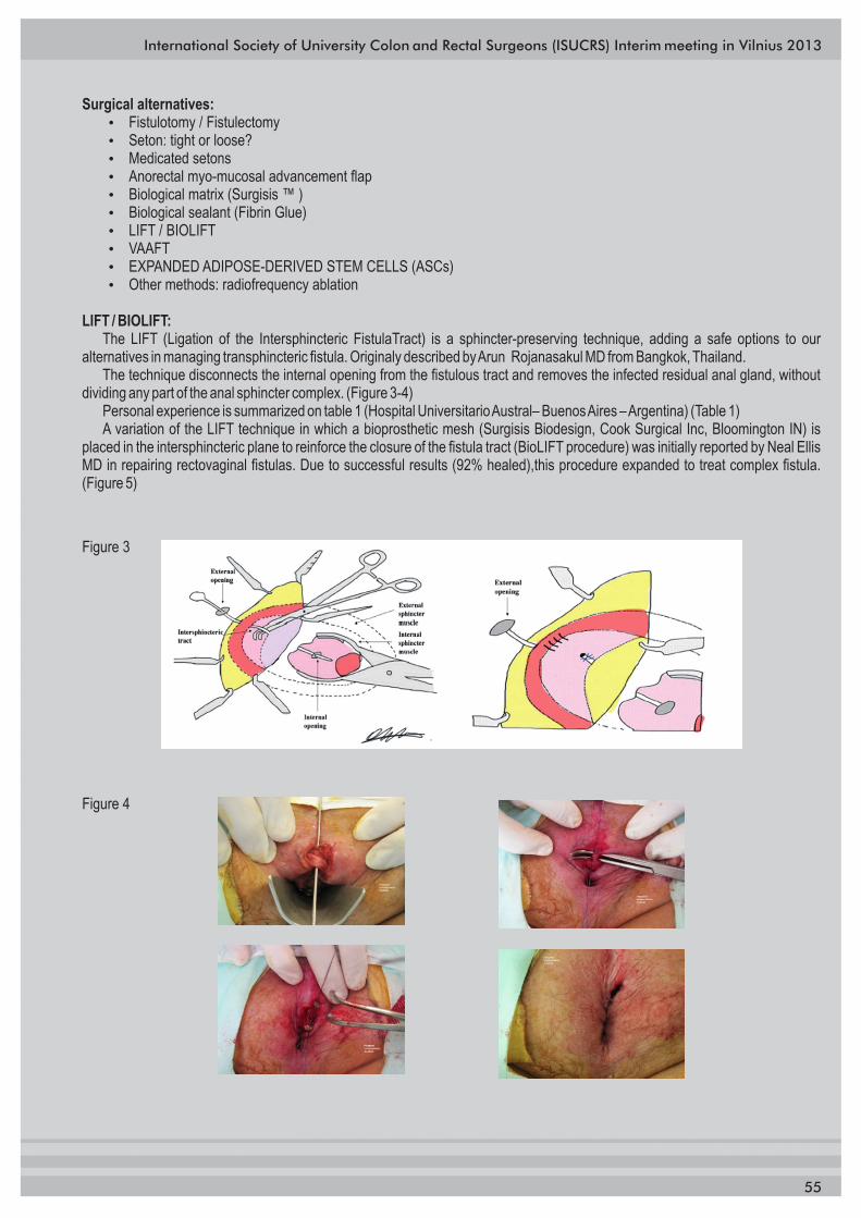
International Society of University Colon and Rectal Surgeons (ISUCRS) Interim meeting in Vilnius 2013
55
Surgical alternatives:� Fistulotomy / Fistulectomy� Seton: tight or loose?� Medicated setons� Anorectal myo-mucosal advancement flap� Biological matrix (Surgisis ™ )� Biological sealant (Fibrin Glue)� LIFT / BIOLIFT� VAAFT� EXPANDED ADIPOSE-DERIVED STEM CELLS (ASCs)� Other methods: radiofrequency ablation
LIFT / BIOLIFT:The LIFT (Ligation of the Intersphincteric FistulaTract) is a sphincter-preserving technique, adding a safe options to our
alternatives in managing transphincteric fistula. Originaly described by Arun Rojanasakul MD from Bangkok, Thailand. The technique disconnects the internal opening from the fistulous tract and removes the infected residual anal gland, without
dividing any part of the anal sphincter complex. (Figure 3-4) Personal experience is summarized on table 1 (Hospital Universitario Austral– Buenos Aires – Argentina) (Table 1)A variation of the LIFT technique in which a bioprosthetic mesh (Surgisis Biodesign, Cook Surgical Inc, Bloomington IN) is
placed in the intersphincteric plane to reinforce the closure of the fistula tract (BioLIFT procedure) was initially reported by Neal Ellis MD in repairing rectovaginal fistulas. Due to successful results (92% healed),this procedure expanded to treat complex fistula. (Figure 5)
Figure 3
Figure 4
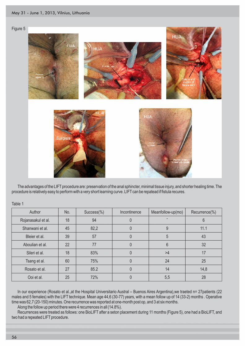
May 31 - June 1, 2013, Vilnius, Lithuania
56
Figure 5
The advantages of the LIFT procedure are: preservation of the anal sphincter, minimal tissue injury, and shorter healing time. The procedure is relatively easy to perform with a very short learning curve. LIFT can be repatead if fistula recures.
Table 1
Author No. Success(%) Incontinence Meanfollow-up(mo) Recurrence(%)
Rojanasakul et al. 18 94 0 - 6
Shanwani et al. 45
39
82,2
57
0
0
9
5
11.1
43Bleier et al.
Aboulian et al.
Sileri et al.
Tsang et al.
Rosato et al.
Ooi et al.
22
18
60
27
25
77
83%
75%
85.2
72%
0
0
0
0
0
6
>4
24
14
5,5
32
17
25
14,8
28
In our experience (Rosato et al.,at the Hospital Universitario Austral – Buenos Aires Argentina),we traeted n= 27patients (22 males and 5 females) with the LIFT technique. Mean age 44,6 (30-77) years, with a mean follow up of 14 (33-2) months . Operative time was 62,7 (20-150) minutes. One recurrence was reported at one-month post op, and 3 at six months.
Along the follow up period there were 4 recurrences in all (14.8%). Recurrences were treated as follows: one BioLIFT after a seton placement during 11 months (Figure 5), one had a BioLIFT, and
two had a repeated LIFT procedure.

International Society of University Colon and Rectal Surgeons (ISUCRS) Interim meeting in Vilnius 2013
57
Suggested Reading:Rojanasakul A, Pattanaarun J, Sahakitrungruang C, Tantiphlachiva
K. Total anal sphincter saving technique for fistula-in-ano; the ligation of intersphincteric fistula tract. J Med Assoc Thai 2007; 90:581-6.
Rojanasakul A. LIFT procedure: a simplified technique for fistula in ano. Tech Coloproctol 2009;13:237-40.
Shanwani A, Nor AM, Amri N. Ligation of the intersphincteric fistula tract (LIFT): a sphincter-saving technique for fistula-in-ano.Dis Colon Rectum 2010;53:39-42.
Bleier JI, Moloo H, Goldberg SM. Ligation of the intersphincteric fistula tract: an effective new technique for complex fistulas. Dis Colon Rectum 2010;53:43-6.
Aboulian A, Kaji A, Kumar R. Early results of ligation of intersphincteric
fitula tract for fistula-in-ano [abstract]. In: 2010 ASCRS Annual Meeting; 2010 May 15-19; Minneapolis, MN, USA. Arlington Heights, IL; American Society of Colon and Rectal Surgeons; 2010;138.
Tan K, Tan I, Koh DC, Tsang C. Long-term results of ligation of intersphincteric fistula tract (LIFT) technique in the management of anal fistula [abstract]. In: 2010 ASCRS Annual Meeting; 2010 May 15-19; Minneapolis, MN, USA. Arlington Heights, IL; American Society of Colon and Rectal Surgeons; 2010;155.
Kee Ho Song. New Techniques for Treating an Anal Fistula.J Korean Soc Coloproctol 2012;28(1):7-12
Grozna E, Carballo F, Canelas A, Bun M, Rotholtz N, Chwat C , Rosato G.
Resultados del uso de la ligadura interesfinteriana de trayecto fistuloso (LIFT) para el tratamiento de fístulas perianales complejas. experiencia inicial multicéntrica.36º Congreso Argentino de Coloproctologia.Nov 2011.
P. Sileri, L. Franceschilli, G. Angelucci, S. D'Ugo, G. Milito, F. Cadeddu, I. Selvaggio, S. Lazzaro, A. Gaspari. Ligation of the intersphincteric fistula tract (LIFT) to treat anal fistula: early results from a prospective observational study Techniques in Coloproctology (11 November 2011), pp. 1-4.
K. Ooi, I. Skinner, M. Croxford, I. Faragher, S. McLaughlin. Managing fistula-in-ano with ligation of the intersphincteric fistula tract procedure: the Western Hospital experience. Colorectal Dis. 2012 May;14(5):599-603

May 31 - June 1, 2013, Vilnius, Lithuania
58
SURGICAL MANAGEMENT OF RADIATION-INDUCED RECTOVAGINAL FISTULAS
Seung-Kook Sohn, MD, PhD. Department of surgery, Yonsei University College of Medicine, Seoul,
Korea
Rectovaginal fistulas (RVFs) represent an often devastating condition in patients and a challenge for surgeons. Successful
management of this condition must take into account a variety of variables including the etiology, size, and location of the fistula. In
addition, the condition of the involved tissues and overall medical condition of the patient are of considerable importance. These
fistulas can develop from a multitude conditions, including obstetrical trauma, inflammatory bowel disease, carcinoma, radiation,
diverticulitis, and infectious processes, and as a result of postsurgical procedures.
Especially, radiation-induced RVFs are one of the most complicated type. Iatrogenic RVFs occur after pelvic surgery and
radiotherapy for the treatment of rectal or gynecologic malignancy. The RVFs from late radiation injury were developed in 0.5~5% of
patients after pelvic radiotherapy. In spite of various surgical techniques, there is still no clear guideline regarding the management
of these fistulas.
The repair of radiation-induced RVFs is very difficult because of tissue damage and finally, spontaneous healing of extensive
defect of normal tissue is extremely rare. Actually, it was reported that spontaneous closure of RVFs was observed in only 3 of 17
patients.
There are two basic principles in the treatment of RVFs. First, due to the high pressure of rectal side, the rectal side repair must be
imperious. Second, it is very important to interpose a viable tissue between the rectum and the vagina. From this concept, the gracilis
muscle flap was introduced. The gracilis muscle provides a well vascularized rotational flap without any effect on the strength or
range of motion of the lower limb. According to a recent report, 100% success rate of their two patients with preoperative
chemoradiation followed by surgery was achieved using gracilis muscle transposition.
In our institute, we reported clinical analysis of 62 rectovaginal fistula patients from various causes. In this analysis, the type of
RVFs was classified to simple and complex type according to their location, size and etiology. Most complex RVFs including 17
patients with radiation-induced RVFs were repaired through abdominal approach or tissue transposition. With an average follow up
of 20 months, 58.1% of all RVFs were completely healed.
We also reported clinical results of coloanal anastomosis in radiation-induced RVFs in 8 patients. All 8 patients concurrently
received irradiation and induction chemotherapy due to primary gynecologic malignancy. For 7 out of 8 patients underwent low
anterior resection with handsewn coloanal anastomosis without diverting stomas. During the median follow-up period of 25 months,
two patients developed anal stenosis and in terms of sphincter function, there were 1 patient with urgency, 1 with gas incontinence
and 1 with night staining. Moreover, according to the data from 1997 to 2012in our institute, 7 out of 9 patients with radiation-induced
RVFs were completely repaired by low anterior resection with handsewn coloanal anastomosis.
In conclusion, rectovaginal fistulas, especially caused by pelvic radiation, present a distressing problem for the patient and a
challenge for the treating physician. From our experiences, if anastomosis is performed between normal and healthy tissues after
complete removal of irradiation-damaged tissues, then one-staged handsewn coloanal anastomosis after low anterior resection
without diverting stomas may guarantee favorable surgical and functional outcomes.

International Society of University Colon and Rectal Surgeons (ISUCRS) Interim meeting in Vilnius 2013
59

May 31 - June 1, 2013, Vilnius, Lithuania
60
STIMULATED GRACILOPLASTY FOLLOWING ABDOMINAL PERINEAL RESECTION (APR)
Francis Seow-Choen. Medical Director, Fortis Colorectal Hospital, Singapore
Abdominal Perineal Resection (APR) is still the standard of care when rectal cancers are very low and not responsive to radio-
chemotherapy. However this entails a removal of the anus with the construction of a permanent stoma. Long or even short term
stomas are associated with physical, psychological and social consequences.
Pickrell was the first to use the gracilis for treatment of anal incontinence in 1952 and Simonsen used it first to restore anal
function after APR. The Gracilis however is a skeletal muscle and prone to fatigue and therefore cannot be relied upon to maintain
continence 24 hours a day seven days a week. Electrical stimulation of this muscle had been shown to convert the muscle to a slow
twitch muscle enabling titanic contraction which in turn can be turned off by a switch.
We proposed and have used the stimulated graciloplasty following APR three years or so after the initial surgery when the risk of
recurrence is low. Such patients who are still motivated to regain perineal annual function should be offered stimulated graciloplasty
as some will have good or acceptable results and thereby improve their lives.

International Society of University Colon and Rectal Surgeons (ISUCRS) Interim meeting in Vilnius 2013
61

May 31 - June 1, 2013, Vilnius, Lithuania
62
HIGH MACRO RUBBER-BAND LIGATURE
Jose A Reis Neto *, Jos A Reis Junior **, Odorino H Kagohara **, Joaquim Simões Neto **, Sergio O Banci ***, Luciane H Oliveira ***
* Head Professor of Surgery at Catholic University of Campinas, Prof. Honoris Causa EDABOL, PhD, SCP,FACS,FRCS, MD** Master of Surgery, Clinica Reis Neto, Catholic University Campinas, FBSCP,MD*** Assistant of Coloproctology, Clinica Reis Neto,FBSCP, MD
Purpose.
The goal of a rubber band ligature is to promote fibrosis of the submucosa with subsequent fixation of the anal epithelium to the
underlying sphincter. Following this principle a new technique of ligature was developed based in two aspects:
1. macro banding: to have a better fibrosis and fixation by banding a bigger volume of mucosa and
2. higher ligature: to have this fixation at the origin of the hemorrhoidal cushion displacement.
Methods.
1634 patients with internal hemorrhoidal disease grade II or III were treated by the technique called High Macro-rubber band. There
was no distinction as to age, gender and race.
Procedure.
The hemorrhoidal cushion must be ligated higher in the anal canal (4 cm above the pectinate line). The mucosa, previously injected
with 2% lidocaine, is gently suctioned at the same time that the rubber band device is slowly moved downward, parallel to the
anoscope, for just a small distance. This maneuver facilitates the suction of a great volume of mucosa and avoids the discomfort
originated from the suction (Figures 5, 6). It is preferable to treat all the hemorrhoids in one single session (maximum of three). When
using the macro rubber-band, it is preferable to band all the existent hemorrhoids at different levels, to avoid stricture of the anal
canal. One ligature is placed on the right anterior zone at four centimeters above the pectinate line, next one on the right posterior
zone at five centimeters above the pectinate line and the last one on the left lateral zone at three or at six centimeters above the
pectinate line (depending of the volume of hemorrhoidal cushion that has to be treated). According to the volume of the mucosa
suctioned and treated, better will be the fibrosis and the fixation of the hemorrhoidal cushion. Sequential single banding, if
necessary, can be performed but at least 30 days should elapse between the sessions.
Results.
All expressed data are derived from our personal experience. Were analyzed the immediate, mediate and long term results obtained
in 1634 patients with hemorrhoidal disease classified as grade II or III. There was no distinction as to age, gender and race. The
analysis was retrospective, without any comparison with conventional banding or infrared coagulation. The period of evaluation
extended from one to twelve years. Evaluation was based on previous symptoms referred by the patient; most frequently bleeding
and prolapse, sometimes aching discomfort after defecation. Follow-up protocol includes a routine of five returns: one week after
the procedure, three weeks, 2 months, 6 months and one year. After the last one year return, if the patient was asymptomatic and
with a normal anoscopy, surveillance was performed by telephone, once a year. The nurse, who attended the procedure, asked for
recurrence of bleeding or prolapse. All the information was noted on the patient inventory. Only patients with a minimum of two years
follow-up were included in the study.
Of the 1634 patients treated, 1275 (78%) have three areas treated in one single session, 279 (17%) have two areas and 80 (4,9%)
have just one zone treated in one single session. Immediately after the procedure, some patients may presents aching discomfort or
tenesmus; to prevent these symptoms it is recommended to utilize an enema of 10 ml of 2% lidocaine (without adrenaline) at the end
of the procedure. After the 4th day of the procedure, these manifestations of tenesmus declined until it disappears.
In this series no skin tag was resected with the banding, but the presence of anal skin tags does not contraindicates the macro rubber
banding; if necessary they can be removed with local assisted anesthesia at the same time that the macro is performed or this skin
tag resection can be performed in another session to avoid post-banding pain.
e

International Society of University Colon and Rectal Surgeons (ISUCRS) Interim meeting in Vilnius 2013
63
The analysis of the immediate results showed peri-anal edema in 27 patients (1,6%), intense post-banding pain (need for parenteral
analgesia) in 27 (1,6%), immediate tenesmus in 14 patients (0,8%) and urinary retention in two male patients (0,1%). Patients with
anal edema had a painful post-banding period.
Light bleeding on defecation was observed in 14 patients (0,8%) in the first post-banding week. More persistent bleeding was
observed in three patients (0,18%), during the second week after the procedure. All of them were examined under local anesthesia,
with spontaneous stop of the bleeding.
On later surveillance, hundred and ninety five patients (11,9%) complained of anal symptoms, including itching, aching discomfort
after defecation and bleeding or prolapse, but only sixty nine (4,2%) showed recurrence of the disease on a new medical evaluation.
Symptomatic recurrence rate of 4,2% (69 patients). The majority of these patients (51 – 73,9%) belonging to the group that have had
just one zone banded. Recurrence in the group with three zones banded was of 21,7% (15 patients of the 69).
All patients with symptomatic recurrence (69) were treated by a new session of macro rubber-banding. None of the patients
developed anal or rectal sepsis. None of the patients needed hospitalization for the observed complications.
Conclusions.
The High Macro rubber-banding technique represents an alternative method for the treatment of hemorrhoidal disease grades II or
III, with good results at a low cost. The analysis of the observed results showed a small incidence of minor complications, with a high
index of symptomatic relief.
May 31 - June 1, 2013, Vilnius, Lithuania
64
OUTPATIENT CLINIC HEMORRHOIDS SURGERY
Pravin J.Gupta. NAGPUR, INDIA
Background.
A persistent search for an ideal treatment for hemorrhoids is on since time immortal. The aim is to minimize postoperative discomfort,
avoid complications, and provide early recovery and return to work. With better understanding of the importance of hemorrhoidal
cushions, the search is on for preservation of the same while minimizing recurrence and relapse. In the last decade, many new
inclusions have been made to address symptomatic hemorrhoids, which mainly include the staplers, harmonic scalpel, ligaSure,
doppler guided dearterialization, radiofrequency surgery etc. The main aim of introduction of these procedures is to improve patient
satisfaction while minimizing intra and post operative complications.
Objective.
This review gives an update on various treatment options for symptomatic hemorrhoids which can be performed in the office or on
outpatient basis.
Conclusion.
From a therapeutic standpoint, the practitioner has access to a wide range of treatment methods. The multiplicity of treatment
modalities is proof in itself that none is greatly superior to other. Each practitioner therefore must be in a position to offer the greatest
possible range of treatment options in order not to be slave to one particular method, with the risk of using it in situations where it may
not really be indicated. So it is always better to tailor the procedure according to the patient rather than opting for a copybook
approach
Keywords.
Hemorrhoids, office procedure, outpatient surgery.

International Society of University Colon and Rectal Surgeons (ISUCRS) Interim meeting in Vilnius 2013
65

May 31 - June 1, 2013, Vilnius, Lithuania
66
CURRENT STATUS OF LIGASURE HEMORRHOIDECTOMY
Daniel Markaryan, Petr Tsarkov (Department of coloproctology and endoscopic surgery, Faculty of
postgraduate professional education of physicians, Department of colorectal surgery, University Hospital
#2, Clinical Center, First Moscow State Medical University n.a. I.M. Sechenov,1/1 Pogodinskaya str.
119435 Moscow, RUSSIA)
for hemorrhoids
The LigaSure vessel sealing system is a bipolar electrothermal device that seals blood vessels through an
optimized combination of pressure and radiofrequency.
ease of use, reduced bleeding and operative time, less wound healing
time, postoperative pain and complication rate and earlier return to work.
The use of radiofrequency energy instead of conventional diathermy in surgical treatment first described in
2001. Since then, many randomized, trials have been conducted to evaluate its efficacy, compared to conventional diathermy
hemorrhoidectomy.
Its use has the advantage of reduced lateral thermal damage of surrounding
tissues. This topic summarize data from most influential trials and meta-analysis to define the role of LigaSure hemorrhoidectomy in
modern anorectal surgery.
The overwhelming majority of publications concluded that LigaSure hemorrhoidectomy has same effectiveness and safety as
conventional diathermy, but also has several advantages like
Because of all this advantages Ligasure™
hemorrhoidectomy can be recomended as a standard for day-care procedure.

International Society of University Colon and Rectal Surgeons (ISUCRS) Interim meeting in Vilnius 2013
67

May 31 - June 1, 2013, Vilnius, Lithuania
68
EXCISIONAL HEMORRHOIDECTOMY - IS IT STILL THE GOLD STANDARD?
Tomas Poskus, MD, PhD. Vilnius University Hospital “Santariskiu Klinikos“
Ideal treatment for hemorrhoids would be the one that effectively provides cure with minimal discomfort, is safe, has low chance
of recurrence in the long term, can be done as an outpatient and with minimal interruption of daily activities and early return to work
and at a minimal cost, it should be simple to learn and use.
There is significant body of evidence in the literature on various treatment methods for hemorrhoids. Conservative treatment
options - flanvonoids and fiber do reduce symptoms of hemorrhoids, however they do not reduce hemorrhoidal prolapse.
Invasive non-operative procedures, of which rubber band ligation is most commonly used, are significantly better compared to
hemorrhoidectomy in terms of post-procedure pain, however they are also significantly less effective, and also not suitable when
hemorrhoidal symptoms are attributed to the external component of hemorrhoids.
Recent metaanalysis compared open and closed hemorrhoidectomy procedures: open hemorrhoidectomy seems to be faster
and simpler to perform, whereas closed technique results in faster wound healing. Overall, there is no difference between
effectiveness of open and closed techniques in the long term and it is almost perfect.
Ligasure hemorrhoidectomy seems to improve operative performance – it reduces blood loss, decreases operative time but
does not seem to improve distant outcomes.
New techniques of hemorrhoid surgery have been studied, although not all of them equally well. Stapled hemorrhoidopexy has
been evaluated in multiple well designed randomized controlled trials and reviewed by several metaanalyses. It has definite early
benefits: shorter operative time, less postoperative pain and less urinary retention, faster return to regular activities. However, long
term results – recurrence of the prolapse and residual skin tags - are all better in standard hemorrhoidectomy group.
Transanal hemorrhoidal dearterialization produces similar results to stapled hemorrhoidopexy but with less postoperative pain.
In conclusion, multiple randomized controlled trials and meta-analyses confirm that excisional hemorrhoidectomy is the most
effective method of treatment, however it is associated with significant postoperative discomfort. So it unfortunately does not satisfy
all the requirements for an ideal procedure to treat hemorrhoids.

International Society of University Colon and Rectal Surgeons (ISUCRS) Interim meeting in Vilnius 2013
69

May 31 - June 1, 2013, Vilnius, Lithuania
70
PPH. IS IT A REAL GOLD STANDARD FOR HEMORRHOIDAL DISEASE?
Ricardo Escalante. Universidad Central de Venezuela, Loira Medical Center, Caracas, Venesuela
Most patients with hemorrhoidal disease may be treated conservatively along the years. Several surgical options have been
proposed, including closed open and semiclosed hemorrhoidectomy, radiofrequency (LigaSure), stapled haemorrhoidopexy (PPH)
with or without excision of anal tags, and doppler hemorrhoidal artery ligation.
In our private center we consider the circular stapled mucosectomy as the standard therapy for the treatment of symptomatic
third-degree haemorrhoids and mucosal prolapsed.
Randomized prospective trials and metanalyses have been carried out with the aim of finding the gold standard operation. PPH
carries less postoperative pain and a shorter convalescence than conventional procedure. On the other hand, while carrying a
higher rate of complications, it may be responsible of the so-called "PPH syndrome", consisting of proctalgia, tenesmus and
urgency. Occasional recto-vaginal fistulas have been described after PPH, if not even of rectal perforation and other life-threatening
complications.
We review the different alternatives in the surgical management for hemorrhoidal disease. Some societies guidelines
recommend a tailored surgery, i.e., the use of different procedures according to the grade of haemorrhoids, which suggests that
patients should be operated by a specialist colorectal surgeon, able to perform different surgeries and to deal with complications and
failures.

International Society of University Colon and Rectal Surgeons (ISUCRS) Interim meeting in Vilnius 2013
71

May 31 - June 1, 2013, Vilnius, Lithuania
72
HAL RAR (HEMOROIDAL ARERIAL LIGATION – RECTO ANAL REPAIR) IN THE
TREATMENT OF HEMORROIDAL DISEASE
Guillermo Rosato MD MAAC MSACP FASCRS MISUCRS – Hospital Universitario Austal – Buenos Aires-
ArgentinaPablo Piccinini MD MAAC MSACP – Hospital Universitario CEMIC – Buenos Aires –Argentina
Hemorrhoids are the most common pathology of coloproctology consultation.
Between 10-20% of this patients may require a surgical approach.
Depending on the type of prolapse, hemorrhoids are classified into four grades:
Grades
I do not prolapse
II prolapse during straining, but reduce spontaneously
III require manual reduction
IV are irreducible
After failure of conservative measures, symptomatic or prolapsed hemorroids (mainly Grades III and IV ) may require surgery, to
achieve curative treatment.
Surgical treatments options for hemorroids have expanded since Milligan –Morgan , Ferguson and Park's operations.
Alternatives include instruments such as: scalpel, scissors, diathermy, radio frequency, laser, harmonic scalpels, cryosurgery,
staples, ecodoppler and laser and ecodoppler and suture.
Technology had made possible the advent of new procedures with different kind of instruments. Tissue resection may not be
necessary with some of this modern tools, eventually seeking for less post operative discomfort.
Hemorroidal surgery has an incidence of early (pain, post op bleeding, king tag swelling, urinary retention) and late (anal
stenosis, formation of skin tags, anal fissure, recurrence) complications shared almost by all procedures.
Kazumasa Morinaga et al. (1995) from Japan, reported this novel technique: HAL (Hemorroidal Artery Ligation), for the first time,
based upon an understanding of the pathogenesis and arterial inflow to hemorrhoids. Since then its use expanded to South East
Asia, India, Europe , and the United States. Thereafter, a mucopexy, as described by Farag and Hussein, but developed and
published by Scheyer on 2008 (RAR : Recto Anal Repair ) was added, in order to reduce remaining mucosal redundancy.
The procedure is now known as HAL/RAR (Hemorrhoid Artery Ligation/Recto Anal Repair- A.M.I. GmbH,Austria)
In Argentina, local FDA has approved its use since June 2012 and is commercially available since October 2012.
This new hemorrhoid treatment procedure is minimally invasive .Is carried out on an area that is relatively less sensitive to pain. It
does not involve tissue removal. HAL (Hemorrhoid Artery Ligation ) uses doppler technology to find the blood flow within the small
artery feeding the dilated hemorrhoid to be treated. Once the artery is identified, a reabsorbable suture in a figure of eight
(Polyglycolic acid) is placed around it. The suturing restricts the blood flow, which in time, will result in the shrinkage of the
hemorrhoid and relief of symptoms as: bleeding.
For larger Grade IV hemorrhoids, internal component is sutured and lifted back into the anal canal by means of a running
absorbable suture (RAR= Recto Anal Repair). This hemorrhoidal lift treatment may also reduce the external hemorrhoids, solving
the second most important symptom, which is prolapse.
The sutures will dissolve over a short period of time (6-8 weeks).
No tissue resection takes place and all sutures are placed above the dentate line, in order to prevent postoperative discomfort.
Patients may resume their normal activities within few days, and as with other hemorrhoid treatment surgeries, avoiding physical
efforts is recommended for around four weeks.

International Society of University Colon and Rectal Surgeons (ISUCRS) Interim meeting in Vilnius 2013
73
HAL being performed Hemorroid for RAR procedure
Print-out of points of HAL Procedure concluded RAR
Pre op of HAL RAR Post op of HAL RAR
Due to the amount of procedures to treat hemorroidal disease it is wise to provide the most effective surgical treatment and
choose the appropriate technique tailored to the individual patients' clinical symptoms.
Needle holder and knot pusher
Handle and anoscope(doppler incorporated)

May 31 - June 1, 2013, Vilnius, Lithuania
74
General considerations regarding HAL - RAR� Surgeons who would like to introduce this practice to their surgical activity should be trained and supervised by an
experienced coloproctologist. Training should include: observation and practice on a bowel or anal model.
� Ten to twenty cases, seems to be enough for the learning curve,pending skills and surgical experience.
� Procedures to compare with should include: mucosal banding, stapled anopexy.
� Reports of early complications are similar to other invasive treatment options.
� HAL- RAR appears to be as safe as conventional surgery and has potentially fewer risks.Suggested readingThe superior rectal artery and its branching pattern with regard to its clinical influence on ligation techniques for internal hemorrhoids.Aigner F, Bodner G, Conrad F, Mbaka G, Kreczy A, Fritsch H.Am J Surg. 2004 Jan;187(1):102-8.Anatomical branches of the superior rectal artery in the distal rectum. Colorectal Disease.Volume 11, Issue 9, pages 967–971, November 2009Farag A. Pile suture: a new technique for the treatment of haemorrhoids. Br J Surg. 1978; 65: 293-5.Morinaga K, Hasuda K, Ikeda T, A Novel Therapy for Internal Hemorrhoids: Ligation of the Hemorrhoidal Artery Wirth a Newly evised Instrument (Moricorn) in Conjunction Wirth a Doppler Flowmeter. Am. Journal of Gastroenterology, April 1995, Volume 90, No. 4: 610-613Satzinger U, Feil W, Glaser K. Recto anal repair (RAR): a viablenew treatment option for high-grade hemorrhoids. One year results of a prospective study. Pelviperineology. 2009;28:37– 42.Hussein AM. Ligation-anopexy for treatment of advanced hemorrhoidal disease. Dis Colon Rectum 2001;44:1887-1890.Doppler-guided hemorrhoidal artery ligation in the management of symptomatic hemorrhoids.Ramírez JM, Aguilella V, Elía M, Gracia JA, Martínez M.Rev Esp Enferm Dig. 2005 Feb;97(2):97-103Doppler-guided hemorrhoidal artery ligation.Scheyer M, Antonietti E, Rollinger G, Mall H, Arnold S.Am J Surg. 2006 Jan;191(1):89-93.Two-center experience in the treatment of hemorrhoidal disease using Doppler-guided hemorrhoidal artery ligation: functional results after 1-year follow-up.

International Society of University Colon and Rectal Surgeons (ISUCRS) Interim meeting in Vilnius 2013
75
Wałega P, Scheyer M, Kenig J, Herman RM, Arnold S, Nowak M, Cegielny T.Surg Endosc. 2008 Nov;22(11):2379-83. Doppler-guided recto-anal repair: a combination of haemorroidal arterial ligation and transrectal mucopexy for minimally invasive treatment of haemorroidal disease (all grades). Scheyer M,Antonietti E,Arnold S.JCD December 2008 .Vol 23,Issue 4 .pp17-21.Trans-anal disarterization of internal hemorrhoids under Doppler control with mucopexy and lifting in treatment of stage III-IV hemorrhoids.Zagriadskiĭ EA.Khirurgiia (Mosk). 2009;(2):52-8.Conventional haemorrhoidectomy, stapled haemorrhoidectomy, Doppler guided haemorrhoidectomy artery ligation; post operative pain and anorectal manometric assessment.Khafagy W, El Nakeeb A, Fouda E, Omar W, Elhak NG, Farid M, Elshobaky M. Hepatogastroenterology. 2009 Jul-Aug;56(93):1010-5.Doppler-guided haemorrhoidal artery ligation with recto anal repair: a new technique for the treatment of symptomatic haemorrhoids.Forrest NP, Mullerat J, Evans C, Middleton SB. Int J Colorectal Dis. 2010 Oct;25(10):1251-6.Evaluation of transanal hemorrhoidal dearterialization as a minimally invasive therapeutic approach to hemorrhoids.Ratto C, Donisi L, Parello A, Litta F, Doglietto GB.Dis Colon Rectum. 2010 May;53(5):803-11.DG-RAR (Doppler-guided recto-anal repair): a new mini invasive technique in the treatment of prolapsed hemorrhoids (grade III-IV): preliminary report.Testa A, Torino G, Gioia A.Int Surg. 2010 Jul-Sep;95(3):265-9.Early Experience of Doppler-Guided Hemorrhoidal Artery Ligationand Rectoanal Repair (DG-HAL & RAR) for the Treatment of Symptomatic Hemorrhoids. SungWook Cho, Ryung-Ah Lee, Soon Sup Chung, Kwang Ho Kim, M.D.. J Korean Surg Soc 2010;78:23-28Transanal haemorrhoidal artery echodoppler ligation and anopexy (THD) is effective for II and III degree haemorrhoids: a prospective multicentric study.Infantino A, Bellomo R, Dal Monte PP, Salafia C, Tagariello C, Tonizzo CA, Spazzafumo L, Romano G, Altomare DF.Colorectal Dis. 2010 Aug;12(8):804-9.Doppler-guided hemorrhoidal artery ligation and rectoanal repair (HAL-RAR) for the treatment of grade IV hemorrhoids: long-term results in 100 consecutive patients.Faucheron JL, Poncet G, Voirin D, Badic B, Gangner Y.Dis Colon Rectum. 2011 Feb;54(2):226-31. Doppler-guided hemorrhoidal laser procedure for the treatment of symptomatic hemorrhoids: experimental background and short-term clinical results of a new mini-invasive treatment. Giamundo P, Cecchetti W, Esercizio L, Fantino G, Outcome of stapled haemorrhoidopexy versus doppler-guided haemorrhoidal artery ligation for grade III haemorrhoids.Giordano P.Tech Coloproctol. 2011 Sep;15(3):257-8Doppler-guided hemorrhoidal laser procedure for the treatment of symptomatic hemorrhoids: experimental background and short-term clinical results of a new mini-invasive treatment.Giamundo P, Cecchetti W, Esercizio L, Fantino G Geraci M, Lombezzi R, Pittaluga M, Tibaldi L, Torre G, Valente M.Surg Endosc. 2011 May;25(5):1369-75.Doppler-guided hemorrhoidal artery ligation does not offer any advantage over suture ligation of grade 3 symptomatic hemorrhoids.Gupta PJ, Kalaskar S, Taori S, Heda PS.Tech Coloproctol. 2011 Dec;15(4):439-44.Hemorrhoidal artery ligation procedure with or without Doppler transducer in grade II and III hemorrhoidal disease: a blinded randomized clinical trial.Schuurman JP, Borel Rinkes IH, Go PM.Ann Surg. 2012 May;255(5):840-5.DG-RAR for the treatment of symptomatic grade III and grade IV haemorrhoids: a 12-month multi-centre,prospective observational study. S. Roka, D. Gold, P. Walega, S. Lancee, E. Zagriadsky, A. Testa, A. N . Kukreja, A. Salat. Eur Surg (2013) 45:26–30

May 31 - June 1, 2013, Vilnius, Lithuania
76
PATIENT DELAYING FACTORS IN THE DIAGNOSIS OF SYMPTOMATIC RECTAL CANCER
Ruiz-Healy,F; Arroyo-Cazarez,M. Service of Coloproctology and Unit of Coloproctology. Centro
Hospitalario Sanatorio Durango, Mexico City, Mexico
Introduction.
Despite information, modern diagnostic tools, exams and protocols, symptomatic patients with rectal cancer continue to-date;
present diagnostic delay and frequently are seen in advance stages of disease. Patient delay plays an important part in late
diagnosis.
Purpose.
Analyze patient delay of symptomatic rectal cancer treated in a Coloproctology service within a private teaching hospital in Mexico
City.
Methods.
A retrospective chart review on patients with rectal cancer from January 2006 to December 2010 was performed. Inclusion criteria
consisted of histological diagnosis of adenocarcinoma and tumor limited within 15 cm of anal verge. Exclusion criteria consisted of
age younger of 40 years and recurrent cancer. Parameters included age and sex, signs and symptoms, patient behavior, and time
from onset of symptoms to first visit to a physician. Results were compared to world literature identified from electronic databases.
Results.
67 patients were included within the protocol. (m/f 40/27) and mean age of 57 (range, 42-93) years. Most common symptom were
bleeding (76%). Other symptoms included rectal pain (27%); constipation (9%) and diarrhea (5%). Vague abdominal symptoms,
weight loss, rectal secretion and fecal impaction were also reported. Forty five percent of patients had multiple symptoms. Patient's
behavior responded in two ways. Seven directly consulted a physician and sixty self-diagnosed their symptoms. Assumed
diagnosis was hemorrhoidal diseases in fifty two, colitis in five, intestinal parasitosis in four, intestinal constipation in three, anal
fissure in two and prostate problems in one patient. Initial treatment was with over-the-counter medications. These included
hemorrhoidal creams, medication for colitis, intestinal parasitosis and laxatives. Patient delay from first symptoms to consulting a
physician, ranged from four days to twelve months, with a mean average of 25 weeks.
Conclusions.
Findings of this study demonstrated that 88 % of patients erroneously credited their symptoms to common colorectal diseases.
Patients delaying diagnosis of rectal cancer, is a concern that has remained almost constant for decades in world literature. Health
education on initial symptoms of rectal cancer should be actively emphasized in the general population.

International Society of University Colon and Rectal Surgeons (ISUCRS) Interim meeting in Vilnius 2013
77

May 31 - June 1, 2013, Vilnius, Lithuania
78
TREATMENT STRATEGY OF EARLY COLORECTAL CANCER: WHAT CAN BE DONE?
Konishi F, Togashi K (Departmetn of Surgery Nerima Hikarigaoka Hospital Tokyo Japan AND
Department of Colorectal Surgery/ endoscopy Aizu General Hospital)
Purpose.
The management of T1 colorectal cancer has been a controversial issue in the treatment of colorectal cancer in early stages. The
purpose of this report is to clarify the process of determining the selection of treatment for early stage colorectal cancer (Tis T1).
Methods and Results.
Firstly the colonoscopic appearances under meticulous observation are important findings that will determine whether colonoscopic
resection or bowel resection should be done as an initial treatment. Magnifying colonoscopy is also an important tool to estimate the
grade of invasion in the submucosal. These endoscopic findings were analyzed in 162 cases. When pit pattern V is seen it was
considered to be the sign of deep invasion in the submucosal. However, there is a limitation in making the colonoscopic diagnosis,
therefore, when the assessment as for the depth of invasion is unclear on colonoscopy, colonoscopic resection is usually performed
when it is technically feasible. The indication for bowel resection will then be determined according to the histological findings of the
resected specimens. Our histological analysis of T1 stage cancer, substantial invasion depth in the submucosal, lymphatic channel
invasion, moderate to poor differentiation, invasive front on histology (budding) were significant risk factors for the estimating lymph
node metastasis.
Methods of Endoscopic resection of early stage colorectal cancer.
Endoscopic mucosal resection (EMR) is a useful and safe method to remove relatively large sessile lesions. In this method, saline or
hyaluronic acid solution is infused in the submucosa to lift up the lesion. After the infusion and the lift up, a snare wire will be place
around the tumor and the lesion will be removed by the usage of electrocoagulation. Endoscopic submucosal dissection (ESD) is
used to remove even larger sessile lesions. In this method, after the injection of hyaluronic acid, the mucosa surrounding the lesion
will be circumferentially divided by the usage of “needle knife” or “hook knife”. Subsequently the submucosa will be dissected by
using similar devices from the distal side to the proximal side. Although this is a time consuming and tedious procedure, it can well be
tolerated by the patients. The important advantage of ESD is the en block resection which will lead to a better treatment than piece
meal resection. Although the time for this method is usually long, the procedure is less invasive than bowel resection.
Conclusions.
Colonoscopic findings are important for the determination of the depth of invasion in the submucosa prior to resection. When
endoscopic resection is performed the presence of one or more of the above histological features is considered to be the indication
for bowel resection. The treatment of T1 stage cancer should be discussed in a team of endoscopists, surgeons and
histopathologists to choose an optimal method of treatment.

International Society of University Colon and Rectal Surgeons (ISUCRS) Interim meeting in Vilnius 2013
79

May 31 - June 1, 2013, Vilnius, Lithuania
80
INITIAL RESULTS AFTER LOCAL EXCISION FOLLOWED WITH POSTOPERATIVE
RADIATION FOR UT1N0M0 RECTAL CANCER
Ashari LH, Alsanea N. Section of Colon & Rectal Surgery, King Faisal Specialist Hospital & Research
Center-Riyadh, Saudi Arabia
Purpose.
Assess local recurrence after local excision and postoperative radiation for non-metastatic ultrasonographic stage of T1N0M0 rectal
cancer.
Methods.
The colorectal database at King Faisal Specialist Hospital & Research Center-Riyadh was reviewed for all rectal cancer cases that
underwent local excision for rectal cancer and postoperative radiation for ultrasonographic stage T1N0M0 for the period 2003-2011.
Local excision was performed only in patients who could not undergo a sphincter-saving procedure or were not fit to have major
radical surgery due to sever co-morbidities. The demographic data, clinical, pathologic, recurrence and survival data were also
reviewed.
Results.
There were 11 men and one woman. Mean age at presentation was 62 years. The tumors were 3.6 cm away from the anal verge.
After resection, the tumors had a median diameter of 3.5 cm (range: 1.0- 5.0 cm). All the tumors had favorable pathology and
negative resection margins. The tumor cells were never less than 3 mm from any of the resection margins. All resections were full
thickness. One sample contained 2 negative lymph nodes. Median Follow up was 36 months. All patients received adjuvant
radiation. One tumor recurred in a lymph node in the meso-rectum; recurrence 8%. Overall 3-year survival was 100%, 3-year
disease free survival was 91%.
Conclusion.
Local excision followed with postoperative radiation is associated with low recurrence rate and acceptable survival.

International Society of University Colon and Rectal Surgeons (ISUCRS) Interim meeting in Vilnius 2013
81

May 31 - June 1, 2013, Vilnius, Lithuania
82
OUTCOMES OF EARLY RECTAL CANCER AT TATA MEMORIAL CENTRE
Shailesh V. Shrikhande, Gaurav Chaubal, Ashwin Desouza, Mahesh Goel. GI and HPB Service,
Department of Surgical Oncology, Tata Memorial Centre, Mumbai, India
Introduction.
The incidence of colorectal cancer in India is low but increasing in urban population. Prognosis of patients with early rectal cancer is
significantly better than those who present at an advanced stage. The purpose of this study was to analyse our results of early rectal
cancer.
Methods.
Patients were divided into 3 groups. Group 1 comprised of early rectal cancers (T1,T2, N0), Group 2 comprised of patients who were
downstaged to stages T1 or T2 stage after neoadjuvant chemoradiation and group 3 comprised of advanced rectal cancers
(T3,T4,N1). A retrospective analysis of a prospectively maintained database was done. Recurrence was studied in the 3 groups.
Results.
From May 2010 to February 2013, a total of 335 cases underwent resection for carcinoma rectum. Group 1 comprised of 16 (4.8%)
cases, group 2 comprised of 146 (43.5%) cases and group 3 comprised of 173 (51.6%) cases. In Group 1, 7 (43.8%) underwent
anterior resection, 3 (18.8%) underwent low anterior resection, 2 (12.5%) underwent ultralow anterior resection and 4(25%)
underwent APR. In Group 2, 22 (14.9%) underwent anterior resection, 21(14.2%) underwent low anterior resection, 24(16.2%)
underwent ultralow anterior resection, 72(48.6%) underwent APR, 2 (1.4%) underwent Hartmann's and 5(3.4%) underwent total
proctocolectomy. In Group 3, 46(26.6%) underwent anterior resection, 26 (15%) underwent low anterior resection, 16 (9.2%)
underwent ultralow anterior resection, 66 (38.2%) underwent APR, 13(7.5%) underwent Hartmann's and 5(2.9%) underwent total
proctocolectomy. Mean follow up was 18 months. In group 1, no patient developed recurrence, in group 2, 7(4.8%) developed
systemic recurrence but no local recurrence and in group 3, 25 (7 local; 18 systemic) (14.4%) developed recurrence. Patients in
group 1 had the best prognosis followed by those of group 2 and group 3.
Conclusion.
Our results highlight the need for increased awareness to diagnose early rectal cancers and the crucial role of neo-adjuvant
chemoradiation in improving outcomes in resectable but advanced rectal cancers.

International Society of University Colon and Rectal Surgeons (ISUCRS) Interim meeting in Vilnius 2013
83

May 31 - June 1, 2013, Vilnius, Lithuania
84
IS THERE A PLACE FOR SELECTIVE USE OF NEOADJUVANT RADIATION WITHOUT
CHEMOTHERAPY
J.A. Reis Neto *, J.A. Reis Junior **, O. Kagohara** & J.Simões Neto***, Sérgio O Banci***, Luciane H.
Oliveira ***. Clínica Reis Neto* Head Professor of Surgery at Catholic University of Campinas, Prof. Honoris Causa EDABOL,PhD, SCP,FACS,FRCS, MD** Master of Surgery, Clinica Reis Neto, Catholic University Campinas, FBSCP,MD*** Assistant of Coloproctology Clinica Reis Neto,FBSCP, MD
(Key words: Rectal Cancer, Cancer of the Lower Rectum, Irradiation, Preoperative Radiotherapy, Local Recurrence, Mortality,
Survival )
Aims.
The prognosis on treatment of the cancer of the rectum has changed in the last fifteen years. The main cause for those results is the
decrease in the incidence of local recurrence. Local recurrence is directly related to the number of undifferentiated cells and to the
grade of wall invasion. So any kind of treatment that would diminish the number of undifferentiated cells and the size or the tumor
wall penetration would certainly decrease the local recurrence rate, lengthening the interval free from cancer and, perhaps,
modifying the long term survival rate. Neo-adjuvant radiotherapy has been used combined with chemotherapy for cancer of the
lower rectum, stage III and in some cases of stage II tumours. Neo-adjuvant radiotherapy alone could be selected for patients stage I
and elderly patients.
Methodology.
Between 1978 and 2012 , a total of 75 patients with cancer of the lower rectum classified as Stage I tumours (T1/T2 NoMo ), and a
group of 10 patients, over 90 years, were treated by pre-operative radiotherapy and selected for this study. There was no gender,
race and age distinction. The same protocol was used in all the patients – 4500 cGy, 200 cGy /day, during 4 consecutive weeks
(anterior and posterior pelvic fields) by means of a Linear Megavoltage Accelerator ( 25 MeV ).
Preoperative dosage of CEA, after cancer diagnosis, colonoscopy and abdominal tomography were performed in all the patients to
stage the tumor. Endorectal ultrasound was performed in all patients treated after 1993. Pelvic MR was performed in all patients
after 1998.
Size and infiltration of the tumor were thus evaluated before and after the irradiation. Every patient had biopsies (a minimum of 10) of
the tumor taken at the time of the diagnosis and another 4 to 8 weeks after the completion of the radiotherapy. The number of
undifferentiated cells were exhaustively noted and the results compared, in both biopsies. Broders' classification was equally
observed and the results obtained, before and after radiotherapy, registered and compared.
Protocol:
post-radiotherapy protocol included:
1. proctoscopy and digital examination immediately, 4 weeks and 8 weeks after the end of the irradiation treatment, to evaluate
tumor volume and wall infiltration;
2. endorectal ultrasound was performed 8 weeks after the end of the irradiation treatment;
3. CEA , 4 weeks and 8 weeks after the end of the irradiation treatment;
4. when clinical assessment suggested local recurrence, Pelvic MR was accomplished;
5. it was considered local recurrence the presence of residual tumour after 8 weeks;
6. only patients with residual tumour after 8 weeks were selected for surgery;
7. surgery varies according to residual tumour infiltration (T1 or T2).
Results.
From the 75 patients classified as stage I, 27 were T1NoMo and 48 were T2NoMo. All the patients classified as Stage I, T1NoMo, had
no residual tumour detected after irradiation therapy. Of the 48 patients classified as Stage I, T2NoMo, 40 had no residual tumour
after irradiation; 8 were submitted to FTLE (total local excision). One had recurrence of the tumour 3 years after the surgery.
As a fact, the variation in the number of undifferentiated cells before and after irradiation was significantly proved; the number of
tumors with a high grade of undifferentiated cells shows a negative variation after irradiation showing a significant statistical
reduction in the number of undifferentiated cells .

International Society of University Colon and Rectal Surgeons (ISUCRS) Interim meeting in Vilnius 2013
85
At the same time, as a corollary, the number of tumors with a low grade of differentiated cells at diagnosis shows an effective increase
after irradiation, with a positive variation.
Statistical analysis of the whole group showed that preoperative radiotherapy does decrease frequency of undifferentiated cells. The
results observed in this study confirmed that after the irradiation an involution of the tumor size and volume occurred, detectable not
only by endorectal ultrasound (ERUS), but also by digital and endoscopic examinations. Of the 27 patients with stage I tumors
classified as T1 none needed further treatment; the tumor disappeared after the radiotherapy. Of the 48 patients with stage I T2
tumors only 8 needed further surgery – full local total excision. Of the 10 patients aged 90 years, only one had to be submitted to
surgery.
Conclusion.
Preoperative radiotherapy is really effective in reducing the number of undifferentiated cells and in diminishing the tumor volume and
the carcinomatous infiltration of the rectal wall for patients stage I tumours and for aged patients.

May 31 - June 1, 2013, Vilnius, Lithuania
86
ULTRALOW ANTERIOR RESECTION IN ERA OF NEOADJUVANT THERAPY
Shailesh V. Shrikhande, Gaurav Chaubal, Ashwin Desouza, Mahesh Goel. GI and HPB Service,
Department of Surgical Oncology, Tata Memorial Centre, Mumbai, India
Introduction.
For patients with low rectal cancer, abdomino-perineal resection (APR) and low anterior resection (LAR) was considered as the
procedure of choice. However ultra low anterior resection (ULAR) with sphincter preservation is also now considered as a safe
procedure with equivalent oncological outcomes. The purpose of this study was to compare our results for the 3 procedures done for
low rectal cancers.
Methods.
A retrospective analysis of a prospectively maintained databases was done. Low rectal cancers were defined as those lying within 7
cm from the anal verge. An analysis of their surgical outcomes was done.
Results.
From May 2010 to February 2013, a total of 226 cases underwent resection for low rectal cancers. 140(61.9%) patients underwent
APR, 47(20.8%) underwent LAR and 39(17.2%) underwent ULAR. In the ULAR group, the mean age was 47 + 15.3 years (14 – 73
years). Mean hospital stay was 8.7 + 4.7 days (7 – 25 days). Mean distance from anal verge was 4.9 + 1.2 cm ( 3 – 8 cm). Mean blood
loss was 490 + 320 ml ( 100 – 1500 ml). Out of the 39 cases, 1 was done laparoscopically and 1 was laparoscopic assisted. Double
Stapled anastomosis was done in all cases. A covering stoma was done in 32 cases (82.1%). The complication grade was Grade 0
(79%), Grade 1 (2.6%), Grade 2 (10.3%), Grade 3b (7.7%) (Clavein Dindo Classification). In grade 3b, two patients had an
anastomotic leak and one patient developed recto-vaginal fistula. 33 cases (84.6%) received neoadjuvant chemoradiation. Out of
the 39 cases, 2 (5%) had T1 tumours, 10 (25%) had T2, 16 (41%) had T3 and no residual tumour was found in 11 (25%) cases. The
average lymph node yield was 9.1 + 5.9 (0 – 22). Average number of positive nodes was 1.5 + 2.8 (0 – 13). Mean follow up was 18
months. None of the patients had local recurrence. However, 2 patients developed liver metastasis during follow up period.
There was no perioperative mortality in any group. The morbidity rate was 14.2% in APR group, 12.8% in LAR group and 20.5% in
ULAR group. 17 (12.1%) patients had recurrence in APR group, 5(10.6%) in LAR group and 2 (5.1%) in ULAR group.
Conclusion.
ULAR should only be done in selected patients with lower T stage and those who are termed good responders after neoadjuvant
chemoradiation in a multidisciplinary setting. They can be done safely without compromising on oncologic outcomes.

International Society of University Colon and Rectal Surgeons (ISUCRS) Interim meeting in Vilnius 2013
87
May 31 - June 1, 2013, Vilnius, Lithuania
88
SURVIVAL AFTER INDUCTION CHEMOTHERAPY FOLLOWED WITH ADJUVANT
CHEMOTHERAPY AND RADICAL SURGERY FOR INITIALLY UNRESECTABLE RECTAL CANCER
Nasser Al-Sanea. Section of Colon & Rectal Surgery, King Faisal Specialist Hospital & Research Center-
Riyadh, Saudi Arabia
Purpose.
To identify the characteristics of patients who can achieve cure after radical resection for locally advanced non metastatic rectal
cancer (T4 N0-2 M0).
Study Population.
Patients who underwent curative radical treatment for non-metastatic locally advanced rectal cancer T4N0-2M0 from 2000 till 2009
at King Faisal Specialist Hospital and Research Center-Riyadh.
Methods & Design.
The colorectal database at King Faisal Specialist Hospital and Research Center-Riyadh was reviewed for all rectal cancer patients
with the pathologic stage of T4 N0-2 M0 who underwent curative treatment. Data collected included demography and clinical,
radiologic and pathologic characteristics. Treatment, local recurrence, distal metastasis and survival data were also collected.
Results.
There were nineteen patients, 13 women and 6 men. The median age at presentation was 52 years (range: 29-80), All patients were
deemed unresectable at presentation and had at least one pelvic organ involved outside the rectum. They were subjected to 6 cycles
of XELOX (Capecitabine and Oxaliplatin) followed up with neoadjuvant chemoradiation using Capecitabine. Abdominoperineal and
anterior resection were performed in 10 & 9 patients respectively. All proved to be T4 N0-2 on histology. Complete surgical resection
was achieved and documented by pathology in 15 patients (R0). Non favorable prognostic pathologic signs (mucinous, signet ring
cells, lymphovascular or preneural invasion, poor differentiation) were noted in 9 patients. The median diameter of the tumors was
3.5 cm (range, 2-9cm). There was no case of complete pathologic response to neoadjuvant chemoradiation treatment. Five patients
had more than one organ invaded by tumor. All patients received adjuvant chemotherapy in the form of XELOX.
Overall 3-year survival rate was 73%, disease-free 3-year survival rate was 36%. Both were significantly lower in patients with non-
favorable pathology (p < 0.0003). Reduction in size by 30% after induction chemotherapy is an indicator of improved survival
(p<0.05). Survival was not significantly different between R0 and R1 groups of patients with similar pathology.
Conclusion.
Non-favorable pathology is associated with lower overall & disease free survival regardless of completeness of resection.

International Society of University Colon and Rectal Surgeons (ISUCRS) Interim meeting in Vilnius 2013
89
May 31 - June 1, 2013, Vilnius, Lithuania
90
COLORECTAL CANCER AND LIVER METASTASES: POSSIBLE STRATEGIES
E. De Raffele, M. Mirarchi, S. Vaccari, S. Palumbo, B. Cola
Department of Emergency, General Surgery and Transplants. S.Orsola University Hospital of Bologna.
Bologna. ITALY
Colorectal cancer (CRC) is still one of the leading causes of cancer-related death in the Western countries. Liver metastases
(CRLM) are present in nearly 15 to 25% of individuals with newly diagnosed colorectal cancer and will develop in up to 50% of
patients during the course of their disease.
When liver metastases are considered resectable after an adequate preoperative evaluation, radical liver resection still remains
the only potentially curative therapy, with reported 5- and 10-year actuarial survival rates of 17% to 35% and 16% to 23%,
respectively. More recent series describe 5-year overall survival rates of 37% to 58% after hepatectomy.
Most of the criteria defining resectability of CRLM in the past, such as the size, location, number of intrahepatic metastases and
the presence of extrahepatic disease, have been largely abandoned. The current criteria focus on what should be left after liver
resection: at present, the definition of resectability includes a complete resection with clear surgical margins on microscopy (R0
resection), without compromising postoperative liver function because of the insufficiency of either the remaining liver volume or
biliary and venous vascularization and drainage.
The systematic use of intraoperative ultrasonography (IOUS) to evaluate the tumour-vessels relationship and to guide hepatic
resection has been demonstrated to decrease the extent of the liver resection both for primary and metastatic liver tumours without
prejudice on oncological radicality: the extensive use of IOUS guidance during liver surgery permits to minimize the resection
margins, to avoid the need for strict anatomic approaches and to reduce the rate of major hepatectomies without increase of local
recurrence rates in the long-term follow-up.
. One-stage IOUS guided liver resection has
been proven to be effective also in selected patients with multiple bilobar CRLM usually considered for portal vein
ligation/embolization and subsequent major hepatectomy or for two-stage hepatectomy.
The optimal timing for surgical resection in case of synchronous presentation of primary CRC and liver metastases has long
been controversial. In the past decades most investigators have recommended a staged approach with initial resection of the
primary tumour followed by liver resection, usually reserved to patients without evidence of disease progression during
chemotherapy. In patients with large synchronous CRLM and uncomplicated primary cancer, the reversal of the traditional strategy
has been recently proposed, to avoid the risk of progression of the liver tumour to unresectability. Proponents of staged strategies
advance evidence for increased morbidity and mortality associated with simultaneous procedures.
In recent years however an increasing number of studies have shown good results for elective simultaneous procedures, which
have the striking advantage of avoiding a second laparotomy along with the opportunity of an earlier initiation of adjuvant therapy,
even though the cumulative risks of the two procedures need to be adequately considered. As a matter of fact, the paradigm for the
surgical management of synchronous CRLM has begun to change, and the planned extent of hepatic resection seem to represent
the most important determinant of whether colorectal and hepatic procedures should be performed simultaneously. Our experience
in this field shows that simultaneous colorectal and liver resection represent an adequate surgical strategy in selected patients with
resectable colorectal cancer and resectable synchronous CRLM. A staged approach is still advisable in patients requiring urgent
colorectal resection because of complicated CRC, while all the other patients should be theoretically considered for simultaneous
resection: in these cases a systematic approach to CRLM which considers the need of reducing the extent of hepatectomy while
preserving oncological radicality with the use of intraoperative ultrasonography should represent the best option to reduce the
perioperative risks.
In case of vascular invasion of the right hepatic vein or the middle hepatic vein, more
conservative procedures have been described as an alternative to right hepatectomy or trisegmentectomy, based on the extensive
use of colour Doppler IOUS and of sonographic guidance of parenchymal resection

International Society of University Colon and Rectal Surgeons (ISUCRS) Interim meeting in Vilnius 2013
91

May 31 - June 1, 2013, Vilnius, Lithuania
92
INDUCTION THERAPY OF COLORECTAL CANCER LIVER METASTASES: CURRENT
EVIDENCE AND VUOI EXPERIENCE
Liver is the most common and often the only location of colorectal cancer metastases. For patients with liver limited disease
multimodality treatment combining intensive chemotherapy, radical resection, ablative techniques enables to reach 32% ten year
survival. When treating non resectable metastatic colorectal cancer patient median overall survival reaches only two years.
Unfortunately only 10-20% of liver metastases are initially resectable. Induction therapy is being used to enhance respectability.
Ideal induction therapy regimen must have high response rate because it directly influence resection rate. Chemotherapy duplets or
triplets in combination with biologic therapy are mostly used regimens for induction therapy. Response rates of such combinations
differ from 12 to 70%. The choice of regimen is done regarding biological markers such as KRAS status, patient condition. The most
effective regimen for wt KRAS patients according CELIM trial is FOLFIRI/FOLFOX with cetuximab showing response rate up to 70%
and increasing resection rate from 3 to 60% with acceptable toxicity profile.
These results reflects in our clinical practice. We had 3 patients receiving FOLFIRI with cetuximab. The regimen was safe, all the
patients achieved partial response an all were refered for curative resection. Partial radiological response in one patient turn out to
be complete pathological response with no vital cancer cells in fibrotic and necrotic mass.
Cetuximab with chemotherapy is optimal induction treatment option for wt KRAS colorectal cancer patients with liver
metastases.
Edita Baltruskeviciene, Monika DrobnienVilnius University Oncology Institute
e

International Society of University Colon and Rectal Surgeons (ISUCRS) Interim meeting in Vilnius 2013
93

ISBN 978-609-435-003-0
9 786094 350030