Embed Size (px)
Citation preview
Interplay between DNA sequence and negativesuperhelicity drives R-loop structuresRobert Stolza,b,1, Shaheen Sulthanaa,1, Stella R. Hartonoa, Maika Maliga,b, Craig J. Benhamc,d,e,2, and Frederic Chedina,e,2
aDepartment of Molecular and Cellular Biology, University of California, Davis, CA 95616; bIntegrative Genetics and Genomics Graduate Group, Universityof California, Davis, CA 95616; cDepartment of Mathematics, University of California, Davis, CA 95616; dDepartment of Biomedical Engineering, Universityof California, Davis, CA 95616; and eGenome Center, University of California, Davis, CA 95616
Edited by Philip C. Hanawalt, Stanford University, Stanford, CA, and approved February 4, 2019 (received for review November 13, 2018)
R-loops are abundant three-stranded nucleic-acid structures thatform in cis during transcription. Experimental evidence suggeststhat R-loop formation is affected by DNA sequence and topology.However, the exact manner by which these factors interact todetermine R-loop susceptibility is unclear. To investigate this, wedeveloped a statistical mechanical equilibrium model of R-loopformation in superhelical DNA. In this model, the energy involvedin forming an R-loop includes four terms—junctional and base-pairing energies and energies associated with superhelicity andwith the torsional winding of the displaced DNA single strandaround the RNA:DNA hybrid. This model shows that the significantenergy barrier imposed by the formation of junctions can be over-come in two ways. First, base-pairing energy can favor RNA:DNAover DNA:DNA duplexes in favorable sequences. Second, R-loops,by absorbing negative superhelicity, partially or fully relax the restof the DNA domain, thereby returning it to a lower energy state.In vitro transcription assays confirmed that R-loops cause plasmidrelaxation and that negative superhelicity is required for R-loopsto form, even in a favorable region. Single-molecule R-loop foot-printing following in vitro transcription showed a strong agree-ment between theoretical predictions and experimental mappingof stable R-loop positions and further revealed the impact of DNAtopology on the R-loop distribution landscape. Our results clarify theinterplay between base sequence and DNA superhelicity in control-ling R-loop stability. They also reveal R-loops as powerful and revers-ible topology sinks that cells may use to nonenzymatically relievesuperhelical stress during transcription.
R-loop | DNA topology | transcription | modeling
R-loops are three-stranded nucleic-acid structures that formwhen the nascent RNA transcript hybridizes with the tem-
plate DNA strand, leaving the nontemplate strand unpaired (1,2). The sensitivity of this unpaired strand to nondenaturing bi-sulfite treatment allows the occurrence and locations of R-loopsto be determined on single DNA molecules (3). When applied tochromosomes extracted from mammalian cells, this approachhas shown that R-loops can be long, often spanning severalhundred base pairs, and reaching maximal lengths of up to 2 ki-lobases (3–5). Genomic profiling studies using the S9.6 anti-RNA:DNA hybrid antibody have established that R-loops areprevalent, covering 3–5% of the genomic space in organismsfrom yeasts (6–9) to plants (10) and mammals (4, 11–14). GlobalR-loop maps show that R-loop formation does not result fromrandom trapping of the nascent RNA. Instead, R-loops are ob-served over tens of thousands of broadly conserved hotspots thatare enriched at gene ends (10, 11, 15, 16).Although R-loops are abundant and biologically relevant (17),
relatively little is known about the factors that determine theiroccurrence. They form efficiently in G-rich transcripts (18, 19),owing to the high thermodynamic stability of riboG:deoxyCRNA:DNA hybrids (20–23). GC-rich DNA sequences that arealso GC-skewed (i.e., show strand asymmetry in the distribution ofG and C bases) are prone to R-loop formation both at endogenousgenomic loci (3–5) and upon in vitro transcription (3, 4, 18, 19).
G clusters, in particular, were shown to be strong initiationpoints for RNA strand invasion, leading to R-loop formationupon extension (24, 25).Experimental evidence suggests that R-loop formation is also
affected by DNA topology. In both Escherichia coli and yeast,hypernegative supercoiling resulting from Topo1 inactivationleads to an increased frequency of R-loops in highly transcribedribosomal regions (26–29). In human cells, transient knock-down of Top1 also leads to R-loop gains at rDNA sequencesand over long highly transcribed genes where supercoil dissi-pation was constrained (30). The negative superhelicity com-monly found in bacterial DNA or transiently imposed behindthe RNA polymerase in eukaryotes (31–33) is thought to favorR-loop formation. However, the exact manner by which DNAtopology interacts with sequence-based physicochemical prop-erties to determine R-loop susceptibility has not been examinedto date.Here, we present an equilibrium statistical mechanical model
that analyzes R-loop susceptibilities in superhelical DNA se-quences. Our approach is similar to that used to treat othersuperhelically driven transitions, such as strand separation andB/Z transitions (34–39). We have used this model to describe theinterplay between DNA topology and base sequence in stabiliz-ing R-loops. In vitro transcription assays and single-moleculeR-loop footprinting establish that negative superhelicity andDNA sequence together regulate R-loop stability and determine
Significance
Three-stranded R-loop structures form during transcriptionwhen the nascent RNA transcript rehybridizes to the templateDNA strand. This creates an RNA:DNA hybrid and forces thenontemplate DNA strand into a single-stranded, looped-outstate. R-loops form universally over conserved hotspot regions.To date, the physicochemical bases underlying R-loop forma-tion remain unclear. Using a “first-principle” mathematical ap-proach backed by experimental validation, we elucidated therelative contributions of DNA sequence and DNA topology toR-loop formation. Our work provides a quantitative assess-ment of the energies underlying R-loop formation and of theirinterplay. It further reveals these structures as important reg-ulators of the DNA topological state.
Author contributions: C.J.B. and F.C. designed research; R.S. and S.S. performed research;M.M. contributed new reagents/analytic tools; R.S., S.S., S.R.H., C.J.B., and F.C. analyzeddata; and C.J.B. and F.C. wrote the paper.
The authors declare no conflict of interest.
This article is a PNAS Direct Submission.
This open access article is distributed under Creative Commons Attribution License 4.0(CC BY).1R.S. and S.S. contributed equally to this work.2To whom correspondence may be addressed. Email: [email protected] or [email protected].
This article contains supporting information online at www.pnas.org/lookup/suppl/doi:10.1073/pnas.1819476116/-/DCSupplemental.
Published online March 8, 2019.
6260–6269 | PNAS | March 26, 2019 | vol. 116 | no. 13 www.pnas.org/cgi/doi/10.1073/pnas.1819476116
Dow
nloa
ded
by g
uest
on
Nov
embe
r 17
, 202
1
the R-loop distribution landscape. Our model suggests that theseeffects are due to the ability of R-loops to absorb negativesuperhelicity, thereby returning the DNA fiber to a more ener-getically favorable partially or fully relaxed state. R-loops withmore favorable base-pairing properties require less energy returnfrom DNA relaxation, while R-loop formation over less-favorableregions requires significantly more negative superhelicity for theirformation and stability.
ResultsAn Energy-Based Model for R-Loops. Equilibrium statistical me-chanics assesses the frequencies with which alternate molecularconformations occur in a population at equilibrium, based ontheir energetics (40). The relative frequency of a state s varies withthe free energy G(s) of that state, according to its Boltzmannfactor e-G(s)/RT, where R is the gas constant and T the absolutetemperature.Here, we have applied this approach to R-loops. We consid-
ered a duplex DNA sequence containing N base pairs that issuperhelically constrained with a specified linking difference α.In principle, an R-loop of any length can occur at any positionwithin this topological domain, provided the cRNA exists. Oncea free energy is assigned to each state, including the state with noR-loop, equilibrium values of any parameters of interest (e.g., R-loop lengths, probabilities, or energies) can be computed asdescribed in SI Appendix. This model is not concerned with how
R-loops dynamically arise or dissipate, but only with their rela-tive stabilities at the given level of superhelicity.Suppose a specific state has an R-loop that contains m DNA:
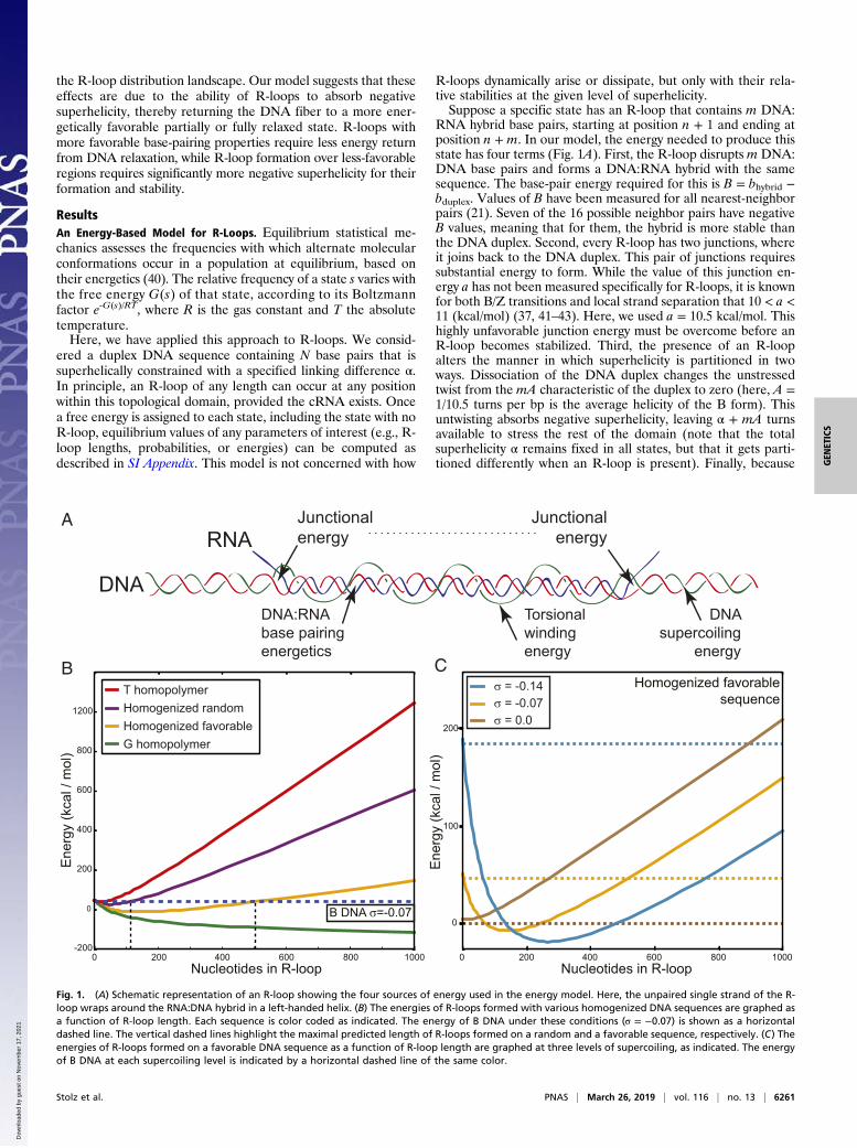
RNA hybrid base pairs, starting at position n + 1 and ending atposition n + m. In our model, the energy needed to produce thisstate has four terms (Fig. 1A). First, the R-loop disrupts m DNA:DNA base pairs and forms a DNA:RNA hybrid with the samesequence. The base-pair energy required for this is B = bhybrid −bduplex. Values of B have been measured for all nearest-neighborpairs (21). Seven of the 16 possible neighbor pairs have negativeB values, meaning that for them, the hybrid is more stable thanthe DNA duplex. Second, every R-loop has two junctions, whereit joins back to the DNA duplex. This pair of junctions requiressubstantial energy to form. While the value of this junction en-ergy a has not been measured specifically for R-loops, it is knownfor both B/Z transitions and local strand separation that 10 < a <11 (kcal/mol) (37, 41–43). Here, we used a = 10.5 kcal/mol. Thishighly unfavorable junction energy must be overcome before anR-loop becomes stabilized. Third, the presence of an R-loopalters the manner in which superhelicity is partitioned in twoways. Dissociation of the DNA duplex changes the unstressedtwist from the mA characteristic of the duplex to zero (here, A =1/10.5 turns per bp is the average helicity of the B form). Thisuntwisting absorbs negative superhelicity, leaving α + mA turnsavailable to stress the rest of the domain (note that the totalsuperhelicity α remains fixed in all states, but that it gets parti-tioned differently when an R-loop is present). Finally, because
A
B DNA σ=-0.07
Nucleotides in R-loop0 200 400 600 800 1000
Ene
rgy
(kca
l / m
ol)
0
200
400
600
800
1200
-200
Ene
rgy
(kca
l / m
ol)
Nucleotides in R-loop
0
100
200
0 200 400 600 800 1000
BHomogenized favorable
sequence
RNA
DNA:RNA base pairingenergetics
Junctionalenergy
Junctionalenergy
Torsionalwindingenergy
DNA supercoiling
energy C
DNA
Homogenized randomT homopolymer
G homopolymerHomogenized favorable σ = 0.0
σ = -0.07σ = -0.14
Fig. 1. (A) Schematic representation of an R-loop showing the four sources of energy used in the energy model. Here, the unpaired single strand of the R-loop wraps around the RNA:DNA hybrid in a left-handed helix. (B) The energies of R-loops formed with various homogenized DNA sequences are graphed asa function of R-loop length. Each sequence is color coded as indicated. The energy of B DNA under these conditions (σ = −0.07) is shown as a horizontaldashed line. The vertical dashed lines highlight the maximal predicted length of R-loops formed on a random and a favorable sequence, respectively. (C) Theenergies of R-loops formed on a favorable DNA sequence as a function of R-loop length are graphed at three levels of supercoiling, as indicated. The energyof B DNA at each supercoiling level is indicated by a horizontal dashed line of the same color.
Stolz et al. PNAS | March 26, 2019 | vol. 116 | no. 13 | 6261
GEN
ETICS
Dow
nloa
ded
by g
uest
on
Nov
embe
r 17
, 202
1
single-stranded DNA is flexible, the unpaired nontemplatestrand can wind around the DNA:RNA hybrid with helicity τ(radians per base). This leaves a residual superhelicity αr = α +mA − mτ/2π to superhelically deform the domain. The free en-ergies associated with these last two effects are both quadratic,with coefficients K for superhelicity and torsional stiffness C forthe winding of the unpaired strand. If we let these two effectsequilibrate, then the total free energy G(s) associated to thisstate is:
GðsÞ=
8>>><>>>:
12Kα2,m= 0
a+Xmi=1
Bðn+ iÞ+ 2π2CK4π2C+Km
ðα+mAÞ2,m> 0. [1]
(The state with no R-loop corresponds to m = 0.) The values ofall of the energy parameters used here were taken directly fromthe literature; none were optimized to fit data. The full model,including the derivation of this equation and full set of parame-ter values used, is presented in SI Appendix.We developed an algorithm to perform calculations based on
this model for DNA domains having any base sequence and anylevel of superhelicity. The topological domain may be eitherclosed circular like a plasmid or a portion of a superhelicallyconstrained loop. The entire domain may be regarded as sus-ceptible to R-looping or only a portion of it. The C++ imple-mentation of this algorithm, called R-looper, enumerates R-loopstates, calculates their energies from Eq. 1, and assigns a Boltzmannfactor to each. This information is used to calculate the equi-librium probability of each state as a function of superhelicityand DNA sequence. Other equilibrium properties can then becalculated at each position, including the probability of R-loopformation and average values of R-loop lengths and free ener-gies. In this implementation, we only considered states having atmost one R-loop because the high value of the junction energya strongly disfavors states with multiple R-loops.
R-Loop Equilibria Are Sensitive to both DNA Sequence and Topology.To assess the role of sequence in R-loop stability, we ran ourmodel on various uniform sequences at superhelical densityσ = −0.07 (σ = α/NA) and compared the free energy of R-loopsof each length to the free energy of the B-form state having noR-loop (Fig. 1B). We used four sequences spanning the rangefrom least to most favorable. The most unfavorable sequence forR-loops is a T homopolymer, for which the base pair energy is
B = +0.8 kcal/mol. In this case, R-loops of all lengths werepredicted to be energetically disfavored relative to the B-formstate. Next, we used a homogenized random sequence, for whichthe base-pair energy was set at the constant value of B =+0.23 kcal/mol, corresponding to the average energy of all16 nearest-neighbor pairs. In this sequence and level of super-helicity, short R-loops of lengths up to ∼100 bp were energeti-cally favored over the B form. By contrast, R-loops of lengths upto 500 bp were energetically favored for a homogenized favor-able sequence (B = −0.15 kcal/mol, corresponding to the averageenergy of all seven favorable nearest-neighbor pairs). R-loopswere favored over B DNA at all lengths for the most favor-able, G homopolymer sequence (B = −0.36 kcal/mol).Our model also predicted that R-loops are highly sensitive to
DNA superhelicity. Fig. 1C plots the energy of R-loops for thehomogenized favorable sequence at three levels of negativesuperhelicity. In each case, the energy of the state with no R-loop(B DNA) is represented by the horizontal dotted line of the samecolor. Although R-loops of length up to 500 bp are favored inthis sequence at σ = −0.07, the model predicted that no R-loopstates are energetically favored over the B form when the samesequence is relaxed (σ = 0). When a highly negative superhelicaldensity was imposed (σ = −0.14), R-loops were predicted to behighly favored, with lengths up to 1,400 bp. Thus, our modelpredicts that both DNA sequence and topology strongly influ-ence R-loop formation.To analyze the relationship between DNA topology and R-
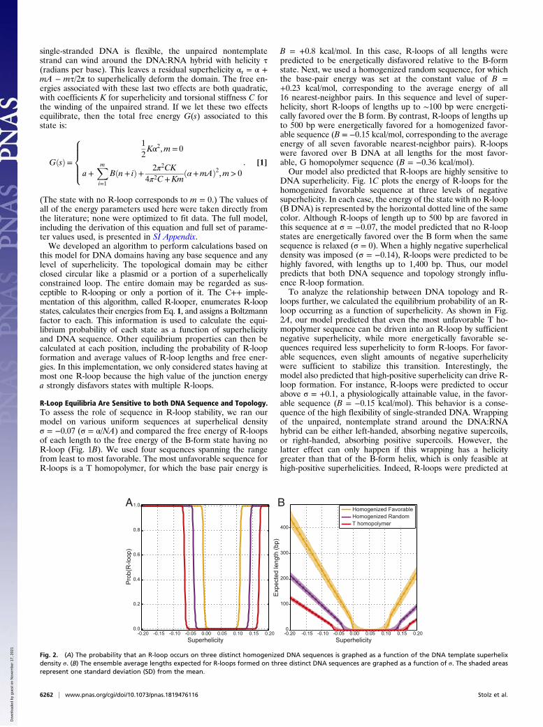
loops further, we calculated the equilibrium probability of an R-loop occurring as a function of superhelicity. As shown in Fig.2A, our model predicted that even the most unfavorable T ho-mopolymer sequence can be driven into an R-loop by sufficientnegative superhelicity, while more energetically favorable se-quences required less superhelicity to form R-loops. For favor-able sequences, even slight amounts of negative superhelicitywere sufficient to stabilize this transition. Interestingly, themodel also predicted that high-positive superhelicity can drive R-loop formation. For instance, R-loops were predicted to occurabove σ = +0.1, a physiologically attainable value, in the favor-able sequence (B = −0.15 kcal/mol). This behavior is a conse-quence of the high flexibility of single-stranded DNA. Wrappingof the unpaired, nontemplate strand around the DNA:RNAhybrid can be either left-handed, absorbing negative supercoils,or right-handed, absorbing positive supercoils. However, thelatter effect can only happen if this wrapping has a helicitygreater than that of the B-form helix, which is only feasible athigh-positive superhelicities. Indeed, R-loops were predicted at
A B
Pro
b(R
-loop
)
0.0
0.2
0.4
0.6
0.8
1.0
Exp
ecte
d le
ngth
(bp)
400
300
200
100
0
Superhelicity0.00 0.05 0.10 0.15 0.20-0.05-0.10-0.15-0.20
Superhelicity0.00 0.05 0.10 0.15 0.20-0.05-0.10-0.15-0.20
T homopolymerHomogenized RandomHomogenized Favorable
Fig. 2. (A) The probability that an R-loop occurs on three distinct homogenized DNA sequences is graphed as a function of the DNA template superhelixdensity σ. (B) The ensemble average lengths expected for R-loops formed on three distinct DNA sequences are graphed as a function of σ. The shaded areasrepresent one standard deviation (SD) from the mean.
6262 | www.pnas.org/cgi/doi/10.1073/pnas.1819476116 Stolz et al.
Dow
nloa
ded
by g
uest
on
Nov
embe
r 17
, 202
1
moderate levels of negative superhelicity, but only at muchhigher levels of positive superhelicity (SI Appendix, Fig. S1).Our model also predicted that increasing levels of negative
DNA superhelicity stabilize R-loops of increasing size (Fig. 2B).For all tested sequences, R-loops started growing in length assoon as they became favorable. However, for energetically less-favorable sequences, R-loops required more superhelicity toinitiate and grew more slowly. In the homogenized favorablesequence at σ = −0.07, the predicted ensemble average R-looplength was 180 bp. By contrast, the unfavorable T homopolymersequence was only beginning to experience short R-loops (av-erage length of 25 bp) at this superhelix density. While R-loopformation became possible at high-positive superhelicity, theaverage lengths of the structures remained much shorter thanthose predicted for the corresponding level of negative super-helicity (Fig. 2B). Thus, increasing superhelicity was predicted toincrease both the probability and length of R-loop structures,with other factors being fixed.
Relaxation of DNA Superhelicity by R-Loop Formation. According toour model, R-loops are favored at equilibrium, primarily becausethey absorb negative superhelicity through base unpairing andstrand twisting, thereby allowing the rest of the domain to relax.R-loops become favored at superhelicities where the base-pairing energies B and the superhelical relaxation together are
enough to overcome the unfavorable junction free energy a.Beyond that point, longer R-loops occur at higher negativesuperhelicity because they provide more relaxation.To test these predictions, we applied the equilibrium model to
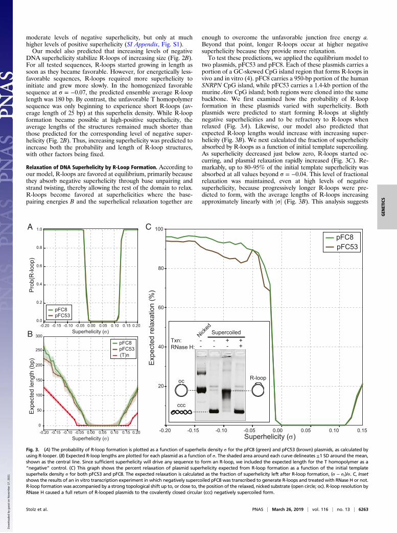
two plasmids, pFC53 and pFC8. Each of these plasmids carries aportion of a GC-skewed CpG island region that forms R-loops invivo and in vitro (4). pFC8 carries a 950-bp portion of the humanSNRPN CpG island, while pFC53 carries a 1.4-kb portion of themurine Airn CpG island; both regions were cloned into the samebackbone. We first examined how the probability of R-loopformation in these plasmids varied with superhelicity. Bothplasmids were predicted to start forming R-loops at slightlynegative superhelicities and to be refractory to R-loops whenrelaxed (Fig. 3A). Likewise, our model also predicted thatexpected R-loop lengths would increase with increasing super-helicity (Fig. 3B). We next calculated the fraction of superhelicityabsorbed by R-loops as a function of initial template supercoiling.As superhelicity decreased just below zero, R-loops started oc-curring, and plasmid relaxation rapidly increased (Fig. 3C). Re-markably, up to 80–95% of the initial template superhelicity wasabsorbed at all values beyond σ = −0.04. This level of fractionalrelaxation was maintained, even at high levels of negativesuperhelicity, because progressively longer R-loops were pre-dicted to form, with the average lengths of R-loops increasingapproximately linearly with jσj (Fig. 3B). This analysis suggests
A C
B
Exp
ecte
d le
ngth
(bp)
pFC53pFC8
250
200
150
100
0
50
300
0.00 0.05 0.10 0.15 0.20-0.05-0.10-0.15-0.20Superhelicity ()
0.00 0.05 0.10 0.15 0.20-0.05-0.10-0.15-0.20Superhelicity ()
Pro
b(R
-loop
)
0.0
0.2
0.4
0.6
0.8
1.0
pFC53pFC8
(T)n
Exp
ecte
d re
laxa
tion
(%)
Superhelicity ()0.00 0.05 0.10-0.05-0.10-0.15-0.20
80
60
40
20
pFC53pFC8
100
0.15
Nicked
+++
-- -- -Txn:
RNase H:
R-loopoc
ccc
Supercoiled
Fig. 3. (A) The probability of R-loop formation is plotted as a function of superhelix density σ for the pFC8 (green) and pFC53 (brown) plasmids, as calculated byusing R-looper. (B) Expected R-loop lengths are plotted for each plasmid as a function of σ. The shaded area around each curve delineates ±1 SD around the mean,shown as the central line. Since sufficient superhelicity will drive any sequence to form an R-loop, we included the expected length for the T homopolymer as a“negative” control. (C) This graph shows the percent relaxation of plasmid superhelicity expected from R-loop formation as a function of the initial templatesuperhelix density σ for both pFC53 and pFC8. The expected relaxation is calculated as the fraction of superhelicity left after R-loop formation, (σ − σr)/σ. C, Insetshows the results of an in vitro transcription experiment in which negatively supercoiled pFC8 was transcribed to generate R-loops and treated with RNase H or not.R-loop formation was accompanied by a strong topological shift up to, or close to, the position of the relaxed, nicked substrate (open circle; oc). R-loop resolution byRNase H caused a full return of R-looped plasmids to the covalently closed circular (ccc) negatively supercoiled form.
Stolz et al. PNAS | March 26, 2019 | vol. 116 | no. 13 | 6263
GEN
ETICS
Dow
nloa
ded
by g
uest
on
Nov
embe
r 17
, 202
1
that R-loop formation significantly relaxes the superhelical stresson a susceptible plasmid. These findings are entirely consistentwith the well-known behavior of R-loops in in vitro transcriptionassays, where R-loop formation is measured by the gel retarda-tion resulting from the topological relaxation of the negativelysupercoiled substrate (Fig. 3 C, Inset) (3, 4, 18, 44).
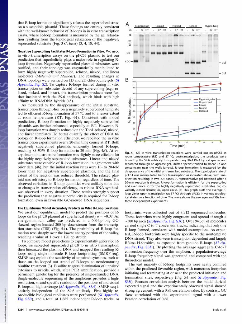
Negative Supercoiling Facilitates R-Loop Formation in Vitro.We usedin vitro transcription assays on the pFC53 plasmid to test ourprediction that superhelicity plays a major role in regulating R-loop formation. Negatively supercoiled plasmid substrates werepurified, and their topology was enzymatically manipulated toform highly negatively supercoiled, relaxed, nicked, and linearmolecules (Materials and Methods). The resulting changes inDNA topology were verified on 1D and 2D chloroquine gels (SIAppendix, Fig. S2). To capture R-loops formed during in vitrotranscription on substrates devoid of any supercoiling (e.g., re-laxed, nicked, and linear), the transcription products were fur-ther incubated with the S9.6 antibody, which binds with highaffinity to RNA:DNA hybrids (45).As measured by the disappearance of the initial substrate,
transcription through Airn on a negatively supercoiled templateled to efficient R-loop formation at 37 °C and to a lesser extentat room temperature (RT; Fig. 4A). Consistent with modelpredictions, R-loop formation on highly negatively supercoiledplasmids was further enhanced, especially at RT. However, R-loop formation was sharply reduced on the Top1-relaxed, nicked,and linear templates. To better quantify the effect of DNA to-pology on R-loop formation efficiency, we repeated the in vitrotranscription experiments over a 20-min time course at RT. Bothnegatively supercoiled plasmids efficiently formed R-loops,reaching 85–95% R-loop formation in 20 min (Fig. 4B). At ev-ery time point, structure formation was slightly more efficient forthe highly negatively supercoiled substrates. Linear and nickedsubstrates were capable of R-loop formation, in agreement withprior data (46), but the initial reaction rates were 10- to 20-foldlower than for negatively supercoiled plasmids, and the finalextent of the reaction was reduced threefold. The relaxed plas-mid was refractory to R-loop formation, regardless of time. Theobserved variations in R-loop quantities were unlikely to be dueto changes in transcription efficiency, as robust RNA synthesiswas observed in every situation. These results strongly supportthe prediction that negative superhelicity is required for R-loopformation, even in favorable GC-skewed DNA sequences.
The Equilibrium Model Accurately Predicts in Vitro R-Loop Locations.We used our equilibrium model to predict the positions of R-loops on the pFC8 plasmid at superhelical density σ = −0.07. Anenergy-minimum valley was predicted in a 600-bp-long GC-skewed region located 200 bp downstream from the transcrip-tion start site (TSS) (Fig. 5A). The probability of R-loop for-mation rose sharply over the lowest energy portion of the valley,reaching a value of 1 over a 120 bp stretch.To compare model predictions to experimentally generated R-
loops, we subjected supercoiled pFC8 to in vitro transcription,then linearized the plasmid DNA and mapped the resulting R-loops using single-molecule R-loop footprinting (SMRF-seq).SMRF-seq exploits the sensitivity of unpaired cytosines, such asthose on the looped out strand of R-loops, to nondenaturingbisulfite treatment (3). Bisulfite triggers deamination of unpairedcytosines to uracils, which, after PCR amplification, provide apermanent genetic tag for the presence of single-stranded DNA.Single-molecule sequencing of the amplicons provides a high-resolution, strand-specific readout of the positions of individualR-loops at high coverage (SI Appendix, Fig. S3A). SMRF-seq isentirely independent of the S9.6 antibody. Five highly re-producible biological replicates were performed (SI Appendix,Fig. S3B), and a total of 1,885 independent R-loop tracks, or
footprints, were collected out of 3,912 sequenced molecules.These footprints were highly congruent and spread through a∼300 bp area (SI Appendix, Fig. S3C). Over 94.3% of moleculescarried single C-to-T conversion tracks, indicating that only oneR-loop formed, consistent with model assumptions. As expec-ted, R-loop footprints were highly specific to the nontemplateDNA strand. They also were transcription-dependent and largelyRNase H-sensitive, as expected from genuine R-loops (SI Ap-pendix, Fig. S3D). By plotting the average aggregate C-to-Tconversion frequency over the amplicon, a population-averageR-loop frequency signal was generated and compared with thetheoretical model.The vast majority of R-loop footprints were neatly confined
within the predicted favorable region, with numerous footprintsinitiating and terminating at or near the predicted initiation andtermination sites, respectively (Fig. 5A and SI Appendix, Fig.S3E). Pearson correlation analysis between the model-derivedexpected signal and the experimentally observed signal showedstrong agreement, with a 0.93 correlation value. By contrast, GCskew correlated with the experimental signal with a lowerPearson correlation of 0.66.
Txn:Supercoiled Relaxed Nicked Linear Hyper-Neg
0
10
20
30
40
50
60
70
80
90
100
0 2 4 6 8 10 12 14 16 18 20
% R
-loop
pro
duct
Time (min)
Hyper-negativeSupercoiledLinearNickedRelaxed
- 37 RT - 37 RT - 37 RT - 37 RT - 37 RT
A
B
ccc
oc
cut
cut
shift
Fig. 4. (A) In vitro transcription reactions were carried out on pFC53 atroom temperature (RT) and 37 °C; posttranscription, the products werebound by the S9.6 antibody to supershift any RNA:DNA hybrid species andseparated through an agarose gel. Shifted species tended to smear up andconcentrate near the wells (arrow). R-loop formation is measured by thedisappearance of the initial untranscribed substrate. The topological state ofpFC53 was manipulated before transcription as indicated above, with line-arization resulting in two cut bands. A representative gel obtained after a20-min reaction is shown. R-loop formation is efficient for the supercoiledand even more so for the highly negatively supercoiled substrates. ccc, co-valently closed circular; oc, open circle. (B) This graph plots the average R-loop yields upon transcription (at 37 °C) through pFC53 in various topolog-ical states, as a function of time. The curve shows the averages and SDs fromthree independent experiments.
6264 | www.pnas.org/cgi/doi/10.1073/pnas.1819476116 Stolz et al.
Dow
nloa
ded
by g
uest
on
Nov
embe
r 17
, 202
1
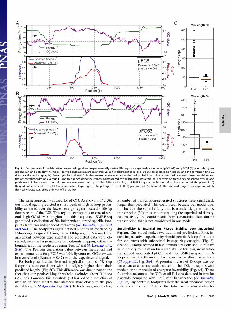
The same approach was used for pFC53. As shown in Fig. 5B,our model again predicted a sharp peak of high R-loop proba-bility centered over the lowest energy region located ∼400 bpdownstream of the TSS. This region corresponds to one of sev-eral high-GC-skew subregions in this sequence. SMRF-seqgenerated a collection of 564 independent, strand-specific foot-prints from two independent replicates (SI Appendix, Figs. S3Dand S4A). The footprints again defined a series of overlappingR-loop signals spread through an ∼300-bp region. A remarkableagreement between experimental and predicted data were ob-served, with the large majority of footprints mapping within theboundaries of the predicted region (Fig. 5B and SI Appendix, Fig.S4B). The Pearson correlation value between theoretical andexperimental data for pFC53 was 0.94. By contrast, GC skew wasless correlated (Pearson = 0.42) with the experimental signal.For both plasmids, the observed length distributions of R-loop
footprints were consistent with, but slightly higher than, theirpredicted lengths (Fig. 5C). This difference was due in part to thefact that our peak-calling threshold excludes short R-loops(<30 bp). Lowering this threshold (10 bp) led to a reduction ofmedian observed lengths that matched more closely to the pre-dicted lengths (SI Appendix, Fig. S4C). In both cases, nonetheless,
a number of transcription-generated structures were significantlylonger than predicted. This could occur because our model doesnot include the superhelicity that is transiently generated bytranscription (30), thus underestimating the superhelical density.Alternatively, this could result from a dynamic effect duringtranscription that is not considered in our model.
Superhelicity Is Essential for R-Loop Stability over SuboptimalRegions. Our model makes two additional predictions. First, in-creasing negative superhelicity should permit R-loop formationfor sequences with suboptimal base-pairing energies (Fig. 2).Second, R-loops formed in less-favorable regions should requiresuperhelicity to maintain their stability. To test this, we in vitro-transcribed supercoiled pFC53 and used SMRF-seq to map R-loops either directly on circular molecules or after linearization(SI Appendix, Fig. S6A). A prominent class of R-loops was de-tected on circular molecules closer to the TSS, in regions withmodest or poor predicted energetic favorability (Fig. 6A). Thesefootprints accounted for 33% of all R-loops detected in circularplasmids, compared with 6.2% after linearization (SI Appendix,Fig. S5). By contrast, footprints over the most favorable regiononly accounted for 56% of the total on circular molecules
Ene
rgy
(kca
l/mol
)P
roba
bilit
y C to T
FrequencyG
C skew
0
20
40
500.5
-0.5
0
30
10
1.0
0.8
0.6
0.4
0.2
0.0
1.0
0.8
0.6
0.4
0.2
0.0
Ene
rgy
(kca
l/mol
)P
roba
bilit
y
GC
skew
Position (bp)
10
30
40
0.5
-0.5
0
20
1.0
0.8
0.6
0.4
0.2
0.0
1.0
0.8
0.6
0.4
0.2
0.0
50
1800300 600 900 1200 1500
250 500 1000 1250 1500750
EnergyGC skew
B
A
Pearson’s: 0.9273p-value < 0.001
pFC8
Pearson’s: 0.9450p-value < 0.001
pFC53
Position (bp)
Leng
th (b
p)
600
500
400
300
200
100
0Obs. Exp.
C Min length 30
Leng
th (b
p)
400
350
300
250
200
150
100
50
Obs. Exp.
Min length 30
C to T
Frequency
Expected (model)Observed (C to T)
0
Expected (model)Observed (C to T)
EnergyGC skew
0
60
Fig. 5. Comparison of model-derived expected signal and experimentally derived R-loops for negatively supercoiled pFC8 (A) and pFC53 (B) plasmids. Uppergraphs in A and B display the model-derived ensemble average energy value for all predicted R-loops at any given base pair (green) and the corresponding GCskew for the region (purple). Lower graphs in A and B display ensemble average model-derived probability of R-loop formation at each base pair (blue) andthe observed population average R-loop frequency along the region, as measured by the bisulfite-induced C-to-T conversion frequency measured over R-looppeaks (red). In both cases, transcription was conducted on supercoiled DNA molecules, and SMRF-seq was performed after linearization of the plasmid. (C)Boxplots of observed (Obs., left) and predicted (Exp., right) R-loop lengths for pFC8 (Upper) and pFC53 (Lower). The minimal lengths for experimentallyderived R-loops was arbitrarily cut off at 30 bp.
Stolz et al. PNAS | March 26, 2019 | vol. 116 | no. 13 | 6265
GEN
ETICS
Dow
nloa
ded
by g
uest
on
Nov
embe
r 17
, 202
1
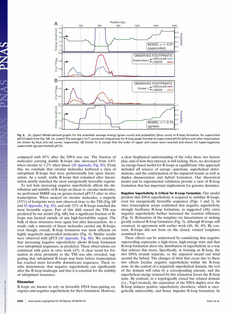
compared with 85% after the DNA was cut. The fraction ofmolecules carrying double R-loops also decreased from 4.8%when circular to 1.2% when linear (SI Appendix, Fig. S5). Fromthis, we conclude that circular molecules harbored a class ofsuboptimal R-loops that were preferentially lost upon lineari-zation. As a result, stable R-loops that remained after lineari-zation mostly matched the most energetically favorable regions.To test how increasing negative superhelicity affects the dis-
tribution and stability of R-loops on linear vs. circular molecules,we performed SMRF-seq on gyrase-treated pFC53 after in vitrotranscription. When assayed on circular molecules, a majority(55%) of footprints were now observed close to the TSS (Fig. 6Band SI Appendix, Fig. S5), and only 32% of R-loops matched themost favorable region. Part of this shift toward the TSS waspredicted by our model (Fig. 6B), but a significant fraction of R-loops was located outside of any high-favorability region. Thebulk of these structures were again lost after linearization. As aresult, only a minority of linear molecules carried any R-loops,even though, overall, R-loop formation was most efficient onhighly negatively supercoiled molecules (Fig. 4). Similar resultswere observed with pFC8 (SI Appendix, Fig. S6). We concludethat increasing negative superhelicity allows R-loop formationover suboptimal sequences, as predicted. These observations areconsistent with prior in vitro work (47). A clear trend for for-mation in close proximity to the TSS was also revealed, sug-gesting that suboptimal R-loops may form before transcriptionhas reached more favorable downstream sequences. These re-sults demonstrate that negative superhelicity can significantlyalter the R-loop landscape and that it is essential for the stabilityof suboptimal structures.
DiscussionR-loops are known to rely on favorable DNA base-pairing en-ergetics and negative superhelicity for their formation. However,
a clear biophysical understanding of the roles these two factorsplay, and of how they interact, is still lacking. Here, we developedan energy-based model for R-loops at equilibrium. Our approachincluded all sources of energy—junctions, superhelical defor-mations, and the conformation of the unpaired strand, as well asduplex disassociation and hybrid formation. Our theoreticalmodel and its experimental validation provide a view of R-loopformation that has important implications for genome dynamics.
Negative Superhelicity Is Critical for R-Loop Formation. Our modelpredicts that DNA superhelicity is required to stabilize R-loops,even for energetically favorable sequences (Figs. 1 and 2). Invitro transcription assays confirmed that negative superhelicitystrongly facilitates R-loop formation, as suggested (48); extranegative superhelicity further increased the reaction efficiency(Fig. 4). Relaxation of the template via linearization or nickinggreatly reduced R-loop formation (Fig. 3), although R-loops stilloccurred, in agreement with earlier work (46, 48, 49). By con-trast, R-loops did not form on the closed, relaxed templatesexamined here.These effects can be understood by considering that negative
supercoiling represents a high-stress, high-energy state and thatR-loop formation alters the distribution of superhelicity in a waythat relieves this stress. Specifically, in forming an R-loop, thetwo DNA strands separate, so the unpaired strand can windaround the hybrid. The changes of twist that occur due to thesetwo effects localize negative superhelicity within the R-loopzone. In the context of a negatively superhelical domain, the restof the domain will relax by a corresponding amount, and thesuperhelical energy returned by this relaxation favors the R-loopstate. By contrast, in a topologically closed but relaxed domain(i.e., Top1-treated), the separation of the DNA duplex over theR-loop induces positive superhelicity elsewhere, which is ener-getically unfavorable, and hence impedes R-loop formation. In
C to
T F
req.
C to
T F
req.
0.6
0.4
0.2
0.0
Ene
rgy
(kca
l/mol
)
75
100
50
125
MODEL: = -0.14(gyrase)
B
1800300 600 900 1200 15000
0.6
0.4
0.2
0.0
LINEARCIRCULAR
OBSERVED FOOTPRINTSSupercoiled
OBSERVED FOOTPRINTS
LINEARCIRCULARGyrase
Position (bp)
Ene
rgy
(kca
l/mol
)P
robability 10
30
40
20
1.0
0.8
0.6
0.4
0.2
0.0
50
60
MODEL: = -0.07(supercoiled)
A
Probability
1.0
0.8
0.6
0.4
0.2
0.0
Fig. 6. (A, Upper) Model-derived graphs for the ensemble average energy (green curve) and probability (blue curve) of R-loop formation for supercoiledpFC53 (data from Fig. 5B). (A, Lower) The average C-to-T conversion frequencies for R-loop peaks formed on supercoiled pFC53 before and after linearizationare shown by blue and red curves, respectively. (B) Similar to A, except that the order of Upper and Lower were reversed and shown for hypernegativelysupercoiled (gyrase-treated) pFC53.
6266 | www.pnas.org/cgi/doi/10.1073/pnas.1819476116 Stolz et al.
Dow
nloa
ded
by g
uest
on
Nov
embe
r 17
, 202
1
agreement, R-loop formation induced upon transcription of anextremely favorable (AGGAG)28 repeat sequence on relaxedsubstrates forced the DNA into a positively supercoiled state(46). For nicked and linear DNA, the excess positive supercoilsinduced by R-loop formation are eliminated by rapid strandrotation, so superhelicity neither impedes nor facilitates thisreaction. The lack of ambient superhelicity, however, preventsenergy return from DNA relaxation, and therefore R-loop for-mation is significantly lower on these substrates (Fig. 4).Intriguingly, our model predicts that a high level of positive
superhelicity also favors R-loops (Figs. 2 and 3). Similar behaviorhas been noted for strand separation (50, 51). Whether thisprediction is borne out in vitro or in vivo remains to be tested.We note that positive superhelicity may hinder transcriptionelongation (52). It also remains unclear if the displaced ssDNAcan sterically wind around the RNA:DNA hybrid enough topermit relaxation of positive supercoils. Such winding wouldrequire the single strand to stretch nearly to its physical limit, andin these conditions, the energetics might be different from thoseassumed in the model. Furthermore, very few genomic loci areexpected to experience levels of positive superhelicity sufficientto drive R-loops. Such regions are likely limited to loci betweennearby convergent highly transcribed genes or convergently ad-vancing transcription and replication forks. Even in these rarecases, the positively supercoiled region would be located aheadof the RNA polymerase and therefore would be untranscribed;as such, there would be no complementary transcript available toform an R-loop, unless the RNA polymerase undergoes back-tracking. Overall, although R-loop formation at high positivesuperhelical densities is possible according to our model, itsoccurrence and relevance to in vivo situations remains tobe established.
R-Loop Distribution Patterns Are Governed by the Interplay BetweenSequence, Superhelicity, and Proximity to the TSS. In our model, thesubstantial energy needed to make a pair of junctions is the mainbarrier to R-loop formation. This barrier can only be overcomeby a combination of superhelical relaxation and favorable base-pairing energetics. Our model predicts that for moderately fa-vorable DNA sequences, only modest levels of negative super-helicity (σ ≅ −0.02) are sufficient to stabilize R-loops. Theselevels occur during normal physiological processes in all organ-isms. In mesophilic bacteria, gyrase activity maintains genomicDNA at unconstrained superhelical densities that can reachbeyond σ = −0.05 (53). In eukaryotes, DNA replication, tran-scription, and chromatin-remodeling transiently introduce neg-ative superhelicity (54, 55). During transcription, DNA isnegatively supercoiled to a density of σ = −0.07 in a regionextending 1,500 bp behind the advancing RNA polymerase (32,56), which is where R-loops are thought to form. Thus, the to-pological requirements revealed here are compatible with R-loop formation in vivo.Our model and its experimental validation established that
superhelicity, together with DNA sequence, cooperate to regu-late both the positions and the stability of R-loops. Consistentwith the well-known association of R-loops with GC skew in vivoand in vitro (3, 4, 16, 24), our model predicts that R-loops willtend to concentrate over the most energetically favorable se-quences, ensuring maximal energy return from RNA:DNA base-pairing. These regions tend to be G-rich. However, GC skew byitself was found to be only moderately predictive of R-loop lo-cations (Fig. 5). Single-molecule R-loop footprinting revealedthat stable R-loops—i.e., those that persist after linearization ofthe circular molecules on which they were generated—show re-markable agreement in position with model-predicted favorableregions (Fig. 5). Thus, cotranscriptional R-loop formation invitro, an inherently dynamic process, follows, at least in part, theenergy landscape highlighted by our equilibrium model. Our
model also explicitly predicts that the position of R-loop-proneregions will shift with varying levels of superhelicity. More fa-vorable sequences require less energy return from superhelicityand can therefore transition into R-loops at lower negative su-perhelical densities. By contrast, R-loops formed over sub-optimal sequences are expected to require more energy returnfrom superhelicity. At its extreme, any sequence, however un-favorable, could transition into an R-loop, provided sufficientnegative supercoiling exists (Figs. 2 and 3). Experimental vali-dation showed that TSS-proximal R-loops formed over un-favorable regions significantly increased in representation whenthe superhelical density of the plasmid template was increasedupon gyrase treatment (Fig. 6 and SI Appendix, Figs. S5 and S6).Thus, increased negative superhelicity drives R-loop formationover unfavorable regions, consistent with prior in vitro work (47).Finally, our model predicted that R-loops should show differ-ential stability in the face of a loss of superhelicity. R-loopsformed over favorable regions should remain more stable be-cause base-pairing energy provides significant anchoring. Bycontrast, the stability of R-loops formed over less favorableregions should be strongly compromised by relaxation. Theseexpectations were validated by measuring the distribution of R-loop footprints generated on circular molecules after lineariza-tion of the template (Fig. 6 and SI Appendix, Figs. S5 and S6).TSS-proximal R-loops matching poorly favorable sequenceswere sharply reduced after linearization. By contrast, R-loopsformed over favorable regions remained mostly stable andaccounted for the majority of structures postcleavage. Our worktherefore demonstrates that superhelicity can dramaticallyaffect the R-loop landscape, both in terms of their formationand stability.Our data also confirmed a propensity for R-loops to form in
proximity to the 5′ end of the nascent transcript (48, 57), evenwhen the sequence was suboptimal and the resulting R-loopsunstable. This suggests that R-loops that form first becausethey are closest to the TSS may benefit from a dynamic advan-tage during transcription. This class of unstable, dynamic R-loops may provide promoter-proximal topological relief, even ifit is to the detriment of stable R-loop formation at more favor-able regions located downstream.
R-Loops as Reversible Superhelical Stress Relievers. Before ourwork, the underwound state of negatively superhelical DNA wasunderstood to favor R-loops by facilitating the ability of thenascent RNA to invade duplex DNA (1). However, superhelicalduplex destabilization is confined to the AT-richest regions of adomain (58), whereas most R-loops occur in G-rich locations.Thus, supercoiling-induced strand separation may not be strictlynecessary for R-loop initiation. Our energy-based equilibriummodel provides an alternative, more quantitative explanation forhow negative superhelicity facilitates R-loops. Both in silico andin vitro (Fig. 3), a major effect of R-loop formation is to return anegatively supercoiled DNA domain to an energetically morefavorable, nearly relaxed state. Thus, negative superhelicity fa-vors R-loops because these structures relax superhelical stresses.It is useful to contrast R-loop-mediated relaxation with that
provided by nucleosomal winding. In vitro, R-loops of lengths<150 bp (Fig. 5) efficiently relaxed 3- to 4-kb negatively super-coiled plasmids (Fig. 3). Assuming a superhelical density ofσ = −0.05, these R-loops absorbed a large fraction of the 15–20 negative supercoils present on these plasmids. By comparison,a nucleosome occupying ∼146 bp of DNA absorbs one negativesupercoil (59–61). Thus, R-loops are at least an order of mag-nitude more effective than nucleosomes at absorbing negativesuperhelicity. Single-molecule R-loop profiling studies haveshown that genomic R-loops often extend for several hundredbase pairs, and kilobase R-loops have been detected at lowerfrequencies (3–5). R-loops having these lengths are capable of
Stolz et al. PNAS | March 26, 2019 | vol. 116 | no. 13 | 6267
GEN
ETICS
Dow
nloa
ded
by g
uest
on
Nov
embe
r 17
, 202
1
absorbing striking amounts of negative superhelicity and there-fore can act as efficient superhelical stress relievers over longdomains. Importantly, the superhelicity stored in an R-loop isimmediately returned to the DNA fiber upon R-loop resolution.As shown in Fig. 3C, the R-loop-mediated relaxation of plasmidDNA is entirely shifted back to the negatively supercoiled stateby RNase H treatment.The ability of R-loops to efficiently sequester and release
negative superhelicity may be relevant for several processes.First, it will influence the landscape of non-B DNA structures insurrounding regions. Indeed, other alternative DNA structures,such as strand separations, cruciforms, and B/Z transitions, alsocan be superhelically driven (34–39). In effect, all of the possibleDNA structural transitions, including R-loops, will compete to-gether because the superhelical relief caused by the formation ofany one structure will inhibit the formation of all others. Giventheir lengths, stabilities, and remarkable capacity for absorbingsuperhelicity, R-loops are expected to play a major role in thesecompetitions. Second, since negative supercoiling favors strandopening, which is required for the initiation of transcription andof DNA replication (62), these processes may be strongly influ-enced by the formation or resolution of nearby R-loops. CpGisland promoters, which serve both as active promoters and asearly, efficient DNA replication origins (63), are R-loop hotspots(4). More broadly, the identification of replication “initiationzones” near gene ends (64) matches well with the known favoredlocations for R-loops at the beginnings and ends of genes. Fi-nally, the sequestering or release of negative superhelicity by R-loop formation or resolution could influence the landscape ofprotein–DNA interactions, since proteins such as transcriptionfactors and nucleosomes preferentially bind negatively super-coiled DNA (65, 66). Indeed, the release of negative super-helicity upon R-loop resolution may permit efficient nucleosomeredeposition locally, since R-loops are most likely nucleosome-free as long as they persist (11, 67).
Finally, the observation that negative DNA superhelicity en-hances the formation and stability of R-loops suggests that ge-nomic sites where R-loops occur exist under topological tension.If so, genome-wide R-loop maps may indirectly report on locallevels of superhelicity. CpG island promoters experience re-peated topological strain caused by transcription initiation, in-cluding abortive transcription and nucleosome remodeling (32,56). The observation of strong R-loop hotspots at these sites isconsistent with R-loops providing a nonenzymatic method toregulate that strain. The fact that CpG island promoters acrossvertebrates contain R-loop-favorable sequences (4, 15) suggeststhat these loci may have evolved to take advantage of R-loop-mediated torsional stress relief. Interestingly, the 3′ ends of nu-merous genes are strong R-loop hotspots (11). These regionsmay therefore also experience significant topological tension,perhaps linked to transcription termination. Overall, our worksuggests that R-loops can provide ways to distribute and managetopological stresses in eukaryotic genomes.
Materials and MethodsFor more information on the derivation of the equilibrium model, includingthe implementation of the model in the R-looper algorithm, please consultthe SI Appendix. The SI Appendix also includes information regarding re-agents, chemicals, and procedures related to plasmid DNA extraction andtopological manipulations and in vitro transcription assays. Single-moleculeR-loop footprinting assays are also described in the SI Appendix.
ACKNOWLEDGMENTS. We thank Marc Nadal, Vincent Vanoosthuyse, andmembers of the F.C. laboratory for critical feedback and valuable discus-sion. This work was supported by National Institutes of Health Grant R01-GM120607 (to F.C.). M.M. was supported in part by National ScienceFoundation Graduate Research Fellowship Grant 1650042 and by NationalInstitute of General Medical Sciences Biomolecular Technology PredoctoralTraining Program Grant T32-GM008799. R.S. was supported in part byNational Science Foundation CAREER Grant DMS1057284 (to Dr. MarielVazquez).
1. Santos-Pereira JM, Aguilera A (2015) R loops: New modulators of genome dynamics
and function. Nat Rev Genet 16:583–597.2. Belotserkovskii BP, Tornaletti S, D’Souza AD, Hanawalt PC (2018) R-loop generation
during transcription: Formation, processing and cellular outcomes. DNA Repair (Amst)
71:69–81.3. Yu K, Chedin F, Hsieh CL, Wilson TE, Lieber MR (2003) R-loops at immunoglobulin class
switch regions in the chromosomes of stimulated B cells. Nat Immunol 4:442–451.4. Ginno PA, Lott PL, Christensen HC, Korf I, Chédin F (2012) R-loop formation is a dis-
tinctive characteristic of unmethylated human CpG island promoters. Mol Cell 45:
814–825.5. Huang FT, Yu K, Hsieh CL, Lieber MR (2006) Downstream boundary of chromosomal
R-loops at murine switch regions: Implications for the mechanism of class switch re-
combination. Proc Natl Acad Sci USA 103:5030–5035.6. Wahba L, Costantino L, Tan FJ, Zimmer A, Koshland D (2016) S1-DRIP-seq identifies
high expression and polyA tracts as major contributors to R-loop formation. Genes
Dev 30:1327–1338.7. Chan YA, et al. (2014) Genome-wide profiling of yeast DNA:RNA hybrid prone sites
with DRIP-chip. PLoS Genet 10:e1004288.8. El Hage A, Webb S, Kerr A, Tollervey D (2014) Genome-wide distribution of RNA-DNA
hybrids identifies RNase H targets in tRNA genes, retrotransposons and mitochondria.
PLoS Genet 10:e1004716.9. Hartono SR, et al. (2018) The affinity of the S9.6 antibody for double-stranded RNAs
impacts the accurate mapping of R-loops in fission yeast. J Mol Biol 430:272–284.10. Xu W, et al. (2017) The R-loop is a common chromatin feature of the Arabidopsis
genome. Nat Plants 3:704–714.11. Sanz LA, et al. (2016) Prevalent, dynamic, and conserved R-loop structures associate
with specific epigenomic signatures in mammals. Mol Cell 63:167–178.12. Lim YW, Sanz LA, Xu X, Hartono SR, Chédin F (2015) Genome-wide DNA hypo-
methylation and RNA:DNA hybrid accumulation in Aicardi-Goutières syndrome. eLife
4:e08007.13. Chen PB, Chen HV, Acharya D, Rando OJ, Fazzio TG (2015) R loops regulate promoter-
proximal chromatin architecture and cellular differentiation. Nat Struct Mol Biol 22:
999–1007.14. Stork CT, et al. (2016) Co-transcriptional R-loops are the main cause of estrogen-
induced DNA damage. eLife 5:e17548.15. Hartono SR, Korf IF, Chédin F (2015) GC skew is a conserved property of unmethylated
CpG island promoters across vertebrates. Nucleic Acids Res 43:9729–9741.
16. Ginno PA, Lim YW, Lott PL, Korf I, Chédin F (2013) GC skew at the 5′ and 3′ ends ofhuman genes links R-loop formation to epigenetic regulation and transcription ter-mination. Genome Res 23:1590–1600.
17. Chédin F (2016) Nascent connections: R-loops and chromatin patterning. TrendsGenet 32:828–838.
18. Reaban ME, Griffin JA (1990) Induction of RNA-stabilized DNA conformers by tran-scription of an immunoglobulin switch region. Nature 348:342–344.
19. Daniels GA, Lieber MR (1995) RNA:DNA complex formation upon transcription ofimmunoglobulin switch regions: Implications for the mechanism and regulation ofclass switch recombination. Nucleic Acids Res 23:5006–5011.
20. Ratmeyer L, Vinayak R, Zhong YY, Zon G, Wilson WD (1994) Sequence specific ther-modynamic and structural properties for DNA.RNA duplexes. Biochemistry 33:5298–5304.
21. Huppert JL (2008) Thermodynamic prediction of RNA-DNA duplex-forming regions inthe human genome. Mol Biosyst 4:686–691.
22. Roberts RW, Crothers DM (1992) Stability and properties of double and triple helices:Dramatic effects of RNA or DNA backbone composition. Science 258:1463–1466.
23. Sugimoto N, et al. (1995) Thermodynamic parameters to predict stability of RNA/DNAhybrid duplexes. Biochemistry 34:11211–11216.
24. Roy D, Lieber MR (2009) G clustering is important for the initiation of transcription-induced R-loops in vitro, whereas high G density without clustering is sufficientthereafter. Mol Cell Biol 29:3124–3133.
25. Roy D, Yu K, Lieber MR (2008) Mechanism of R-loop formation at immunoglobulinclass switch sequences. Mol Cell Biol 28:50–60.
26. Drolet M, Bi X, Liu LF (1994) Hypernegative supercoiling of the DNA template duringtranscription elongation in vitro. J Biol Chem 269:2068–2074.
27. Massé E, Phoenix P, Drolet M (1997) DNA topoisomerases regulate R-loop formationduring transcription of the rrnB operon in Escherichia coli. J Biol Chem 272:12816–12823.
28. Massé E, Drolet M (1999) Escherichia coli DNA topoisomerase I inhibits R-loop for-mation by relaxing transcription-induced negative supercoiling. J Biol Chem 274:16659–16664.
29. El Hage A, French SL, Beyer AL, Tollervey D (2010) Loss of topoisomerase I leads to R-loop-mediated transcriptional blocks during ribosomal RNA synthesis. Genes Dev 24:1546–1558.
30. Manzo SG, et al. (2018) DNA topoisomerase I differentially modulates R-loops acrossthe human genome. Genome Biol 19:100.
31. Liu LF, Wang JC (1987) Supercoiling of the DNA template during transcription. ProcNatl Acad Sci USA 84:7024–7027.
6268 | www.pnas.org/cgi/doi/10.1073/pnas.1819476116 Stolz et al.
Dow
nloa
ded
by g
uest
on
Nov
embe
r 17
, 202
1
32. Kouzine F, et al. (2013) Transcription-dependent dynamic supercoiling is a short-range genomic force. Nat Struct Mol Biol 20:396–403.
33. Kouzine F, Levens D (2007) Supercoil-driven DNA structures regulate genetic trans-actions. Front Biosci 12:4409–4423.
34. Fye RM, Benham CJ (1999) Exact method for numerically analyzing a model of localdenaturation in superhelically stressed DNA. Phys Rev E 59:3408–3426.
35. Zhabinskaya D, Madden S, Benham CJ (2015) SIST: Stress-induced structural transi-tions in superhelical DNA. Bioinformatics 31:421–422.
36. Zhabinskaya D, Benham CJ (2013) Competitive superhelical transitions involvingcruciform extrusion. Nucleic Acids Res 41:9610–9621.
37. Zhabinskaya D, Benham CJ (2011) Theoretical analysis of the stress induced B-Ztransition in superhelical DNA. PLoS Comput Biol 7:e1001051.
38. Wang H, Benham CJ (2008) Superhelical destabilization in regulatory regions of stressresponse genes. PLoS Comput Biol 4:e17.
39. Wang H, Kaloper M, Benham CJ (2006) SIDDBASE: A database containing the stress-induced DNA duplex destabilization (SIDD) profiles of complete microbial genomes.Nucleic Acids Res 34:D373–D378.
40. Moore JW (1972) Physical Chemistry (Prentice-Hall, Englewood Cliffs, NJ), 4th Ed, pp180–185.
41. Bauer WR, Benham CJ (1993) The free energy, enthalpy and entropy of native and ofpartially denatured closed circular DNA. J Mol Biol 234:1184–1196.
42. Peck LJ, Wang JC (1983) Energetics of B-to-Z transition in DNA. Proc Natl Acad Sci USA80:6206–6210.
43. Benham CJ (1987) Energetics of superhelicity and of B-Z transitions in superhelicalDNA. Cell Biophys 10:193–204.
44. Daniels GA, Lieber MR (1995) Strand specificity in the transcriptional targeting ofrecombination at immunoglobulin switch sequences. Proc Natl Acad Sci USA 92:5625–5629.
45. Phillips DD, et al. (2013) The sub-nanomolar binding of DNA-RNA hybrids by thesingle-chain Fv fragment of antibody S9.6. J Mol Recognit 26:376–381.
46. Reaban ME, Lebowitz J, Griffin JA (1994) Transcription induces the formation of astable RNA.DNA hybrid in the immunoglobulin alpha switch region. J Biol Chem 269:21850–21857.
47. Baaklini I, et al. (2008) Hypernegative supercoiling inhibits growth by causing RNAdegradation. J Bacteriol 190:7346–7356.
48. Roy D, Zhang Z, Lu Z, Hsieh CL, Lieber MR (2010) Competition between the RNAtranscript and the nontemplate DNA strand during R-loop formation in vitro: A nickcan serve as a strong R-loop initiation site. Mol Cell Biol 30:146–159.
49. Zheng KW, et al. (2017) Superhelicity constrains a localized and R-loop-dependentformation of G-quadruplexes at the upstream region of transcription. ACS Chem Biol12:2609–2618.
50. Benham CJ (1979) Torsional stress and local denaturation in supercoiled DNA. Proc
Natl Acad Sci USA 76:3870–3874.51. Lau PP, Gray HB, Jr (1979) Extracellular nucleases of Alteromonas espejiana BAL 31.IV.
The single strand-specific deoxyriboendonuclease activity as a probe for regions of
altered secondary structure in negatively and positively supercoiled closed circular
DNA. Nucleic Acids Res 6:331–357.52. Gartenberg MR, Wang JC (1992) Positive supercoiling of DNA greatly diminishes
mRNA synthesis in yeast. Proc Natl Acad Sci USA 89:11461–11465.53. Drlica K (1992) Control of bacterial DNA supercoiling. Mol Microbiol 6:425–433.54. Gilbert N, Allan J (2014) Supercoiling in DNA and chromatin. Curr Opin Genet Dev 25:
15–21.55. Baranello L, Levens D, Gupta A, Kouzine F (2012) The importance of being supercoiled:
How DNA mechanics regulate dynamic processes. Biochim Biophys Acta 1819:632–638.56. Kouzine F, Sanford S, Elisha-Feil Z, Levens D (2008) The functional response of up-
stream DNA to dynamic supercoiling in vivo. Nat Struct Mol Biol 15:146–154.57. Chen L, et al. (2017) R-ChIP using inactive RNase H reveals dynamic coupling of R-
loops with transcriptional pausing at gene promoters. Mol Cell 68:745–757.e5.58. Benham CJ (1996) Duplex destabilization in superhelical DNA is predicted to occur at
specific transcriptional regulatory regions. J Mol Biol 255:425–434.59. Finch JT, et al. (1977) Structure of nucleosome core particles of chromatin. Nature 269:
29–36.60. Luger K, Mäder AW, Richmond RK, Sargent DF, Richmond TJ (1997) Crystal structure
of the nucleosome core particle at 2.8 A resolution. Nature 389:251–260.61. Corless S, Gilbert N (2016) Effects of DNA supercoiling on chromatin architecture.
Biophys Rev 8:245–258.62. Tabuchi H, Hirose S (1988) DNA supercoiling facilitates formation of the transcription
initiation complex on the fibroin gene promoter. J Biol Chem 263:15282–15287.63. Prioleau MN (2009) CpG islands: Starting blocks for replication and transcription. PLoS
Genet 5:e1000454.64. Petryk N, et al. (2016) Replication landscape of the human genome. Nat Commun 7:
10208.65. Mizutani M, Ohta T, Watanabe H, Handa H, Hirose S (1991) Negative supercoiling of
DNA facilitates an interaction between transcription factor IID and the fibroin gene
promoter. Proc Natl Acad Sci USA 88:718–722.66. Patterton HG, von Holt C (1993) Negative supercoiling and nucleosome cores. I. The
effect of negative supercoiling on the efficiency of nucleosome core formation in
vitro. J Mol Biol 229:623–636.67. Dunn K, Griffith JD (1980) The presence of RNA in a double helix inhibits its in-
teraction with histone protein. Nucleic Acids Res 8:555–566.
Stolz et al. PNAS | March 26, 2019 | vol. 116 | no. 13 | 6269
GEN
ETICS
Dow
nloa
ded
by g
uest
on
Nov
embe
r 17
, 202
1