Embed Size (px)
Citation preview
Interpretation of Benign Gastric Mucosal Lesions Using Narrow Band Imaging Paghadhar et al.THIEME
106 Review Article
Interpretation of Benign Gastric Mucosal Lesions Using Narrow-Band ImagingSameer Paghadhar1 Mayank Jain2 B. Mahadevan1 Jayanthi Venkataraman3
1Department of Gastroenterology, Gleneagles Global Health City, Chennai, Tamil Nadu, India
2Department of Gastroenterology, Arihant Hospital and Research Centre, Indore, Madhya Pradesh, India
3Department of Gastroenterology, Sri Ramachandra Medical College, Porur, Chennai, Tamil Nadu, India
Address for correspondence Mayank Jain, MD, DNB, Department of Gastroenterology, Arihant Hospital and Research Centre 283-A Gumasta Nagar, Indore 452009, Madhya Pradesh, India (e-mail: [email protected]).
The major drawback of conventional white light endoscopy (WLE) is that it lacks accu-racy in diagnosis and differentiation of various benign and premalignant mucosal gas-trointestinal lesions. To overcome this, image-enhanced endoscopy techniques, which provide high-definition images with good resolution and contrast enhancement, have been developed. One such technique is narrow-band imaging (NBI). NBI functions by filtering the illumination light. The red component of the standard red, green, and blue filters is rejected and the selected bandwidth of the blue and green light is trans-mitted. The present review highlights the role of NBI in diagnosis of benign gastric lesions like atrophic gastritis, Helicobacter pylori–related gastritis, intestinal metaplasia, and other rarer conditions. NBI is a simple procedure which does not require any addi-tional equipment and does not have a long learning curve. Use of NBI in daily practice is likely to improve detection of mucosal abnormalities.
Abstract
Keywords ► endoscopy ► gastric ► imaging ► mucosa
DOI https://doi.org/ 10.1055/s-0040-1713553 ISSN 0976-5042.
©2020 Society of Gastrointestinal Endoscopy of India
IntroductionSince the invention of flexible fiber-optic endoscope in 1957, several modifications and improved techniques have been developed to enhance the diagnostic yield of the endoscopy procedure. The major drawback of conventional white light endoscopy (WLE) is that it lacks accuracy in diagnosis and differentiation of various benign and premalignant muco-sal gastrointestinal lesions.1 To overcome this, image-en-hanced endoscopy techniques providing high-definition images with good resolution and contrast enhancement have been developed. One such technique is narrow-band imaging (NBI).2
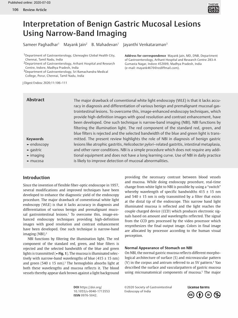
NBI functions by filtering the illumination light. The red component of the standard red, green, and blue filters is rejected and the selected bandwidth of the blue and green lights is transmitted (►Fig. 1). The mucosa is illuminated selec-tively with narrow-band wavelengths of blue (415 ± 15 nm) and green (540 ± 15 nm).3 The hemoglobin absorbs light at both these wavelengths and mucosa reflects it. The blood vessels thereby appear dark brown against a light background
providing the necessary contrast between blood vessels and mucosa. While doing endoscopy procedure, real-time change from white light to NBI is possible by using a “switch” whereby wavelength of specific bandwidths 415 ± 15 nm and 540 ± 15 nm is only transmitted by a filter that exists at the distal tip of the endoscope. This narrow band light illuminated mucosa is reflected and the light reaches the couple charged device (CCD) which produces electronic sig-nals based on amount and wavelengths reflected. The signal from the CCD gets processed by the video processor which resynthesizes the final output image. Colors in final image are allocated by processor according to the human visual perception.
Normal Appearance of Stomach on NBIOn NBI, the normal gastric mucosa reflects different morpho-logical architecture of surface (S) and microvascular pattern (V) in the corpus and antrum referred to as SV pattern.4 Yao described the surface and vascularpattern of gastric mucosa using microanatomical components of mucosa.5 The major
J Digest Endosc 2020;11:106–111
Published online: 2020-07-03
107Interpretation of Benign Gastric Mucosal Lesions Using Narrow Band Imaging Paghadhar et al.
Journal of Digestive Endoscopy Vol. 11 No. 2/2020
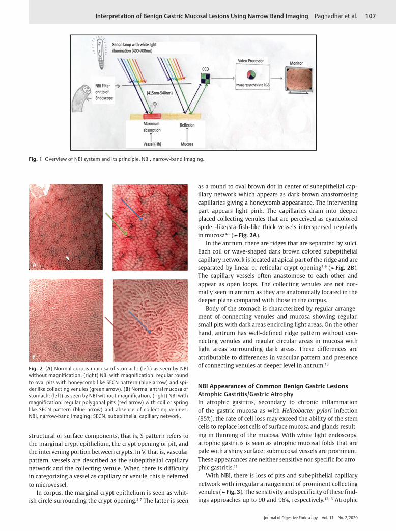
structural or surface components, that is, S pattern refers to the marginal crypt epithelium, the crypt opening or pit, and the intervening portion between crypts. In V, that is, vascular pattern, vessels are described as the subepithelial capillary network and the collecting venule. When there is difficulty in categorizing a vessel as capillary or venule, this is referred to microvessel.
In corpus, the marginal crypt epithelium is seen as whit-ish circle surrounding the crypt opening.5-7 The latter is seen
as a round to oval brown dot in center of subepithelial cap-illary network which appears as dark brown anastomosing capillaries giving a honeycomb appearance. The intervening part appears light pink. The capillaries drain into deeper placed collecting venules that are perceived as cyancolored spider-like/starfish-like thick vessels interspersed regularly in mucosa4-8 (►Fig. 2A).
In the antrum, there are ridges that are separated by sulci. Each coil or wave-shaped dark brown colored subepithelial capillary network is located at apical part of the ridge and are separated by linear or reticular crypt opening7-9 (►Fig. 2B). The capillary vessels often anastomose to each other and appear as open loops. The collecting venules are not nor-mally seen in antrum as they are anatomically located in the deeper plane compared with those in the corpus.
Body of the stomach is characterized by regular arrange-ment of connecting venules and mucosa showing regular, small pits with dark areas encircling light areas. On the other hand, antrum has well-defined ridge pattern without con-necting venules and regular circular areas in mucosa with light areas surrounding dark areas. These differences are attributable to differences in vascular pattern and presence of connecting venules at deeper level in antrum.10
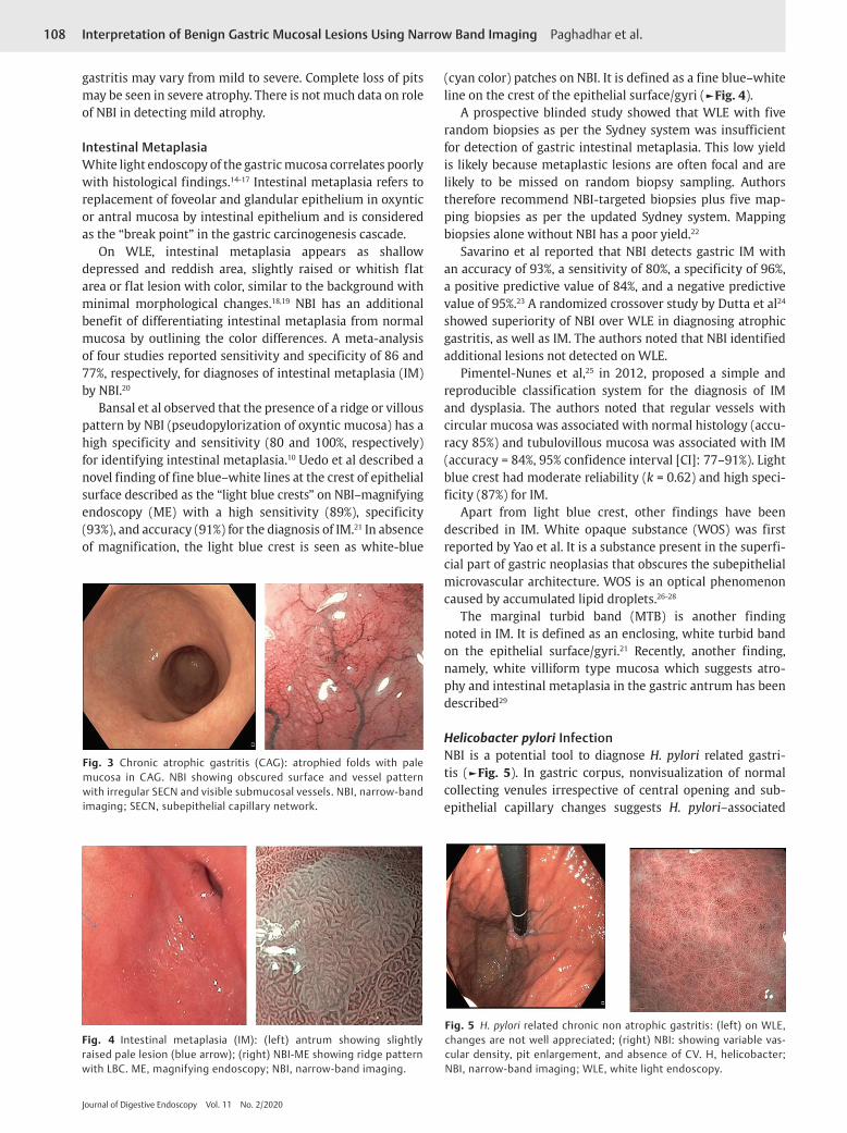
NBI Appearances of Common Benign Gastric LesionsAtrophic Gastritis/Gastric AtrophyIn atrophic gastritis, secondary to chronic inflammation of the gastric mucosa as with Helicobacter pylori infection (85%), the rate of cell loss may exceed the ability of the stem cells to replace lost cells of surface mucosa and glands result-ing in thinning of the mucosa. With white light endoscopy, atrophic gastritis is seen as atrophic mucosal folds that are pale with a shiny surface; submucosal vessels are prominent. These appearances are neither sensitive nor specific for atro-phic gastritis.11
With NBI, there is loss of pits and subepithelial capillary network with irregular arrangement of prominent collecting venules (►Fig. 3). The sensitivity and specificity of these find-ings approaches up to 90 and 96%, respectively.12,13 Atrophic
Fig. 1 Overview of NBI system and its principle. NBI, narrow-band imaging.
Fig. 2 (A) Normal corpus mucosa of stomach: (left) as seen by NBI without magnification, (right) NBI with magnification: regular round to oval pits with honeycomb like SECN pattern (blue arrow) and spi-der like collecting venules (green arrow). (B) Normal antral mucosa of stomach: (left) as seen by NBI without magnification, (right) NBI with magnification: regular polygonal pits (red arrow) with coil or spring like SECN pattern (blue arrow) and absence of collecting venules. NBI, narrow-band imaging; SECN, subepithelial capillary network.
108 Interpretation of Benign Gastric Mucosal Lesions Using Narrow Band Imaging Paghadhar et al.
Journal of Digestive Endoscopy Vol. 11 No. 2/2020
gastritis may vary from mild to severe. Complete loss of pits may be seen in severe atrophy. There is not much data on role of NBI in detecting mild atrophy.
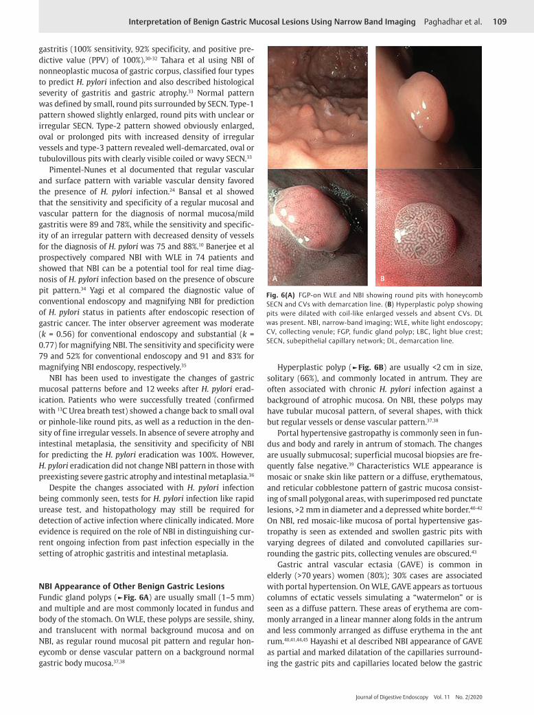
Intestinal MetaplasiaWhite light endoscopy of the gastric mucosa correlates poorly with histological findings.14-17 Intestinal metaplasia refers to replacement of foveolar and glandular epithelium in oxyntic or antral mucosa by intestinal epithelium and is considered as the “break point” in the gastric carcinogenesis cascade.
On WLE, intestinal metaplasia appears as shallow depressed and reddish area, slightly raised or whitish flat area or flat lesion with color, similar to the background with minimal morphological changes.18,19 NBI has an additional benefit of differentiating intestinal metaplasia from normal mucosa by outlining the color differences. A meta-analysis of four studies reported sensitivity and specificity of 86 and 77%, respectively, for diagnoses of intestinal metaplasia (IM) by NBI.20
Bansal et al observed that the presence of a ridge or villous pattern by NBI (pseudopylorization of oxyntic mucosa) has a high specificity and sensitivity (80 and 100%, respectively) for identifying intestinal metaplasia.10 Uedo et al described a novel finding of fine blue–white lines at the crest of epithelial surface described as the “light blue crests” on NBI–magnifying endoscopy (ME) with a high sensitivity (89%), specificity (93%), and accuracy (91%) for the diagnosis of IM.21 In absence of magnification, the light blue crest is seen as white-blue
(cyan color) patches on NBI. It is defined as a fine blue–white line on the crest of the epithelial surface/gyri (►Fig. 4).
A prospective blinded study showed that WLE with five random biopsies as per the Sydney system was insufficient for detection of gastric intestinal metaplasia. This low yield is likely because metaplastic lesions are often focal and are likely to be missed on random biopsy sampling. Authors therefore recommend NBI-targeted biopsies plus five map-ping biopsies as per the updated Sydney system. Mapping biopsies alone without NBI has a poor yield.22
Savarino et al reported that NBI detects gastric IM with an accuracy of 93%, a sensitivity of 80%, a specificity of 96%, a positive predictive value of 84%, and a negative predictive value of 95%.23 A randomized crossover study by Dutta et al24 showed superiority of NBI over WLE in diagnosing atrophic gastritis, as well as IM. The authors noted that NBI identified additional lesions not detected on WLE.
Pimentel-Nunes et al,25 in 2012, proposed a simple and reproducible classification system for the diagnosis of IM and dysplasia. The authors noted that regular vessels with circular mucosa was associated with normal histology (accu-racy 85%) and tubulovillous mucosa was associated with IM (accuracy = 84%, 95% confidence interval [CI]: 77–91%). Light blue crest had moderate reliability (k = 0.62) and high speci-ficity (87%) for IM.
Apart from light blue crest, other findings have been described in IM. White opaque substance (WOS) was first reported by Yao et al. It is a substance present in the superfi-cial part of gastric neoplasias that obscures the subepithelial microvascular architecture. WOS is an optical phenomenon caused by accumulated lipid droplets.26-28
The marginal turbid band (MTB) is another finding noted in IM. It is defined as an enclosing, white turbid band on the epithelial surface/gyri.21 Recently, another finding, namely, white villiform type mucosa which suggests atro-phy and intestinal metaplasia in the gastric antrum has been described29
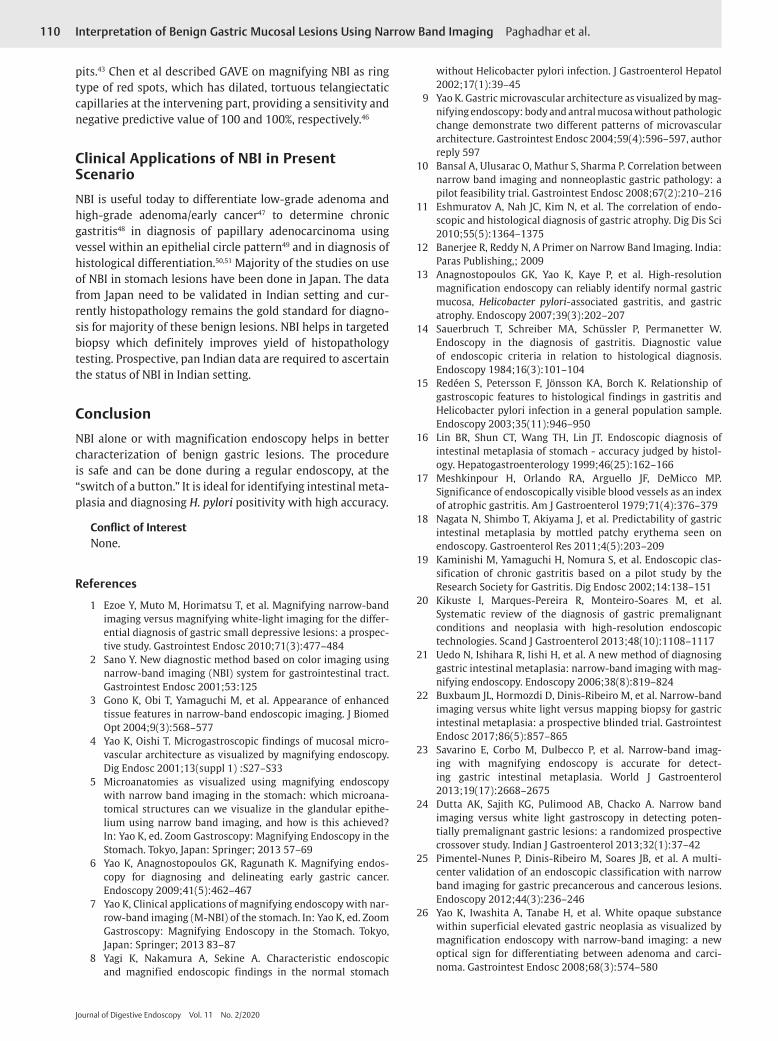
Helicobacter pylori InfectionNBI is a potential tool to diagnose H. pylori related gastri-tis (►Fig. 5). In gastric corpus, nonvisualization of normal collecting venules irrespective of central opening and sub-epithelial capillary changes suggests H. pylori–associated
Fig. 3 Chronic atrophic gastritis (CAG): atrophied folds with pale mucosa in CAG. NBI showing obscured surface and vessel pattern with irregular SECN and visible submucosal vessels. NBI, narrow-band imaging; SECN, subepithelial capillary network.
Fig. 4 Intestinal metaplasia (IM): (left) antrum showing slightly raised pale lesion (blue arrow); (right) NBI-ME showing ridge pattern with LBC. ME, magnifying endoscopy; NBI, narrow-band imaging.
Fig. 5 H. pylori related chronic non atrophic gastritis: (left) on WLE, changes are not well appreciated; (right) NBI: showing variable vas-cular density, pit enlargement, and absence of CV. H, helicobacter; NBI, narrow-band imaging; WLE, white light endoscopy.
109Interpretation of Benign Gastric Mucosal Lesions Using Narrow Band Imaging Paghadhar et al.
Journal of Digestive Endoscopy Vol. 11 No. 2/2020
gastritis (100% sensitivity, 92% specificity, and positive pre-dictive value (PPV) of 100%).30-32 Tahara et al using NBI of nonneoplastic mucosa of gastric corpus, classified four types to predict H. pylori infection and also described histological severity of gastritis and gastric atrophy.33 Normal pattern was defined by small, round pits surrounded by SECN. Type-1 pattern showed slightly enlarged, round pits with unclear or irregular SECN. Type-2 pattern showed obviously enlarged, oval or prolonged pits with increased density of irregular vessels and type-3 pattern revealed well-demarcated, oval or tubulovillous pits with clearly visible coiled or wavy SECN.33
Pimentel-Nunes et al documented that regular vascular and surface pattern with variable vascular density favored the presence of H. pylori infection.24 Bansal et al showed that the sensitivity and specificity of a regular mucosal and vascular pattern for the diagnosis of normal mucosa/mild gastritis were 89 and 78%, while the sensitivity and specific-ity of an irregular pattern with decreased density of vessels for the diagnosis of H. pylori was 75 and 88%.10 Banerjee et al prospectively compared NBI with WLE in 74 patients and showed that NBI can be a potential tool for real time diag-nosis of H. pylori infection based on the presence of obscure pit pattern.34 Yagi et al compared the diagnostic value of conventional endoscopy and magnifying NBI for prediction of H. pylori status in patients after endoscopic resection of gastric cancer. The inter observer agreement was moderate (k = 0.56) for conventional endoscopy and substantial (k = 0.77) for magnifying NBI. The sensitivity and specificity were 79 and 52% for conventional endoscopy and 91 and 83% for magnifying NBI endoscopy, respectively.35
NBI has been used to investigate the changes of gastric mucosal patterns before and 12 weeks after H. pylori erad-ication. Patients who were successfully treated (confirmed with 13C Urea breath test) showed a change back to small oval or pinhole-like round pits, as well as a reduction in the den-sity of fine irregular vessels. In absence of severe atrophy and intestinal metaplasia, the sensitivity and specificity of NBI for predicting the H. pylori eradication was 100%. However, H. pylori eradication did not change NBI pattern in those with preexisting severe gastric atrophy and intestinal metaplasia.36
Despite the changes associated with H. pylori infection being commonly seen, tests for H. pylori infection like rapid urease test, and histopathology may still be required for detection of active infection where clinically indicated. More evidence is required on the role of NBI in distinguishing cur-rent ongoing infection from past infection especially in the setting of atrophic gastritis and intestinal metaplasia.
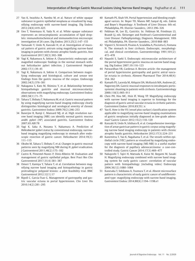
NBI Appearance of Other Benign Gastric LesionsFundic gland polyps (►Fig. 6A) are usually small (1–5 mm) and multiple and are most commonly located in fundus and body of the stomach. On WLE, these polyps are sessile, shiny, and translucent with normal background mucosa and on NBI, as regular round mucosal pit pattern and regular hon-eycomb or dense vascular pattern on a background normal gastric body mucosa.37,38
Hyperplastic polyp (►Fig. 6B) are usually <2 cm in size, solitary (66%), and commonly located in antrum. They are often associated with chronic H. pylori infection against a background of atrophic mucosa. On NBI, these polyps may have tubular mucosal pattern, of several shapes, with thick but regular vessels or dense vascular pattern.37,38
Portal hypertensive gastropathy is commonly seen in fun-dus and body and rarely in antrum of stomach. The changes are usually submucosal; superficial mucosal biopsies are fre-quently false negative.39 Characteristics WLE appearance is mosaic or snake skin like pattern or a diffuse, erythematous, and reticular cobblestone pattern of gastric mucosa consist-ing of small polygonal areas, with superimposed red punctate lesions, >2 mm in diameter and a depressed white border.40-42 On NBI, red mosaic-like mucosa of portal hypertensive gas-tropathy is seen as extended and swollen gastric pits with varying degrees of dilated and convoluted capillaries sur-rounding the gastric pits, collecting venules are obscured.43
Gastric antral vascular ectasia (GAVE) is common in elderly (>70 years) women (80%); 30% cases are associated with portal hypertension. On WLE, GAVE appears as tortuous columns of ectatic vessels simulating a “watermelon” or is seen as a diffuse pattern. These areas of erythema are com-monly arranged in a linear manner along folds in the antrum and less commonly arranged as diffuse erythema in the antrum.40,41,44,45 Hayashi et al described NBI appearance of GAVE as partial and marked dilatation of the capillaries surround-ing the gastric pits and capillaries located below the gastric
Fig. 6(A) FGP-on WLE and NBI showing round pits with honeycomb SECN and CVs with demarcation line. (B) Hyperplastic polyp showing pits were dilated with coil-like enlarged vessels and absent CVs. DL was present. NBI, narrow-band imaging; WLE, white light endoscopy; CV, collecting venule; FGP, fundic gland polyp; LBC, light blue crest; SECN, subepithelial capillary network; DL, demarcation line.
110 Interpretation of Benign Gastric Mucosal Lesions Using Narrow Band Imaging Paghadhar et al.
Journal of Digestive Endoscopy Vol. 11 No. 2/2020
pits.43 Chen et al described GAVE on magnifying NBI as ring type of red spots, which has dilated, tortuous telangiectatic capillaries at the intervening part, providing a sensitivity and negative predictive value of 100 and 100%, respectively.46
Clinical Applications of NBI in Present ScenarioNBI is useful today to differentiate low-grade adenoma and high-grade adenoma/early cancer47 to determine chronic gastritis48 in diagnosis of papillary adenocarcinoma using vessel within an epithelial circle pattern49 and in diagnosis of histological differentiation.50,51 Majority of the studies on use of NBI in stomach lesions have been done in Japan. The data from Japan need to be validated in Indian setting and cur-rently histopathology remains the gold standard for diagno-sis for majority of these benign lesions. NBI helps in targeted biopsy which definitely improves yield of histopathology testing. Prospective, pan Indian data are required to ascertain the status of NBI in Indian setting.
ConclusionNBI alone or with magnification endoscopy helps in better characterization of benign gastric lesions. The procedure is safe and can be done during a regular endoscopy, at the “switch of a button.” It is ideal for identifying intestinal meta-plasia and diagnosing H. pylori positivity with high accuracy.
Conflict of InterestNone.
References
1 Ezoe Y, Muto M, Horimatsu T, et al. Magnifying narrow-band imaging versus magnifying white-light imaging for the differ-ential diagnosis of gastric small depressive lesions: a prospec-tive study. Gastrointest Endosc 2010;71(3):477–484
2 Sano Y. New diagnostic method based on color imaging using narrow-band imaging (NBI) system for gastrointestinal tract. Gastrointest Endosc 2001;53:125
3 Gono K, Obi T, Yamaguchi M, et al. Appearance of enhanced tissue features in narrow-band endoscopic imaging. J Biomed Opt 2004;9(3):568–577
4 Yao K, Oishi T. Microgastroscopic findings of mucosal micro-vascular architecture as visualized by magnifying endoscopy. Dig Endosc 2001;13(suppl 1) :S27–S33
5 Microanatomies as visualized using magnifying endoscopy with narrow band imaging in the stomach: which microana-tomical structures can we visualize in the glandular epithe-lium using narrow band imaging, and how is this achieved? In: Yao K, ed. Zoom Gastroscopy: Magnifying Endoscopy in the Stomach. Tokyo, Japan: Springer; 2013 57–69
6 Yao K, Anagnostopoulos GK, Ragunath K. Magnifying endos-copy for diagnosing and delineating early gastric cancer. Endoscopy 2009;41(5):462–467
7 Yao K, Clinical applications of magnifying endoscopy with nar-row-band imaging (M-NBI) of the stomach. In: Yao K, ed. Zoom Gastroscopy: Magnifying Endoscopy in the Stomach. Tokyo, Japan: Springer; 2013 83–87
8 Yagi K, Nakamura A, Sekine A. Characteristic endoscopic and magnified endoscopic findings in the normal stomach
without Helicobacter pylori infection. J Gastroenterol Hepatol 2002;17(1):39–45
9 Yao K. Gastric microvascular architecture as visualized by mag-nifying endoscopy: body and antral mucosa without pathologic change demonstrate two different patterns of microvascular architecture. Gastrointest Endosc 2004;59(4):596–597, author reply 597
10 Bansal A, Ulusarac O, Mathur S, Sharma P. Correlation between narrow band imaging and nonneoplastic gastric pathology: a pilot feasibility trial. Gastrointest Endosc 2008;67(2):210–216
11 Eshmuratov A, Nah JC, Kim N, et al. The correlation of endo-scopic and histological diagnosis of gastric atrophy. Dig Dis Sci 2010;55(5):1364–1375
12 Banerjee R, Reddy N, A Primer on Narrow Band Imaging. India: Paras Publishing,; 2009
13 Anagnostopoulos GK, Yao K, Kaye P, et al. High-resolution magnification endoscopy can reliably identify normal gastric mucosa, Helicobacter pylori-associated gastritis, and gastric atrophy. Endoscopy 2007;39(3):202–207
14 Sauerbruch T, Schreiber MA, Schüssler P, Permanetter W. Endoscopy in the diagnosis of gastritis. Diagnostic value of endoscopic criteria in relation to histological diagnosis. Endoscopy 1984;16(3):101–104
15 Redéen S, Petersson F, Jönsson KA, Borch K. Relationship of gastroscopic features to histological findings in gastritis and Helicobacter pylori infection in a general population sample. Endoscopy 2003;35(11):946–950
16 Lin BR, Shun CT, Wang TH, Lin JT. Endoscopic diagnosis of intestinal metaplasia of stomach - accuracy judged by histol-ogy. Hepatogastroenterology 1999;46(25):162–166
17 Meshkinpour H, Orlando RA, Arguello JF, DeMicco MP. Significance of endoscopically visible blood vessels as an index of atrophic gastritis. Am J Gastroenterol 1979;71(4):376–379
18 Nagata N, Shimbo T, Akiyama J, et al. Predictability of gastric intestinal metaplasia by mottled patchy erythema seen on endoscopy. Gastroenterol Res 2011;4(5):203–209
19 Kaminishi M, Yamaguchi H, Nomura S, et al. Endoscopic clas-sification of chronic gastritis based on a pilot study by the Research Society for Gastritis. Dig Endosc 2002;14:138–151
20 Kikuste I, Marques-Pereira R, Monteiro-Soares M, et al. Systematic review of the diagnosis of gastric premalignant conditions and neoplasia with high-resolution endoscopic technologies. Scand J Gastroenterol 2013;48(10):1108–1117
21 Uedo N, Ishihara R, Iishi H, et al. A new method of diagnosing gastric intestinal metaplasia: narrow-band imaging with mag-nifying endoscopy. Endoscopy 2006;38(8):819–824
22 Buxbaum JL, Hormozdi D, Dinis-Ribeiro M, et al. Narrow-band imaging versus white light versus mapping biopsy for gastric intestinal metaplasia: a prospective blinded trial. Gastrointest Endosc 2017;86(5):857–865
23 Savarino E, Corbo M, Dulbecco P, et al. Narrow-band imag-ing with magnifying endoscopy is accurate for detect-ing gastric intestinal metaplasia. World J Gastroenterol 2013;19(17):2668–2675
24 Dutta AK, Sajith KG, Pulimood AB, Chacko A. Narrow band imaging versus white light gastroscopy in detecting poten-tially premalignant gastric lesions: a randomized prospective crossover study. Indian J Gastroenterol 2013;32(1):37–42
25 Pimentel-Nunes P, Dinis-Ribeiro M, Soares JB, et al. A multi-center validation of an endoscopic classification with narrow band imaging for gastric precancerous and cancerous lesions. Endoscopy 2012;44(3):236–246
26 Yao K, Iwashita A, Tanabe H, et al. White opaque substance within superficial elevated gastric neoplasia as visualized by magnification endoscopy with narrow-band imaging: a new optical sign for differentiating between adenoma and carci-noma. Gastrointest Endosc 2008;68(3):574–580
111Interpretation of Benign Gastric Mucosal Lesions Using Narrow Band Imaging Paghadhar et al.
Journal of Digestive Endoscopy Vol. 11 No. 2/2020
27 Yao K, Iwashita A, Nambu M, et al. Nature of white opaque substance in gastric epithelial neoplasia as visualized by mag-nifying endoscopy with narrow-band imaging. Dig Endosc 2012;24(6):419–425
28 Ueo T, Yonemasu H, Yada N, et al. White opaque substance represents an intracytoplasmic accumulation of lipid drop-lets: immunohistochemical and immunoelectron microscopic investigation of 26 cases. Dig Endosc 2013;25(2):147–155
29 Yamasaki Y, Uedo N, Kanzaki H, et al. Investigation of muco-sal pattern of gastric antrum using magnifying narrow-band imaging in patients with chronic atrophic fundic gastritis. Ann Gastroenterol 2017;30(3):302–308
30 Yagi K, Nakamura A, Sekine A. Characteristic endoscopic and magnified endoscopic findings in the normal stomach with-out helicobacter pylori infection. J Gastroenterol Hepatol 2002;17(1):39–45
31 Yagi K, Nakamura A, Sekine A. Comparison between magni-fying endoscopy and histological, culture and urease test findings from the gastric mucosa of the corpus. Endoscopy 2002;34(5):376–381
32 Nakagawa S, Kato M, Shimizu Y, et al. Relationship between histopathologic gastritis and mucosal microvascularity: observations with magnifying endoscopy. Gastrointest Endosc 2003;58(1):71–75
33 Tahara T, Shibata T, Nakamura M, et al. Gastric mucosal pattern by using magnifying narrow-band imaging endoscopy clearly distinguishes histological and serological severity of chronic gastritis. Gastrointest Endosc 2009;70(2):246–253
34 Banerjee R, Ramji C, Mansard MJ, et al. High resolution nar-row band imaging (NBI) can identify normal gastric mucosa andH. pylori (HP) associated gastritis. Gastrointest Endosc 2007;65:AB178
35 Yagi K, Saka A, Nozawa Y, Nakamura A. Prediction of Helicobacter pylori status by conventional endoscopy, narrow- band imaging magnifying endoscopy in stomach after endo-scopic resection of gastric cancer. Helicobacter 2014;19(2): 111–115
36 Okubo M, Tahara T, Shibata T, et al. Changes in gastric mucosal patterns seen by magnifying NBI during H. pylori eradication. J Gastroenterol 2011;46(2):175–182
37 Castro R, Pimentel-Nunes P, Dinis-Ribeiro M. Evaluation and management of gastric epithelial polyps. Best Pract Res Clin Gastroenterol 2017;31(4):381–387
38 Omori T, Kamiya Y, Tahara T, et al. Correlation between mag-nifying narrow band imaging and histopathology in gastric protruding/or polypoid lesions: a pilot feasibility trial. BMC Gastroenterol 2012;12(1):17
39 Ripoll C, Garcia-Tsao G. Management of gastropathy and gas-tric vascular ectasia in portal hypertension. Clin Liver Dis 2010;14(2):281–295
40 Kamath PS, Shah VH, Portal hypertension and bleeding esoph-ageal varices. In: Boyer TD, Manns MP, Sanyal AJ, eds. Zakim and Boyer’s Hepatology: A Textbook of Liver Disease. 6th ed. Philadelphia, PA: Elsevier Saunders,; 2012 296–326
41 Feldman M, Lee EL, Gastritis. In: Feldman M, Friedman LS, Brandt LJ, eds. Sleisenger and Fordtran’s Gastrointestinal and Liver Disease: Pathophysiology, Diagnosis, Management. 10th ed. Philadelphia, PA: Elsevier Saunders,; 2010 868–883
42 Vigneri S, Termini R, Piraino A, Scialabba A, Pisciotta G, Fontana N. The stomach in liver cirrhosis. Endoscopic, morphologi-cal, and clinical correlations. Gastroenterology 1991;101(2): 472–478
43 Hayashi S, Saeki S. Endoscopic microvascular architecture of the portal hypertensive gastric mucosa on narrow band imag-ing. Dig Endosc 2007;19:116–123
44 Patwardhan VR, Cardenas A. Review article: the management of portal hypertensive gastropathy and gastric antral vascu-lar ectasia in cirrhosis. Aliment Pharmacol Ther 2014;40(4): 354–362
45 Kamath PS, Lacerda M, Ahlquist DA, McKusick MA, Andrews JC, Nagorney DA. Gastric mucosal responses to intrahepatic porto-systemic shunting in patients with cirrhosis. Gastroenterology 2000;118(5):905–911
46 Chen PH, Hou MC, Hsin IF, Wang YP. Magnifying endoscopy with narrow band imaging is superior to histology for the diagnosis of gastric antral vascular ectasia in cirrhotic patients. Gastrointest Endosc 2016;83(5S) :x
47 Yao K. How is the VS (vessel plus surface) classification system applicable to magnifying narrow-band imaging examinations of gastric neoplasias initially diagnosed as low-grade adeno-mas? Gastric Cancer 2012;15(2):118–120
48 Kanzaki H, Uedo N, Ishihara R, et al. Comprehensive investiga-tion of areae gastricae pattern in gastric corpus using magnify-ing narrow band imaging endoscopy in patients with chronic atrophic fundic gastritis. Helicobacter 2012;17(3):224–231
49 Kanemitsu T, Yao K, Nagahama T, et al. The vessels within epi-thelial circle (VEC) pattern as visualized by magnifying endos-copy with narrow-band imaging (ME-NBI) is a useful marker for the diagnosis of papillary adenocarcinoma: a case-con-trolled study. Gastric Cancer 2014;17(3):469–477
50 Nakayoshi T, Tajiri H, Matsuda K, Kaise M, Ikegami M, Sasaki H. Magnifying endoscopy combined with narrow band imag-ing system for early gastric cancer: correlation of vascular pattern with histopathology (including video) Endoscopy 2004;36(12):1080–1084
51 Kanesaka T, Sekikawa A, Tsumura T, et al. Absent microsurface pattern is characteristic of early gastric cancer of undifferenti-ated type: magnifying endoscopy with narrow-band imaging. Gastrointest Endosc 2014;80(6):1194–1198.e1