Embed Size (px)
Citation preview

HAL Id: inserm-00543644https://www.hal.inserm.fr/inserm-00543644
Submitted on 6 Dec 2010
HAL is a multi-disciplinary open accessarchive for the deposit and dissemination of sci-entific research documents, whether they are pub-lished or not. The documents may come fromteaching and research institutions in France orabroad, or from public or private research centers.
L’archive ouverte pluridisciplinaire HAL, estdestinée au dépôt et à la diffusion de documentsscientifiques de niveau recherche, publiés ou non,émanant des établissements d’enseignement et derecherche français ou étrangers, des laboratoirespublics ou privés.
Intracoronary autologous mononucleated bone marrowcell infusion for acute myocardial infarction: results of
the randomized multicenter BONAMI trial.Jérôme Roncalli, Frédéric Mouquet, Christophe Piot, Jean-Noel Trochu,
Philippe Le Corvoisier, Yannick Neuder, Thierry Le Tourneau, Denis Agostini,Virginia Gaxotte, Catherine Sportouch, et al.
To cite this version:Jérôme Roncalli, Frédéric Mouquet, Christophe Piot, Jean-Noel Trochu, Philippe Le Corvoisier, et al..Intracoronary autologous mononucleated bone marrow cell infusion for acute myocardial infarction:results of the randomized multicenter BONAMI trial.. European Heart Journal, Oxford UniversityPress (OUP): Policy B, 2011, 32 (14), pp.1748-57. �10.1093/eurheartj/ehq455�. �inserm-00543644�

Eur Heart J . Author manuscript
Page /1 12
Intracoronary autologous mononucleated bone marrow cell infusion foracute myocardial infarction: results of the randomized multicenterBONAMI trialJ r me Roncalli é ô 1 2 # , Fr d ric Mouquet é é 3 # , Christophe Piot 4 , Jean-Noel Trochu 5 , Philippe Le Corvoisier 6 7 , Yannick Neuder 8 ,Thierry Le Tourneau 3 5 , Denis Agostini 9 , Virginia Gaxotte 10 , Catherine Sportouch 4 , Michel Galinier 1 2 , Dominique P. Crochet 5 ,Emmanuel Teiger 6 7 , Marie-Jeanne Richard 11 , Anne-Sophie Polge 3 , Jean-Paul Beregi 10 , Alain Manrique 12 , Didier Carrie 1 2 ,Sophie Susen 11 13 , Bernard Klein 4 , Angelo Parini 2 , Guillaume Lamirault 5 , Pierre Croisille 14 , H l ne Rouard é è 15 , Philippe Bourin 16 , Jean-Michel Nguyen 17 , B atrice Delasalle é 5 , G rald Vanzetto é 8 , Eric Van Belle 3 , Patricia F. Lemarchand 5 *
F d ration de Cardiologie 1 é é CHU Toulouse , Hopital de Rangueil, 31403 Toulouse, FR
I2MR, Institut de m decine mol culaire de Rangueil 2 é é INSERM : U858 , IFR31 , IFR150 , Universit Paul Sabatier - Toulouse III é , InstitutLouis Bugnard 1, avenue Jean Poulhes BP 84225 31432 Toulouse Cedex 4,FR
Interface sang vaisseaux et r paration cardiovasculaire 3 é CHRU Lille , Universit du Droit et de la Sant - Lille II : EA2693 é é , PoleCardiologie, 59037 Lille Cedex, FR
Bioth rapie des cellules souches normales et canc reuses 4 é é INSERM : U847 , Institut de recherche en bioth rapie é , Universit Montpellier I é , CHRU Montpellier , IRB - CHRU Saint-Eloi, 80 Avenue Augustin Fliche 34295 MONTPELLIER Cedex 5,FR
Institut du thorax 5 INSERM : U915 , Universit de Nantes é , IFR26 , 8 quai moncousu, BP 70721, 44007 Nantes Cedex 1, FR
Institut Mondor de Recherche Biom dicale 6 é INSERM : U955 , Universit Paris XII Val de Marne é , IFR10 , 94010 Cr teil, FRé
CIC - CHU Henri Mondor 7 INSERM : CIC6 , Universit Paris XII Val de Marne é , Hopital Henri Mondor, 51 Av du Mal de Lattre deTassigny 94000 Cr teil, FRé
Radiopharmaceutiques biocliniques 8 INSERM : U877 , Universit Joseph Fourier - Grenoble I é , Facult de m decine et de pharmacieé édomaine de la merci B timent Jean Roget 38700 La Tronche, FRâ
Groupe coeur et isch mie 9 é CHU Caen , Universit de Caen : EA3212 é , 14032 Caen Cedex, FR
Therapies Interventionnelles Assistees Par l Image et la Simulation 10 ' INSERM : U703 , Universit du Droit et de la Sant - Lille II é é , CHRUde Lille, Avenue Oscar Lambret 59037 Lille Cedex, FR
Institut d oncologie/d veloppement Albert Bonniot de Grenoble 11 ' é INSERM : U823 , CHU Grenoble , EFS , Universit Joseph Fourier -é Grenoble I , Institut Albert Bonniot, BP170, 38042 Grenoble Cedex 9, FR
Centre Cyceron 12 CNRS : UMR6551 , Universit de Caen é , CEA : DSV , INSERM , CHU Caen , Boulevard Becquerel, BP 5229, 14074Caen CEDEX, FR
P le d h matologie-Transfusion 13 ô ' é CHRU Lille , Universit du Droit et de la Sant - Lille II é é , Lille,FR
CREATIS, Centre de recherche et d applications en traitement de l image et du signal 14 ' ' INSERM : U630 , Universit Claude Bernard - Lyoné I , Institut National des Sciences Appliqu es de Lyon é , CNRS : UMR5220 , cole Sup rieure Chimie Physique lectronique de Lyon É é É , 69621
Villeurbanne cedex, FR
Bioing nierie cellulaire, tissulaire et sanguine à vis e th rapeutique 15 é é é Etablissement Fran ais du Sang (EFS) ç , Universit Paris XII Val deé Marne: EA3952 , Cr teil, F-94000,FRé
Laboratoire de th rapie cellulaire 16 é Etablissement Fran ais du Sang (EFS) ç , 31027 Toulouse Cedex 3, FR
P le d Information M dicale, Evaluation et Sant Publique - PIMESP 17 ô ' é é CHU Nantes , Universit de Nantes é , Hopital saint-Jacques, 44093Nantes Cedex 1, FR*Correspondence should be adressed to: Patricia Lemarchand <[email protected] >
# Drs Roncalli and Mouquet contributed equally to this work.
AbstractAims
Intracoronary administration of autologous bone marrow cells (BMCs) leads to a modest improvement in cardiac function, but the
effect on myocardial viability is unknown. The aim of this randomized multicenter study was to evaluate the effect of BMC therapy
on myocardial viability in patients with decreased left ventricular ejection fraction (LVEF) after acute myocardial infarction (AMI)
and to identify predictive factors for improvement of myocardial viability.
Methods and Results
One-hundred one patients with AMI and successful reperfusion, LVEF 45 , and decreased myocardial viability (resting≤ %Tl201-SPECT) were randomized to either a control group (n 49) or a BMC group (n 52). Primary endpoint was improvement of= =
Eur Heart J . Author manuscript
Page /2 12
myocardial viability 3 months after AMI. Baseline mean LVEF measured by radionuclide angiography was 36.3 6.9 . BMC± %infusion was performed 9.3 1.7 days after AMI. Myocardial viability improved in 16/47 (34 ) patients in the BMC group compared± %to 7/43 (16 ) in the control group (p 0.06). The number of non-viable segments becoming viable was 0.8 1.1 in the control group% = ±and 1.2 1.5 in the BMC group (p 0.13). Multivariate analysis including major post-AMI prognostic factors showed a significant± =improvement of myocardial viability in BMC . control group (p 0.03). Moreover, a significant adverse role for active smoking (pvs = =0.04) and a positive trend for microvascular obstruction (p 0.07) were observed.=
Conclusions
Intracoronary autologous BMC administration to patients with decreased LVEF after AMI was associated with improvement of
myocardial viability in multivariate but not in univariate analysis. A large multicenter international trial is warranted to further– –document the efficacy of cardiac cell therapy and better define a group of patients that will benefit from this therapy.
Author Keywords Myocardial infarction ; Cells ; Smoking
Introduction
More than 7 years ago, an initial report on the clinical application of mononucleated bone marrow-derived cells (BMCs) in patients
with acute myocardial infarction (AMI) opened up a new era of regenerative cardiology and brought with it great enthusiasm .Since[1 ]then, numerous clinical trials have been carried out with the aim of assessing the efficacy and safety of stem cell therapy. Improvement of
left ventricular ejection fraction (LVEF) was shown in REPAIR-AMI study but the benefit was transient in BOOST, , while other[2 ] [3 4 ]trials were unsuccessful .Although randomized trials reported mixed results, meta-analyses have shown a significant improvement in[5 –7 ]cardiac function assessed by LVEF after cell therapy .Some of the discrepancies between these large trials could be explained by[8 ]differences in criteria for patient selection. There is some evidence to suggest that BMC-based therapy is more effective at improving
LVEF after AMI in patients with decreased LVEF and in patients whose treatment is delayed at least 5 days after AMI , .Lifestyle may[2 8 ]also affect clinical outcome following cell therapy. Indeed, smoking habit is a major factor contributing to reduced number and function of
circulating progenitor cells in patients with coronary artery disease .Most of clinical trials have focused on changes in ejection fraction[9 ]to evaluate the efficiency of cell therapy. However, it has been shown that myocardial viability represents a reliable parameter for
prediction of recovery of cardiac function after revascularization .We carried out a prospective randomized open label blinded endpoint[10 ]evaluation (PROBE) study to assess the efficacy of mononucleated autologous BMC intracoronary injection in patients with AMI and low
LVEF. Our primary objective was to evaluate the effect of BMC therapy on myocardial viability and to identify predictive factors for
improvement of myocardial viability.
MethodsPatient selection
Patients admitted to the University Hospitals of Cr teil, Grenoble, Lille, Montpellier, Nantes and Toulouse, between December 2004éand January 2007, with an ST-segment elevation myocardial infarction (STEMI) were screened for inclusion in the BONAMI trial.
Screening criteria were: age 18 75 years, a successful percutaneous coronary intervention (PCI) with bare metal stent implantation–performed on the culprit lesion during the 24 h after the onset of symptoms, and LVEF <50 assessed by echocardiography. The main%exclusion criteria are listed in . The ethics review board of Nantes University Hospital approved theSupplementary material online
protocol, and the study was conducted in accordance with the Declaration of Helsinki. All subjects gave informed consent.
Study design and treatment randomization
The study design is shown in . Day 0 was defined as the day of occurrence of STEMI. Screened patients underwentFigure 1
radionuclide angiography (RNA) and resting 4 h thallium-201-gated-single-photon-emission computed tomography (SPECT) between day
1 and day 4. Randomization criteria were defined as follows: LVEF 45 assessed by RNA and absence of myocardial viability in at least≤ %2/17 contiguous segments by SPECT. The Center of Clinical Research University Hospital Nantes provided consecutively numbered
sealed envelopes for all participant centers. Patients were randomly assigned in a 1:1 ratio to either the control group or BMC group using
permuted-block randomization stratified according to center, diabetes status and time to PCI after the onset of AMI ( 12 or >12 h) on day≤4 7. We used a stratified randomization model because diabetes is known to impact on BMC and late reperfusion influences the– [11 ]prognosis after AMI. In addition, baseline echocardiography and cardiac magnetic resonance imaging (MRI) were performed on day 4 7–in both groups.
Neither bone marrow aspiration nor sham injection was performed in the control group. In the BMC group, bone marrow aspiration
and intracoronary BMC injection were performed from day 7 10. Three months after STEMI, echocardiography, SPECT, RNA, cardiac–MRI and coronary angiography were repeated.

Eur Heart J . Author manuscript
Page /3 12
Harvest and transfer of bone marrow cells
Bone marrow harvest (50 mL) and cell preparation, flow cytometry analysis of BMCs, and cell administration were performed
according to standard procedures and are described in .Supplementary material online
Cardiac imaging
Three independent core imaging laboratories, blinded to treatment assignment, performed all measurements (prime investigator of the
RNA and SPECT core lab: D. Agostini, MD, Caen; echo: Th. Le Tourneau, MD, PhD, Lille; IRM: J.P. Beregi, MD, Lille, France).
Doppler echocardiography
Images were recorded prospectively and analyzed offline at the core echo laboratory of the study in a blinded fashion. Left ventricular
(LV) volumes and LVEF were calculated using the Simpson s biplane method in apical 4- and 2- chamber views. LV volumes were’indexed to body surface area.
Radionuclide angiography and single-photon-emission computed tomography
RNA was carried out in all patients to determine LVEF at rest (measured by Multiple Gated Acquisition scan). Thallium-201 SPECT
imaging was performed according to a rest-redistribution protocol, using a standard camera to assess myocardial viability. Left ventricular
segmentation in 17 segments as defined by the American Heart Association was used for all analyses. The activity of each LV segment
was expressed as the mean activity of all pixels belonging to this sector divided by the highest value of pixel activity in the myocardium.
Each segment was defined as viable (uptake > 60 ), non-viable (uptake <50 ), or equivocal (uptake 50 60 ) (see % % – % Supplementary
).material online
Cardiac magnetic resonance imaging
Cardiac MRI was performed for evaluating infarct size and the presence of microvascular obstruction using late contrast-enhanced
images acquired 10 min after injection of 0.2 mmol/kg Gd-DTPA (Dotarem, Guerbet, France). LV function, volumes and regional
contractility were also assessed (see ).Supplementary material online
Coronary angiography
Patients underwent coronary angiography to assess the degree of restenosis in the stented segment of the infarct-related artery (IRA) at
3 months follow-up (see ).Supplementary material online
Endpoints
The primary endpoint - - was defined as a gain of at least 2/17 viable segments 3 months afterimprovement of myocardial viability
STEMI, assessed by resting 4 h thallium-201-gated-SPECT. As prespecified, major cardiovascular (CV) risk factors (age, gender,
dyslipidemia, cigarette smoking, hypertension, diabetes), and post-MI prognostic factors (time to PCI, LVEF, infarct size, microvascular
obstruction) were also tested for their association with improvement of myocardial viability 3 months after BMC administration.
Secondary prespecified endpoints included changes in LVEF evaluated by RNA, MRI, and echocardiography; changes in LV end-diastolic
and end-systolic volumes (EDV and ESV) at the time of and 3 months after AMI; infarct size by MRI; and binary restenosis by coronary
angiography. Improvement of myocardial viability was also assessed on a segment-by-segment basis, counting the number of non-viable
segment becoming viable.
Statistical analysis
The baseline characteristics were recorded for each treatment arm and compared using Student s t test and Fisher s exact test.’ ’Categorical variables were presented as count and percent, continuous variables as mean and standard deviation, median with interquartile
ranges. Association of BMC injection with the primary endpoint was assessed by a Cochran-Mantel-Haenszel test adjusted for
stratification factors (diabetes status and time to PCI after the onset of STEMI ( 12 or >12 h)). Univariate analysis of major CV risk and≤post-MI prognosis factors was performed on the primary endpoint, except for diabetes and time to PCI as they were stratification
randomization parameters. In a second step, CV risk factors (age, gender, tobacco status, hypertension, dyslipidemia, diabetes), time to
PCI after the onset of AMI ( 12 or >12 h) and the presence of microvascular obstruction at MRI were introduced into a multivariate≤logistic regression analysis (odds ratio with 95 CI). Mann-Whitney and Wilcoxon signed rank tests were used to assess secondary%endpoints.
Safety of BMC administration was analyzed using prespecified clinical endpoints and included major adverse cardiovascular events
(MACEs) defined as death, re-hospitalization for heart failure, and ischemic events. Other clinical events were assessed as a post-hoc
analysis. Only the first event for each patient was included in the analysis.
Eur Heart J . Author manuscript
Page /4 12
All analyses were performed using SAS 9.1 statistical software. p value < 0.05 was considered statistically significant. This study was
registered with clinicaltrials.gov: number NCT00200707.
ResultsEnrolment and baseline characteristics
A total of 122 patients with successfully reperfused STEMI by PCI and stent implantation within 24 h of the onset of chest pain gave
written informed consent and was enrolled in the study. Twenty-one patients were excluded before randomization ( ) because theyFigure 1
did not meet the randomization criteria. Forty-nine of the remaining patients were randomly assigned to the control group and 52 to the
BMC group. Both groups were well matched with respect to baseline characteristics, procedural characteristics of reperfusion therapy, and
concomitant pharmacological therapy during the study ( ). Baseline recordings were obtained 4 2 days after myocardial infarctionTable 1 ±for RNA, 5.3 2.6 days for SPECT, 7 2.2 days for echocardiography, and 7.1 2.3 days for MRI. There were no significant differences± ± ±in baseline parameters including EDV, ESV, LVEF, microvascular obstruction, and infarct size between the two groups ( ).Table 1
Moreover, 71.8 of patients presented at least one segment with microvascular obstruction assessed by MRI after reperfusion therapy. The%mean number of segments with microvascular obstruction was 2.4 2.1 and was similar in both groups (p 0.94).± =
Forty-four patients completed the follow-up in the control group and 48 in the BMC group. In the control group, two patients were
withdrawn from the study before day 7 (one patient required injections of steroids for angioneurotic edema and the other because of a
post-MI ventricular septal defect that might have interfered with recovery of cardiac function). Three further patients refused to complete
the 3-month follow-up. In the BMC group, two patients were excluded before bone marrow aspiration because of a transient ischemic
attack and a randomization error; one patient did not have bone marrow aspiration because of thrombopenia induced by a GP2b3a
inhibitor. Of the remaining 49 patients in the BMC group, one died 1-month post-MI and one refused to complete the 3-month follow-up.
Characterization of the cell therapy product
Bone marrow aspiration and intracoronary cell injection were performed on the same day for each patient, at a mean of 9.3 1.7 days±after PCI. The mean delay between BMC preparation and intracoronary injection was 5 h 28 min 1 h 30 min. The injected cell number±was 98.3 8.7 10 autologous mononucleated BMCs, with a mean percentage of viable cells of 98.4 1.1 ( ).± × 6 ± % Table 2
Myocardial viability
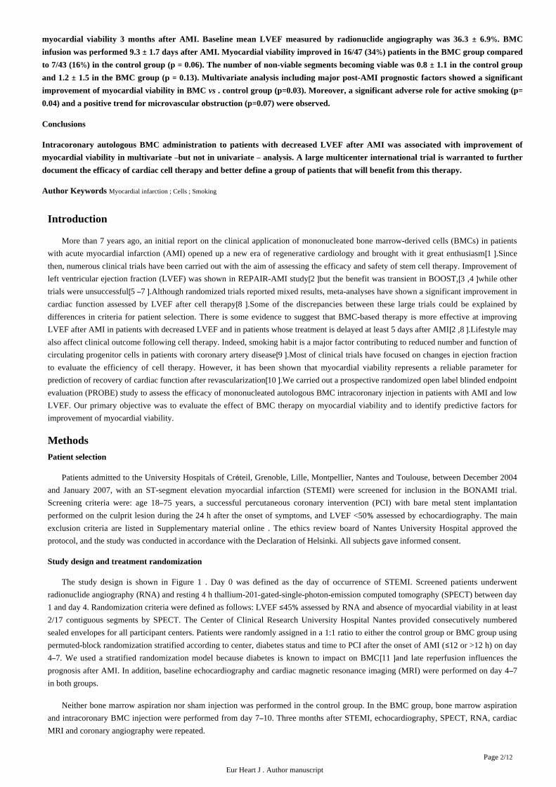
To analyze the primary end-point of improvement of myocardial viability ( ), SPECT images were obtained from 43 and 47Figure 2A
patients in the control and BMC groups, respectively.
At 3 months after BMC infusion, 7/43 (16 ) patients in the control group compared to 16/47 (34 ) patients in the BMC group% %improved their myocardial viability (p 0.06) ( ). The same trend was observed when improvement of viability was defined as a= Figure 2B
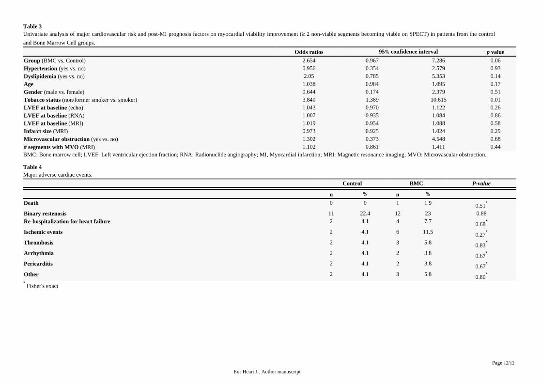
gain of at least three viable segments (3/43, 7 in the control group vs. 10/47, 21 in the BMC group, p 0.07). Univariate analysis of CV% % =risk factors and major post-MI prognosis factors showed that smoking status at the time of AMI was the only significant factor related to
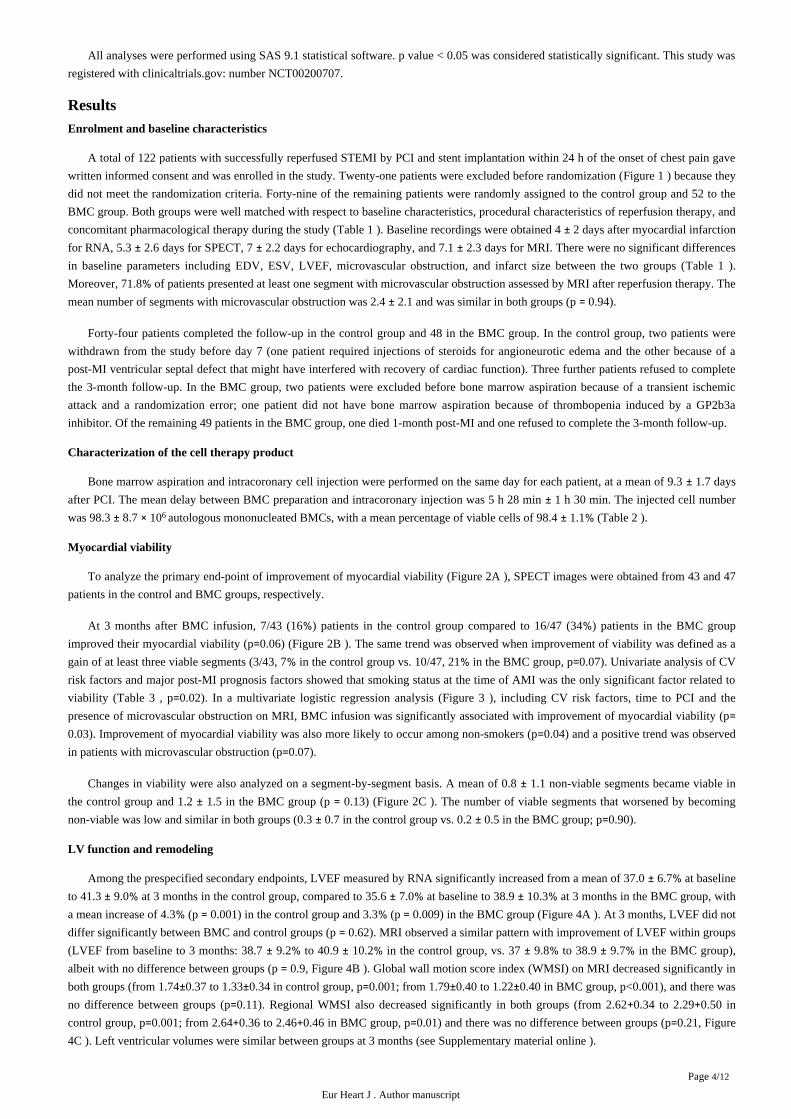
viability ( , p 0.02). In a multivariate logistic regression analysis ( ), including CV risk factors, time to PCI and theTable 3 = Figure 3
presence of microvascular obstruction on MRI, BMC infusion was significantly associated with improvement of myocardial viability (p=0.03). Improvement of myocardial viability was also more likely to occur among non-smokers (p 0.04) and a positive trend was observed=in patients with microvascular obstruction (p 0.07).=
Changes in viability were also analyzed on a segment-by-segment basis. A mean of 0.8 1.1 non-viable segments became viable in±the control group and 1.2 1.5 in the BMC group (p 0.13) ( ). The number of viable segments that worsened by becoming± = Figure 2C
non-viable was low and similar in both groups (0.3 0.7 in the control group vs. 0.2 0.5 in the BMC group; p 0.90).± ± =
LV function and remodeling
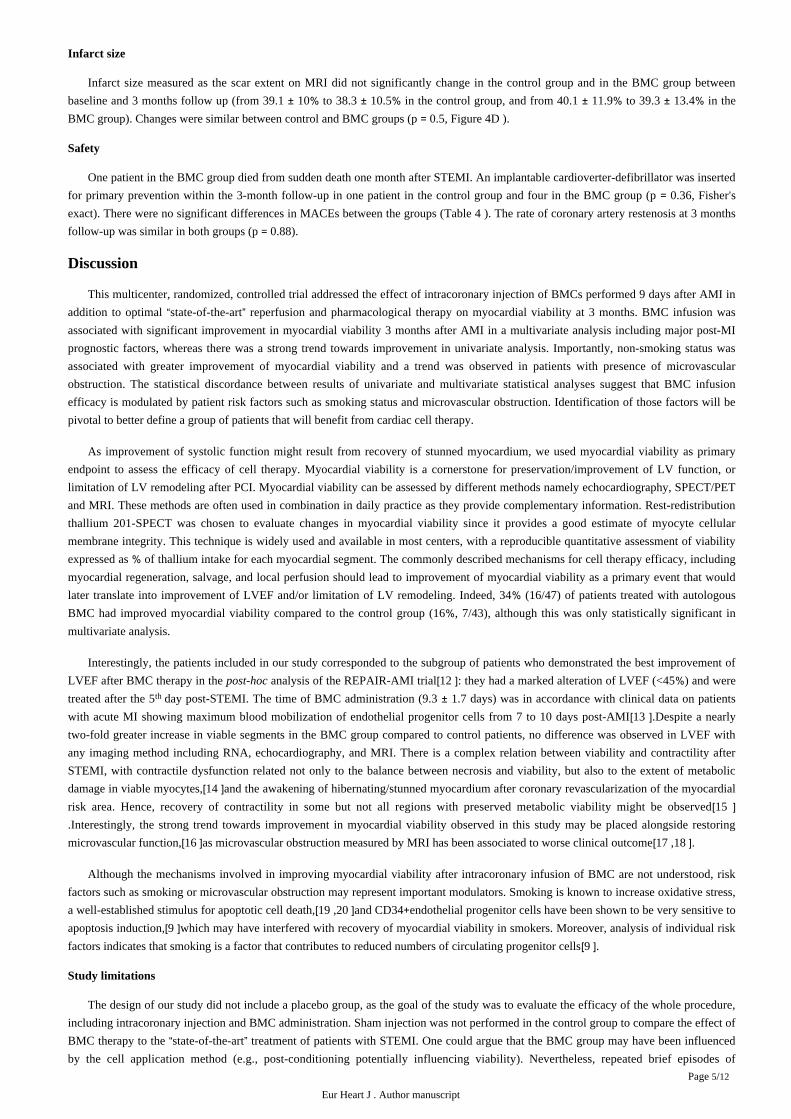
Among the prespecified secondary endpoints, LVEF measured by RNA significantly increased from a mean of 37.0 6.7 at baseline± %to 41.3 9.0 at 3 months in the control group, compared to 35.6 7.0 at baseline to 38.9 10.3 at 3 months in the BMC group, with± % ± % ± %a mean increase of 4.3 (p 0.001) in the control group and 3.3 (p 0.009) in the BMC group ( ). At 3 months, LVEF did not% = % = Figure 4A
differ significantly between BMC and control groups (p 0.62). MRI observed a similar pattern with improvement of LVEF within groups=(LVEF from baseline to 3 months: 38.7 9.2 to 40.9 10.2 in the control group, vs. 37 9.8 to 38.9 9.7 in the BMC group),± % ± % ± % ± %albeit with no difference between groups (p 0.9, ). Global wall motion score index (WMSI) on MRI decreased significantly in= Figure 4B
both groups (from 1.74 0.37 to 1.33 0.34 in control group, p 0.001; from 1.79 0.40 to 1.22 0.40 in BMC group, p<0.001), and there was± ± = ± ±no difference between groups (p 0.11). Regional WMSI also decreased significantly in both groups (from 2.62 0.34 to 2.29 0.50 in= + +control group, p 0.001; from 2.64 0.36 to 2.46 0.46 in BMC group, p 0.01) and there was no difference between groups (p 0.21, = + + = = Figure
). Left ventricular volumes were similar between groups at 3 months (see ).4C Supplementary material online
Eur Heart J . Author manuscript
Page /5 12
Infarct size
Infarct size measured as the scar extent on MRI did not significantly change in the control group and in the BMC group between
baseline and 3 months follow up (from 39.1 10 to 38.3 10.5 in the control group, and from 40.1 11.9 to 39.3 13.4 in the± % ± % ± % ± %BMC group). Changes were similar between control and BMC groups (p 0.5, ).= Figure 4D
Safety
One patient in the BMC group died from sudden death one month after STEMI. An implantable cardioverter-defibrillator was inserted
for primary prevention within the 3-month follow-up in one patient in the control group and four in the BMC group (p 0.36, Fisher s= ’exact). There were no significant differences in MACEs between the groups ( ). The rate of coronary artery restenosis at 3 monthsTable 4
follow-up was similar in both groups (p 0.88).=
Discussion
This multicenter, randomized, controlled trial addressed the effect of intracoronary injection of BMCs performed 9 days after AMI in
addition to optimal state-of-the-art reperfusion and pharmacological therapy on myocardial viability at 3 months. BMC infusion was“ ”associated with significant improvement in myocardial viability 3 months after AMI in a multivariate analysis including major post-MI
prognostic factors, whereas there was a strong trend towards improvement in univariate analysis. Importantly, non-smoking status was
associated with greater improvement of myocardial viability and a trend was observed in patients with presence of microvascular
obstruction. The statistical discordance between results of univariate and multivariate statistical analyses suggest that BMC infusion
efficacy is modulated by patient risk factors such as smoking status and microvascular obstruction. Identification of those factors will be
pivotal to better define a group of patients that will benefit from cardiac cell therapy.
As improvement of systolic function might result from recovery of stunned myocardium, we used myocardial viability as primary
endpoint to assess the efficacy of cell therapy. Myocardial viability is a cornerstone for preservation/improvement of LV function, or
limitation of LV remodeling after PCI. Myocardial viability can be assessed by different methods namely echocardiography, SPECT/PET
and MRI. These methods are often used in combination in daily practice as they provide complementary information. Rest-redistribution
thallium 201-SPECT was chosen to evaluate changes in myocardial viability since it provides a good estimate of myocyte cellular
membrane integrity. This technique is widely used and available in most centers, with a reproducible quantitative assessment of viability
expressed as of thallium intake for each myocardial segment. The commonly described mechanisms for cell therapy efficacy, including%myocardial regeneration, salvage, and local perfusion should lead to improvement of myocardial viability as a primary event that would
later translate into improvement of LVEF and/or limitation of LV remodeling. Indeed, 34 (16/47) of patients treated with autologous%BMC had improved myocardial viability compared to the control group (16 , 7/43), although this was only statistically significant in%multivariate analysis.
Interestingly, the patients included in our study corresponded to the subgroup of patients who demonstrated the best improvement of
LVEF after BMC therapy in the analysis of the REPAIR-AMI trial : they had a marked alteration of LVEF (<45 ) and werepost-hoc [12 ] %treated after the 5 day post-STEMI. The time of BMC administration (9.3 1.7 days) was in accordance with clinical data on patientsth ±with acute MI showing maximum blood mobilization of endothelial progenitor cells from 7 to 10 days post-AMI .Despite a nearly[13 ]two-fold greater increase in viable segments in the BMC group compared to control patients, no difference was observed in LVEF with
any imaging method including RNA, echocardiography, and MRI. There is a complex relation between viability and contractility after
STEMI, with contractile dysfunction related not only to the balance between necrosis and viability, but also to the extent of metabolic
damage in viable myocytes, and the awakening of hibernating/stunned myocardium after coronary revascularization of the myocardial[14 ]risk area. Hence, recovery of contractility in some but not all regions with preserved metabolic viability might be observed[15 ].Interestingly, the strong trend towards improvement in myocardial viability observed in this study may be placed alongside restoring
microvascular function, as microvascular obstruction measured by MRI has been associated to worse clinical outcome , .[16 ] [17 18 ]
Although the mechanisms involved in improving myocardial viability after intracoronary infusion of BMC are not understood, risk
factors such as smoking or microvascular obstruction may represent important modulators. Smoking is known to increase oxidative stress,
a well-established stimulus for apoptotic cell death, , and CD34 endothelial progenitor cells have been shown to be very sensitive to[19 20 ] +apoptosis induction, which may have interfered with recovery of myocardial viability in smokers. Moreover, analysis of individual risk[9 ]factors indicates that smoking is a factor that contributes to reduced numbers of circulating progenitor cells .[9 ]
Study limitations
The design of our study did not include a placebo group, as the goal of the study was to evaluate the efficacy of the whole procedure,
including intracoronary injection and BMC administration. Sham injection was not performed in the control group to compare the effect of
BMC therapy to the state-of-the-art treatment of patients with STEMI. One could argue that the BMC group may have been influenced“ ”by the cell application method (e.g., post-conditioning potentially influencing viability). Nevertheless, repeated brief episodes of
Eur Heart J . Author manuscript
Page /6 12
inflation-deflation of the angioplasty balloon reduced infarct size only when performed immediately after re-opening of the culprit
coronary artery .In our trial, the intracoronary cell therapy procedure was performed 9.3 1.7 days after the IRA has been reopened.[21 ] ±We also acknowledge our study has a short-term follow-up; however, long-term effects of BMC therapy are controversial and our aim[4 ]was to investigate short-term effect on myocardial viability in an effort to better understand the underlying mechanisms of cardiac cell
therapy after AMI.
Conclusion
Intracoronary autologous BMC administration to patients with decreased LVEF after AMI was associated with improvement of
myocardial viability in multivariate but not in univariate analysis. The results of our multivariate analysis generate hypotheses about the– –potential role of active smoking and microvascular obstruction in cardiac cell therapy efficacy that should be taken into account for
designing large international trials that will address the clinical relevance of BMC administration.
Acknowledgements:
Funding
This work was supported in part by a PHRC (Programme Hospitalier de Recherche Clinique) from the French Department of Health, and
grants from the Association Fran aise contre les Myopathies and the Fondation de France. There was no relationship with industry.ç
The authors thank all hematologists, surgeons, echocardiographers, radiologists, nuclear physicians, and research technicians involved in the
study.
Footnotes:is available at European Heart Journal online.Supplementary material
Conflict of interest The authors declare no conflict of interest.
URL: . Unique identifier NCT00200707.Clinical Trial Registration Information: http://www.clinicaltrials.gov
References: 1 . Strauer BE , Brehm M , Zeus T , Kostering M , Hernandez A , Sorg RV , Kogler G , Wernet P . Repair of infarcted myocardium by autologous intracoronary mononuclear
bone marrow cell transplantation in humans . Circulation . 2002 ; 106 : 1913 - 1918 2 . Schachinger V , Erbs S , Elsasser A , Haberbosch W , Hambrecht R , Holschermann H , Yu J , Corti R , Mathey DG , Hamm CW , Suselbeck T , Assmus B , Tonn T ,
Dimmeler S , Zeiher AM . Intracoronary bone marrow-derived progenitor cells in acute myocardial infarction . N Engl J Med . 2006 ; 355 : 1210 - 1221 3 . Wollert KC , Meyer GP , Lotz J , Ringes-Lichtenberg S , Lippolt P , Breidenbach C , Fichtner S , Korte T , Hornig B , Messinger D , Arseniev L , Hertenstein B , Ganser A
, Drexler H . Intracoronary autologous bone-marrow cell transfer after myocardial infarction: the BOOST randomised controlled clinical trial . Lancet . 2004 ; 364 : 141 - 148 4 . Meyer GP , Wollert KC , Lotz J , Steffens J , Lippolt P , Fichtner S , Hecker H , Schaefer A , Arseniev L , Hertenstein B , Ganser A , Drexler H . Intracoronary bone
marrow cell transfer after myocardial infarction: eighteen months follow-up data from the randomized, controlled BOOST (BOne marrOw transfer to enhance ST-elevation’ infarct regeneration) trial . Circulation . 2006 ; 113 : 1287 - 1294
5 . Janssens S , Dubois C , Bogaert J , Theunissen K , Deroose C , Desmet W , Kalantzi M , Herbots L , Sinnaeve P , Dens J , Maertens J , Rademakers F , Dymarkowski S , Gheysens O , Van Cleemput J , Bormans G , Nuyts J , Belmans A , Mortelmans L , Boogaerts M , Van de Werf F . Autologous bone marrow-derived stem-cell transfer in
patients with ST-segment elevation myocardial infarction: double-blind, randomised controlled trial . Lancet . 2006 ; 367 : 113 - 121 6 . Lunde K , Solheim S , Aakhus S , Arnesen H , Abdelnoor M , Egeland T , Endresen K , Ilebekk A , Mangschau A , Fjeld JG , Smith HJ , Taraldsrud E , Grogaard HK ,
Bjornerheim R , Brekke M , Muller C , Hopp E , Ragnarsson A , Brinchmann JE , Forfang K . Intracoronary injection of mononuclear bone marrow cells in acute myocardial infarction . N Engl J Med . 2006 ; 355 : 1199 - 1209
7 . Tendera M , Wojakowski W , Ruzyllo W , Chojnowska L , Kepka C , Tracz W , Musialek P , Piwowarska W , Nessler J , Buszman P , Grajek S , Breborowicz P , Majka M
, Ratajczak MZ . Intracoronary infusion of bone marrow-derived selected CD34 CXCR4 cells and non-selected mononuclear cells in patients with acute STEMI and reduced+ +left ventricular ejection fraction: results of randomized, multicentre Myocardial Regeneration by Intracoronary Infusion of Selected Population of Stem Cells in Acute
Myocardial Infarction (REGENT) Trial . Eur Heart J . 2009 ; 30 : 1313 - 1321 8 . Martin-Rendon E , Brunskill SJ , Hyde CJ , Stanworth SJ , Mathur A , Watt SM . Autologous bone marrow stem cells to treat acute myocardial infarction: a systematic
review . Eur Heart J . 2008 ; 29 : 1807 - 1818 9 . Vasa M , Fichtlscherer S , Aicher A , Adler K , Urbich C , Martin H , Zeiher AM , Dimmeler S . Number and migratory activity of circulating endothelial progenitor cells
inversely correlate with risk factors for coronary artery disease . Circ Res . 2001 ; 89 : E1 - E7 10 . Senior R , Kaul S , Lahiri A . Myocardial viability on echocardiography predicts long-term survival after revascularization in patients with ischemic congestive heart
failure . J Am Coll Cardiol . 1999 ; 33 : 1848 - 1854 11 . Dimmeler S , Zeiher AM . Vascular repair by circulating endothelial progenitor cells: the missing link in atherosclerosis? . J Mol Med . 2004 ; 82 : 671 - 677 12 . Dill T , Schachinger V , Rolf A , Mollmann S , Thiele H , Tillmanns H , Assmus B , Dimmeler S , Zeiher AM , Hamm C . Intracoronary administration of bone
marrow-derived progenitor cells improves left ventricular function in patients at risk for adverse remodeling after acute ST-segment elevation myocardial infarction: results of the Reinfusion of Enriched Progenitor cells And Infarct Remodeling in Acute Myocardial Infarction study (REPAIR-AMI) cardiac magnetic resonance imaging substudy . Am
Heart J . 2009 ; 157 : 541 - 547 13 . Shintani S , Murohara T , Ikeda H , Ueno T , Sasaki K , Duan J , Imaizumi T . Augmentation of postnatal neovascularization with autologous bone marrow transplantation
. Circulation . 2001 ; 103 : 897 - 903 14 . Beygui F , Le Feuvre C , Helft G , Maunoury C , Metzger JP . Myocardial viability, coronary flow reserve, and in-hospital predictors of late recovery of contractility
following successful primary stenting for acute myocardial infarction . Heart . 2003 ; 89 : 179 - 183 15 . Panza JA , Dilsizian V , Laurienzo JM , Curiel RV , Katsiyiannis PT . Relation between thallium uptake and contractile response to dobutamine. Implications regarding
myocardial viability in patients with chronic coronary artery disease and left ventricular dysfunction . Circulation . 1995 ; 91 : 990 - 998 16 . Erbs S , Linke A , Schachinger V , Assmus B , Thiele H , Diederich KW , Hoffmann C , Dimmeler S , Tonn T , Hambrecht R , Zeiher AM , Schuler G . Restoration of
microvascular function in the infarct-related artery by intracoronary transplantation of bone marrow progenitor cells in patients with acute myocardial infarction: the Doppler Substudy of the Reinfusion of Enriched Progenitor Cells and Infarct Remodeling in Acute Myocardial Infarction (REPAIR-AMI) trial . Circulation . 2007 ; 116 : 366 - 374
17 . Wu KC , Zerhouni EA , Judd RM , Lugo-Olivieri CH , Barouch LA , Schulman SP , Blumenthal RS , Lima JA . Prognostic significance of microvascular obstruction by magnetic resonance imaging in patients with acute myocardial infarction . Circulation . 1998 ; 97 : 765 - 772

Eur Heart J . Author manuscript
Page /7 12
18 . Hombach V , Grebe O , Merkle N , Waldenmaier S , Hoher M , Kochs M , Wohrle J , Kestler HA . Sequelae of acute myocardial infarction regarding cardiac structure and function and their prognostic significance as assessed by magnetic resonance imaging . Eur Heart J . 2005 ; 26 : 549 - 557
19 . Dimmeler S , Zeiher AM . Reactive oxygen species and vascular cell apoptosis in response to angiotensin II and pro-atherosclerotic factors . Regul Pept . 2000 ; 90 : 19 -25
20 . Li AE , Ito H , Rovira II , Kim KS , Takeda K , Yu ZY , Ferrans VJ , Finkel T . A role for reactive oxygen species in endothelial cell anoikis . Circ Res . 1999 ; 85 : 304 -310
21 . Staat P , Rioufol G , Piot C , Cottin Y , Cung TT , L Huillier ’ I , Aupetit JF , Bonnefoy E , Finet G , Andre-Fouet X , Ovize M . Postconditioning the human heart .
Circulation . 2005 ; 112 : 2143 - 2148
Figure 1Patient enrolment and outcomes. PCI: percutaneous coronary intervention; LVEF: left ventricular ejection fraction; SPECT:
single-photon-emission computed tomography; RNA: radionuclide angiography; MRI: magnetic resonance imaging; FU: follow-up.

Eur Heart J . Author manuscript
Page /8 12
Figure 2SPECT myocardial viability at baseline and 3 months after myocardial infarction. (A) Cardiac polar map example of myocardial viability
improvement using SPECT from baseline to 3 months. Baseline evaluation revealed a large viability defect in the ventricular septum from the
apex to the base. Evaluation, after 3 months, showed an improvement in myocardial viability of the septum from the apex to the base
segments. (B) percentage of patients in the control and BMC groups with an increase of 2/17 viable segments. (C) Increase in viable segment≥number/patient (median with interquartile ranges). Adjusted for diabetes and time to revascularization ( 12 or >12 h).* ≤
Figure 3Multivariate logistic regression analysis for improvement of at least 2 non-viable segments becoming viable (n 77). OR: Odds ratio; CI:=Confidence interval; p: p-value.

Eur Heart J . Author manuscript
Page /9 12
Figure 4Evaluation of left ventricular ejection fraction (LVEF) and scar extent at baseline and 3 months. (A) LVEF evaluated by radionuclide
angiography. (B) LVEF evaluated by MRI. (C) Regional wall motion score index (WMSI) on cardiac MRI. (D) Scar extent evaluated by
MRI.

Eur Heart J . Author manuscript
Page /10 12
Table 1Baseline characteristics of the control group and patients who received intracoronary injection of bone marrow mononucleated cells (BMCs).
Baseline characteristic Control (n 49)= BMC (n 52)= valuep
Age (years), mean SD± 55 11± 56 12± 0.72
Male, n ( )% 44 (89.8) 42 (80.8) 0.20
Body mass index (kg/m ), mean SD2 ± 26 3.8± 26 3.4± 0.66
Risk factors, n ( )% Hypertension 17 (34.7) 18 (34.6) 0.58
Hyperlipidemia 17 (34.7) 24 (46.2) 0.24
Diabetes 9 (18.4) 11 (21.2) 0.73
Current smoker (tobacco in the last 3 months before AMI) 26 (53.1) 28 (53.8) 0.94
Family history of CAD 22 (44.9) 17 (32.7) 0.21
Previous history, n ( )% Atrial fibrillation 0 (0) 0 (0) -
Ventricular arrhythmia 0 (0) 2 (3.8) 0.49*
Supraventricular arrhythmia 0 (0) 1 (1.9) 0.33*
Stroke 0 (0) 2 (3.8) 0.23*
Peripheral arterial occlusive disease 2 (4.1) 1 (1.9) 0.61*
Heart failure 1 (2.0) 1 (1.9) 0.74*
MI treatment, % Time to revascularization (<12 h) 75.5 75.0 0.95
Infarct-related artery (LAD) 95.7 91.8 0.68*
TIMI flow 2 3 after PCI– 100 97.9 0.46
At admission Killip, % 0.72*
1 or 2 93.3 95.8
3 or 4 6.7 4.2
Heart rate (bpm), mean SD± 83 17± 83 17± 0.95
Mean systolic arterial pressure (mmHg), mean SD± 126 22± 125 35± 0.94
Mean diastolic arterial pressure (mmHg), mean SD± 79 14± 81 19± 0.49
Mean LVEF, (RNA) SD% ± 37.0 6.7± 35.6 7.0± 0.29
LVEF <30 , n ( )% % 40 (85.1) 41 (82) 0.68
Cardiac imaging, mean SD± EDV (echo), mL/m2 56.9 13.1± 57.9 15.6± 0.72
ESV (echo), mL/m2 34.6 10.0± 36.6 12.9± 0.40
LVEF (echo), % 39.8 7.0± 38.1 7.9± 0.26
Infarct size (MRI), % 39.4 10.0± 40.1 11.9± 0.83

Eur Heart J . Author manuscript
Page /11 12
Microvascular obstruction (MRI), segment# 2.38 2.1± 2.42 2.2± 0.94
# patients with MVO ( )% 32/42 (76.2) 29/43 (67.4) 0.47
Non-viable segments (SPECT), segment# 4.6 2.5± 5.6 2.7± 0.10
Treatments at 3 months follow-up, n ( )% Control (n 43)= BMC (n 47)= valuep
Aspirin/clopidogrel 43 (100) 47 (100) 0.73 Beta-blockers 43 (100) 46 (97.9) 0.34
ACE inhibitors/angiotensin II receptor 90.7 (39) 95.7 (45) 0.42
Statins 43 (100) 47 (100) 0.70
Diuretics 12 (28.0) 17 (36.2) 0.40
Aldosterone antagonists 17 (39.6) 11 (23.4) 0.10
Anticoagulants 9 (20.9) 7 (14.9) 0.46AMI: acute myocardial infarction; MI: myocardial infarction; CAD: coronary artery disease; LAD: left anterior descending artery; TIMI: thrombolysis in myocardial infarction; PCI: percutaneouscoronary intervention; SD: standard deviation; LVEF: left ventricular ejection fraction; EDV: end-diastolic volume; ESV: end-systolic volume; MRI: magnetic resonance imaging; MVO: Microvascularobstruction; SPECT: resting 4 h thallium-201-gated-single-photon-emission computed tomography; ACE: angiotensin-converting enzyme. * Fisher s exact.’
Table 2Characteristics of the cell therapy product.
Characteristic n Mean SD±
Number of mononucleated cells ( 10 )× 6 49 98.3 8.7±Viability of cells ( )% 49 98.4 1.1±Surface markers (FACS analysis)
CD34 /CD45 ( ) + + % 43 1.2 0.4±
CD34 /CD133 /CD45 ( ) + + + % 42 1.1 0.4±
CD34 /KDR ( ) + + % 36 0.02 0.01±
CD34 /CXCR4 /CD45 ( ) + + + % 43 1.1 0.4±
Colony forming unit capacityHematopoietic colonies (CFU/1 10 BMCs)× 5 38 84.3 63.8±CFU: colony-forming units; BMCs: bone marrow cells.
Eur Heart J . Author manuscript
Page /12 12
Table 3Univariate analysis of major cardiovascular risk and post-MI prognosis factors on myocardial viability improvement ( 2 non-viable segments becoming viable on SPECT) in patients from the control≥and Bone Marrow Cell groups.
Odds ratios 95 confidence interval% valuep
(BMC vs. Control)Group 2.654 0.967 7.286 0.06(yes vs. no)Hypertension 0.956 0.354 2.579 0.93(yes vs. no)Dyslipidemia 2.05 0.785 5.353 0.14
Age 1.038 0.984 1.095 0.17(male vs. female)Gender 0.644 0.174 2.379 0.51
(non/former smoker vs. smoker)Tobacco status 3.840 1.389 10.615 0.01(echo)LVEF at baseline 1.043 0.970 1.122 0.26(RNA)LVEF at baseline 1.007 0.935 1.084 0.86(MRI)LVEF at baseline 1.019 0.954 1.088 0.58
(MRI)Infarct size 0.973 0.925 1.024 0.29(yes vs. no)Microvascular obstruction 1.302 0.373 4.548 0.68
(MRI) segments with MVO # 1.102 0.861 1.411 0.44BMC: Bone marrow cell; LVEF: Left ventricular ejection fraction; RNA: Radionuclide angiography; MI, Myocardial infarction; MRI: Magnetic resonance imaging; MVO: Microvascular obstruction.
Table 4Major adverse cardiac events.
Control BMC P-value
n % n %Death 0 0 1 1.9 0.51*
Binary restenosis 11 22.4 12 23 0.88Re-hospitalization for heart failure 2 4.1 4 7.7 0.68*
Ischemic events 2 4.1 6 11.5 0.27*
Thrombosis 2 4.1 3 5.8 0.83*
Arrhythmia 2 4.1 2 3.8 0.67*
Pericarditis 2 4.1 2 3.8 0.67*
Other 2 4.1 3 5.8 0.80* * Fisher s exact’