Embed Size (px)
Citation preview
CASE REPORT PEER REVIEWED | OPEN ACCESS
www.edoriumjournals.com
International Journal of Case Reports and Images (IJCRI)International Journal of Case Reports and Images (IJCRI) is an international, peer reviewed, monthly, open access, online journal, publishing high-quality, articles in all areas of basic medical sciences and clinical specialties.
Aim of IJCRI is to encourage the publication of new information by providing a platform for reporting of unique, unusual and rare cases which enhance understanding of disease process, its diagnosis, management and clinico-pathologic correlations.
IJCRI publishes Review Articles, Case Series, Case Reports, Case in Images, Clinical Images and Letters to Editor.
Website: www.ijcasereportsandimages.com
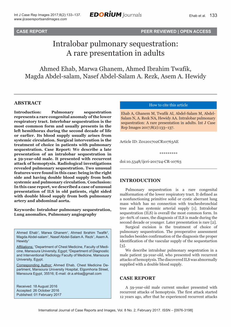
Intralobar pulmonary sequestration: A rare presentation in adults
Ahmed Ehab, Marwa Ghanem, Ahmed Ibrahim Twafik, Magda Abdel-salam, Nasef Abdel-Salam A. Rezk, Asem A. Hewidy
ABSTRACT
Introduction: Pulmonary sequestration represents a rare congenital anomaly of the lower respiratory tract. Interlobar sequestration is the most common form and usually presents in the left hemithorax during the second decade of life or earlier. Its blood supply usually arises from systemic circulation. Surgical intervention is the treatment of choice in patients with pulmonary sequestration. Case Report: We describe a late presentation of an intralobar sequestration in a 59-year-old male. It presented with recurrent attack of hemoptysis. Radiological investigations revealed pulmonary sequestration. Two unusual features were found in this case: being in the right side and having double blood supply from both systemic and pulmonary circulation. Conclusion: In this case report, we described a case of unusual presentation of ILS in old patients, right sided with double blood supply from both pulmonary artery and abdominal aorta.
(This page in not part of the published article.)
International Journal of Case Reports and Images, Vol. 8 No. 2, February 2017. ISSN – [0976-3198]
Int J Case Rep Images 2017;8(2):133–137. www.ijcasereportsandimages.com
Ehab et al. 133
CASE REPORT PEER REVIEWED | OPEN ACCESS
Intralobar pulmonary sequestration: A rare presentation in adults
Ahmed Ehab, Marwa Ghanem, Ahmed Ibrahim Twafik, Magda Abdel-salam, Nasef Abdel-Salam A. Rezk, Asem A. Hewidy
ABSTRACT
Introduction: Pulmonary sequestration represents a rare congenital anomaly of the lower respiratory tract. Interlobar sequestration is the most common form and usually presents in the left hemithorax during the second decade of life or earlier. Its blood supply usually arises from systemic circulation. Surgical intervention is the treatment of choice in patients with pulmonary sequestration. Case Report: We describe a late presentation of an intralobar sequestration in a 59-year-old male. It presented with recurrent attack of hemoptysis. Radiological investigations revealed pulmonary sequestration. Two unusual features were found in this case: being in the right side and having double blood supply from both systemic and pulmonary circulation. Conclusion: In this case report, we described a case of unusual presentation of ILS in old patients, right sided with double blood supply from both pulmonary artery and abdominal aorta.
Keywords: Intralobar pulmonary sequestration, Lung anomalies, Pulmonary angiography
Ahmed Ehab1, Marwa Ghanem1, Ahmed Ibrahim Twafik2, Magda Abdel-salam1, Nasef Abdel-Salam A. Rezk1, Asem A. Hewidy1
Affiliations: 1Department of Chest Medicine, Faculty of Medi-cine, Mansoura University, Egypt; 2Department of Diagnostic and Interventional Radiology Faculty of Medicine, Mansoura University, Egypt.Corresponding Author: Ahmed Ehab, Chest Medicine De-partment, Mansoura University Hospital, Elgomhoria Street, Mansoura Egypt, 35516; E-mail: [email protected]
Received: 18 August 2016Accepted: 26 October 2016Published: 01 February 2017
How to cite this article
Ehab A, Ghanem M, Twafik AI, Abdel-Salam M, Abdel-Salam N, A. Rezk NA, Hewidy AA. Intralobar pulmonary sequestration: A rare presentation in adults. Int J Case Rep Images 2017;8(2):133–137.
Article ID: Z01201702CR10763AE
*********
doi:10.5348/ijcri-201724-CR-10763
INTRODUCTION
Pulmonary sequestration is a rare congenital malformation of the lower respiratory tract. It defined as a nonfunctioning primitive solid or cystic aberrant lung mass which has no connection with tracheobronchial tree and has systemic arterial supply [1]. Intralobar sequestration (ILS) is overall the most common form. In 50–60% of cases, the diagnosis of ILS is made during the second decade or younger. Later presentation is rare [2].
Surgical excision is the treatment of choice of pulmonary sequestration. The preoperative assessment includes besides confirmation of the diagnosis the proper identification of the vascular supply of the sequestartion [3].
We describe intralobar pulmonary seqestration in a male patient 59-year-old, who presented with recurrent attacks of hemoptysis. The discovered ILS was abnormally supplied with a double blood supply.
CASE REPORT
A 59-year-old male current smoker presented with recurrent attacks of hemoptysis. The first attack started 12 years ago, after that he experienced recurrent attacks

International Journal of Case Reports and Images, Vol. 8 No. 2, February 2017. ISSN – [0976-3198]
Int J Case Rep Images 2017;8(2):133–137. www.ijcasereportsandimages.com
Ehab et al. 134
of blood-tinged sputum (average 3 attacks per year). The patient was first investigated in the primary health care facility where chest X-ray (Figure 1) was done and was interpreted as a case of bronchitis despite the obvious right para cardiac opacity. The patient was treated with supportive measures. Then the patient was admitted to a general hospital due to another attack of hemoptysis with syncope. Computed tomography scan of thorax with contrast revealed right lower lobe opacity with calcification (Figure 2). Due to the suspicious of malignancy, ultrasonographic-guided aspiration was done twice and the pathological examination revealed fibrous tissue with dilated vascular spaces. Finally, the patient was referred to our chest medicine department for further evaluation and bronchoscopy. After revision of his history and chest radiology, the pulmonary sequestration was suspected besides other benign lung lesions and CT angiography was ordered before any further interventions. It revealed evidence of right intralobular pulmonary sequestration. It has dual arterial supply from both right pulmonary artery and multiple branches from aorta arise, the venous drainage into right pulmonary vein (Figure 2 and Figure 3). Finally, the patient was transferred to the surgical department for surgical intervention, but the patient refused to perform any interventions.
DISCUSSION
Pulmonary sequestration was first described by Pryce in 1946. Its name was derived from the Latin verb ‘sequestrate’ which means to set apart. It can be defined as a developmental lung disease with non-functioning pulmonary tissue; which has no communication with the bronchial tree and receive a systemic blood flow [4].
Pulmonary sequestration is classified into three subtypes:
• Intralobar pulmonary sequestration (located within normal lung lobe and has its own visceral pleura),
• Extra lobar pulmonary (located outside the lung lobe and has its visceral pleura), and
• Bronchopulmonary- foregut malformation (BPFM) which is a rare variant of sequestration and is connected to the gastrointestinal tract [5].
The incidence of the pulmonary sequestration is rare and represented about 0.15–6.4% of all congenital pulmonary malformations [6]. Generally, ILS is the most common form of pulmonary sequestration. It represents about 75–90% with no sex differentiation [5].
The embryologic basis of the pulmonary sequestration remained unclear. Many possibilities had been suggested. The first referred to very early abnormality in the development during lung bud formation. Another
Figure 1: Chest X ray: Right paracardiac shadow.
Figure 2: (A) Computed tomography scan of chest with contrast reveled right lower opacity, and (B) Chest angiography with appearance of the aberrant blood supply of pulmonary sequestration.
Figure 3: Computed tomography angiography (volume rendering) show the arterial blood supply of pulmonary sequestration from both right pulmonary artery and multiple branches from aorta.

International Journal of Case Reports and Images, Vol. 8 No. 2, February 2017. ISSN – [0976-3198]
Int J Case Rep Images 2017;8(2):133–137. www.ijcasereportsandimages.com
Ehab et al. 135
theory suggested mechanical separation of a portion of the developing lung due to compression or traction by aberrant vascular structure or inadequate pulmonary blood flow. However, the mechanical theory cannot fully explain all types of the pulmonary sequestration especially BPFM [7]. The third theory suggested that ILS may be an acquired rather than developmental lesion [8]. Recently, researchers found that the abnormal vascular development in the pulmonary arterial blood supply can lead to retention and proliferation of the nascent systemic capillary network [1].
Intralobar sequestration (ILS) usually presents in patients less than 20 years of age in 50% of the patients and rarely found in patients older than 50 years. Lower lobe above the diaphragm is typically the mostly affected area and left side is common in 55–60% of the patients [2, 4].
Herein, we describe presentation of ILS in a male patient 59-year-old which is not only atypical due to the old age but also due to the atypical site in the right lower lobe. The time of ILS presentation is variable. It usually present in late childhood or adolescence. It presented with recurrent lower respiratory tract infection. Hemoptysis and chest pain were also reported. It may be also asymptomatic and discovered in routine chest radiology in 15.5% of the patients with ILS [6]. In rare cases, heart failure occurs due to high flow through the anomalous artery [1].
The radiological assessment aimed not only to confirm the sequestration diagnosis, but also to evaluate its vascular supply for further management. Besides the chest radiography, ultrasonography, computed tomography scan and magnetic resonance imaging scan, the relatively recent introduced angiography represent the diagnostic tool of choice to define the vascular supply prior to any surgical intervention [3]. Single feeding blood supply was detected in 71% of the patients. It arises from the thoracic aorta in 36% [6].
The intralobar sequestration can be classified according to Pryce et al. into:
• type 1 presence of aberrant artery without sequestration,
• type 2 the aberrant artery supply the sequestration as well as the adjacent normal lung and type
• type 3 the aberrant supply only the sequestration [2].
The main management of the pulmonary sequestration in symptomatic patient is surgical resection for curative purposes. Proper identification and ligation of the feeding vessels is crucial. While in asymptomatic patients with ILS, the surgery is also recommended to prevent recurrent infections and the unfavorable cardiac influence caused by the existing aortopulmonary shunt. Recently, published data has introduced the emerging successful role of video-assisted thoracoscopic surgery (VATS) for pulmonary sequestration resection despite the difficulties in surgical dissection due to recurrent inflammation and
fibrosis owing to the recurrent infections [9]. Arterial embolization of the feeding vessels has been also reported [10].
CONCLUSION
The interesting points in our case include the late presentation of the patient (59-year-old), unusual right-sided Intralobar sequestration (ILS) and the double blood supply from both pulmonary artery and abdominal aorta. Finally, we should emphasize the importance of proper interpretation of the chest radiology by the primary medical care providers as a standard tool of chest examination for early and adequate diagnosis of catastrophic hidden chest diseases.
*********
Author ContributionsAhmed Ehab – Substantial contributions to conception and design, Acquisition of data, Analysis and interpretation of data, Drafting the article, Revising it critically for important intellectual content, Final approval of the version to be publishedMarwa Ghanem – Analysis and interpretation of data, Revising it critically for important intellectual content, Final approval of the version to be publishedAhmed Ibrahim Twafik – Analysis and interpretation of data, Revising it critically for important intellectual content, Final approval of the version to be publishedMagda Abdel-salam – Analysis and interpretation of data, Revising it critically for important intellectual content, Final approval of the version to be publishedNasef Abdel-Salam A. Rezk – Analysis and interpretation of data, Revising it critically for important intellectual content, Final approval of the version to be publishedAsem A. Hewidy – Analysis and interpretation of data, Revising it critically for important intellectual content, Final approval of the version to be published
GuarantorThe corresponding author is the guarantor of submission.
Conflict of InterestAuthors declare no conflict of interest.
Copyright© 2017 Ahmed Ehab et al. This article is distributed under the terms of Creative Commons Attribution License which permits unrestricted use, distribution and reproduction in any medium provided the original author(s) and original publisher are properly credited. Please see the copyright policy on the journal website for more information.

International Journal of Case Reports and Images, Vol. 8 No. 2, February 2017. ISSN – [0976-3198]
Int J Case Rep Images 2017;8(2):133–137. www.ijcasereportsandimages.com
Ehab et al. 136
REFERENCES
1. Hertzenberg C, Daon E, Kramer J. Intralobar pulmonary sequestration in adults: Three case reports. J Thorac Dis 2012 Oct;4(5):516–9.
2. Hirai S, Hamanaka Y, Mitsui N, Uegami S, Matsuura Y. Surgical treatment of infected intralobar pulmonary sequestration: A collective review of patients older than 50 years reported in the literature. Ann Thorac Cardiovasc Surg 2007 Oct;13(5):331–4.
3. Kang M, Khandelwal N, Ojili V, Rao KL, Rana SS. Multidetector CT angiography in pulmonary sequestration. J Comput Assist Tomogr 2006 Nov-Dec;30(6):926–32.
4. Alizadeh E, Suliman H. Intralobular pulmonary sequestration. JBR-BTR 2013 Jul-Aug;96(4):208–9.
5. Stocker JT, Drake RM, Madewell JE. Cystic and congenital lung disease in the newborn. Perspect Pediatr Pathol 1978;4:93–154.
6. Van Raemdonck D, De Boeck K, Devlieger H, et al. Pulmonary sequestration: A comparison between pediatric and adult patients. Eur J Cardiothorac Surg 2001 Apr;19(4):388–95.
7. Kravitz RM. Congenital malformations of the lung. Pediatr Clin North Am 1994 Jun;41(3):453–72.
8. Holder PD, Langston C. Intralobar pulmonary sequestration (a nonentity?). Pediatr Pulmonol 1986 May-Jun;2(3):147–53.
9. Kestenholz PB, Schneiter D, Hillinger S, Lardinois D, Weder W. Thoracoscopic treatment of pulmonary sequestration. Eur J Cardiothorac Surg 2006 May;29(5):815–8.
10. Cho MJ, Kim DY, Kim SC, Kim KS, Kim EA, Lee BS. Embolization versus surgical resection of pulmonary sequestration: Clinical experiences with a thoracoscopic approach. J Pediatr Surg 2012 Dec;47(12):2228–33.
ABOUT THE AUTHORS
Article citation: Ehab A, Ghanem M, Twafik AI, Abdel-Salam M, Abdel-Salam N, A. Rezk NA, Hewidy AA. Intralobar pulmonary sequestration: A rare presentation in adults. Int J Case Rep Images 2017;8(2):133–137.
Ahmed Ehab is Assistant lecturer of chest medicine at chest medicine department, Faculty of medicine, Mansoura University, Mansoura, Egypt. He earned undergraduate degree (M.B.B.Ch) and postgraduate degree (MD in Chest Medicine) from from Mansoura University, Mansoura, Egypt. He has published two papers and one book. His research interests include bronchoscopic intervention, medical thoracoscopy, and respiratory critical care. E-mail: [email protected]
Magda Abdel-Salam is Chest medicine Assistant Professor at Chest medicine department, Faculty of medicine, Mansoura University, Mansoura, Egypt. She earned undergraduate degree (M.B.B.Ch) and postgraduate degree (MD in Chest Medicine) from Mansoura University, Mansoura, Egypt.E-mail: [email protected]
Marwa Ghanem is Chest medicine resident at Chest medicine department, Faculty of medicine, Mansoura University, Mansoura Egypt. She earned the undergraduate degree (M.B.B.Ch) from Mansoura University, Mansoura, Egypt). Her research interests include interventional bronchoscopy and respiratory critical care.E-mail: [email protected]
Ahmed Ibrahim Twafik is lecturer of diagnostic and interventional Radiology at Department of diagnostic and interventional radiology, faculty of medicine, Mansoura University, Mansoura, Egypt. He earned undergraduate degree (M.B.B.Ch) and postgraduate degree (MD in Radiology) from Mansoura University, Mansoura, Egypt. He has published one research paper and one book. His interest includes interventional radiology.E-mail: [email protected]
International Journal of Case Reports and Images, Vol. 8 No. 2, February 2017. ISSN – [0976-3198]
Int J Case Rep Images 2017;8(2):133–137. www.ijcasereportsandimages.com
Ehab et al. 137
Nasef Abdel-Salam A. Rezk is Chest medicine Assistant Professor at Chest Medicine Department, Faculty of medicine, Mansoura University, Mansoura, Egypt. He earned undergraduate degree (M.B.B.Ch) and postgraduate degree (MD in Chest Medicine) from Department of chest medicine, faculty of medicine, Mansoura University, Mansoura, Egypt. He has published ten research papers in national and international academic journals. E-mail: [email protected]
Asem A. Hewidy is Chest medicine Assistant Professor at Chest Medicine Department, Faculty of Medicine, Mansoura University, Mansoura, Egypt. He earned undergraduate degree (M.B.B.Ch) and postgraduate degree (MD in Chest Medicine) from Chest Medicine Department, Faculty of Medicine, Mansoura University, Mansoura, Egypt. He has published eleven research papers in national and international academic journals. His research interests include interventional bronchoscopy, and thoracoscopy. E-mail: [email protected]
Access full text article onother devices
Access PDF of article onother devices
EDORIUM JOURNALS AN INTRODUCTION
Edorium Journals: On Web
About Edorium JournalsEdorium Journals is a publisher of high-quality, open ac-cess, international scholarly journals covering subjects in basic sciences and clinical specialties and subspecialties.
Edorium Journals www.edoriumjournals.com
Edorium Journals et al.
Edorium Journals: An introduction
Edorium Journals Team
But why should you publish with Edorium Journals?In less than 10 words - we give you what no one does.
Vision of being the bestWe have the vision of making our journals the best and the most authoritative journals in their respective special-ties. We are working towards this goal every day of every week of every month of every year.
Exceptional servicesWe care for you, your work and your time. Our efficient, personalized and courteous services are a testimony to this.
Editorial ReviewAll manuscripts submitted to Edorium Journals undergo pre-processing review, first editorial review, peer review, second editorial review and finally third editorial review.
Peer ReviewAll manuscripts submitted to Edorium Journals undergo anonymous, double-blind, external peer review.
Early View versionEarly View version of your manuscript will be published in the journal within 72 hours of final acceptance.
Manuscript statusFrom submission to publication of your article you will get regular updates (minimum six times) about status of your manuscripts directly in your email.
Our Commitment
Favored Author programOne email is all it takes to become our favored author. You will not only get fee waivers but also get information and insights about scholarly publishing.
Institutional Membership programJoin our Institutional Memberships program and help scholars from your institute make their research accessi-ble to all and save thousands of dollars in fees make their research accessible to all.
Our presenceWe have some of the best designed publication formats. Our websites are very user friendly and enable you to do your work very easily with no hassle.
Something more...We request you to have a look at our website to know more about us and our services.
We welcome you to interact with us, share with us, join us and of course publish with us.
Browse Journals
CONNECT WITH US
Invitation for article submissionWe sincerely invite you to submit your valuable research for publication to Edorium Journals.
Six weeksYou will get first decision on your manuscript within six weeks (42 days) of submission. If we fail to honor this by even one day, we will publish your manuscript free of charge.*
Four weeksAfter we receive page proofs, your manuscript will be published in the journal within four weeks (31 days). If we fail to honor this by even one day, we will pub-lish your manuscript free of charge and refund you the full article publication charges you paid for your manuscript.*
This page is not a part of the published article. This page is an introduction to Edorium Journals and the publication services.
* Terms and condition apply. Please see Edorium Journals website for more information.