Embed Size (px)
Citation preview
J. Cell Sci. 22, 325-334 (1976) 325Printed in Great Britain
INTRANUCLEAR MEMBRANOUS INCLUSIONS
IN OOCYTES OF A VIVIPAROUS TELEOST
(XIPHOPHORUS HELLERI)
CARLOS AZEVEDO*
Department of Histology and Embryology,Faculty of Medicine of Oporto, Oporto, Portugal
SUMMARY
Intranuclear inclusions were observed in oocytes of Xiphophorus helleri during prophase I.In osmium-fixed leptotene nuclei, the inclusions were made up of groups of membrane-limitedvesicles or tubules with pale contents, situated near the inner nuclear membrane with whichsome of them exhibited apparent continuities. In zygotene nuclei, larger vesicles also appearedbounded by two or three membranes and containing tubules apparently invaginated from theirwalls. In pachytene-dictyate nuclei most vesicular bodies had a wall formed by stratified mem-branes, or were entirely made up of membranous whorls. In glutaraldehyde-osmium fixedmaterial some of these myelin-like bodies showed a peculiar arrangement, consisting of con-centric bands each containing thick inner dense lamellae 20-3-0 nm thick and a 50-nm outerlamella.
It is suggested that these inclusion bodies arise from the inner nuclear membrane of oocyteswhen cells start to grow intensely during prophase I. The bodies seem to become more complexat late prophase, probably by association of individual vesicles and the occurrence of multiplemembrane imaginations, which may be related to active metabolic phenomena taking placeat this stage in oocytes.
INTRODUCTION
During a study of oogenesis in Xiphophorus helleri, a viviparous Teleost (Azevedo,1974), membranous inclusions were observed in the nuclei of oocytes undergoingmeiotic prophase I. The inclusions were made up of simple vesicles or tubulo-vesicular composite structures at leptotene-zygotene stages. In pachytene, diploteneand dictyate cells, most vesicular bodies were bound by concentric membranes orentirely made up of membrane whorls. The evolution of these bodies through meiosisand the fact that the more complex ones occurred in late prophase when oocytesgrow intensely and are the sites of active metabolic phenomena suggested that thebodies might have a functional significance.
MATERIALS AND METHODS
We studied the ovaries of 18 normal females of Xiphophorus helleri Heckel (Poeciliidaefamily) from birth to 7 months of age. When animals are about 15 days old, oocytes startentering prophase I to reach sexual maturation at 7 months of age. During this time interval
* Assistant-professor in the Bio-Medical Institute of the Universiy of Oporto, Oporto,Portugal.
326 C. Azevedo
it is possible to find cells at the various stages of oogenesis in the ovary. Animals have beensacrificed at different times throughout the last 2 years without preference for any particularseason or month.
In 6 animals, the ovaries were fixed by immersion in 2 % osmium tetroxide in 01 M phos-phate buffer containing 005 % calcium chloride, pH 7-2, for 2 h at 4 °C. In the remaininganimals, the ovaries were fixed in 2 % glutaraldehyde in the same buffer for 2 h at 4 °C,washed overnight and postfixed in 2 % osmium tetroxide for 2 h at 4 °C. The pieces wereembedded in Araldite (Glauert & Glauert, 1958) or in Epon 812 (Luft, 1961). Semithin serialsections were cut and each one was immediately stained with methylene blue-azur II(Richardson, Jarett & Finke, i960). When intranuclear bodies appeared in a semithin section,ultrathin sections of the same block were then cut. These sections were double stained withaqueous 2-5% uranyl acetate for 20 min plus aqueous lead hydroxide (Karnovsky, 1961) for10 min or lead citrate (Reynolds, 1963) for 15 min, and examined in an electron microscopeJeol 100 B operated at 80 kV. The specimen was tilted up to 300 from the horizontal by meansof the manual goniometer stage IB-1004.
RESULTS
Osmium-fixed material
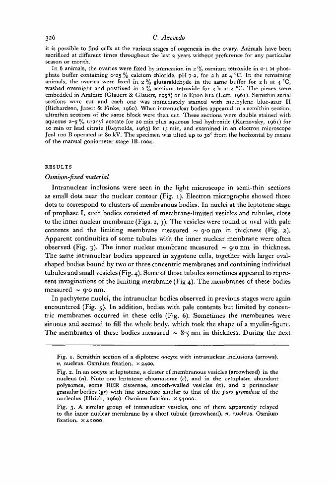
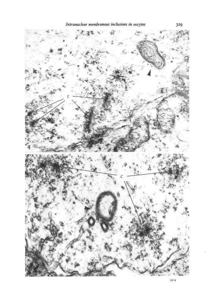
Intranuclear inclusions were seen in the light microscope in semi-thin sectionsas small dots near the nuclear contour (Fig. 1). Electron micrographs showed thosedots to correspond to clusters of membranous bodies. In nuclei at the leptotene stageof prophase I, such bodies consisted of membrane-limited vesicles and tubules, closeto the inner nuclear membrane (Figs. 2, 3). The vesicles were round or oval with palecontents and the limiting membrane measured ~ 9-0 nm in thickness (Fig. 2).Apparent continuities of some tubules with the inner nuclear membrane were oftenobserved (Fig. 3). The inner nuclear membrane measured ~ 9-0 nm in thickness.The same intranuclear bodies appeared in zygotene cells, together with larger oval-shaped bodies bound by two or three concentric membranes and containing individualtubules and small vesicles (Fig. 4). Some of those tubules sometimes appeared to repre-sent invaginations of the limiting membrane (Fig 4). The membranes of these bodiesmeasured ~ 9-0 nm.
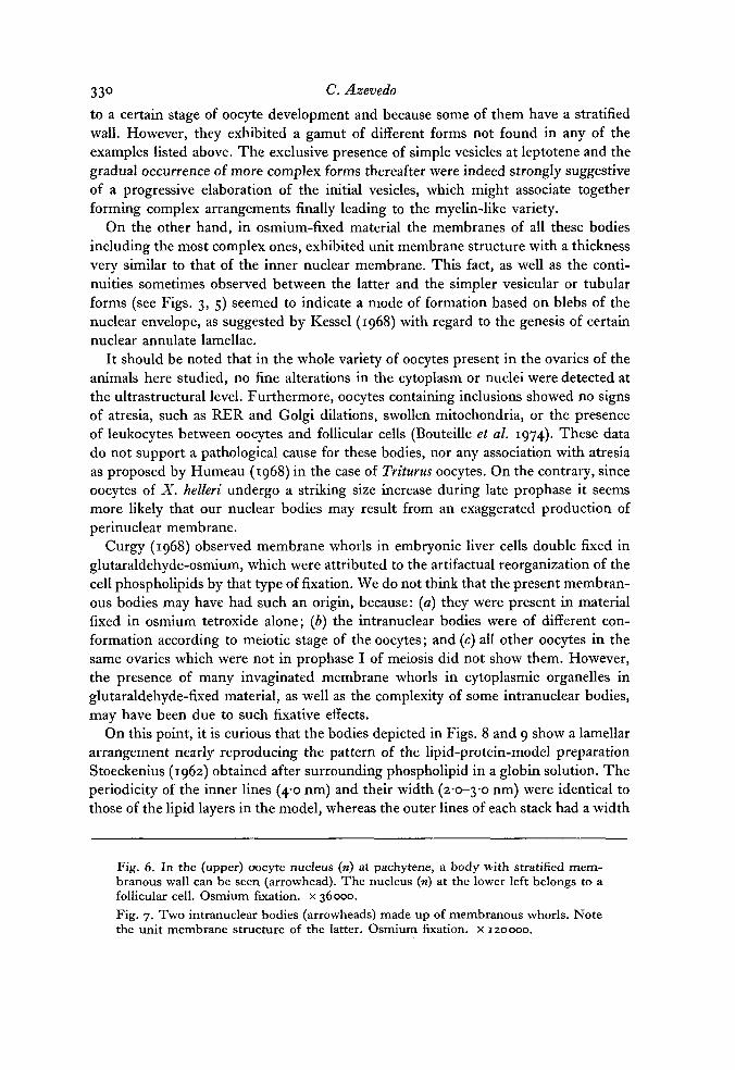
In pachytene nuclei, the intranuclear bodies observed in previous stages were againencountered (Fig. 5). In addition, bodies with pale contents but limited by concen-tric membranes occurred in these cells (Fig. 6). Sometimes the membranes weresinuous and seemed to fill the whole body, which took the shape of a myelin-figure.The membranes of these bodies measured ~ 8-5 nm in thickness. During the next
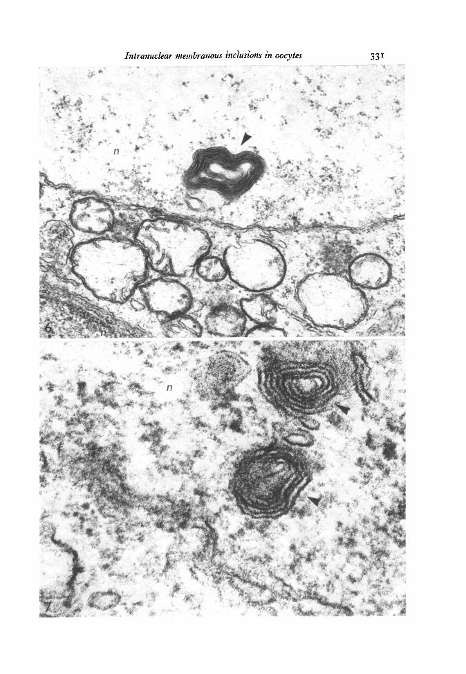
Fig. 1. Semithin section of a diplotene oocyte with intranuclear inclusions (arrows).n, nucleus. Osmium fixation, x 2400.Fig. 2. In an oocyte at leptotene, a cluster of membranous vesicles (arrowhead) in thenucleus (n). Note one leptotene chromosome (c), and in the cytoplasm abundantpolysomes, some RER cisternae, smooth-walled vesicles (v), and 2 perinucleargranular bodies {gr) with fine structure similar to that of the pars granulosa of thenucleolus (Ulrich, 1969). Osmium fixation, x 54000.Fig. 3. A similar group of intranuclear vesicles, one of them apparently relayedto the inner nuclear membrane by a short tubule (arrowhead), n, nucleus. Osmiumfixation, x AS 000.
Intranuclear membranous inclusions in oocytes 327
328 C. Azevedo
stages of diplotene and dictyate these myelin-like forms were often observed (Fig. 8)while simple vesicles, connected or not connected with the inner nuclear membrane,became rare. Intranuclear bodies were found in meiotic oocytes from all fish studied;they were absent from cells which were not undergoing meiosis.
Glutar aldehyde-osmium fixation
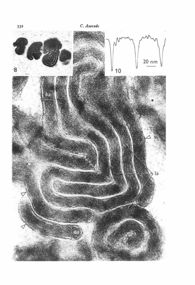
During leptotene-zygotene the images were the same as those observed in osmium-fixed material. In pachytene-dictyate cells some of the myelin-like bodies had a morecomplex arrangement of the membranes (Figs. 8, 9). These bodies were formed by4-6 curved concentric wide bands, 40-0-50-0 nm thick, separated by 4-0-8-0 nm spaces.At high magnification the bands were seen to consist of 8-14 dense lines alternatingwith light spaces. The 2 outer lines of each band were 5-0 nm thick, the inner ones2-0-3-0 nm and the intervening spaces 1-0-2-0 nm thick (Fig. 9). The lines werestacked within each band with a periodicity of 4-0 nm (Fig. 9).
Cells with nuclear inclusions, whatever the meiotic stage in which they werefound, in addition contained mitochondria with membranous whorls in the matrixor at the periphery; the membranes of these were continuous with the limiting mem-branes of the mitochondria. Similar whorls appeared in connexion with Golgi andRER cisternae, though these were much rarer. Such whorls were not found inosmium-fixed material. Intranuclear bodies were found in double-fixed oocytes of8 animals out of 12.
DISCUSSION
A number of intranuclear organelles, such as the ribonucleoproteic interchromatinand perichromatin granules or the proteinaceous simple nuclear bodies, appear to beof general occurrence in normal eukaryotic cells (Bouteille, Laval & Dupuy-Coin,1974; Monneron & Bernhard, 1969). Other nuclear structures have been shown onlyin certain cells and their presence seems to be highly dependent on the physiologicalstate of the cell. Among the latter are the intranuclear rodlets, frequent in neurons(Feldman & Peters, 1972) at certain development stages (Masurovsky, Benitez, Kim& Murray, 1970); the intranuclear annulate lamellae observed mainly in differentiatinggerm cells of invertebrates (Folliot, 1968; Hsu, 1967; Kessel, 1968); and inclusionsformed by concentric membranes recently described in young oocytes of Triturushelveticus (Humeau, 1968), in tumoral glial cells (Tripier, Berard & Toca, 1974) andin renal tubular cells of lead-poisoned rats (Franke & Scheer, 1974).
The bodies described here partly belong in this last category since they are confined
Fig. 4. A zygotene nucleus (n) with 2 chromosome pairs (c) and a membranous bodycontaining tubules (arrowhead). Osmium fixation, x 54000.Fig. 5. At pachytene, numerous simple vesicles and tubules close to the innernuclear membrane (*) and 3 vesicles (arrowhead) with 2 or 3 membranes at the walland a tubule in the interior of the larger vesicle. Of the 3 pairs of chromosomes (c)the synaptonemic complex is evident in the one situated on the right. Osmium fixation,x 37800.
Intranuclear membranous inclusions in oocytes 329
±*xArJft:&>>
.*. *-n\*a*
r
J» .'-
?^J• • ^ . • A v
KSfc..
330 C. Azevedo
to a certain stage of oocyte development and because some of them have a stratifiedwall. However, they exhibited a gamut of different forms not found in any of theexamples listed above. The exclusive presence of simple vesicles at leptotene and thegradual occurrence of more complex forms thereafter were indeed strongly suggestiveof a progressive elaboration of the initial vesicles, which might associate togetherforming complex arrangements finally leading to the myelin-like variety.
On the other hand, in osmium-fixed material the membranes of all these bodiesincluding the most complex ones, exhibited unit membrane structure with a thicknessvery similar to that of the inner nuclear membrane. This fact, as well as the conti-nuities sometimes observed between the latter and the simpler vesicular or tubularforms (see Figs. 3, 5) seemed to indicate a mode of formation based on blebs of thenuclear envelope, as suggested by Kessel (1968) with regard to the genesis of certainnuclear annulate lamellae.
It should be noted that in the whole variety of oocytes present in the ovaries of theanimals here studied, no fine alterations in the cytoplasm or nuclei were detected atthe ultrastructural level. Furthermore, oocytes containing inclusions showed no signsof atresia, such as RER and Golgi dilations, swollen mitochondria, or the presenceof leukocytes between oocytes and follicular cells (Bouteille et al. 1974). These datado not support a pathological cause for these bodies, nor any association with atresiaas proposed by Humeau (1968) in the case of Triturus oocytes. On the contrary, sinceoocytes of X. helleri undergo a striking size increase during late prophase it seemsmore likely that our nuclear bodies may result from an exaggerated production ofperinuclear membrane.
Curgy (1968) observed membrane whorls in embryonic liver cells double fixed inglutaraldehyde-osmium, which were attributed to the artifactual reorganization of thecell phospholipids by that type of fixation. We do not think that the present membran-ous bodies may have had such an origin, because: (a) they were present in materialfixed in osmium tetroxide alone; (b) the intranuclear bodies were of different con-formation according to meiotic stage of the oocytes; and (c) all other oocytes in thesame ovaries which were not in prophase I of meiosis did not show them. However,the presence of many invaginated membrane whorls in cytoplasmic organelles inglutaraldehyde-fixed material, as well as the complexity of some intranuclear bodies,may have been due to such fixative effects.
On this point, it is curious that the bodies depicted in Figs. 8 and 9 show a lamellararrangement nearly reproducing the pattern of the lipid-protein-model preparationStoeckenius (1962) obtained after surrounding phospholipid in a globin solution. Theperiodicity of the inner lines (4-0 nm) and their width (2-0-3-o nm) were identical tothose of the lipid layers in the model, whereas the outer lines of each stack had a width
Fig. 6. In the (upper) oocyte nucleus (n) at pachytene, a body with stratified mem-branous wall can be seen (arrowhead). The nucleus (n) at the lower left belongs to afollicular cell. Osmium fixation, x 36000.Fig. 7. Two intranuclear bodies (arrowheads) made up of membranous whorls. Notethe unit membrane structure of the latter. Osmium fixation, x 120000.
Intranuclear membranous inclusions in oocytes
•*. -•&•: i_. - -*& \.^ >! *•', -' •'.'".i9(f*'if
332 C. Azevedo
Intranuclear membranous inclusions in oocytes 333
(~ 5-0 nm) comparable to that of the protein film absorbed on to the outermost phos-pholipid layers in the Stoeckenius (1962) preparation. Although this elaborate arrange-ment may be a consequence of a glutaraldehyde artifact, it may also be argued thatonly glutaraldehyde would be capable of adequately preserving the protein com-ponents of the outer layer of such bands.
Finally, it should be emphasized that the larger and more elaborate bodies occurredat late prophase when cells not only grow rapidly, but also start storing lipid andprotein yolk, while intense cytoplasmic exchanges of nucleolar material are indicatedby the occurrence of perinuclear extrusion bodies (Fig. 6). The intranuclear bodiesdescribed here may thus result not only from exaggerated membrane growth, butmay also represent special sites of metabolic phenomena taking place at this particulartime in the life of the cell.
We would like to express our gratitude to Professor A. Coimbra for receiving us in his Depart-ment and for his valuable suggestions and guidance. We thank Dr A. Gautier (Universityof Lausanne, Switzerland) for his criticisms, and Professor R. Salema (Institute of Botany,University of Oporto) for making the microdensitometer readings. This work was supportedby Project PMC-3 from the Instituto de Alta Cultura, Lisbon, Portugal.
REFERENCESAZEVEDO, C. (1974). Evolution des enveloppes ovocytaires, au cours de l'ovogenese, chez un
Teleosteen vivipare, Xiphophorus helleri. jf. Microscopie 21, 43-54.BOUTEILLE, M., LAVAL, M. & DUPUY-COIN, A. M. (1974). Nuclear structure. Localization
of nuclear functions as revealed by ultrastructural autoradiography and cytochemistry.In The Cell Nucleus, vol. 1 (ed. H. Busch), pp. 5-71. New York and London: AcademicPress.
CURGY, J.-J. (1968). Influence du mode de fixation sur la possibility d'observer des estructuresmyeliniques dans les hepatocytes d'embryons de poulet. J. Microscopie 7, 63-80.
FELDMAN, M. L. & PETERS, A. (1972). Intranuclear rods and sheets in rat cochlear nucleus.J. Neurocytology 1, 109-127.
FOLLIOT, R. (1968). Les lamelles annelees intranucleaires des cellules du tissu germinal maleavant la meiose chez Philaenus spumarius L. (Insecte Homoptere). Z. Zellforsch. mikrosk.Anat. 92, 115-129.
FRANKE, W. W. & SCHEER, M. (1974). Nuclear structure. Structures and functions of thenuclear envelope. In The Cell Nucleus, vol. 1 (ed. H. Busch), pp. 219-347. New York andLondon: Academic Press.
GLAUERT, A. M. & GLAUERT, R. H. (1958). Araldite as an embedding medium for electronmicroscopy. J. biophys. biochem. Cytol. 4, 191-194.
Hsu, W. S. (1967). The origin of annulate lamellae in the oocyte of the ascidian, Bolteniavillosa Stimpson. Z. Zellforsch. mikrosk. Anat. 82, 376-390.
Fig. 8. Three inclusion bodies in a dictyate nucleus formed by concentric dense bands.Glutaraldehyde-osmium fixation, x 33000.Fig. 9. High magnification of the same type of bodies depicted in the anterior figureshowing the wide bands (6a) composed of tightly dense lines (/a) the outer ones ofwhich are thicker (hollow arrowheads). A grazing section shows blurred membraneprofiles (*). Glutaraldehyde-osmium fixation, x 225000.Fig. 10. Densitometric reading of the bands, each small peak corresponding to adense line.
334 C. Azevedo
HUMEAU, C. (1968). Formations membranaires dans le noyau et le cytoplasme perinucleairedue jeune ovocyte de Triturus helveticus Raz. C.r. Seanc. Soc. Biol. 162, 183-186.
KARNOVSKY, M. J. (1961). Simple methods for 'staining' with lead at high pH in electronmicroscopy. .7. biophys. biochem. Cytol. n , 729-732.
KESSEL, R. G. (1968). Annulate lamellae. J. Ultrastruct. Res., Suppl. 10, 1-82.LUFT, J. H. (1961). Improvements in epoxy embedding methods. J. biophys. biochem. Cytol. 9,
409-414.MASUROVSKY, E. B., BENITEZ, H. H., KIM, S. U. & MURRAY, M. R. (1970). Origin, develop-
ment, and nature of intranuclear rodlets and associated bodies in chicken sympatheticneurons. J. Cell Biol. 44, 172-191.
MONNERON, A. & BERHNARD, W. (1969). Fine structual organization of the interphase nucleusin some mammalian cells. J. Ultrastruct. Res. 27, 266-288.
REYNOLDS, E. S. (1963). The use of lead citrate at high pH as an electron-opaque stain inelectron microscopy. J. Cell Biol. 17, 208-212.
RICHARDSON, K. C, JARETT, T. & FINKE, E. H. (i960). Embedding in epoxy resins for ultra-thin sectioning in electron microscopy. Stain Technol. 35, 313-325.
STOECKENIUS, W. (1962). The molecular structure of lipid-water systems and cell membranemodels studied with the electron microscope. In The Interpretation of Ultrastructure, vol. 1(ed. R. J. C. Harris), pp. 349-367. New York and London: Academic Press.
TRIPIER, M.-F., BERARD, M. & TOCA, M. (1974). 'Membranous nuclear bodies' as possibleintermediate structure between nuclear bodies and invaginations of the nuclear envelope.J. Microscopie 21, 21-30.
ULRICH, E. (1969). fitude des ultrastructures au cours de l'ovogenese d'un poisson teleosteen,le Danio, Brachydanio rerio. J. Microscopie 8, 447-478.
{Received 26 March 1976)
![Intranuclear Compartmentalization of Cyclin E …...[CANCER RESEARCH 61, 1220–1226, February 1, 2001] Intranuclear Compartmentalization of Cyclin E during the Cell Cycle: Disruption](https://img.pdfslide.net/doc/110x75/5f63db5b44239533cf1f413c/intranuclear-compartmentalization-of-cyclin-e-cancer-research-61-1220a1226.jpg)