Embed Size (px)
Citation preview
THIEME
603
Intraoperative Computed Tomography Scanner-Guided Craniovertebral Junction Surgery in a Patient with an Occipitalized C1Mohammad Ashraf1,2, Usman Ahmad Kamboh2 Naveed Ashraf2
1Wolfson School of Medicine, University of Glasgow, Scotland, United Kingdom
2Department of Neurosurgery, Allama Iqbal Medical College, Jinnah Hospital Lahore, Lahore, Pakistan
Address for correspondence Mohammad Ashraf, BMedSci (Hons), 33 Glenmill Crescent, Glasgow G53-7HL, United Kingdom (e-mail: [email protected]).
Craniovertebral junction surgery is associated with unique difficulties. Type 2 odontoid fractures (Anderson and D Alonzo) have a great potential for nonunion and malunion. These fracture patients may require a circumferential decompression and fixation. The addition of intraoperative CT with neuronavigation greatly aids in craniovertebral junc-tion surgery. We operated on a 59-year-old-male with a type 2 fracture with posterior subluxation of C1 anterior arch and a cranially displaced odontoid peg. First, a tran-soral odontoidectomy was performed followed by a craniocervical fixation. Occipital plates and C3–C4 lateral mass screws were used as C1 was discovered to be occipi-talized intraoperatively and atlantoaxial facet joints could not be reduced as discov-ered by intraoperative CT resconstruction. Intraoperative CT scan was crucial to this circumferential decompression and fixation, allowed us to resect the odontoid peg safely and completely and to confirm adequate screw trajectory making this complex surgery easier for us and safer for the patient. The patient was discharged 4 months after admission with stable neurology. Intraoperative CT was fundamental to correct decision making.
Abstract
DOI https://doi.org/ 10.1055/s-0041-1730088 ISSN 0976-3147
© 2021. Association for Helping Neurosurgical Sick People.This is an open access article published by Thieme under the terms of the Creative Commons Attribution-NonDerivative-NonCommercial-License, permitting copying and reproduction so long as the original work is given appropriate credit. Contents may not be used for commercial purposes, or adapted, remixed, transformed or built upon. (https://creativecommons.org/licenses/by-nc-nd/4.0/).Thieme Medical and Scientific Publishers Pvt. Ltd. A-12, 2nd Floor, Sector 2, Noida-201301 UP, India
IntroductionInjuries to the craniovertebral (CV) junction account for a small proportion of spinal cord injuries, are usually associ-ated with high energy trauma and affect the skull base at the occipital bone and the C1 and C2 vertebrae.1,2 The area is anatomically complex, its injuries peculiar and instability of this area can have devastating neurological consequences. As such spinal surgery in this area and its consideration are different from subaxial cervical spinal surgery.2
The Anderson and D Alonzo type II fracture occurs at the base of the odontoid process or superior to the junction with the vertebral body.3 These fractures are usually unstable, and surgery indicated as nonunion rates between 40 and 80% is reported.4,5 We present a case of a 59-year-old male patient who presented with a CV junction instability that indicated an anterior decompression and posterior fixation. We report the use of our intraoperative CT (iCT) scanner with neuronav-igation and its benefit in such a procedure.
J Neurosci Rural Pract 2021;12:603–607.
Keywords ► intraoperative CT scanner ► type 2 odontoid fracture ► occipitalized C1 ► craniocervical junction surgery ► craniovertebral junc-tion surgery
Case Report
published onlineMay 7, 2021
Published online: 2021-05-07
604
Journal of Neurosciences in Rural Practice Vol. 12 No. 3/2021 © 2021. Association for Helping Neurosurgical Sick People
Role of Intraoperative CT Scanner in CV Junction Surgery Ashraf et al.
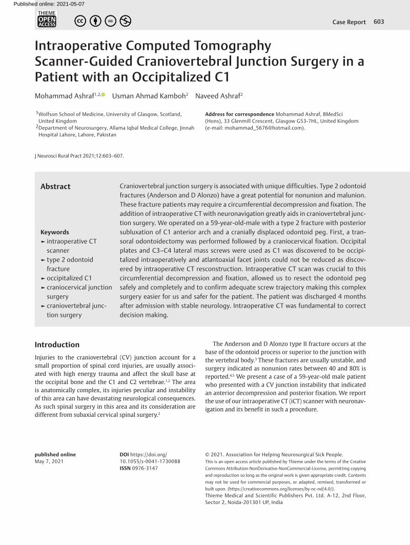
Case HistoryA 59-year-old male presented to our outpatient clinic com-plained of neck pain, quadriparesis, and right-sided torticol-lis. He was involved in a fall from his donkey cart one and a half month prior in a road traffic accident. There was spon-taneous movement in all four limbs and neurological exam-ination revealed intact sensation and normal reflexes, 2+ (Deep Tendon Reflex scale), with motor power of four out of five in all limbs (MRC grading scale). Tone, sensation and gait were intact. Plantar reflex was normal. The patient brought his only previous investigation, a lateral X-ray of cervical spine which revealed instability at the CV junction with a posteriorly displaced anterior arch of C1 and invisible odon-toid peg which was fractured from the body of C2 (►Fig. 1).
Upon admission to our department, Philadelphia Collar was applied the first day followed by skull traction for 2 weeks. After the initial 2 weeks he developed a pin site
infection, the traction was removed, and 1 g of ceftriaxone was initiated twice daily. Upon resolution of infection trac-tion was reapplied at a different site for 1 week. Piroxicam 2 mg twice daily was administered as an analgesic and tizan-idine 2 mg twice daily as a muscle relaxant.
CT scan of the cervical spine with three-dimensional reconstruction revealed a C2 type II fracture (Anderson and D Alonzo) with an anterior and cranial right-sided dis-placement of the odontoid peg and atlantoaxial subluxation causing cervical myelopathy. MRI of cervical spine showed compression and thinning of the cervical spinal cord. The odontoid fracture warranted an anterior decompression and an occipital-cervical fixation which was planned as a two-stage procedure.
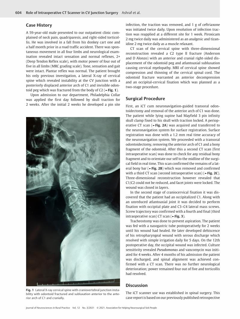
Surgical ProcedureFirst, an iCT cum neuronavigation-guided transoral odon-toidectomy and removal of the anterior arch of C1 was done. The patient while lying supine had Mayfield 3 pin infinity skull clamp fixed to his skull with traction locked. A periop-erative CT scan (►Fig. 2A) was acquired and transferred to the neuronavigation system for surface registration. Surface registration was done with a 1.2 mm real time accuracy of the neuronavigation system. We proceeded with a transoral odontoidectomy, removing the anterior arch of C1 and a bony fragment of the odontoid. After this a second CT scan (first intraoperative scan) was done to check for any residual bony fragment and to orientate our self to the midline of the surgi-cal field in real time. This scan confirmed the remains of a lat-eral bony bar (►Fig. 2B) which was removed and confirmed with a third CT scan (second intraoperative scan) (►Fig. 2C). Three-dimensional reconstruction however revealed that C1/C2 could not be reduced, and facet joints were locked. The wound was closed in layers.
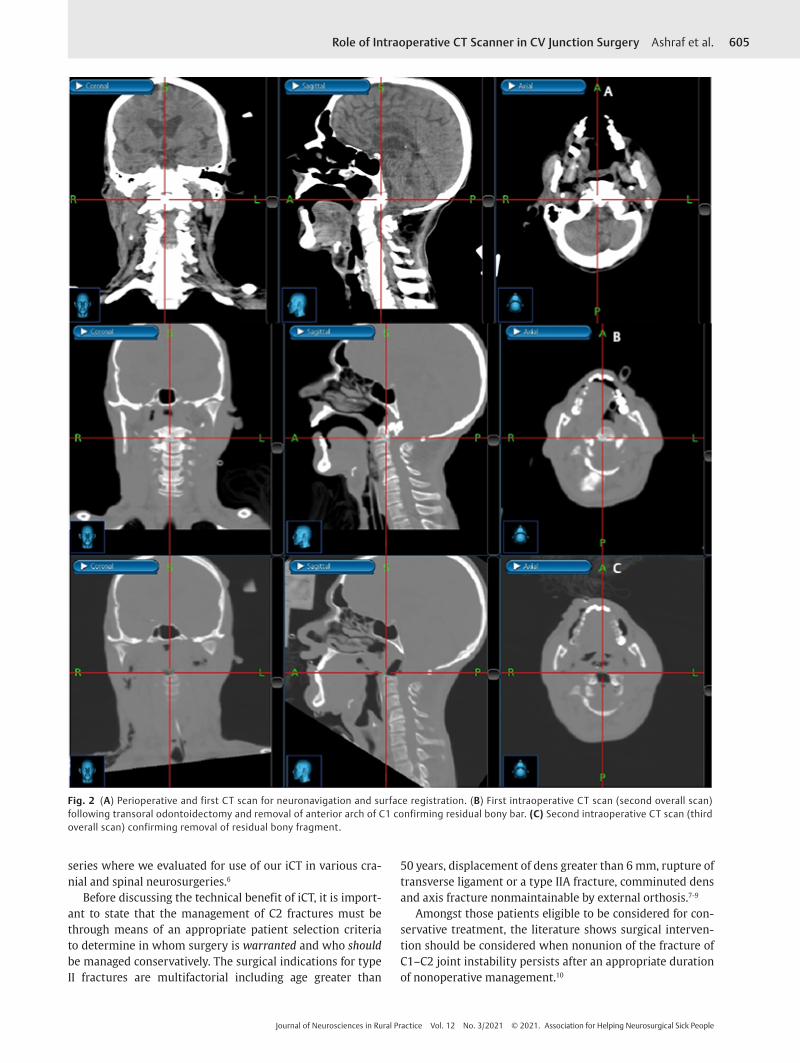
In the second stage of craniocervical fixation it was dis-covered that the patient had an occipitalized C1. Along with an unreduced atlantoaxial joint it was decided to perform fixation with occipital plate and C3–C4 lateral mass screws. Screw trajectory was confirmed with a fourth and final (third intraoperative scan) CT scan (►Fig. 3).
Tracheostomy was done to prevent aspiration. The patient was fed with a nasogastric tube postoperatively for 2 weeks until his wound had healed. He later developed dehiscence of his retropharyngeal wound with serous discharge which resolved with simple irrigation daily for 5 days. On the 12th postoperative day, the occipital wound was infected. Culture sensitivity revealed Pseudomonas and vancomycin was initi-ated for 4 weeks. After 4 months of his admission the patient was discharged, and spinal alignment was achieved con-firmed with a CT scan. There was no further neurological deterioration; power remained four out of five and torticollis had resolved.
DiscussionThe iCT scanner use was established in spinal surgery. This case report is based on our previously published retrospective
Fig. 1 Lateral X-ray cervical spine with craniovertebral junction insta-bility with odontoid fractured and subluxation anterior to the ante-rior arch of C1 and cranially.
605Role of Intraoperative CT Scanner in CV Junction Surgery Ashraf et al.
Journal of Neurosciences in Rural Practice Vol. 12 No. 3/2021 © 2021. Association for Helping Neurosurgical Sick People
series where we evaluated for use of our iCT in various cra-nial and spinal neurosurgeries.6
Before discussing the technical benefit of iCT, it is import-ant to state that the management of C2 fractures must be through means of an appropriate patient selection criteria to determine in whom surgery is warranted and who should be managed conservatively. The surgical indications for type II fractures are multifactorial including age greater than
50 years, displacement of dens greater than 6 mm, rupture of transverse ligament or a type IIA fracture, comminuted dens and axis fracture nonmaintainable by external orthosis.7-9
Amongst those patients eligible to be considered for con-servative treatment, the literature shows surgical interven-tion should be considered when nonunion of the fracture of C1–C2 joint instability persists after an appropriate duration of nonoperative management.10
Fig. 2 (A) Perioperative and first CT scan for neuronavigation and surface registration. (B) First intraoperative CT scan (second overall scan) following transoral odontoidectomy and removal of anterior arch of C1 confirming residual bony bar. (C) Second intraoperative CT scan (third overall scan) confirming removal of residual bony fragment.
606
Journal of Neurosciences in Rural Practice Vol. 12 No. 3/2021 © 2021. Association for Helping Neurosurgical Sick People
Role of Intraoperative CT Scanner in CV Junction Surgery Ashraf et al.
The iCT was of great help and a reassurance during the transoral approach to the odontoid. While operating in a narrow space and dealing with the complex anatomy of the CV junction which is particularly important due to the prox-imity of neurovascular structures, the accuracy required is within millimeters. This is especially so because of the ver-tebral artery in relation to the C1 and C2 vertebral bodies, the position of which may have been displaced because of the subluxation of these vertebrae. The incidence of verte-bral artery damage overall is 1.4% in cervical spinal surgery; however, it can be as high as 8% when performing posterior instrumented upper cervical spinal surgery.11,12 Although some authors advocate a perioperative CT angiogram to chart the course of V3 segment of the vertebral artery as it frequently has an anomalous course at C1/C2,13 but in our case the articular processes of C1 were not purchased and an occipio-C3/C4 fusion was done. Therefore, the need for a perioperative charting was obviated as the vertebral artery is not compromised here.14
Additionally, the iCT allowed us to detect a residual bony fragment and confirm its resection (2b and 2c). Had this not been removed it may have led to the persistence of cervical cord compression and the highly impaired chances of neu-rological stability or recovery. During the posterior fixation, the iCT allowed us to confirm screw trajectory thus achieving complete and safe stabilization of the cervical spine (►Fig. 3). It may be added that this case report warrants further stud-ies to decide whether iCT should be a standard of care in CV junction instabilities and if such patients should be managed at centers equipped with iCT.
ConclusionCenters equipped with iCT scan and neuronavigation have an added benefit to operate in a safer environment and allow for greater precision and better accuracy, especially in the complicated anatomical region of the craniocervical junc-tion. However, to affirm this, larger trials with rigid selection criteria are required to establish just how safe the iCT is and how much benefits are derived from it in CV junction spinal surgery.
Authors’ ContributionsM.A. wrote the first draft and followed up patient for missing data. U.A.H. collected the patient data, performed surgery, and wrote the first draft. N.A. performed surgery, revised the first draft to its final form.
Ethical ApprovalAll procedures performed in studies involving human par-ticipants were in accordance with the ethical standards of the institutional and/or national research committee and with the 1964 Helsinki declaration and its later amend-ments or comparable ethical standards. For this type of study formal ethical review was not required by the insti-tution. The authors certify that they have obtained all appropriate written informed consent.
FundingNone.
Conflict of InterestNone declared.
Fig. 3 Final intraoperative CT scan confirming appropriate screw trajectory and adequate decompression. Top, sagittal sections; bottom, axial sections; right, coronal section.
607Role of Intraoperative CT Scanner in CV Junction Surgery Ashraf et al.
Journal of Neurosciences in Rural Practice Vol. 12 No. 3/2021 © 2021. Association for Helping Neurosurgical Sick People
References
1 Clark JG, Abdullah KG, Mroz TE, Steinmetz MP. Biomechanics of the Craniovertebral Junction. Biomechanics in Applications; 2013. Accessed March 15, 2018 at: https://www.intechopen.com/books/biomechanics-in-applications/biomechanics- of-thecraniovertebral-junction
2 Joaquim AF, Patel AA. Craniocervical traumatic injuries: evaluation and surgical decision making. Global Spine J 2011;1(1):37–42
3 Anderson LD, D’Alonzo RT. Fractures of the odontoid process of the axis. J Bone Joint Surg Am 1974;56(8):1663–1674
4 Robinson Y, Robinson AL, Olerud C. Systematic review on sur-gical and nonsurgical treatment of type II odontoid fractures in the elderly. BioMed Res Int 2014;2014:231948
5 Iyer S, Hurlbert RJ, Albert TJ. Management of odontoid fractures in the elderly: a review of the literature and an evidence-based treatment algorithm. Neurosurgery 2018;82(4):419–430
6 Ashraf M, Choudhary N, Hussain SS, Kamboh UA, Ashraf N. Role of intraoperative computed tomography scanner in modern neurosurgery—an early experience. Surg Neurol Int 2020;11:247
7 Aldrian S, Erhart J, Schuster R, et al. Surgical vs nonop-erative treatment of Hadley type IIA odontoid fractures. Neurosurgery 2012;70(3):676–682, discussion 682–683
8 Greene KA, Dickman CA, Marciano FF, Drabier JB, Hadley MN, Sonntag VK. Acute axis fractures. Analysis of management and outcome in 340 consecutive cases. Spine 1997;22(16):1843–1852
9 Tanki H, Wani AA, Ramzan AU, et al. Conservative manage-ment of craniovertebral junction injuries: still a good option. Surg Neurol Int 2017;8:43
10 Landells CD, Van Peteghem PK. Fractures of the atlas: classifi-cation, treatment and morbidity. Spine 1988;13(5):450–452
11 Rampersaud YR, Moro ER, Neary MA, et al. Intraoperative adverse events and related postoperative complications in spine surgery: implications for enhancing patient safety founded on evidence-based protocols. Spine 2006;31(13):1503–1510
12 Wright NM, Lauryssen C; American Association of Neurological Surgeons/Congress of Neurological Surgeons. Vertebral artery injury in C1-2 transarticular screw fixation: results of a survey of the AANS/CNS section on disorders of the spine and periph-eral nerves. J Neurosurg 1998;88(4):634–640
13 Ekşi MŞ, Toktaş ZO, Yılmaz B, et al. Vertebral artery loops in surgical perspective. Eur Spine J 2016;25(12):4171–4180
14 Khanfour AA, El Sekily NM. Relation of the vertebral artery segment from C1 to C2 vertebrae: an anatomical study. Alexandria J Med 2015;51(2):143–151