Embed Size (px)
Citation preview
Rev Col Bras Cir 2017; 44(6): 603-611
DOI: 10.1590/0100-69912017006010
Intravitreal injection of polysorbate 80: a functional and morphological study
Injeção intravítrea de polissorbato 80: estudo funcional e morfológico
Francisco Max DaMico1; Fábio Gasparin1; Gabriela lourençon ioshiMoto2; thais ZaMuDio iGaMi1; arManDo Da silva cunha Jr.3; silvia liGorio Fialho4; anDre Mauricio liber2; lucy hwa-yue younG5; Dora Fix ventura2.
INTRODUCTION
Drug access to the retina and choroid has always
been a challenge to ophthalmologists due to the
existence of two anatomic barriers (internal and external
blood-retinal barriers) that impairs penetration of drugs
in the posterior segment of ocular bulbus. Treatment of
blindness secondary to most prevalent retina and cho-
roid diseases (macular degeneration related to age and
diabetic retinopathy) has changed dramatically with the
use of intravitreal injection of therapeutic agents in the
posterior segment of ocular bulbus1. Intravitreal injec-
tion of drugs overcomes external blood-retinal barrier
and assures that retina and choroid receive therapeutic
level of drugs, lowering significantly systemic absorp-
tion and consequent toxicity. According to Brazilian and
World legislation, intravitreal injection of drugs is a sur-
gical procedure and must be performed under rigorous
aseptic technique.
Most commonly injected drugs in the vitreous
are monoclonal antibodies (particularly inhibitors of the
vascular endothelium growth factor), corticosteroids
and antibiotics, but, in theory, any drug can be injec-
ted in the vitreous. However, some pharmacological
aspects must be considered, such as the aqueous so-
lubility, pharmacokinetics and biochemical proprieties
of the compounds, as well as their interaction with the
vitreous2.
Polysorbates, a class of non-ionic surfactants,
are very useful excipients in several pharmaceutic for-
mulations for intravenous use with different objectives.
Polysorbates increase drug solubility in suspensions with
low or no-solubility, to obtain aqueous dispersions. In
those cases, surfactant concentration varies from 0.05%
to 0.5%, depending on the solid content of formulation.
Polysorbates also are used in the formula of injectable
solutions to increase absorption of soluble drugs due to
micelle formation. Also, polysorbates are useful to sta-
1 - USP Medical School, Department of Ophthalmology and Otolaryngology, São Paulo, SP, Brazil. 2 - USP Institute of Psychology, Department of Experimental Psychology, São Paulo, SP, Brazil. 3 - UFMG School of Pharmacy, Department of Pharmaceutic Products, Belo Horizonte, MG, Brazil. 4 - Fundação Ezequiel Dias, Technologic Pharmaceutic Development Division, Belo Horizonte, MG, Brazil. 5 - Harvard Medical School, Department of Ophthalmology, Boston, MA, USA.
Original Article
A B S T R A C T
Objective: to determine the functional and morphological effects at rabbits retina of PS80 concentration used in the preparation of intravit-
real drugs. Methods: eleven New Zealand rabbits received a intravitreal injection of 0.1ml of PS80. As control, the contralateral eye of each
rabbit received the same volume of saline. Electroretinography was performed according to a modified protocol, as well as biomicroscopy
and retina mapping before injection and seven and ten days after. Animals were euthanized in the 30th day and the retinas were analyzed
by light microscopy. Results: eyes injected with PS80 did not present clinical signs of intraocular inflammation. Electroretinography did not
show any alteration of extent and implicit time of a and b waves at scotopic and photopic conditions. There were no morphological alter-
ations of retinas at light microscopy. Conclusion: intravitreal injection of PS80 in the used concentration for intravitreal drug preparations
do not cause any functional or morphological alterations of rabbit retinas. These results suggest that PS80 is not toxic to rabbit retinas and
may be safely used in the preparation of new lipophilic drugs for intravitreal injection.
Keywords: Polysorbates. Retina. Electroretinography. Intravitreal Injections. Morphological and Microscopic Findings.
DamicoIntravitreal injection of polysorbate 80: a functional and morphological study604
Rev Col Bras Cir 2017; 44(6): 603-611
bilize proteins in formulas with monoclonal antibodies.
Virtually, all formulas contain polysorbate 20 or 80.
Polysorbate 80, also known as polyoxietilen-
sorbitan-20 mono-oleate or Tween 80® (MW: 428.60,
FM: C24H44O6, aqueous solubility: 5-10g/100ml at
23oC) is a polysorbate used to stabilize aqueous formu-
lations of drugs used topically, intravenously and intra-
vitreal. It is also a solubilizer used in eye drops and an
important component of lipophilic suspended drugs.
Safety of systemic use of PS80 is controversial.
PS80 has no neurotoxicity in newborn rats following ad-
ministration of high oral doses during pregnancy, and
do not cause development disturbances, functional alte-
rations of central nervous system, and alterations of lo-
comotion or of reflexes3. In adult animals, oral intake of
high doses of PS80 is safe in mice, rats, dogs and apes4.
However, intraperitoneal injection of PS80 in newborn
female rats cause morphological and functional altera-
tions of uterus and ovaries5. Also, PS80 may be asso-
ciated to non-immune anaphylactic reaction, following
intravenous administration during pregnancy6.
PS80 effects on the eye surface were studied
in several experimental models. Sub-tenon injection
of PS80 in rabbits caused less toxicity in eye surface
than other commonly used excipients commonly used
in topic formulations for ocular use, such as carboxy-
methylcellulose, polyethylene glycol, benzylic alcohol,
benzalkonium chloride and methylcellulose7. PS80 also
seems to have a protective mechanism in the corneal
epithelium of cells maintained in culture, reducing the
toxicity induced by benzalkonium chloride, a commonly
used excipient used in eye drops8,9.
Formula most used commercially of triamcino-
lone acetonide (TA) contains PS80. TA is a synthetic glu-
cocorticoid with long-lasting effect that has been widely
used in the treatment of retinal diseases by intravitreal
injection, but safety studies show controversial results.
Some in vivo experimental studies suggest that TA intra-
vitreal injection, which formula contains PS80, is safe10-
12. However, other experimental studies suggest that TA
formulation without preservative is less toxic to retina
after intravitreal injection than most common formu-
las13-15. Since TA vehicle formulation has many compou-
nds, such as benzylic alcohol, carboxy-methylcellulose,
PS80, sodium hydroxide and hydrochloric acid, the role
of each compound in retinal toxicity is still uncertain16-18.
Although PS80 is frequently used in the pre-
paration of formulations for ocular use, including drugs
for intravitreal use, its effect on retina after intravitreal
injection has never been studied. The objective of the
present study is to determine functional and morpho-
logical alterations of rabbit retina caused by PS80, at
the same concentration used for the preparation of new
drugs for intravitreal use.
METHODS
Eleven New Zealand non-pigmented rabbits
(weighting from 2 to 3 kg) were used. Animals were
treated according to the recommendations of the As-
sociation for Research in Vision and Ophthalmology
Statement for the Use of Animals in Ophthalmic and Vi-
sion Research. Experiments were approved by the Ethic
Commission of Animal Experimentation of Biomedical
Science Institute of the University of São Paulo #029,
sheet 43, book 2, and by the Ethic Commission of Rese-
arch in Animals of the Psychological Institute of Univer-
sity of São Paulo (#07.2010).
Animals were kept in individual cages in a clear-
dark cycle of 12 hours, and free access to water and food.
Pupils were dilated with tropicamide 0.5% eye drops
and eyes were anesthetized with proxymetacaine eye
drops. Before intravitreal injection, electroretinography
and euthanasia, animals were anesthetized with intra-
muscular injection of ketamine hydrochloride (35mg/
kg) and xylazine hydrochloride (5mg/kg). Animals were
sacrificed by intravenous injection of sodium pentobar-
bital (40mg/kg).
Intravitreal Injection
Right before intravitreal injection, it was per-
formed paracentesis of anterior camera (30G needle),
removing 0.1ml of aqueous humor to avoid significant
increase of ocular pressure. Under direct visualization,
right eye of each animal were submitted to an intravi-
treal injection of 0.1ml of PS80 (0.4% w/v, pH 6.6-6.8,
osmolarity 288-318mOsm/kg H2O) using a 30G needle
attached to a tuberculin syringe. Intravitreal injection
was performed approximately at 3mm posterior to lim-
Damico Intravitreal injection of polysorbate 80: a functional and morphological study 605
Rev Col Bras Cir 2017; 44(6): 603-611
bo. Left eye received an intravitreal injection of sterile
saline and used as control.
Ophthalmologic Exam
Animals were submitted to biomicroscopy
and indirect binocular ophthalmoscopy before and right
after intravitreal injections, repeated in the 7th and 14th
days after injections.
Electroretinography
Both eyes were submitted to full-field elec-
troretinography (ERG) before and after seven and 14
days of injection. For ERG, contact lens were applied
attached to bipolar corneal electrodes in both eyes and
a ground electrode was fixed at the animal ear. Ani-
mals were positioned in a Faraday cage (60x60cm) and
the luminous stimulation was generated by a Ganzfeld
stimulator controlled by a computer system. ERG signs
were amplified and digitalized. Data were analyzed by
LabVIEW® computer software. Luminous stimuli band
was calibrated to vary from 0.3 to 1000 Hz.
The protocol used for ERG acquisition was the
one suggested by the International Society for Clinical
Electrophysiology of Vision (ISCEV)19 modified for acqui-
rement of some additional information in experimental
studies. For obtaining scotopic answers, animals were
adapted in the dark for 30 minutes and were submitted
to stimuli with five different luminous intensity (0.001,
0.01, 0.1, 1 and 10 cd.s/m2). After adaptation for ten
minutes to light, they were submitted to luminous sti-
muli with 1cd.s/m2 with background illumination of
25cd/m2.
A and b waves were recorded and their ampli-
tude and implicit time were analyzed. A wave amplitude
was measured from baseline to minimum amplitude re-
gistered after presentation of stimuli. Implicit time was
measured from the beginning of luminous stimulus until
the a wave peak. B wave amplitude was measured from
a wave peak to b wave peak, and the implicit time of b
wave corresponded to the necessary time for that peak.
ERG dynamic interval at scotopic condition
was evaluated by a graphic of median amplitude ver-
sus luminous stimulus intensity. Curves were obtained
by the equation of Naka-Rushton: V=Vmax. In/Kn + In;
Vmax is the saturation amplitude of b wave, I is the
intensity of luminous stimulus, K is necessary luminous
intensity for obtaining 50% of Vmax and n is the cur-
ve inclination, representing the dynamic interval of the
measured wave.
Morphological Analysis
Animals were sacrificed 30 days after intravi-
treal injections and their eyes were processed for light
microscopy, after euthanasia, posterior eye segments
were fixed in ALFAC solution. After inclusion in paraffin,
they were submitted to 7µm slices that were dyed with
hematoxylin and eosin and analyzed under light micros-
copy. Thickness and retinal organization were analyzed
at retinal inferior medium periphery of all eyes.
Statistical Analysis
Amplitude and implicit times were described
as medium ± standard deviation. Results were analyzed
by ANOVA variance analysis test using repeated measu-
res. Fisher test was used as post hoc test to determine
significant difference among medias identified by ANO-
VA. Naka-Rushton equation parameters (b wave ampli-
tude versus intensity of luminous stimulus) were initially
evaluated by ANOVA variance analysis test of one and
two factors, with adequate Bonferroni correction to the
number of comparisons between groups and intervals.
Differences were considered significant when p was
lower than 0.05.
RESULTS
Clinical Aspects
No alterations were observed at biomicrosco-
py and indirect binocular ophthalmoscopy during the
follow-up period (cataract, cells at anterior and poste-
rior cameras, retinal lesion and endophthalmitis).
Electroretinography
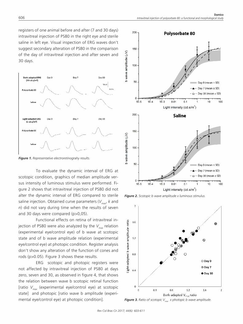
Figure 1 shows scotopic and photopic ERG
DamicoIntravitreal injection of polysorbate 80: a functional and morphological study606
Rev Col Bras Cir 2017; 44(6): 603-611
registers of one animal before and after (7 and 30 days)
intravitreal injection of PS80 in the right eye and sterile
saline in left eye. Visual inspection of ERG waves don’t
suggest secondary alteration of PS80 in the comparison
of the day of intravitreal injection and after seven and
30 days.
Figure 1. Representative electroretinograhy results.
Figure 2. Scotopic b wave amplitude x luminous stimulus.
Figure 3. Ratio of scotopic Vmax x photopic b wave amplitude.
To evaluate the dynamic interval of ERG at
scotopic condition, graphics of median amplitude ver-
sus intensity of luminous stimulus were performed. Fi-
gure 2 shows that intravitreal injection of PS80 did not
alter the dynamic interval of ERG compared to sterile
saline injection. Obtained curve parameters (Vmax, k and
n) did not vary during time when the results of seven
and 30 days were compared (p>0,05).
Functional effects on retina of intravitreal in-
jection of PS80 were also analyzed by the Vmax relation
(experimental eye/control eye) of b wave at scotopic
state and of b wave amplitude relation (experimental
eye/control eye) at photopic condition. Register analysis
don’t show any alteration of the function of cones and
rods (p>0.05). Figure 3 shows these results.
ERG scotopic and photopic registers were
not affected by intravitreal injection of PS80 at days
zero, seven and 30, as observed in figure 4, that shows
the relation between wave b scotopic retinal function
[ratio Vmax (experimental eye/control eye) at scotopic
state] and photopic [ratio wave b amplitude (experi-
mental eye/control eye) at photopic condition].
Damico Intravitreal injection of polysorbate 80: a functional and morphological study 607
Rev Col Bras Cir 2017; 44(6): 603-611
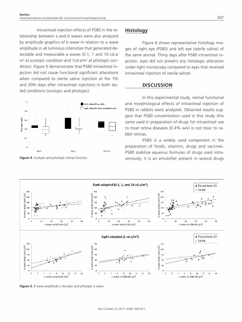
Figure 4. Scotopic and photopic retinal function.
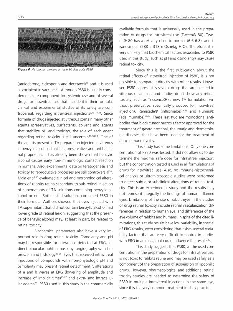
Figure 5. B wave amplitude x stocopic and photopic a wave.
Intravitreal injection effects of PS80 in the re-
lationship between a and b waves were also analyzed
by amplitude graphics of b wave in relation to a wave
amplitude in all luminous intensities that generated de-
tectable and measurable a waves (0.1, 1 and 10 cd.s/
m2 at scotopic condition and 1cd.s/m2 at photopic con-
dition). Figure 5 demonstrates that PS80 intravitreal in-
jection did not cause functional significant alterations
when compared to sterile saline injection at the 7th
and 30th days after intravitreal injections in both tes-
ted conditions (scotopic and photopic).
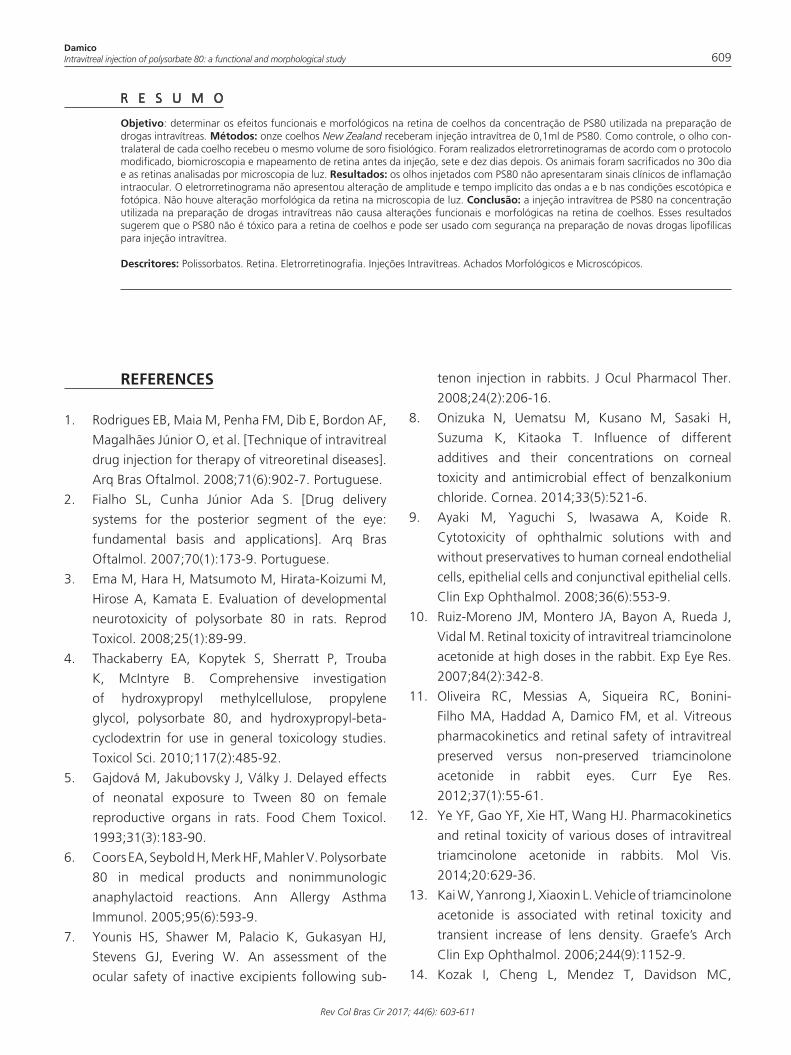
Histology
Figure 6 shows representative histology ima-
ges of right eye (PS80) and left eye (sterile saline) of
the same animal. Thirty days after PS80 intravitreal in-
jection, eyes did not present any histologic alteration
under light microscopy compared to eyes that received
intravitreal injection of sterile salinel.
DISCUSSION
In this experimental study, retinal functional
and morphological effects of intravitreal injection of
PS80 in rabbits were analyzed. Obtained results sug-
gest that PS80 concentration used in this study (the
same used in preparation of drugs for intravitreal use
to treat retina diseases (0.4% w/v) is not toxic to ra-
bbit retinas.
PS80 is a widely used component in the
preparation of foods, vitamins, drugs and vaccines.
PS80 stabilize aqueous formulas of drugs used intra-
venously; it is an emulsifier present in several drugs
DamicoIntravitreal injection of polysorbate 80: a functional and morphological study608
Rev Col Bras Cir 2017; 44(6): 603-611
Figura 6. Histologia retiniana antes e 30 dias após PS80.
(amiodarone, ciclosporin and decetaxel)20 and it is used
as excipient in vaccines21. Although PS80 is usually consi-
dered a safe component for systemic use and of several
drugs for intravitreal use that include it in their formula,
clinical and experimental studies of its safety are con-
troversial, regarding intravitreal injections9,13-15,22. Since
formula of drugs injected at vitreous contain many other
agents (preservatives, surfactants, solvent and agents
that stabilize pH and tonicity), the role of each agent
regarding retinal toxicity is still uncertain16-18,23. One of
the agents present in TA preparation injected in vitreous
is benzylic alcohol, that has preservative and antibacte-
rial proprieties. It has already been shown that benzylic
alcohol causes early non-immunologic contact reaction
in humans. Also, experimental data on teratogenesis and
toxicity to reproductive processes are still controversial24.
Maia et al.15 evaluated clinical and morphological altera-
tions of rabbits retina secondary to sub-retinal injection
of supernatants of TA solutions containing benzylic al-
cohol or not. Both tested solutions contained PS80 in
their formula. Authors showed that eyes injected with
TA supernatant that did not contain benzylic alcohol had
lower grade of retinal lesion, suggesting that the presen-
ce of benzylic alcohol may, at least in part, be related to
retinal toxicity.
Biochemical parameters also have a very im-
portant role in drug retinal toxicity. Osmolarity and pH
may be responsible for alterations detected at ERG, in-
direct binocular ophthalmoscopy, angiography with flu-
orescein and histology25-28. Eyes that received intravitreal
injections of compounds with non-physiologic pH and
osmolarity may present retinal detachment27, alterations
of a and b waves at ERG (lowering of amplitude and
increase of implicit time)25-27 and extra- and intracellu-
lar edema25. PS80 used in this study is the commercially
available formula that is universally used in the prepa-
ration of drugs for intravitreal use (Tween® 80). Twe-
en® 80 has a pH very close to normal (6.6-6.8), and is
iso-osmolar (288 a 318 mOsm/kg H2O). Therefore, it is
very unlikely that biochemical factors associated to PS80
used in this study (such as pH and osmolarity) may cause
retinal toxicity.
Since this is the first publication about the
retinal effects of intravitreal injection of PS80, it is not
possible to compare it directly with other results. Howe-
ver, PS80 is present is several drugs that are injected in
vitreous of animals and studies don’t show any retinal
toxicity, such as Triesence® (a new TA formulation wi-
thout preservative, specifically produced for intravitreal
injection), Remicade® (infliximabe)29-31 and Humira®
(adalimumabe)32-34. These last two are monoclonal anti-
bodies that block tumor necrosis factor approved for the
treatment of gastrointestinal, rheumatic and dermatolo-
gic diseases, that have been used for the treatment of
auto-immune uveitis.
This study has some limitations. Only one con-
centration of PS80 was tested. It did not allow us to de-
termine the maximal safe dose for intravitreal injection,
but the concentration tested is used in all formulations of
drugs for intravitreal use. Also, no immune-histochemi-
cal analysis or ultramicroscopic studies were performed
to detect subtle or subclinical alterations of retinal toxi-
city. This is an experimental study and the results may
not represent integrally the findings of human inflamed
eyes. Limitations of the use of rabbit eyes in the studies
of drug retinal toxicity include retinal vascularization dif-
ferences in relation to human eye, and differences of the
eye volume of rabbits and humans. In spite of the cited li-
mitations, this study results have low variability, in special
of ERG results, even considering that exists several varia-
bility factors that are very difficult to control in studies
with ERG in animals, that could influence the results35.
This study suggests that PS80, at the used con-
centration in the preparation of drugs for intravitreal use,
is not toxic to rabbits retina and may be used safely as a
component of the preparation of suspension of lipophilic
drugs. However, pharmacological and additional retinal
toxicity studies are needed to determine the safety of
PS80 in multiple intravitreal injections in the same eye,
since this is a very common treatment in daily practice.
Damico Intravitreal injection of polysorbate 80: a functional and morphological study 609
Rev Col Bras Cir 2017; 44(6): 603-611
REFERENCES
1. Rodrigues EB, Maia M, Penha FM, Dib E, Bordon AF,
Magalhães Júnior O, et al. [Technique of intravitreal
drug injection for therapy of vitreoretinal diseases].
Arq Bras Oftalmol. 2008;71(6):902-7. Portuguese.
2. Fialho SL, Cunha Júnior Ada S. [Drug delivery
systems for the posterior segment of the eye:
fundamental basis and applications]. Arq Bras
Oftalmol. 2007;70(1):173-9. Portuguese.
3. Ema M, Hara H, Matsumoto M, Hirata-Koizumi M,
Hirose A, Kamata E. Evaluation of developmental
neurotoxicity of polysorbate 80 in rats. Reprod
Toxicol. 2008;25(1):89-99.
4. Thackaberry EA, Kopytek S, Sherratt P, Trouba
K, McIntyre B. Comprehensive investigation
of hydroxypropyl methylcellulose, propylene
glycol, polysorbate 80, and hydroxypropyl-beta-
cyclodextrin for use in general toxicology studies.
Toxicol Sci. 2010;117(2):485-92.
5. Gajdová M, Jakubovsky J, Války J. Delayed effects
of neonatal exposure to Tween 80 on female
reproductive organs in rats. Food Chem Toxicol.
1993;31(3):183-90.
6. Coors EA, Seybold H, Merk HF, Mahler V. Polysorbate
80 in medical products and nonimmunologic
anaphylactoid reactions. Ann Allergy Asthma
Immunol. 2005;95(6):593-9.
7. Younis HS, Shawer M, Palacio K, Gukasyan HJ,
Stevens GJ, Evering W. An assessment of the
ocular safety of inactive excipients following sub-
tenon injection in rabbits. J Ocul Pharmacol Ther.
2008;24(2):206-16.
8. Onizuka N, Uematsu M, Kusano M, Sasaki H,
Suzuma K, Kitaoka T. Influence of different
additives and their concentrations on corneal
toxicity and antimicrobial effect of benzalkonium
chloride. Cornea. 2014;33(5):521-6.
9. Ayaki M, Yaguchi S, Iwasawa A, Koide R.
Cytotoxicity of ophthalmic solutions with and
without preservatives to human corneal endothelial
cells, epithelial cells and conjunctival epithelial cells.
Clin Exp Ophthalmol. 2008;36(6):553-9.
10. Ruiz-Moreno JM, Montero JA, Bayon A, Rueda J,
Vidal M. Retinal toxicity of intravitreal triamcinolone
acetonide at high doses in the rabbit. Exp Eye Res.
2007;84(2):342-8.
11. Oliveira RC, Messias A, Siqueira RC, Bonini-
Filho MA, Haddad A, Damico FM, et al. Vitreous
pharmacokinetics and retinal safety of intravitreal
preserved versus non-preserved triamcinolone
acetonide in rabbit eyes. Curr Eye Res.
2012;37(1):55-61.
12. Ye YF, Gao YF, Xie HT, Wang HJ. Pharmacokinetics
and retinal toxicity of various doses of intravitreal
triamcinolone acetonide in rabbits. Mol Vis.
2014;20:629-36.
13. Kai W, Yanrong J, Xiaoxin L. Vehicle of triamcinolone
acetonide is associated with retinal toxicity and
transient increase of lens density. Graefe’s Arch
Clin Exp Ophthalmol. 2006;244(9):1152-9.
14. Kozak I, Cheng L, Mendez T, Davidson MC,
Objetivo: determinar os efeitos funcionais e morfológicos na retina de coelhos da concentração de PS80 utilizada na preparação de drogas intravítreas. Métodos: onze coelhos New Zealand receberam injeção intravítrea de 0,1ml de PS80. Como controle, o olho con-tralateral de cada coelho recebeu o mesmo volume de soro fisiológico. Foram realizados eletrorretinogramas de acordo com o protocolo modificado, biomicroscopia e mapeamento de retina antes da injeção, sete e dez dias depois. Os animais foram sacrificados no 30o dia e as retinas analisadas por microscopia de luz. Resultados: os olhos injetados com PS80 não apresentaram sinais clínicos de inflamação intraocular. O eletrorretinograma não apresentou alteração de amplitude e tempo implícito das ondas a e b nas condições escotópica e fotópica. Não houve alteração morfológica da retina na microscopia de luz. Conclusão: a injeção intravítrea de PS80 na concentração utilizada na preparação de drogas intravítreas não causa alterações funcionais e morfológicas na retina de coelhos. Esses resultados sugerem que o PS80 não é tóxico para a retina de coelhos e pode ser usado com segurança na preparação de novas drogas lipofílicas para injeção intravítrea.
Descritores: Polissorbatos. Retina. Eletrorretinografia. Injeções Intravítreas. Achados Morfológicos e Microscópicos.
R E S U M O
DamicoIntravitreal injection of polysorbate 80: a functional and morphological study610
Rev Col Bras Cir 2017; 44(6): 603-611
Freeman WR. Evaluation of the toxicity of subretinal
triamcinolone acetonide in the rabbit. Retina.
2006;26(7):811-7.
15. Maia M, Penha FM, Farah ME, Dib E, Príncipe A, Lima
Filho AA, et al. Subretinal injection of preservative-
free triamcinolone acetonide and supernatant vehicle
in rabbits: an electron microscopy study. Graefes
Arch Clin Exp Ophthalmol. 2008;246(3):379-88.
16. Morrison VL, Koh HJ, Cheng L, Bessho K, Davidson
MC, Freeman WR. Intravitreal toxicity of the
kenalog vehicle (benzyl alcohol) in rabbits. Retina.
2006;26(3):339-44.
17. Chang YS, Wu CL, Tseng SH, Kuo PY, Tseng SY.
In vitro benzyl alcohol cytotoxicity: implications for
intravitreal use of triamcinolone acetonide. Exp Eye
Res. 2008;86(6):942-50.
18. Li Q, Wang J, Yang L, Mo B, Zeng H, Wang N, Liu
W. A moephologic study of retinal toxicity induced
by triamcinolone acetonide vehicles in rabbit eyes.
Retina. 2008;28(3):504-10.
19. Marmor MF, Fulton AB, Holder GE, Miyake Y,
Brigell M, Bach M; International Society for Clinical
Electrophysiology of Vision. ISCEV Standard for full-
field clinical electroretinography (2008 update).
Doc Ophthalmol. 2009;118(1):69-77.
20. Strickley RG. Solubilizing excipients in oral and
injectable formulations. Pharm Res. 2004;21(2):201-
30.
21. Fox CB, Haensler J. An update on safety
and immunogenicity of vaccines containing
emulsion-based adjuvants. Expert Rev Vaccines.
2013;12(7):747-58.
22. Zhengyu S, Fang W, Ying F. Vehicle used for
triamcinolone acetonide is toxic to ocular tissues of
the pigmented rabbit. Curr Eye Res. 2009;34(9):769-
76.
23. Patel S, Barnett JM, Kim SJ. Retinal toxicity
of intravitreal polyethylene glycol 400. J Ocul
Pharmacol Ther. 2016;32(2):97-101.
24. Nair B. Final report on the safety assessment of
Benzyl Alcohol, Benzoic Acid, and Sodium Benzoate.
Int J Toxicol. 2001;20 Suppl 3:23-50.
25. Maia M, Margalit E, Lakhanpal R, Tso MO, Grebe
R, Torres G, et al. Effects of intravitreal indocyanine
green injection in rabbits. Retina. 2004;24(1):69-
79.
26. Liang C, Peyman GA, Sun G. Toxicity of intraocular
lidocaine and bupivacaine. Am J Ophthalmol.
1998;125(2):191-6.
27. Marmor MF. Retinal detachment from hyperosmotic
intravitreal injection. Invest Ophthalmol Vis Sci.
1979;18(12):1237-44.
28. Verstraeten TC, Chapman C, Hartzer M, Winkler BS,
Trese MT, Williams GA. Pharmacologic induction of
posterior vitreous detachment in the rabbit. Arch
Ophthalmol. 1993;111(6):849-54.
29. Giansanti F, Ramazzotti M, Vannozzi L, Rapizzi E,
Fiore T, Iaccheri B, et al. A pilot study on ocular
safety of intravitreal infliximab in a rabbit model.
Invest Ophthalmol Vis Sci. 2008;49(3):1151-6.
30. Theodossiadis PG, Liarakos VS, Sfikakis PP, Charonis
A, Agrogiannis G, Kavantzas N, et al. Intravitreal
administration of the anti-TNF monoclonal antibody
infliximab in the rabbit. Graefes Arch Clin Exp
Ophthalmol. 2009;247(2):273-81.
31. Giansanti F, Papucci L, Capaccioli S, Bacherini
D, Vannozzi L, Witort E, et al. Ocular safety of
infliximab in rabbit and cell culture models. J Ocul
Pharmacol Ther. 2010;26(1):65-71.
32. Manzano RP, Peyman GA, Carvounis PE, Kivilcim
M, Khan P, Chevez-Barrios P, et al. Ocular
toxicity of intravitreous adalimumab (Humira) in
the rabbit. Graefe’s Arch Clin Exp Ophthalmol.
2008;246(6):907-11.
33. Manzano RP, Peyman GA, Carvounis PE, Damico
FM, Aguiar RG, Ioshimoto GL, et al. Toxicity of high-
dose intravitreal adalimumab (Humira) in the rabbit.
J Ocul Pharmacol Ther. 2011;27(4):327-31.
34. Myers AC, Ghosh F, Andréasson S, Ponjavic V.
Retinal function and morphology in the rabbit eye
after intravitreal injection of the TNF alpha inhibitor
adalimumab. Curr Eye Res. 2014;39(11):1106-16.
35. Perlman I. Testing retinal toxicity of drugs in
animal models using electrophysiological and
morphological techniques. Doc Ophthalmol.
2009;118(1):3-28.
Received in: 30/07/2017
Accepted for publication: 23/08/2017
Conflict of interest: none.
Damico Intravitreal injection of polysorbate 80: a functional and morphological study 611
Rev Col Bras Cir 2017; 44(6): 603-611
Source of funding: FAPESP 2007/02696-1 FAPESP
2007/56624-1 FAPESP 2014/26818-2 CNPq
150614/2009-8.
Mailing address:
Francisco Max Damico
E-mail: [email protected] / [email protected]