Embed Size (px)
Citation preview

Intrinsic dynamics of enzymes in the unbound state andrelation to allosteric regulationIvet Bahar, Chakra Chennubhotla and Dror Tobi
In recent years, there has been a surge in the number of studies
exploring the relationship between proteins’ equilibrium
dynamics and structural changes involved in function. An
emerging concept, supported by both theory and experiments,
is that under native state conditions proteins have an intrinsic
ability to sample conformations that meet functional
requirements. A typical example is the ability of enzymes to
sample open and closed forms, irrespective of substrate,
succeeded by the stabilization of one form (usually closed)
upon substrate binding. This ability is structure-encoded, and
plays a key role in facilitating allosteric regulation, which
suggests complementing the sequence-encodes-structure
paradigm of protein science by structure-encodes-dynamics-
encodes-function. The emerging connection implies an
evolutionary role in selecting/conserving structures based on
their ability to achieve functional dynamics, and in turn,
selecting sequences that fold into such ‘apt’ structures.
Addresses
Department of Computational Biology, School of Medicine, University of
Pittsburgh, Suite 3064, Biomedical Science Tower 3, 3051 Fifth Avenue,
Pittsburgh, PA 15213, United States
Corresponding author: Bahar, Ivet ([email protected])
Current Opinion in Structural Biology 2007, 17:633–640
This review comes from a themed issue on
Catalysis and regulation
Edited by William N Hunter and Ylva Lindqvist
Available online 19th November 2007
0959-440X/$ – see front matter
# 2007 Elsevier Ltd. All rights reserved.
DOI 10.1016/j.sbi.2007.09.011
IntroductionProteins sample an ensemble of conformations under
equilibrium conditions. This ensemble is broadly distrib-
uted in the denatured state, and it becomes narrowly
distributed – mainly confined to the neighborhood of the
folded state – under native state conditions. Of interest
are those conformations accessible near the global energy
minimum, also called substates when separated by low
energy barriers. Because proteins perform their function
under these conditions, interconversions between these
conformations are potentially functional.
The fact that folded proteins are not static, but undergo
‘wigglings and jigglings’ as put forth by Feynman, is
now well-established. We have indeed come a long
www.sciencedirect.com
way since the idea was first put forward in the pioneering
molecular dynamics (MD) simulations of proteins by
McCammon, Karplus, Wolynes, Levitt, van Gunsteren
and others in the late 1970s and early 1980s. With
recent advances in experiments and theory, increased
evidence is now being provided for the biological func-
tionality of these apparently random motions. Exper-
iments now permit us to visualize the structural
flexibility and heterogeneity of biomolecules and assess
their relevance to catalysis or signaling [1�,2��]. On the
theoretical side, novel coarse-grained models and
methods are providing insights into structure–dynamics
relations on a global, rather than local, scale [3,4�]. The
rapidly accumulating data lead to the emergence of
concepts such as the pre-disposition or intrinsic abilityof proteins to undergo conformational changes required
for function, and a possible evolutionary pressure for
selecting such structures, while also raising new ques-
tions with regard to old concepts.
New questions on old concepts
The first question concerns the conformational changes
observed between the substrate-bound and substrate-
unbound forms of a given enzyme, a phenomenon
broadly referred to as ‘induced fit’ after the original prop-
osition of Koshland [5]. The question is, to what extent
these conformational changes are literally ‘induced’ by
substrate. Would it be possible for the substrate to drivea change if the structure was not pre-disposed to undergo
the change? Does the substrate simply stabilize the ‘fittest’
conformations that already exist in the unbound state,
following the redistribution of a pre-existing population
[6] originally proposed by Weber [7]? Does it essentially
select the lowest-energy-cost pathways away from the
original minimum to optimize its interaction with the
enzyme?
The second question relates to allosteric changes in
conformations usually occurring on a large scale (quatern-
ary changes) stated to be ‘driven’ by local phenomena
like ATP binding or hydrolysis, ligand binding, phos-
phorylation, among others. Again, would it be possible to
elicit cooperative responses, if these were not already
energetically favored by the structure? How similar
are the experimentally known allosteric changes, and
those theoretically predicted to be naturally sampled
by the particular structure?
Third, how can we reconcile the ensemble of confor-
mations accessible near folded state and the two-state
transition observed in many allosteric proteins in accord
Current Opinion in Structural Biology 2007, 17:633–640

634 Catalysis and regulation
Figure 1
Experimental evidence for conformational diversity of folded proteins and comparison with theoretical predictions. (a) Three conformations of
cyclin dependent kinases (CDKs) adopted in the free form (middle), and in the presence of two different substrates, an inhibitor (INK4; left) and
its activator (cylin; right). The corresponding Protein data bank (PDB) codes are 1bi7, 1hcl and 1fin, in reading order. Colors refer to N-lobe (purple),
C-lobe (red), hinge residues (orange) and activation loop (cyan). Both activation and inhibition involve conformational changes in and around the
catalytic cleft. The activation loop (cyan) rotates towards the substrate (not shown). (b) Alternative conformations of HIV-1 RT. RT is composed
of two subunits, p66 and p51 (wheat); the p66 subunit consists of two domains, polymerase and RNase H (blue); and the polymerase domain
contains four subdomains, thumb (red), fingers (blue), palm (pink), connection (green). Comparison of the inhibitor-bound (nevirapine; space filling
Current Opinion in Structural Biology 2007, 17:633–640 www.sciencedirect.com
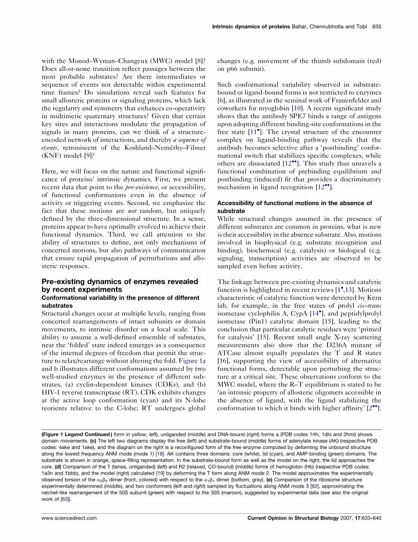
Intrinsic dynamics of proteins Bahar, Chennubhotla and Tobi 635
with the Monod–Wyman–Changeux (MWC) model [8]?
Does all-or-none transition reflect passages between the
most probable substates? Are there intermediates or
sequence of events not detectable within experimental
time frames? Do simulations reveal such features for
small allosteric proteins or signaling proteins, which lack
the regularity and symmetry that enhances co-operativity
in multimeric quaternary structures? Given that certain
key sites and interactions modulate the propagation of
signals in many proteins, can we think of a structure-
encoded network of interactions, and thereby a sequence ofevents, reminiscent of the Koshland–Nemethy–Filmer
(KNF) model [9]?
Here, we will focus on the nature and functional signifi-
cance of proteins’ intrinsic dynamics. First, we present
recent data that point to the pre-existence, or accessibility,
of functional conformations even in the absence of
activity or triggering events. Second, we emphasize the
fact that these motions are not random, but uniquely
defined by the three-dimensional structure. In a sense,
proteins appear to have optimally evolved to achieve their
functional dynamics. Third, we call attention to the
ability of structures to define, not only mechanisms of
concerted motions, but also pathways of communication
that ensure rapid propagation of perturbations and allo-
steric responses.
Pre-existing dynamics of enzymes revealedby recent experimentsConformational variability in the presence of different
substrates
Structural changes occur at multiple levels, ranging from
concerted rearrangements of intact subunits or domain
movements, to intrinsic disorder on a local scale. This
ability to assume a well-defined ensemble of substates,
near the ‘folded’ state indeed emerges as a consequence
of the internal degrees of freedom that permit the struc-
ture to relax/rearrange without altering the fold. Figure 1a
and b illustrates different conformations assumed by two
well-studied enzymes in the presence of different sub-
strates, (a) cyclin-dependent kinases (CDKs), and (b)
HIV-1 reverse transcriptase (RT). CDK exhibits changes
at the active loop conformation (cyan) and its N-lobe
reorients relative to the C-lobe; RT undergoes global
(Figure 1 Legend Continued ) form in yellow; left), unliganded (middle) and
domain movements. (c) The left two diagrams display the free (left) and sub
codes: 4ake and 1ake), and the diagram on the right is a reconfigured form
along the lowest frequency ANM mode (mode 1) [18]. AK contains three do
substrate is shown in orange, space-filling representation. In the substrate-b
core. (d) Comparison of the T (tense, unliganded) (left) and R2 (relaxed, CO
1a3n and 1bbb), and the model (right) calculated [19] by deforming the T fo
observed torsion of the a2b2 dimer (front, colored) with respect to the a1b1
experimentally determined (middle), and two conformers (left and right) sam
ratchet-like rearrangement of the 50S subunit (green) with respect to the 30
work of [63]).
www.sciencedirect.com
changes (e.g. movement of the thumb subdomain (red)
on p66 subunit).
Such conformational variability observed in substrate-
bound or ligand-bound forms is not restricted to enzymes
[6], as illustrated in the seminal work of Frauenfelder and
coworkers for myoglobin [10]. A recent significant study
shows that the antibody SPE7 binds a range of antigens
upon adopting different binding-site conformations in the
free state [11�]. The crystal structure of the encounter
complex on ligand-binding pathway reveals that the
antibody becomes selective after a ‘postbinding’ confor-
mational switch that stabilizes specific complexes, while
others are dissociated [12��]. This study thus unravels a
functional combination of prebinding equilibrium and
postbinding (induced) fit that provides a discriminatory
mechanism in ligand recognition [12��].
Accessibility of functional motions in the absence of
substrate
While structural changes assumed in the presence of
different substrates are common in proteins, what is new
is their accessibility in the absence substrate. Also, motions
involved in biophysical (e.g. substrate recognition and
binding), biochemical (e.g. catalysis) or biological (e.g.
signaling, transcription) activities are observed to be
sampled even before activity.
The linkage between pre-existing dynamics and catalytic
function is highlighted in recent reviews [1�,13]. Motions
characteristic of catalytic function were detected by Kern
lab, for example, in the free states of prolyl cis–transisomerase cyclophilin A, CypA [14�], and peptidylprolyl
isomerase (Pin1) catalytic domain [15], leading to the
conclusion that particular catalytic residues were ‘primed
for catalysis’ [15]. Recent small angle X-ray scattering
measurements also show that the D236A mutant of
ATCase almost equally populates the T and R states
[16], supporting the view of accessibility of alternative
functional forms, detectable upon perturbing the struc-
ture at a critical site. These observations conform to the
MWC model, where the R–T equilibrium is stated to be
‘an intrinsic property of allosteric oligomers accessible in
the absence of ligand, with the ligand stabilizing the
conformation to which it binds with higher affinity’ [2��].
DNA-bound (right) forms a (PDB codes 1rth, 1dlo and 2hmi) shows
strate-bound (middle) forms of adenylate kinase (AK) (respective PDB
of the free enzyme computed by deforming the unbound structure
mains: core (white), lid (cyan), and AMP-binding (green) domains. The
ound form as well as the model on the right, the lid approaches the
-bound) (middle) forms of hemoglobin (Hb) (respective PDB codes:
rm along ANM mode 2. The model approximates the experimentally
dimer (bottom, gray). (e) Comparison of the ribosome structure
pled by fluctuations along ANM mode 3 [62], approximating the
S (maroon), suggested by experimental data (see also the original
Current Opinion in Structural Biology 2007, 17:633–640

636 Catalysis and regulation
Molecular basis of pre-existing dynamics:insights from theory and simulationsThe pre-existence of conformational motions is not
unfamiliar to theoretical and computational biologists.
Yet, their relevance to function is being established only
recently.
Two major groups of studies have been undertaken
toward this goal. The first uses analytical approaches
(e.g. normal mode analysis (NMA) with coarse-grained
(e.g. elastic network) [3,4�]. NMA readily provides a
hierarchy of accessible modes of motion uniquely defined
by the given architecture. The low frequency modes
among them are usually cooperative (collectively invol-
ving large segments/domains/subunits). They also are
most readily accessible, since they are, by definition,
along the direction of the lowest ascent (smallest curva-
ture/force constant) away from the original energy mini-
mum approximated by a harmonic well. As a result, they
conceivably present a favorable, or intrinsically preferred,
mechanism for dissipating the energy increase arising
from external perturbations.
Two classical examples illustrating the relevance of low
frequency modes to functional changes in structure are
shown in Figure 1, panels (c) and (d). Panel (c) displays
the open (left) and closed (middle) forms of E. coli aden-
ylate kinase (AK), both determined by X-ray crystallo-
graphy. The right diagram, on the other hand, displays an
alternative conformation predicted by the anisotropic
network model (ANM [17]) upon deforming the unli-
ganded structure (left) along the slowest mode accessible
in this substrate-free state [18]. Thus the closed form is
readily approached by the open form upon movement
along the direction of the first (energetically easiest)
mode of motion. A similar phenomenon is observed in
hemoglobin (Hb), a prime example of an allosteric protein
(panel c). In this case, the tense (T) (left) structure is
reconfigured to approximate the liganded (R2 (middle))form, by moving along the 2nd slowest mode direction
(right) [19]. Similar results for other systems support the
view of the intrinsic tendency of the unbound proteins to
approach their bound conformations via low frequency
modes [20,21�,22]. An application to ribosome, illustrated
in panel (e), also points to the functional relevance of slow
modes. The low frequency modes predicted by coarse-
grained NMA can now be readily retrieved and viewed for
all PDB structures using recently developed webservers
(e.g. [17]).
The second group of studies is based on MD simulations,
accelerated by advanced algorithms such as replica
exchange, steered MD or simplified models and force
fields. While simulations require significantly longer com-
puting time and may suffer from convergence problems,
their main advantage is in exploring substates that may
not be accessible via NMA. Recent simulations clearly
Current Opinion in Structural Biology 2007, 17:633–640
demonstrate the energetic grounds for the ability of AK to
fluctuate between open and closed forms [23�]. Likewise,
unrestrained MD with implicit solvent shows the ability
of HIV-1 protease to fluctuate between close, semi-open
and fully open states, while predominantly populating the
semi-open state [24�]. Another extensive MD study of a
series of protein–protein complexes showed the existence
of an ensemble of conformers before binding, the stabil-
ization of selected conformers – those distinguished by
shape complementarity – to form complexes, succeeded
by further structural rearrangements [25�]. Simulations
prove particularly useful when combined with NMA or
coarse-grained models [26–31].
A key question, yet to be answered is how the internal
dynamics of enzymes influences reaction rates. Theor-
etical studies on CypA dynamics indicate that a transfer of
energy from first hydration shell and solvent-exposed
regions into the protein interior all the way to the active
site, through a network of vibrations, promotes catalysis
by lowering the energy barrier along the reaction coordi-
nate [32]. Similar observations were made for DHFR
[1�,32]. A link between collective mechanics and inter-
actions at the catalytic site is suggested by the co-local-
ization of the global hinge site with the catalytic site,
shown for a representative set of enzymes using elastic
network models [33]. Another recent ANM study showed
the coupling between domain motions and loop re-
arrangements at the catalytic site of triosephosphate iso-
merase [34]. These studies also drew attention to the
conservation of key residues that concert cooperative
motions [1�,32,33,35,36].
While the coupling between dynamics and function is
recognized, quantitative assessment of the correlation
between intrinsic dynamics and reaction rates remains
to be clarified. The above described changes are physical
events in the neighborhood of the original state. Catalytic
reaction rates are controlled by chemical events farther
along the reaction coordinate; their quantitative assess-
ment requires consideration of free energy paths and
barrier at transition state; and in this respect, the contri-
bution of conformational motions to catalysis has been
challenged [37]. The effect of distant mutations on
DHFR catalytic reaction has been explained quantitat-
ively in terms of the changes in the pre-organization of
polar residues, arising as a consequence of the change in
energy landscape in the presence of mutations [38].
Allosteric changes in conformations:Communication via concerted motionsMany allosteric biomolecules are multimeric and sym-
metric, which enhances the cooperativity of their confor-
mational changes, leading to an all-or-none transition in
accord with the MWC model [2��]. Yet, allostery appears
to be featured by a broad range of proteins [39�] including
single domain proteins, and intermediates or sequential
www.sciencedirect.com

Intrinsic dynamics of proteins Bahar, Chennubhotla and Tobi 637
events are also observed, especially in some small
proteins and ligand-gated ion channels.
The prominent feature in all allosteric proteins appears to
be the accessibility of functional conformers, and the
efficient communication between distant residues (e.g.
ligand-binding and catalytic residues). In nicotinic acetyl-
choline receptor (nAChR), for example, signal transduc-
tion occurs between nACh binding and pore opening sites
separated by 50 A. Such distant couplings may be readily
accounted for by low frequency modes. The NMA of
nAChR indeed showed that pore opening is ensured by
the lowest frequency mode intrinsically favored by the
structure irrespective of neurotransmitter binding [40].
This mode entails a global twist of the quaternary struc-
ture, similar to the pore opening mechanism identified for
a series of potassium channels [41].
Low frequency modes are most effective in spreading
signals, because of their cooperative nature and accessi-
bility with minimal energy requirement. Yet, local effects
may also play a critical role in mediating signals. The
assessment of signal transduction mechanism may there-
fore necessitate the consideration of multiple modes,
including those in the high frequency regime and
mode–mode couplings [1�,32,42,43]. For example, a loop
(b4-a4) has been pointed out in CheY to mediate the
rotational isomerization of substrate-binding tyrosine
Figure 2
Combination of a pre-existing equilibrium and post-binding rearrangement.
accessible via fluctuations in the global energy well (top panel), occasionally
approximated by a harmonic potential (dashed curve) in coarse-grained NMA
dynamic equilibrium before substrate (S) binding, as shown on the left box (b
interaction (conformational selection). The stabilization of the final complex
landscape of the protein E in the presence of the substrate differs from its u
substates in favor of the conformer that binds the substrate.
www.sciencedirect.com
(Y106) succeeding the phosphorylation event 9.5 A apart
[26]. Subsequent simulations showed, however, that the
loop is stabilized by the isomerization of Y106 in an active
conformation [44�], consistent with the population shift
model, and MWC mechanism. Yet, a recently solved
CheY shows the same loop in an intermediate confor-
mation, inviting attention to the accessibility of inter-
mediate conformations [45].
Signal transduction along well-defined pathways mediated
by collective motions emerges now as a common feature of
allosteric proteins [46,47]. Recent theoretical studies
indeed demonstrate the occurrence of key sites with high
allosteric potential [48�,49] or strategically placed hot spot
residues[30], and highlight pathways of allosteric signal
propagation [50] consistent with experimental data [51].
These pathways predominantly involve conserved resi-
dues, as also deduced from sequence-based analysis
[52,53], as illustrated in a recent MD study [54�]. Recent
work also shows how communication pathways relate to
collective motions [55]. Notably, binding sites are reported
to be located at regions that strongly affect the network of
interactions, and such properties have been suggested to
result from evolutionary pressure [56�].
The spread of signals/perturbations via coupled motions,
including in particular the most cooperative (lowest fre-
quency) modes of motions, and conserved residues, thus
The protein (E) originally samples an ensemble of conformations,
involving passages between substates within the well. The well is
. Two conformations/substates are schematically shown, which are in a
ottom). The substrate (S) selects the conformer(s) that allows for optimal
is subject to further rearrangement (induced fit). The energy profile/
nbound form (top right diagram) inducing a shift in the population of the
Current Opinion in Structural Biology 2007, 17:633–640

638 Catalysis and regulation
emerges as a plausible mechanism in allostery. This also
suggests hinge sites in these modes to serve as ‘messen-
gers’ for mediating allosteric communication, a conjecture
to be tested by further studies.
ConclusionIncreasingly larger body of experimental and theoretical
studies concurs on the ability of enzymes and/or allosteric
proteins to sample, even in their inactive or substrate-free
forms, conformations that approximate those required for
biological function. This ability is structure-encoded.
Based on recent observations, can we reconcile the MWC
and KNF models? The accumulating data appear to sup-
port the MWC model, in general. On the other hand, there
exist examples where sequences of events or intermediate
states are reported [45,47,57,58]. While the intrinsic
dynamics pre-dispose the protein to bind its substrate,
the final conformation is usually stabilized upon local
rearrangements induced after binding [12��,16,21�,24�,25�,26,39�,59], suggestive of the mechanism schematically
described in Figure 2, and similar changes in energy land-
scape are entailed by allostery [60]. Therein a pre-existing
equilibrium followed by the selection of the optimal con-
formation by the substrate and an induced fit to stabilize
the final bound form is anticipated. Also, evidence has been
presented for networks of coupled motions that facilitate
enzyme catalysis, energy propagation or signal trans-
mission [1�,30–32,35,36,42,50,51,55,56�], which may elicit
a sequence of events, reminiscent of the KNF model.
Clearly in some systems the sequential events may be
too fast and subtle to be detectable by experiments result-
ing in an apparent all-or-none process dominated by a
highly cooperative single mode of motion.
Irrespective of the kinetics of conformational change, the
intrinsic ability of the protein structures to undergo
conformational changes along directions that enable their
function unequivocally supports the mapping structure -
! dynamics! function. Structures are usually accepted
to be designable when they meet the criterion of being
thermodynamically stable (the lowest energy conformer)
for a diversity of sequences [61]. With the emerging
functionality and robustness (insensitivity to structural
and energetic details) of proteins intrinsic dynamics, the
pre-disposition to perform functional changes in confor-
mation may probably be viewed as another criterion for
designing structures. Structures may have evolved to
‘move’ in the right directions.
References and recommended readingPapers of particular interest, published within the annual period ofreview, have been highlighted as:
� of special interest�� of outstanding interest
1.�
Hammes-Schiffer S, Benkovic SJ: Relating protein motion tocatalysis. Annu Rev Biochem 2006, 75:519-541.
Current Opinion in Structural Biology 2007, 17:633–640
This is an extensive review of experimental and theoretical evidencesupporting the interplay between conformational dynamics of enzymesand their catalytic activities, with focus on two enzymes, dehydrofolatereductase and liver alcohol dehydrogenase .A network of coupledmotions involving both fast thermal vibrations and low frequency modesis pointed out to modulate the interactions between enzyme, substrateand cofactor at the active site, facilitating the hydride transfer reaction.
2.��
Changeux JP, Edelstein SJ: Allosteric mechanisms of signaltransduction. Science 2005, 308:1424-1428.
This is an insightful review of the progress made in the last forty years inunderstanding the allosteric mechanisms of signal tranduction. The basicconcepts underlying the MWC model are summarized, such as the pre-disposition of symmetric oligomeric structures to undergo cooperativechanges and the occurrence of reversible transitions, or spontaneous‘switches’ between discrete conformations, accessible in the absence ofligand. The importance of binding ligands at strategic locations such asinterfaces between subunits or along symmetry axes is emphasized.
3. Bahar I, Rader AJ: Coarse-grained normal mode analysis instructural biology. Curr Opin Struct Biol 2005, 15:586-592.
4.�
Tama F, Brooks CL: Symmetry, form, and shape: guidingprinciples for robustness in macromolecular machines. AnnuRev Biophys Biomol Struct 2006, 35:115-133.
An excellent review where the authors draw attention to the utility of multi-resolution methods based on elastic network models for understandingthe functional dynamics of biological systems. They highlight the role ofshape and form in the defining robust modes of motions.
5. Koshland DE: Application of a theory of enzyme specificity toprotein synthesis. Proc Natl Acad Sci 1958, 44:98-104.
6. Ma B, Shatsky M, Wolfson HJ, Nussinov R: Multiple diverseligands binding at a single protein site: A matter of pre-existingpopulations. Protein Sci 2002, 11:184-197.
7. Weber G: Ligand binding and internal equilibrium in proteins.Biochemistry 1972, 11:864-868.
8. Monod J, Wyman J, Changeux JP: On the nature of allosterictransitions: A plausible model. J Mol Biol 1965, 12:88-118.
9. Koshland DE Jr, Nemethy G, Filmer D: Comparison ofexperimental binding data and theoretical models in proteinscontaining subunits. Biochemistry 1966, 5:365-385.
10. Frauenfelder H, McMahon BH, Austin RH, Chu K, Groves JT: Therole of structure, energy landscape, dynamics, and allostery inthe enzymatic function of myoglobin. Proc Natl Acad Sci U S A2001, 98:2370-2374.
11.�
James LC, Roversi P, Tawfik DS: Antibody multispecificitymediated by conformational diversity. Science 2003,299:1362-1367.
12.��
James LC, Tawfik DS: Structure and kinetics of a transientantibody binding intermediate reveal a kinetic discriminationmechanism in antigen recognition. Proc Natl Acad Sci U S A2005, 102:12730-12735.
Binding of ligands to the antibody SPE7 was analyzed using kineticmeasurements and X-ray crystallography to show that small ligands bindusing the SPE7 pre-existing equilibrium followed by an induced fit thatproduces a high affinity complex. Nonspecific ligands are also able tobind the antibody. However, they are unable to induce the conformationalchanges for optimizing their interaction with SPE7, and consequentlydissociate from the antibody.
13. Kern D, Zuiderweg ER: The role of dynamics in allostericregulation. Curr Opin Struct Biol 2003, 13:748-757.
14.�
Eisenmesser EZ, Millet O, Labeikovsky W, Korzhnev DM, Wolf-Watz M, Bosco DA, Skalicky JJ, Kay LE, Kern D: Intrinsicdynamics of an enzyme underlies catalysis. Nature 2005,438:117-121.
Cyclophilin motions observed during catalysis are detected by NMRrelaxation to be already present in the unbound (free) form of the enzyme.This suggests that the motions necessary for catalysis are an intrinsicproperty of the enzymes.
15. Labeikovsky W, Eisenmesser EZ, Bosco DA, Kern D: Structureand dynamics of pin1 during catalysis by NMR. J Mol Biol 2007,367:1370-1381.
16. Fetler L, Kantrowitz ER, Vachette P: Direct observation insolution of a preexisting structural equilibrium for a mutant of
www.sciencedirect.com

Intrinsic dynamics of proteins Bahar, Chennubhotla and Tobi 639
the allosteric aspartate transcarbamoylase. Proc Natl Acad SciU S A 2007, 104:495-500.
17. Eyal E, Yang LW, Bahar I: Anisotropic network model:systematic evaluation and a new web interface. Bioinformatics2006, 22:2619-2627.
18. Temiz NA, Meirovitch E, Bahar I: Escherichia coli adenylatekinase dynamics: comparison of elastic network modelmodes with mode-coupling (15)N-NMR relaxation data.Proteins 2004, 57:468-480.
19. Xu C, Tobi D, Bahar I: Allosteric changes in protein structurecomputed by a simple mechanical model: hemoglobin T<–>R2transition. J Mol Biol 2003, 333:153-168.
20. Tama F, Sanejouand YH: Conformational change of proteinsarising from normal mode calculations. Protein Eng 2001,14:1-6.
21.�
Tobi D, Bahar I: Structural changes involved in protein bindingcorrelate with intrinsic motions of proteins in the unboundstate. Proc Natl Acad Sci U S A 2005, 102:18908-18913.
In this application of ANM to three different enzyme-substrate systems,the authors show that the motions of enzymes along the global modesfacilitate ligand binding, while final stabilization is achieved by post-binding induced fit.
22. Cui Q, Bahar IE: Normal Mode Analysis. Theory and Applicationsto Biological and Chemical Systems. Edited by Qiang C, Bahar I.Taylor & Francis Group; 2006.
23.�
Lou H, Cukier RI: Molecular dynamics of apo-adenylatekinase: a distance replica exchange method for the freeenergy of conformational fluctuations. J Phys Chem B 2006,110:24121-24137.
The potential of mean force for the reaction coordinate between the openand closed forms of AK is demonstrated to have a rather flat regionbetween the open and relatively closed forms. These results imply thatapo AK can fluctuate between the open and closed conformations in theabsence of its substrate.
24.�
Hornak V, Okur A, Rizzo RC, Simmerling C: HIV-1 protease flapsspontaneously close to the correct structure in simulationsfollowing manual placement of an inhibitor into the open state.J. Am. Chem Soc 2006, 128:2812-2813.
MD simulations of HIV-1 protease demonstrate that the protein exists in adynamic equilibrium between its close (bound), semi-open and open(unbound) conformations in the absence of the ligand.
25.�
Grunberg R, Leckner J, Nilges M: Complementarity ofstructure ensembles in protein–protein binding. Structure2004, 12:2125-2136.
Substates populated by unbound receptor and ligand structures weregenerated by MD simulations for 17 protein complexes, which weresubjected to combinatorial docking computations. Shape complemen-tarity between substates emerges as a major determinant of theselection of particular pairs to form complexes, some of which arefurther rearranged to adopt stable structures. The authors proposed a3-step mechanism for binding: diffusion, free conformer selection andrefolding.
26. Formaneck MS, Ma L, Cui Q: Reconciling the ‘old’ and ‘new’views of protein allostery: a molecular simulation study ofchemotaxis Y protein (CheY). Proteins 2006, 63:846-867.
27. Okazaki K, Koga N, Takada S, Onuchic JN, Wolynes PG: Multiple-basin energy landscapes for large-amplitude conformationalmotions of proteins: Structure-based molecular dynamicssimulations. Proc Natl Acad Sci U S A 2006, 103:11844-11849.
28. Kong Y, Karplus M: The signaling pathway of rhodopsin.Structure 2007, 15:611-623.
29. Trylska J, Tozzini V, Chang CE, McCammon JA: HIV-1 proteasesubstrate binding and product release pathways exploredwith coarse-grained molecular dynamics. Biophys J 2007,92:4179-4187.
30. Yu H, Ma L, Yang Y, Cui Q: Mechanochemical coupling in themyosin motor domain. II. Analysis of critical residues. PLoSComput Biol 2007, 3:e23.
31. Yu H, Ma L, Yang Y, Cui Q: Mechanochemical coupling in themyosin motor domain. I. Insights from equilibrium active-sitesimulations. PLoS Comput Biol 2007, 3:e21.
www.sciencedirect.com
32. Agarwal PK: Role of protein dynamics in reaction rateenhancement by enzymes. J Am Chem Soc 2005,127:15248-15256.
33. Yang LW, Bahar I: Coupling between catalytic site andcollective dynamics: a requirement for mechanochemicalactivity of enzymes. Structure 2005, 13:893-904.
34. Kurkcuoglu O, Jernigan RL, Doruker P: Loop motions oftriosephosphate isomerase observed with elastic networks.Biochemistry 2006, 45:1173-1182.
35. Agarwal PK, Billeter SR, Rajagopalan PT, Benkovic SJ,Hammes-Schiffer S: Network of coupled promotingmotions in enzyme catalysis. Proc Natl Acad Sci U S A 2002,99:2794-2799.
36. Benkovic SJ, Hammes-Schiffer S: A perspective on enzymecatalysis. Science 2003, 301:1196-1202.
37. Olsson MH, Parson WW, Warshel A: Dynamical contributions toenzyme catalysis: critical tests of a popular hypothesis. ChemRev 2006, 106:1737-1756.
38. Liu H, Warshel A: The catalytic effect of dihydrofolate reductaseand its mutants is determined by reorganization energies.Biochemistry 2007, 46:6011-6025.
39.�
Gunasekaran K, Ma B, Nussinov R: Is allostery an intrinsicproperty of all dynamic proteins? Proteins 2004, 57:433-443.
In this review, allostery is proposed to derive from redistribution inpopulation following ligand binding and as a consequence all proteinsare pointed out to be potentially allosteric.
40. Taly A, Delarue M, Grutter T, Nilges M, Le NN, Corringer PJ,Changeux JP: Normal mode analysis suggests a quaternarytwist model for the nicotinic receptor gating mechanism.Biophys. J. 2005, 88:3954-3965.
41. Shrivastava IH, Bahar I: Common mechanism of pore openingshared by five different potassium channels. Biophys J 2006,90:3929-3940.
42. Liu T, Whitten ST, Hilser VJ: Ensemble-based signatures ofenergy propagation in proteins: a new view of an oldphenomenon. Proteins 2006, 62:728-738.
43. Hawkins RJ, McLeish TC: Coupling of global and localvibrational modes in dynamic allostery of proteins. BiophysJ 2006, 91:2055-2062.
44.�
Ma L, Cui Q: The activation mechanism of a signaling protein atatomic resolution from advanced computations. J Am ChemSoc 2007, 129:10261-10268.
To clarify the activation mechanism of CheY as a prototypical signalingmolecule, the authors generate and analyze 160 unbiased activationtrajectories using transition path sampling and free energy simulations.The rotational isomerization of Tyr106 (functional residue at substratebinding site) is shown to be a low-energy-barrier transition readilysampled during simulations, irrespective of the phosphorylation ofThr87. This supports a population shift mechanism in line with MWCmodel, as opposed to the traditional Y-T coupling model which wouldrequire the Thr87 phosphorylation to elicit the isomerization of Tyr106.
45. Dyer CM, Dahlquist FW: Switched or not? The structure ofunphosphorylated CheY bound to the N terminus of FliM.J Bacteriol 2006, 188:7354-7363.
46. Rousseau F, Schymkowitz J: A systems biology perspective onprotein structural dynamics and signal transduction. Curr OpinStruct Biol 2005, 15:23-30.
47. Stock AM, Guhaniyogi J: A new perspective on responseregulator activation. J Bacteriol 2006, 188:7328-7330.
48.�
Ming D, Wall ME: Allostery in a coarse-grained model of proteindynamics. Phys. Rev. Lett. 2005, 95:198103.
This study provides a quantitative measure based on KL divergence forthe change in conformational dynamics induced by ligand binding.
49. Ming D, Wall ME: Interactions in native binding sitescause a large change in protein dynamics. J Mol Biol 2006,358:213-223.
50. Chennubhotla C, Bahar I: Markov propagation of allostericeffects in biomolecular systems: application to GroEL-GroES.Mol Syst Biol 2006, 2:36.
Current Opinion in Structural Biology 2007, 17:633–640

640 Catalysis and regulation
51. Ranson NA, Clare DK, Farr GW, Houldershaw D, Horwich AL,Saibil HR: Allosteric signaling of ATP hydrolysis in GroEL-GroES complexes. Nat Struct Mol Biol 2006, 13:147-152.
52. Lockless SW, Ranganathan R: Evolutionarily conservedpathways of energetic connectivity in protein families. Science1999, 286:295-299.
53. Russ WP, Lowery DM, Mishra P, Yaffe MB, Ranganathan R:Natural-like function in artificial WW domains. Nature 2005,437:579-583.
54.�
Ota N, Agard DA: Intramolecular signaling pathways revealedby modeling anisotropic thermal diffusion. J Mol Biol 2005,351:345-354.
A novel MD simulation method, anisotropic thermal diffusion, is intro-duced and used to examine the long-range propagation of conforma-tional changes that mediate allosteric communication. Successfulapplication to a member of the PDZ domain is presented.
55. Chennubhotla C, Bahar I: Markov propagation of signals inproteins and its relation to equilibrium fluctuations. PLoSComput Biol 2007, 3:1716-1726.
56.�
Liu T, Whitten ST, Hilser VJ: Functional residues serve adominant role in mediating the co-operativity of the proteinensemble. Proc Natl Acad Sci U S A 2007, 104:4347-4352.
An ensemble-based description of proteins is utilized to evaluate theextent to which perturbations at different sites propagate to the entirestructure. Application to a database indicates that perturbations atbinding sites affect the co-operativity between all residues. The authors
Current Opinion in Structural Biology 2007, 17:633–640
suggest that binding residues are inherently located at sites poised toeffectively propagate signals.
57. Ragona L, Catalano M, Luppi M, Cicero D, Eliseo T, Foote J,Fogolari F, Zetta L, Molinari H: NMR dynamic studies suggestthat allosteric activation regulates ligand binding in chickenliver bile acid-binding protein. J Biol Chem 2006, 281:9697-9709.
58. Popovych N, Sun S, Ebright RH, Kalodimos CG: Dynamicallydriven protein allostery. Nat Struct Mol Biol 2006, 13:831-838.
59. Rajamani D, Thiel S, Vajda S, Camacho CJ: Anchor residues inprotein–protein interactions. Proc Natl Acad Sci U S A 2004,101:11287-11292.
60. Swain JF, Gierasch LM: The changing landscape of proteinallostery. Curr Opin Struct Biol 2006, 16:102-108.
61. Li H, Helling R, Tang C, Wingreen N: Emergence of preferredstructures in a simple model of protein folding. Science 1996,273:666-669.
62. Wang Y, Rader AJ, Bahar I, Jernigan RL: Global ribosomemotions revealed with elastic network model. J. Struct Biol2004, 147:302-314.
63. Tama F, Valle M, Frank J, Brooks CL III: Dynamic reorganizationof the functionally active ribosome explored by normal modeanalysis and cryo-electron microscopy. Proc Natl Acad Sci U S A2003, 100:9319-9323.
www.sciencedirect.com