Embed Size (px)
Citation preview
1
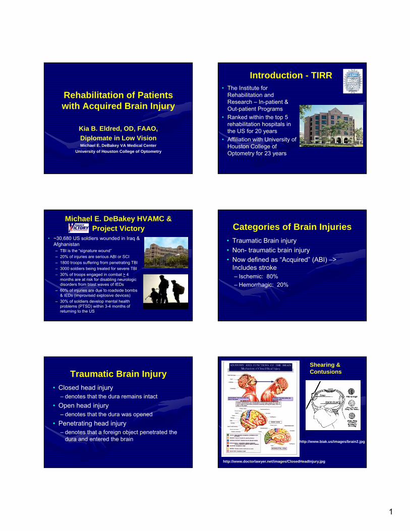
Rehabilitation of Patients with Acquired Brain Injury
Kia B. Eldred, OD, FAAO,
Diplomate in Low VisionMichael E. DeBakey VA Medical Center
University of Houston College of Optometry
Introduction - TIRR• The Institute for
Rehabilitation and Research – In-patient & Out-patient Programs
• Ranked within the top 5 rehabilitation hospitals in the US for 20 years
• Affiliation with University of Houston College of Optometry for 23 years
Michael E. DeBakey HVAMC & Project Victory
• ~30,680 US soldiers wounded in Iraq & Afghanistan– TBI is the “signature wound”
– 20% of injuries are serious ABI or SCI
– 1800 troops suffering from penetrating TBI
– 3000 soldiers being treated for severe TBI
– 30% of troops engaged in combat > 4 months are at risk for disabling neurologic disorders from blast waves of IEDs
– 60% of injuries are due to roadside bombs & IEDs (improvised explosive devices)
– 30% of soldiers develop mental health problems (PTSD) within 3-4 months of returning to the US
Categories of Brain Injuries
• Traumatic Brain injury
• Non- traumatic brain injury
• Now defined as “Acquired” (ABI) –> Includes stroke– Ischemic: 80%
– Hemorrhagic: 20%
Traumatic Brain Injury
• Closed head injury– denotes that the dura remains intact
• Open head injury – denotes that the dura was opened
• Penetrating head injury– denotes that a foreign object penetrated the
dura and entered the brain
http://www.doctorlawyer.net/images/ClosedHeadInjury.jpg
http://www.biak.us/images/brain2.jpg
Shearing & Contusions
2
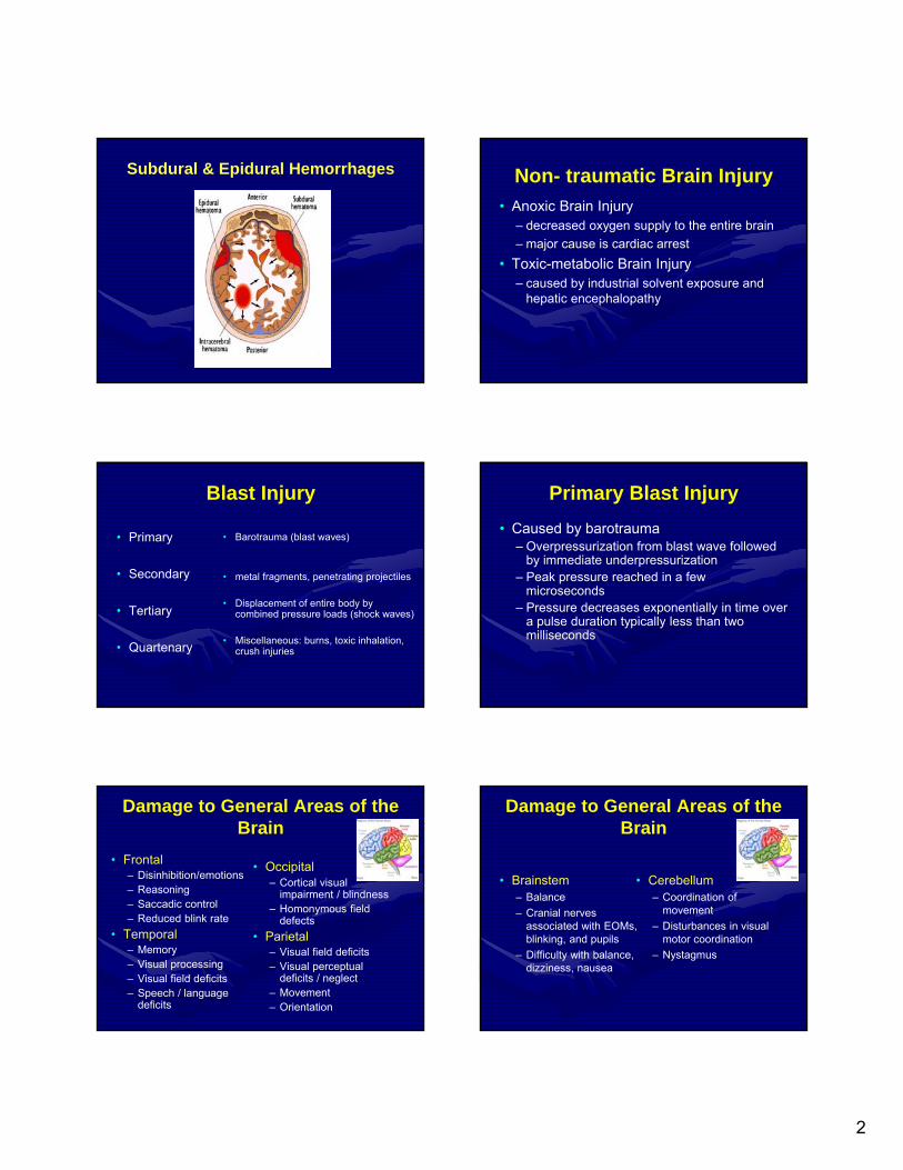
Subdural & Epidural Hemorrhages Non- traumatic Brain Injury
• Anoxic Brain Injury– decreased oxygen supply to the entire brain
– major cause is cardiac arrest
• Toxic-metabolic Brain Injury– caused by industrial solvent exposure and
hepatic encephalopathy
Blast Injury
• Primary
• Secondary
• Tertiary
• Quartenary
• Barotrauma (blast waves)
• metal fragments, penetrating projectiles
• Displacement of entire body by combined pressure loads (shock waves)
• Miscellaneous: burns, toxic inhalation, crush injuries
Primary Blast Injury
• Caused by barotrauma– Overpressurization from blast wave followed
by immediate underpressurization– Peak pressure reached in a few
microseconds– Pressure decreases exponentially in time over
a pulse duration typically less than two milliseconds
Damage to General Areas of the Brain
• Occipital– Cortical visual
impairment / blindness– Homonymous field
defects
• Parietal– Visual field deficits– Visual perceptual
deficits / neglect– Movement– Orientation
• Frontal– Disinhibition/emotions– Reasoning– Saccadic control– Reduced blink rate
• Temporal– Memory– Visual processing– Visual field deficits– Speech / language
deficits
Damage to General Areas of the Brain
• Brainstem– Balance
– Cranial nerves associated with EOMs, blinking, and pupils
– Difficulty with balance, dizziness, nausea
• Cerebellum– Coordination of
movement
– Disturbances in visual motor coordination
– Nystagmus
3
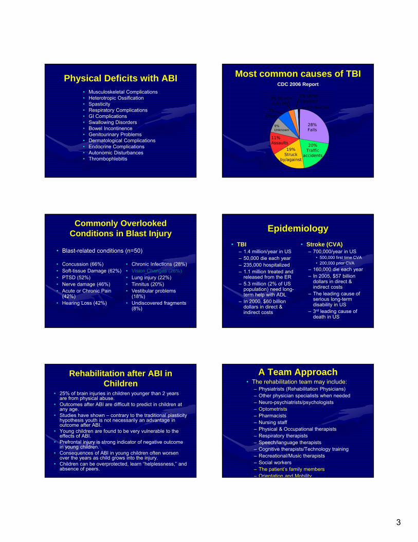
Physical Deficits with ABI• Musculoskeletal Complications• Heterotropic Ossification• Spasticity• Respiratory Complications• GI Complications• Swallowing Disorders• Bowel Incontinence• Genitourinary Problems• Dermatological Complications• Endocrine Complications• Autonomic Disturbances• Thrombophlebitis
Most common causes of TBICDC 2006 Report
Commonly Overlooked Conditions in Blast Injury
• Blast-related conditions (n=50)
• Concussion (66%)• Soft-tissue Damage (62%)• PTSD (52%)• Nerve damage (46%)• Acute or Chronic Pain
(42%)• Hearing Loss (42%)
• Chronic Infections (28%)• Vision Changes (26%)• Lung injury (22%)• Tinnitus (20%)• Vestibular problems
(18%)• Undiscovered fragments
(8%)
Epidemiology
• TBI– 1.4 million/year in US– 50,000 die each year– 235,000 hospitalized– 1.1 million treated and
released from the ER– 5.3 million (2% of US
population) need long-term help with ADL
– In 2000, $60 billion dollars in direct & indirect costs
• Stroke (CVA)– 700,000/year in US
• 500,000 first time CVA• 200,000 prior CVA
– 160,000 die each year– In 2005, $57 billion
dollars in direct & indirect costs
– The leading cause of serious long-term disability in US
– 3rd leading cause of death in US
Rehabilitation after ABI in Children
• 25% of brain injuries in children younger than 2 years are from physical abuse.
• Outcomes after ABI are difficult to predict in children at any age.
• Studies have shown – contrary to the traditional plasticity hypothesis youth is not necessarily an advantage in outcome after ABI.
• Young children are found to be very vulnerable to the effects of ABI.
• Prefrontal injury is strong indicator of negative outcome in young children.
• Consequences of ABI in young children often worsen over the years as child grows into the injury.
• Children can be overprotected, learn “helplessness,” and absence of peers.
A Team Approach• The rehabilitation team may include:
– Physiatrists (Rehabilitation Physicians)– Other physician specialists when needed– Neuro-psychiatrists/psychologists– Optometrists– Pharmacists– Nursing staff– Physical & Occupational therapists– Respiratory therapists– Speech/language therapists– Cognitive therapists/Technology training– Recreational/Music therapists– Social workers– The patient’s family members– Orientation and Mobility
4
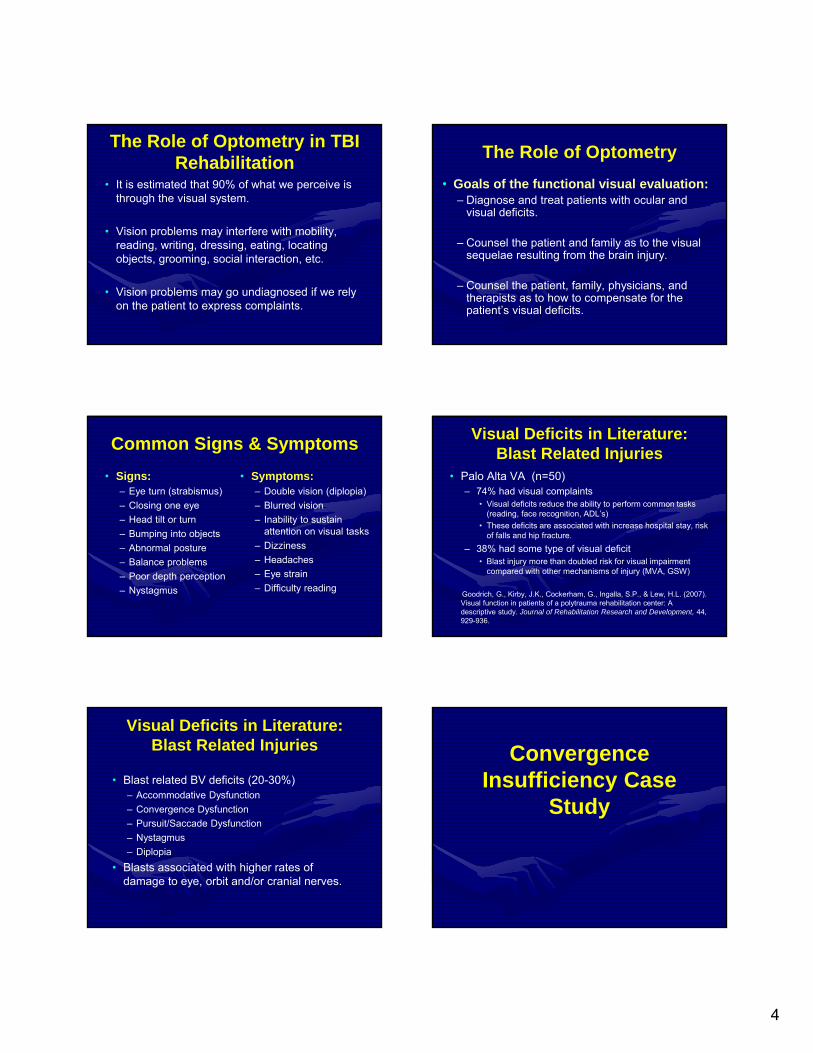
The Role of Optometry in TBI Rehabilitation
• It is estimated that 90% of what we perceive is through the visual system.
• Vision problems may interfere with mobility, reading, writing, dressing, eating, locating objects, grooming, social interaction, etc.
• Vision problems may go undiagnosed if we rely on the patient to express complaints.
The Role of Optometry
• Goals of the functional visual evaluation:– Diagnose and treat patients with ocular and
visual deficits.
– Counsel the patient and family as to the visual sequelae resulting from the brain injury.
– Counsel the patient, family, physicians, and therapists as to how to compensate for the patient’s visual deficits.
Common Signs & Symptoms
• Signs:– Eye turn (strabismus)
– Closing one eye
– Head tilt or turn
– Bumping into objects
– Abnormal posture
– Balance problems
– Poor depth perception
– Nystagmus
• Symptoms:– Double vision (diplopia)
– Blurred vision
– Inability to sustain attention on visual tasks
– Dizziness
– Headaches
– Eye strain
– Difficulty reading
Visual Deficits in Literature: Blast Related Injuries
• Palo Alta VA (n=50)– 74% had visual complaints
• Visual deficits reduce the ability to perform common tasks (reading, face recognition, ADL’s)
• These deficits are associated with increase hospital stay, risk of falls and hip fracture.
– 38% had some type of visual deficit• Blast injury more than doubled risk for visual impairment
compared with other mechanisms of injury (MVA, GSW)
Goodrich, G., Kirby, J.K., Cockerham, G., Ingalla, S.P., & Lew, H.L. (2007). Visual function in patients of a polytrauma rehabilitation center: A descriptive study. Journal of Rehabilitation Research and Development, 44,929-936.
Visual Deficits in Literature: Blast Related Injuries
• Blast related BV deficits (20-30%)– Accommodative Dysfunction
– Convergence Dysfunction
– Pursuit/Saccade Dysfunction
– Nystagmus
– Diplopia
• Blasts associated with higher rates of damage to eye, orbit and/or cranial nerves.
Convergence Insufficiency Case
Study
5
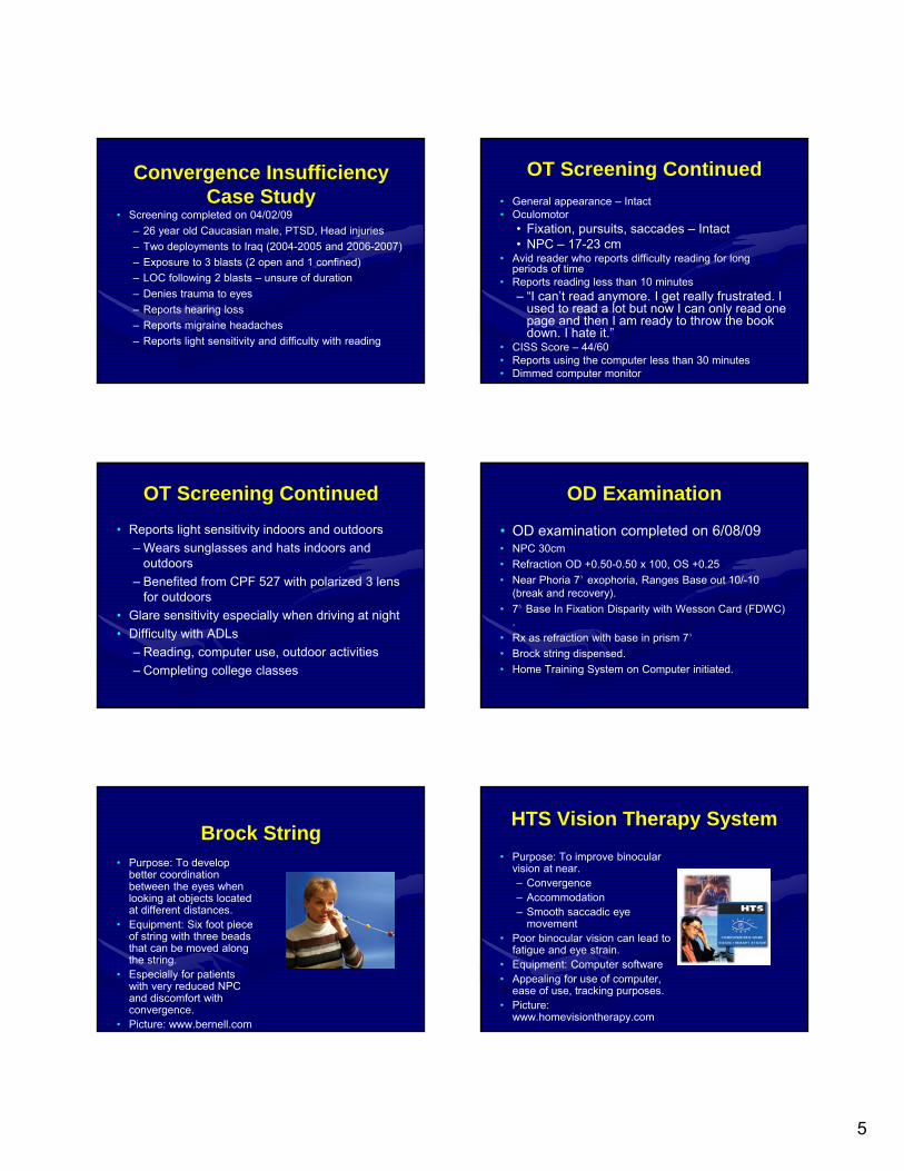
Convergence Insufficiency Case Study
• Screening completed on 04/02/09
– 26 year old Caucasian male, PTSD, Head injuries
– Two deployments to Iraq (2004-2005 and 2006-2007)
– Exposure to 3 blasts (2 open and 1 confined)
– LOC following 2 blasts – unsure of duration
– Denies trauma to eyes
– Reports hearing loss
– Reports migraine headaches
– Reports light sensitivity and difficulty with reading
OT Screening Continued
• General appearance – Intact• Oculomotor
• Fixation, pursuits, saccades – Intact• NPC – 17-23 cm
• Avid reader who reports difficulty reading for long periods of time
• Reports reading less than 10 minutes– “I can’t read anymore. I get really frustrated. I
used to read a lot but now I can only read one page and then I am ready to throw the book down. I hate it.”
• CISS Score – 44/60• Reports using the computer less than 30 minutes• Dimmed computer monitor
OT Screening Continued
• Reports light sensitivity indoors and outdoors
– Wears sunglasses and hats indoors and outdoors
– Benefited from CPF 527 with polarized 3 lens for outdoors
• Glare sensitivity especially when driving at night
• Difficulty with ADLs
– Reading, computer use, outdoor activities
– Completing college classes
OD Examination
• OD examination completed on 6/08/09• NPC 30cm
• Refraction OD +0.50-0.50 x 100, OS +0.25
• Near Phoria 7∆ exophoria, Ranges Base out 10/-10 (break and recovery).
• 7∆ Base In Fixation Disparity with Wesson Card (FDWC) .
• Rx as refraction with base in prism 7∆
• Brock string dispensed.
• Home Training System on Computer initiated.
Brock String• Purpose: To develop
better coordination between the eyes when looking at objects located at different distances.
• Equipment: Six foot piece of string with three beads that can be moved along the string.
• Especially for patients with very reduced NPC and discomfort with convergence.
• Picture: www.bernell.com
HTS Vision Therapy System
• Purpose: To improve binocular vision at near.– Convergence– Accommodation– Smooth saccadic eye
movement • Poor binocular vision can lead to
fatigue and eye strain.• Equipment: Computer software • Appealing for use of computer,
ease of use, tracking purposes.• Picture:
www.homevisiontherapy.com
6
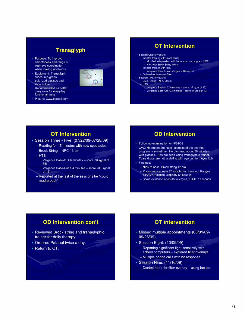
Tranaglyph• Purpose: To improve
smoothness and range of your eye coordination when looking at objects
• Equipment: Tranaglyph slides, red/green polarized glasses and slide holder
• Recommended as better carry over for everyday functional tasks.
• Picture: www.bernell.com
OT Intervention
• Session One: (07/08/09)– Initiated training with Brock String
• Modified independent with home exercise program (HEP)• NPC with Brock String 40cm
– Initiated training with HTS• Vergence Base-In and Vergence Base-Out
– Ordered replacement filters• Session Two: (07/20/09)
– Brock String – NPC 20 cm– HTS
• Vergence Base-In X 3 minutes – score 37 (goal of 35)• Vergence Base-Out X 3 minutes – score 17 (goal of 13)
OT Intervention• Session Three - Five: (07/22/09-07/28/09)
– Reading for 15 minutes with new spectacles
– Brock String – NPC 13 cm
– HTS • Vergence Base-In X 6 minutes – score 34 (goal of
35)
• Vergence Base-Out X 4 minutes – score 20.5 (goal of 13)
– Reported at the last of the sessions he “could read a book”
OD Intervention
• Follow up examination on 8/24/09
• CVC: He reports he hasn’t completed the internet program in sometime. He can read about 30 minutes with glasses. Has not been using tranaglyphic trainer. Tears drops are not assisting with eye comfort, eyes itch.
• Findings:
– NPC to nose, Brock string 12 cm
– Phorometry at near 7^ exophoria, Base out Ranges 14^/12^, Fixation Disparity 6^ base in
– Some evidence of ocular allergies, TBUT 7 seconds
OD Intervention con’t
• Reviewed Brock string and tranaglyphic trainer for daily therapy
• Ordered Patanol twice a day.
• Return to OT.
OT intervention
• Missed multiple appointments (08/01/09-09/28/09)
• Session Eight: (10/09/09)– Reporting significant light sensitivity with
school computers – explored filter overlays
– Multiple phone calls with no response
• Session Nine: (11/16/09)– Denied need for filter overlay – using lap top
7
OD Examination
• Follow up examination on 11/16/09
• CVC: Reports he lost his reading glasses and is having difficulty with reading for school. Has not been working on vision training while in school. Did not receive Patanol and is still having ocular discomfort.
• Findings:
– NPC 8-10cm
– Near Phorometry 7^ exo, BO 28/12, Wesson 6^ BI
– TBUT 8 sec, ocular allergy, ie. Follicles
• Plan prescribed new near Rx with 3^ base in, re-order Patanol.
OD Intervention con’t
• Reviewed Brock string and tranaglyphic trainer for daily therapy
• Ordered Patanol twice a day.
• Return to OT.
How to Classify ABI
Mild, Moderate and Severe
– 1. Glasgow Coma Scale Score is performed after resuscitation in ER
– Scores verbal response, eye opening and motor responses
– Total score is 3 to 15 (modified by intubation)
Scoring GCS
• Score:– (E + M +V) = range 3 to 15
• Mild TBI = 13 to 15 – 99.6% Survive
• Moderate TBI = 9 to 12 – 93% Survive
• Severe TBI = 3 to 8 – 42% Survive
Scoring GCS Post-Traumatic Amnesia
• Definition: period in which a patient’s ability to learn new information is minimal or nonexistent– Duration of PTA after recovery of
consciousness is another measure of severity
– Assessing PTA: patient’s orientation to place & time
8
Post-Traumatic Amnesia Scale
• PTA Duration
• < 5 minutes
• 5 to 60 minutes
• 1 to 24 hours
• 1 to 7 days
• 1 to 4 weeks
• > 4 weeks
• Severity of injury
• very mild
• mild
• moderate
• severe
• very severe
• extremely severe
Damage to Specific Areas of the Brain
• Occipital Lobe
• Parietal Lobe
• Frontal Lobe
• Temporal Lobe
History
– Hospital personnel
– Reliable History
– Communication
– Anosagnosia – a lack of awareness of how one’s own visual system is functioning
Common Medications
• Arousal– Bromocriptine
(Parlodel)
– Methylphenidate (Ritalin)
– Trazodone (anti-depressant to regulate sleep/wake cycle)
• Anti-Seizure– Neurotin – also post-
herpatic neuralgia, neuropathic pain
– Keppra
– Tripleptal
• Spasticity– Baclofen
– Botox
http://braincampus.learnpsychology.com/npsych/pics/aphasia.GIF
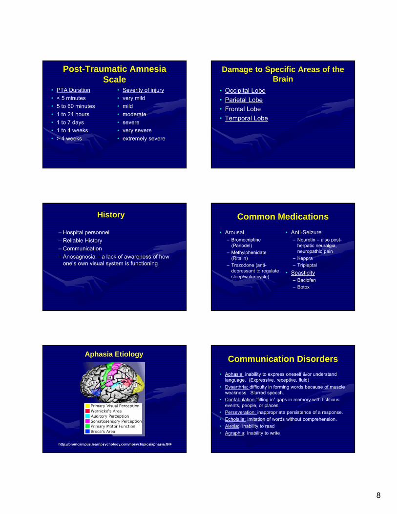
Aphasia EtiologyCommunication Disorders
• Aphasia: inability to express oneself &/or understand language. (Expressive, receptive, fluid)
• Dysarthria: difficulty in forming words because of muscle weakness. Slurred speech.
• Confabulation:”filling in” gaps in memory with fictitious events, people, or places.
• Perseveration: inappropriate persistence of a response.
• Echolalia: Imitation of words without comprehension.
• Alexia: Inability to read
• Agraphia: Inability to write
9
Physical Deficits• Apraxia: Inability to carry out a complex or
skilled movement not due to paralysis, sensory changes, or deficiencies in understanding.
• Ataxia: A problem of muscle coordination. Caused by lesion of the cerebellum or basal ganglia.
• Adiadochokinesia: Inability to stop one movement & follow it with a movement in the opposite direction.
• Paresis: Inability to move part of the body.
Visual Acuity
• Methods of Visual Acuity Assessment– Preferential
Viewing
– Matching – Lea or HOTV
– Verbal • Full lines vs.
Isolation
Visual Acuity
• Traumatic myopia - an increase in myopia which is not present with cycloplegia
• Other common causes of visual acuity reduction– Optic Atrophy– Vitreous Hemorrhage –
Terson’s Syndrome
Refractive Error
Accommodative Disorders
• Accommodative Insufficiency– Use of reading Rx or BF
– May improve over time and with decrease in meds
• Accommodative spasm / Traumatic myopia– Difficult to manage
– May resolve with time
– May need BFs, vision therapy, cycloplegics
• Accommodative infacility– Vision therapy
III Cranial Nerve PalsyCranial Nerve III Palsy• Treat by training
convergence if possible, use of prism or patching. Gross tracking may be helpful
• Resolution time – up to three years
10
IV Cranial Nerve Palsy
• Cranial Nerve IV - May be very subtle and difficult to observe– Park’s three step to diagnose if unilateral. Very
difficult to detect bilateral. Assess with double Maddox rod if greater than 10 degree cyclo-rotation , probably bilateral
• Treatment• Resolution – up to one year• Interactive website• http://www.richmondeye.com/eyevert.asp#et
IV Cranial Nerve Palsies
• May want to encourage head tilt(?) prism training or use of prism. Possible surgical intervention. Could be bilateral problem.
• Avoid tasks in downgaze
VI Cranial Nerve Palsies
• Cranial Nerve VI
• Resolution – up to one year.
VI Cranial Nerve Palsies
• Encourage viewing toward the unaffected eye, near work, gross tracking. Could work with prism jumps, prism on lens, or patching. Gross tracking may be helpful.
• Botox injection to medial rectus may be beneficial
Eye Movement Disorders
Nystagmus
– Vertical – lesion of pons/ midbrain
– Horizontal - MS
Gaze Palsy
Use of CCTV to assist with acquired nystagmus
Patching options
11
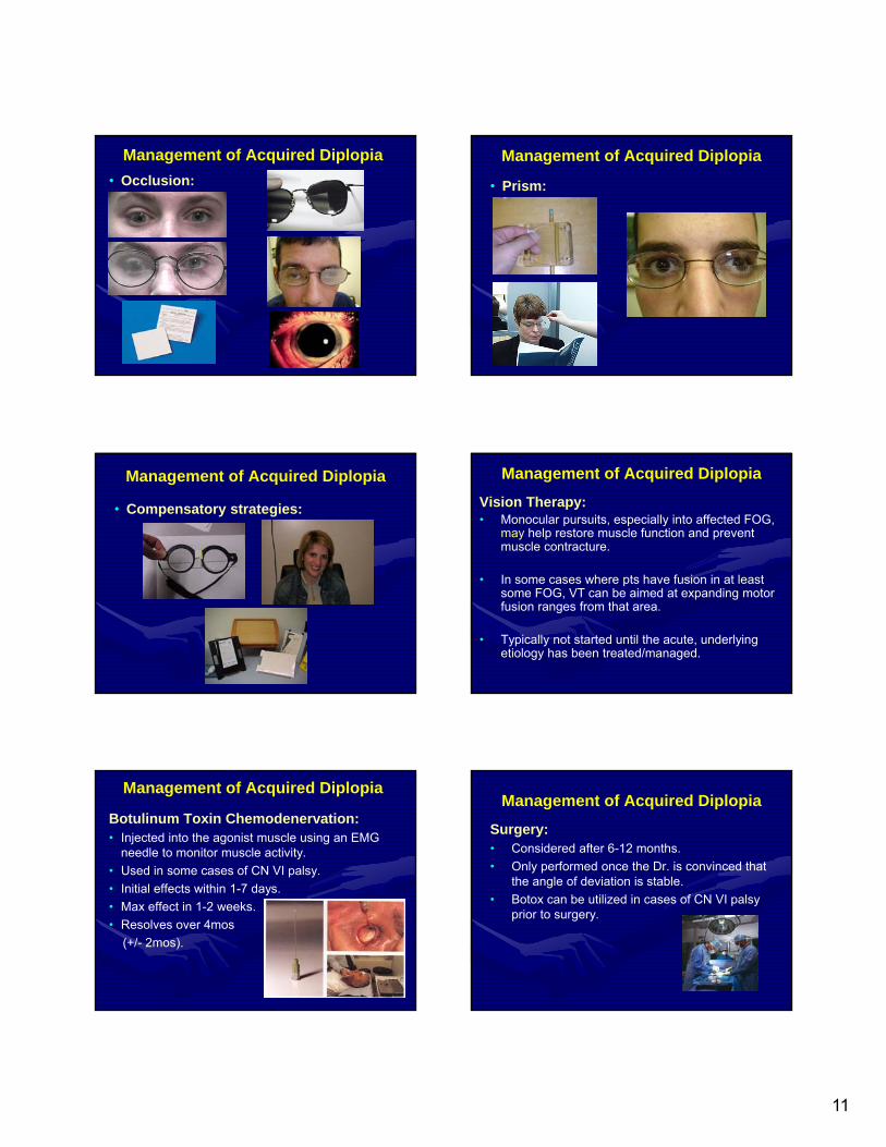
Management of Acquired Diplopia
• Occlusion:
Management of Acquired Diplopia
• Prism:
Management of Acquired Diplopia
• Compensatory strategies:
Management of Acquired Diplopia
Vision Therapy:• Monocular pursuits, especially into affected FOG,
may help restore muscle function and prevent muscle contracture.
• In some cases where pts have fusion in at least some FOG, VT can be aimed at expanding motor fusion ranges from that area.
• Typically not started until the acute, underlying etiology has been treated/managed.
Management of Acquired Diplopia
Botulinum Toxin Chemodenervation:• Injected into the agonist muscle using an EMG
needle to monitor muscle activity.
• Used in some cases of CN VI palsy.
• Initial effects within 1-7 days.
• Max effect in 1-2 weeks.
• Resolves over 4mos
(+/- 2mos).
Management of Acquired Diplopia
Surgery:• Considered after 6-12 months.
• Only performed once the Dr. is convinced that the angle of deviation is stable.
• Botox can be utilized in cases of CN VI palsy prior to surgery.
12
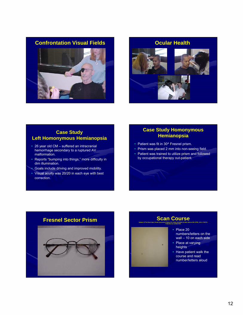
Confrontation Visual Fields Ocular Health
Case Study Left Homonymous Hemianopsia
• 26 year old CM – suffered an intracranial hemorrhage secondary to a ruptured AV malformation.
• Reports “bumping into things,” more difficulty in dim illumination.
• Goals include driving and improved mobility.
• Visual acuity was 20/20 in each eye with best correction.
Case Study Homonymous Hemianopsia
• Patient was fit in 30^ Fresnel prism.
• Prism was placed 2 mm into non-seeing field.
• Patient was trained to utilize prism and followed by occupational therapy out-patient.
Fresnel Sector Prism Scan Course Subtest of The Brain Injury Visual Assessment Battery For Adults (biVABA) by Mary Warren MS, OTR/L, SCLV, FAOTA;
Published by visABILITIES
• Place 20 numbers/letters on the wall – 10 on each side
• Place at varying heights
• Have patient walk the course and read number/letters aloud
13
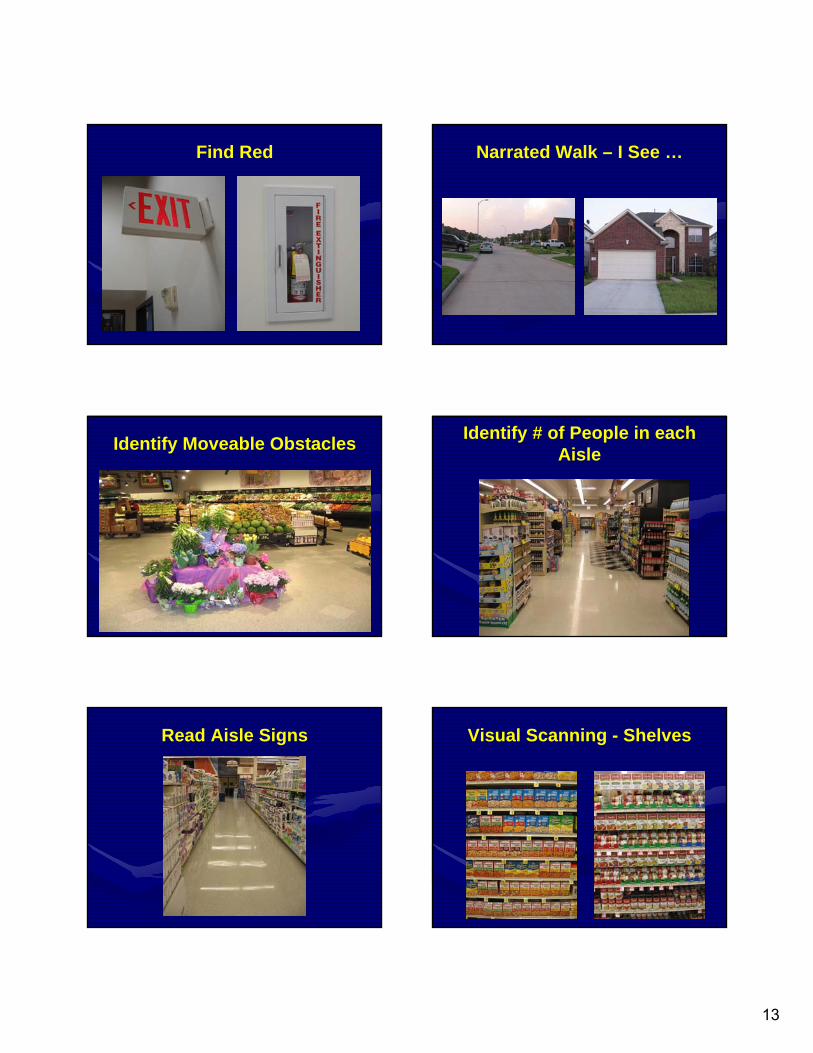
Find Red Narrated Walk – I See …
Identify Moveable ObstaclesIdentify # of People in each
Aisle
Read Aisle Signs Visual Scanning - Shelves
14
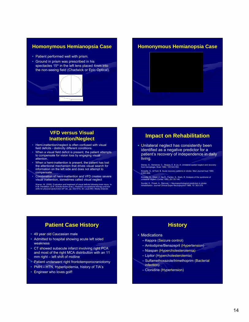
Homonymous Hemianopsia Case
• Patient performed well with prism.
• Ground in prism was prescribed in his spectacles 15^ in the left lens placed 4mm into the non-seeing field (Chadwick or Epic Optical).
Homonymous Hemianopsia Case
VFD versus Visual Inattention/Neglect
• Hemi-inattention/neglect is often confused with visual field deficits - distinctly different conditions.
• When a visual field deficit is present, the patient attempts to compensate for vision loss by engaging visual attention.
• When a hemi-inattention is present, the patient has lost the attentional mechanism that drives visual search for information on the left side and does not attempt to compensate.
• Combination of hemi-inattention and VFD creates severe visual inattention, sometimes called visual neglect
Warren, M. (2006). Evaluation and treatment of visual deficits following brain injury. In H.M. Pendleton, & W. Schultz-Krohn (Eds.), Pedretti’s occupational therapy practice skills for physical dysfunction (6th ed., pp. 532-573). St. Louis MO: Mosby Elsevier.
Impact on Rehabilitation
• Unilateral neglect has consistently been identified as a negative predictor for a patient’s recovery of independence in daily living.
Denes, G., Semenza, C., Stoppa, E. & Lis, A. Unilateral spatial neglect and recovery from hemiplegia. Brain 1982; 105:543-552.
Kinsella, G., & Ford, B. Acute recovery patterns in stroke. Med Journal Aust 1980; 2:663-666.
Kinsella, G., Olver, J., Ng K., Packer, S., Stark, R. Analysis of the syndrome of unilateral neglect. Cortex 1993; 29:135-140.
Sundet, K., Finset, A., Reinvan, I. Neuropsychological predictors in stroke rehabilitation. Journal Clinical Exper Neuropsychol 1988; 10: 363-379.
Patient Case History
• 49 year old Caucasian male
• Admitted to hospital showing acute left sided weakness
• CT showed subacute infarct involving right PCA and most of the right MCA distribution with an 11 mm right – left shift of midline
• Patient underwent right frontotemporocraniotomy
• PMH – HTN, Hyperlipidemia, history of TIA’s
• Engineer who loves golf!
History
• Medications– Keppra (Seizure control)
– Amlodipine/Benazepril (Hypertension)
– Niaspan (Hypercholesterolemia)
– Lipitor (Hypercholesterolemia)
– Sulfamethoxazole/trimethoprim (Bacterial infection)
– Clonidine (Hypertension)
15
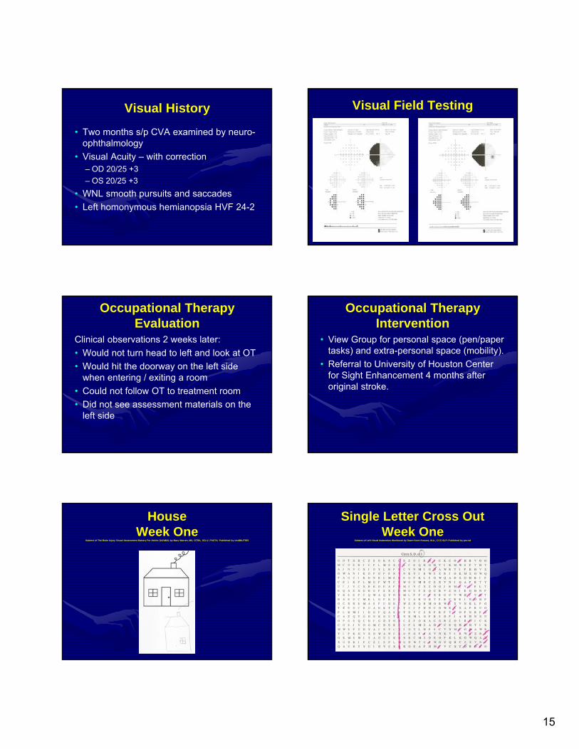
Visual History
• Two months s/p CVA examined by neuro-ophthalmology
• Visual Acuity – with correction– OD 20/25 +3
– OS 20/25 +3
• WNL smooth pursuits and saccades
• Left homonymous hemianopsia HVF 24-2
Visual Field Testing
Occupational Therapy Evaluation
Clinical observations 2 weeks later:
• Would not turn head to left and look at OT
• Would hit the doorway on the left side when entering / exiting a room
• Could not follow OT to treatment room
• Did not see assessment materials on the left side
Occupational Therapy Intervention
• View Group for personal space (pen/paper tasks) and extra-personal space (mobility).
• Referral to University of Houston Center for Sight Enhancement 4 months after original stroke.
HouseWeek One
Subtest of The Brain Injury Visual Assessment Battery For Adults (biVABA) by Mary Warren, MS, OTR/L, SCLV, FAOTA; Published by visABILITIES
Single Letter Cross OutWeek One
Subtest of Left Visual Inattention Workbook by Dawn Scott Knauss, M.A., CCC-SLP; Published by pro-ed
16
Random Plain Circles SimpleSubtest of The Brain Injury Visual Assessment Battery For Adults (biVABA) by Mary Warren, MS, OTR/L, SCLV, FAOTA; Published by visABILITIES
Visit at the University of Houston College of Optometry
• Wearing bifocal contact lenses, some difficulty with insertion and removal
• RE 20/20, LE 20/40
• Left Homonymous Hemianopsia – records were faxed, confirmed with confrontation fields
• Neglect testing completed
• Goals include playing golf and driving
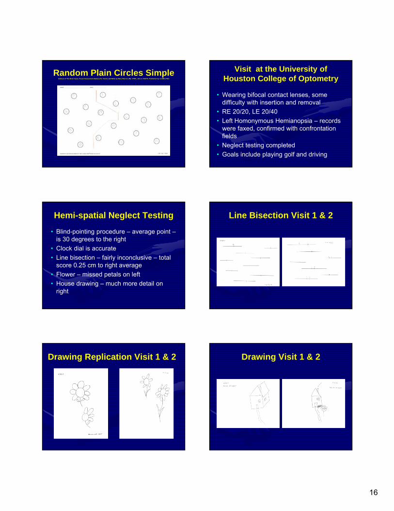
Hemi-spatial Neglect Testing
• Blind-pointing procedure – average point –is 30 degrees to the right
• Clock dial is accurate
• Line bisection – fairly inconclusive – total score 0.25 cm to right average
• Flower – missed petals on left
• House drawing – much more detail on right
Line Bisection Visit 1 & 2
Drawing Replication Visit 1 & 2 Drawing Visit 1 & 2
17
Management
• Initiated treatment with base left yoked prism 20^ magnitude
• Treatment twice a day for 3 weeks with family
• Return for contact lens evaluation and neglect follow-up
Neglect Intervention• Use prompts for missing side - i.e. bold color in
margin for reading, move materials in the room to the affected side.
• Use of kinesthetic feedback is helpful.
• Use of yoked prism training protocol to assist patient to compensate, possibly a more central response. – Rossetti et. al. Nature v395:166-169,1998
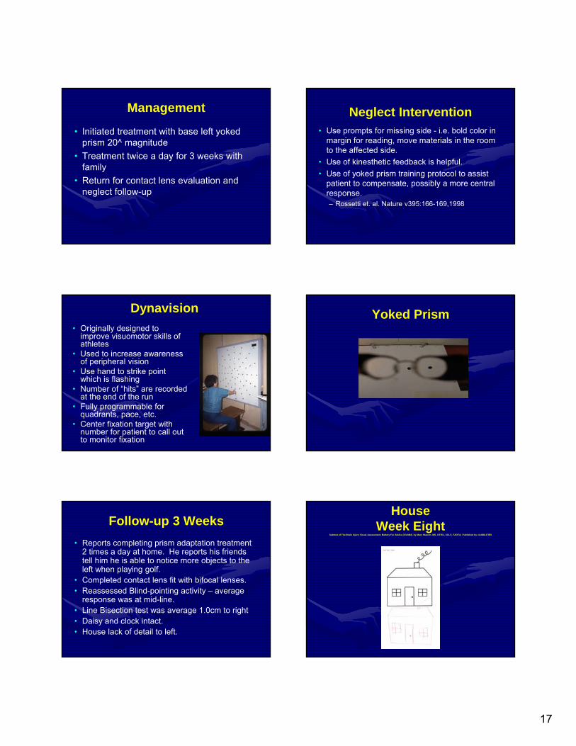
Dynavision
• Originally designed to improve visuomotor skills of athletes
• Used to increase awareness of peripheral vision
• Use hand to strike point which is flashing
• Number of “hits” are recorded at the end of the run
• Fully programmable for quadrants, pace, etc.
• Center fixation target with number for patient to call out to monitor fixation
Yoked Prism
Follow-up 3 Weeks
• Reports completing prism adaptation treatment 2 times a day at home. He reports his friends tell him he is able to notice more objects to the left when playing golf.
• Completed contact lens fit with bifocal lenses. • Reassessed Blind-pointing activity – average
response was at mid-line.• Line Bisection test was average 1.0cm to right• Daisy and clock intact. • House lack of detail to left.
HouseWeek Eight
Subtest of The Brain Injury Visual Assessment Battery For Adults (biVABA) by Mary Warren, MS, OTR/L, SCLV, FAOTA; Published by visABILITIES
18
Single Letter Search CrowdedSubtest of The Brain Injury Assessment Battery For Adults (biVABA) by Mary Warren, MS, OTR/L, SCLV, FAOTA; Published by visABILITIES
Random Plain Circles SimpleSubtest of The Brain Injury Visual Assessment Battery For Adults (biVABA) by Mary Warren MS, OTR/L, SCLV, FAOTA; Published by visABILITIES
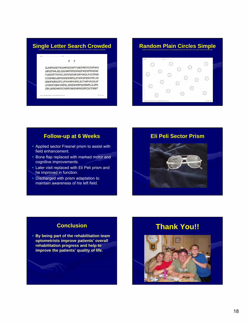
Follow-up at 6 Weeks
• Applied sector Fresnel prism to assist with field enhancement.
• Bone flap replaced with marked motor and cognitive improvements.
• Later visit replaced with Eli Peli prism and he improved in function.
• Discharged with prism adaptation to maintain awareness of his left field.
Eli Peli Sector Prism
Conclusion
• By being part of the rehabilitation team optometrists improve patients’ overall rehabilitation progress and help to improve the patients’ quality of life.
Thank You!!


![Tenaga/denpasar.bpk.go.id/wp-content/uploads/2016/04/Nusa-Bali-12-April-2016.pdf · Tenaga/ Upah Pungut 4 BulanTak ... permohonan legal opinion (tirr-jauan yu ridis] ... Honor untuk](https://img.pdfslide.net/doc/110x75/5d317ec388c9936e768b9e53/tenaga-tenaga-upah-pungut-4-bulantak-permohonan-legal-opinion-tirr-jauan.jpg)