Embed Size (px)
Citation preview
Introduction to Abdominal Radiology
Dr. LeeAnn Pack
Dipl. ACVR
Abdominal Radiography
• Abdominal Preparation– Withhold food for 12-24 hours as needed– Give enema 2-3 hours before study
• Exceptions– Critically ill– Suspect obstruction (acute abdomen)
Indications
• Vomiting
• Abdominal pain
• Hematuria
• Pain on defecation
• Abdominal mass
• Pendulous fluid filled abdomen
• Many many more
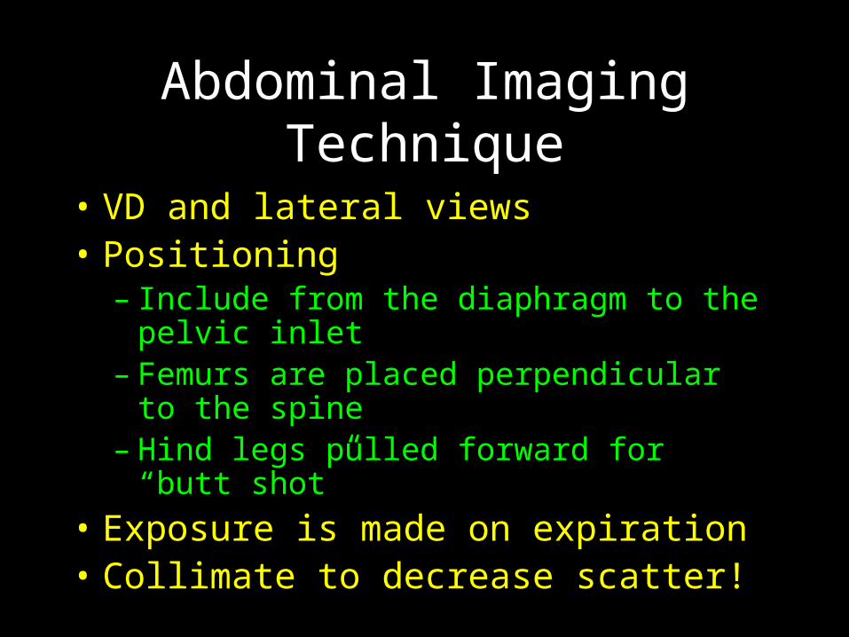
Abdominal Imaging Technique
• VD and lateral views• Positioning
– Include from the diaphragm to the pelvic inlet
– Femurs are placed perpendicular to the spine
– Hind legs pulled forward for “butt shot”
• Exposure is made on expiration• Collimate to decrease scatter!
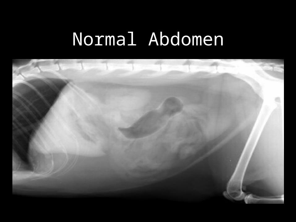
Normal Abdomen
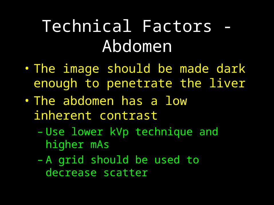
Technical Factors - Abdomen
• The image should be made dark enough to penetrate the liver
• The abdomen has a low inherent contrast– Use lower kVp technique and higher mAs– A grid should be used to decrease scatter
Structures Normally Seen
• Liver • Spleen• Kidneys• Stomach• Duodenum• Small Intestine
• Cecum• Colon• Bladder• Prostate• Retroperitoneal fat
Structures Not Normally Seen
• Gall bladder• Pancreas• Adrenals• Ovaries• Uterus
• Ureters• Lymph nodes• Mesentery• Vasculature
Radiography of the Liver
• Liver size– Normal– Increased– Decreased
• Liver opacity– Increased– decreased
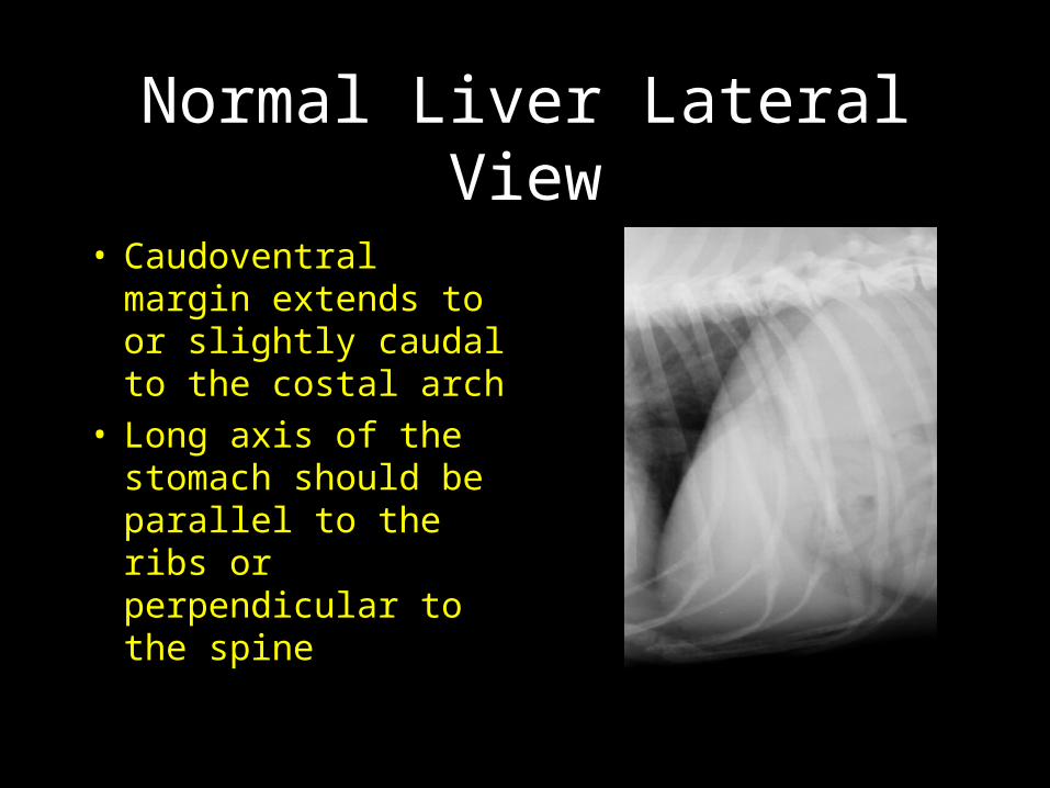
Normal Liver Lateral View
• Caudoventral margin extends to or slightly caudal to the costal arch
• Long axis of the stomach should be parallel to the ribs or perpendicular to the spine

Normal Liver VD View
• Long axis of the stomach is perpendicular to the spine
• Caudal margins of the liver are difficult to visualize on this view
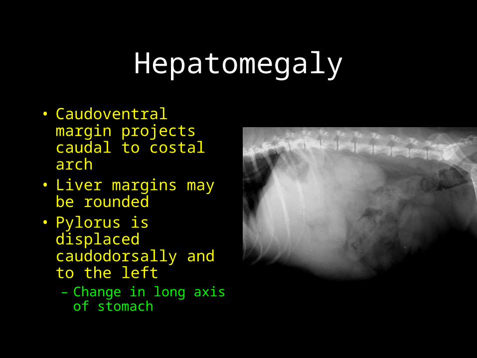
Hepatomegaly
• Caudoventral margin projects caudal to costal arch
• Liver margins may be rounded
• Pylorus is displaced caudodorsally and to the left– Change in long axis
of stomach
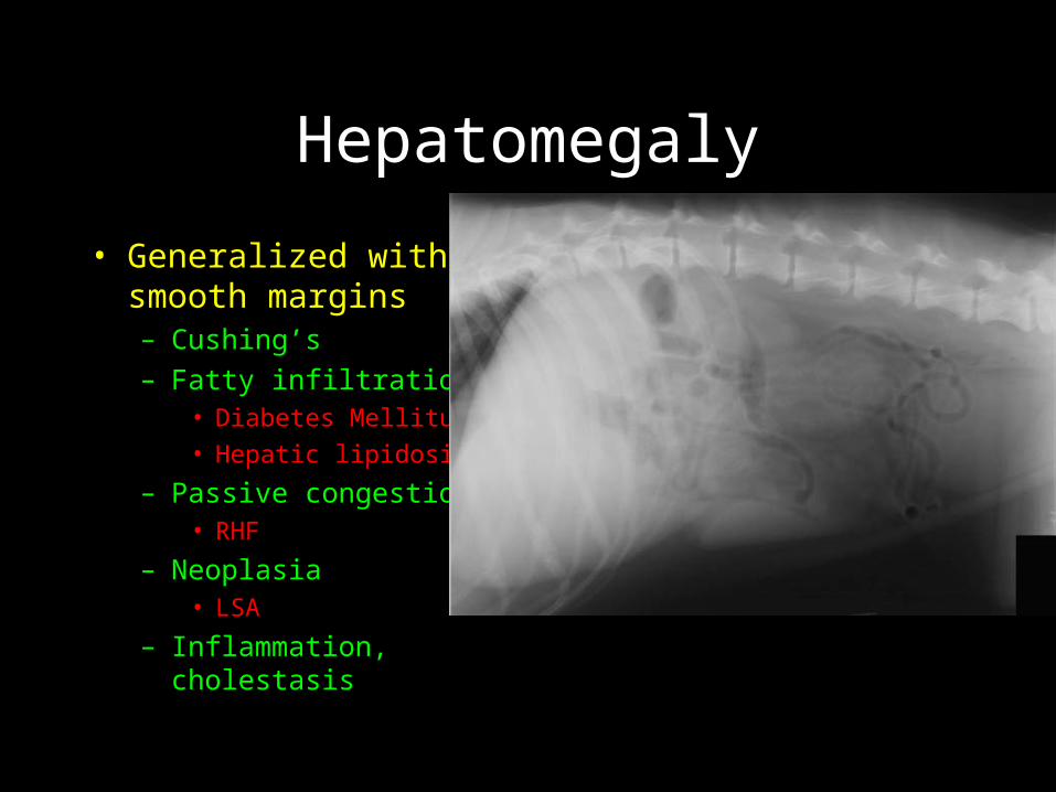
Hepatomegaly
• Generalized with smooth margins– Cushing’s– Fatty infiltration
• Diabetes Mellitus
• Hepatic lipidosis
– Passive congestion• RHF
– Neoplasia• LSA
– Inflammation, cholestasis
Hepatomegaly
• General enlargement lumpy margins– Malignant neoplasia– Nodular hyperplasia
• Focal enlargement– Neoplasia– Nodular hyperplasia– Cysts, abscesses
Microhepatia
• Stomach shifted cranially – especially pylorus– May be functionally normal– Portosystemic shunt– Hepatic fibrosis
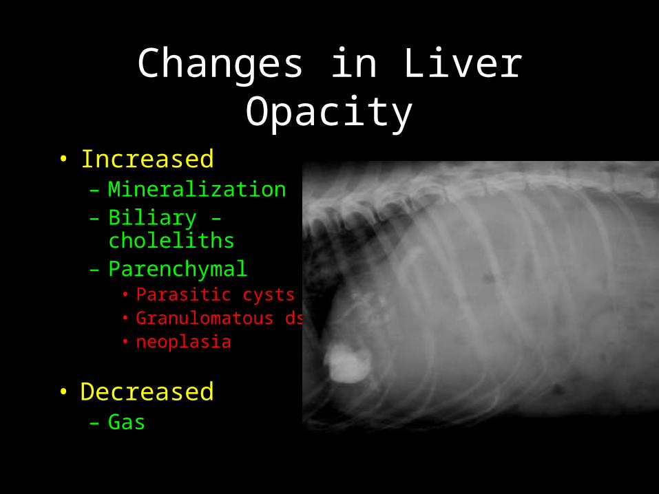
Changes in Liver Opacity
• Increased– Mineralization– Biliary – choleliths– Parenchymal
• Parasitic cysts• Granulomatous ds• neoplasia
• Decreased– Gas
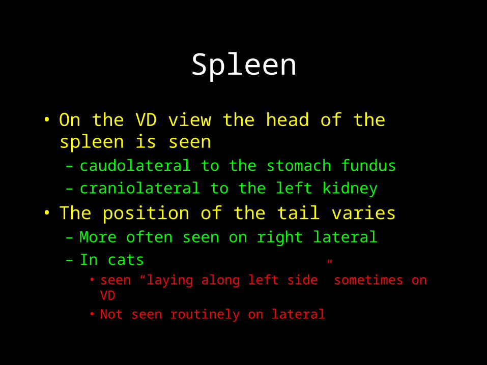
Spleen
• On the VD view the head of the spleen is seen– caudolateral to the stomach fundus– craniolateral to the left kidney
• The position of the tail varies– More often seen on right lateral– In cats
• seen “laying along left side” sometimes on VD• Not seen routinely on lateral
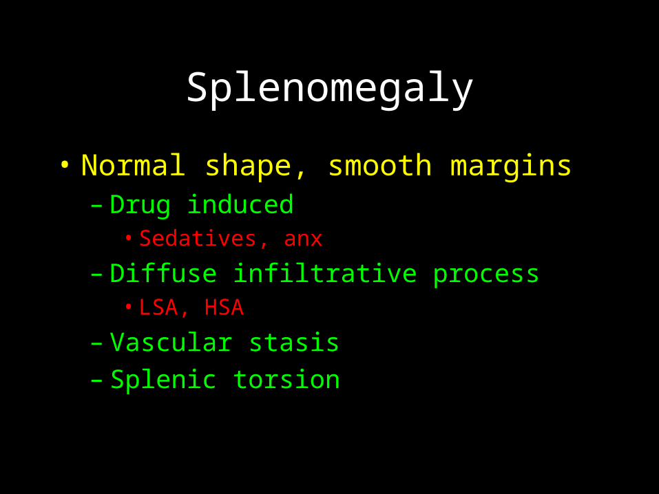
Splenomegaly
• Normal shape, smooth margins– Drug induced
• Sedatives, anx
– Diffuse infiltrative process• LSA, HSA
– Vascular stasis– Splenic torsion
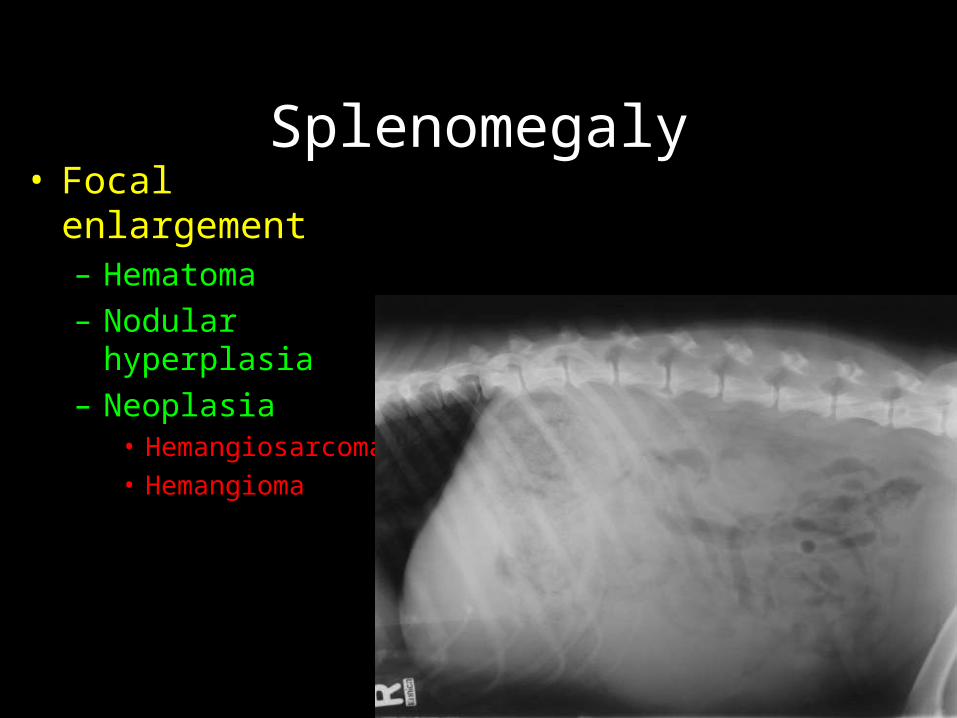
Splenomegaly• Focal enlargement
– Hematoma– Nodular hyperplasia– Neoplasia
• Hemangiosarcoma• Hemangioma
Splenic Masses
• May occur in the head, body or tail
• Located mid abdomen, left or right
• May be very large
• Can cause abdominal organ displacement– Can displace stomach cranially and small
intestines in various direction depending on location
Kidneys
• Right located more cranial than left
• Dogs = 2½-3½ * L2 on VD
• Cats = 2.4-3 * L2 on VD
• Size should only be evaluated on the VD view due to magnification on the lateral
• IV contrast can be used if necessary
Kidneys• Increase in size
– Acute inflammation– Infiltrative process
• LSA
• Decrease in size– Hypoplasia– Fibrosis– Renal failure
• Mineralization – look a kids on both views• Focal change in shape
– ACA
Stomach
• Caudal to liver
• Axis parallel to ribs
• Change in size, shape, mineralized, rugal fold abnormal
• Right vs. Left lateral (air/fluid)
• Foreign bodies, outflow obstruction

Stomach
• Dog – crosses from left to right
• Cat – from left to midline

Which one is Left? Right?
Small Intestine
• Duodenum – fixed along right side
• Jejunum and ileum – position varies
• Normal width = < 3* last rib width
• Contains both air and fluid
• Can not determine wall thickness
• Peyer’s patches, string of pearls

VD Abdomen
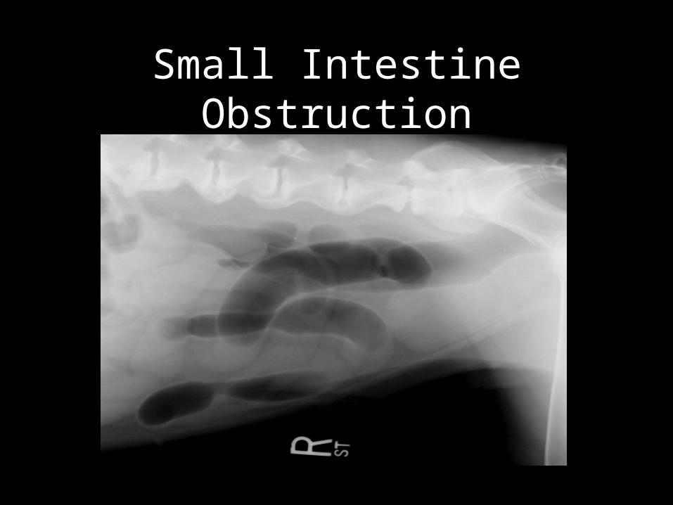
Small Intestine Obstruction
Cecum and Colon
• Cecum– mid right abdomen– Comma shaped –may contain air– Not often seen in cats
• Colon– Ascending, Transverse, Descending– Normal width = < 5 * last rib width
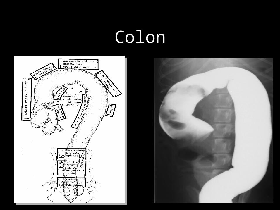
Colon
Urinary Bladder
• Dog – caudal abdomen or pelvic
• Cat – always intra-abdominal
• Vary in size (empty to very distended)
• Bladder wall changes can not be determined on radiographs
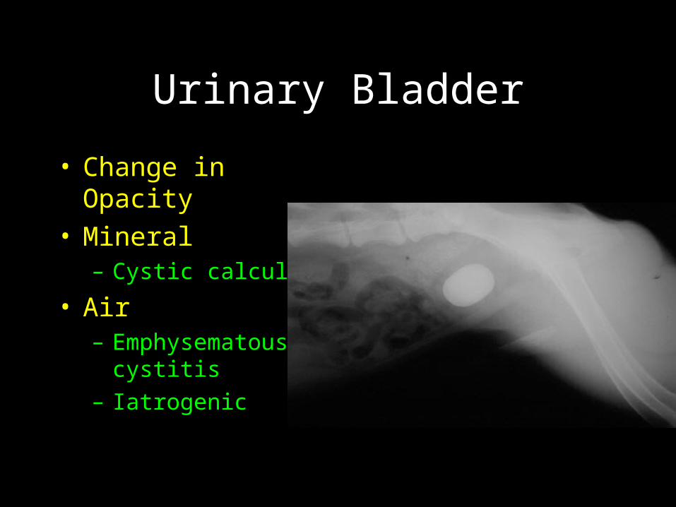
Urinary Bladder
• Change in Opacity• Mineral
– Cystic calculi
• Air– Emphysematous
cystitis– Iatrogenic
Prostate
• Usually well visualized in intact males
• Should be symmetrical with smooth margins
• Enlarged if – > 50% of pelvis inlet width (VD)– >70% of sacro-pubic distance (lateral)
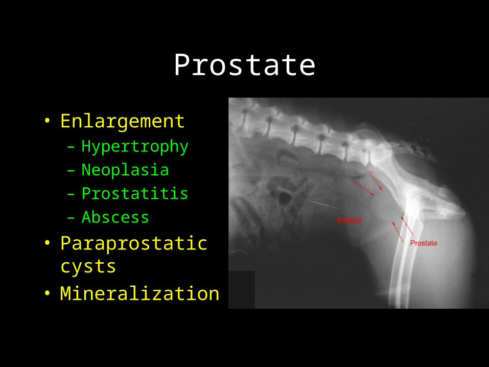
Prostate
• Enlargement– Hypertrophy– Neoplasia– Prostatitis– Abscess
• Paraprostatic cysts• Mineralization
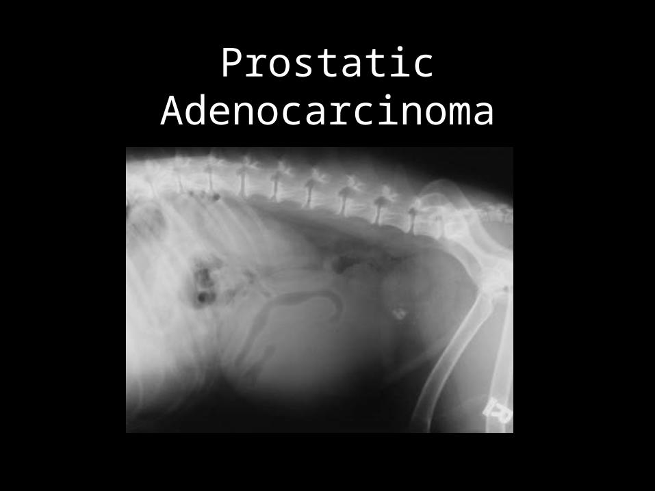
Prostatic Adenocarcinoma
Pancreatitis
• The pancreas is not normally seen• Increased density and decreased
serosal detail in the right cranial quadrant
• Duodenum may be persistently distended with gas (sentinel sign)
• Duodenum can be pushed to the right and pyloroduodenal angle is increased
Adrenal Glands
• Seen only when enlarged or mineralized• Enlargement
– Pheochromocytoma– Cortical carcinoma– Adenoma
• Adrenal mineralization– Dystrophic mineralization of tumors– Mineralization of non neoplastic adrenals
(cats)

Reproductive System
• Uterine enlargement– Metra’s– Gravid uterus
• Ovarian enlargement– Neoplasia
• Enlarged retained testicle– neoplasia
Enlarged Lymph Nodes
• Medial iliac (sublumbar)– Increased opacity (soft tissue) seen in
caudal abdomen ventral to caudal lumbar spine
– May displace colon ventrally
• Mesenteric LNN rarely large enough for radiographic detection
• US is best to evaluate for LAN
Enlarged Medial Iliac LN
• Lymphosarcoma – Most common
• Metastasis from neoplasia in the pelvis canal or further caudally– Prostate– Perineal tumors
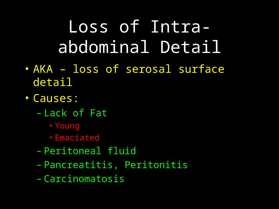
Loss of Intra-abdominal Detail
• AKA – loss of serosal surface detail
• Causes:– Lack of Fat
• Young• Emaciated
– Peritoneal fluid– Pancreatitis, Peritonitis– Carcinomatosis
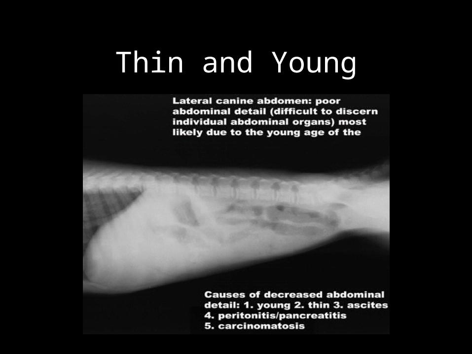
Thin and Young
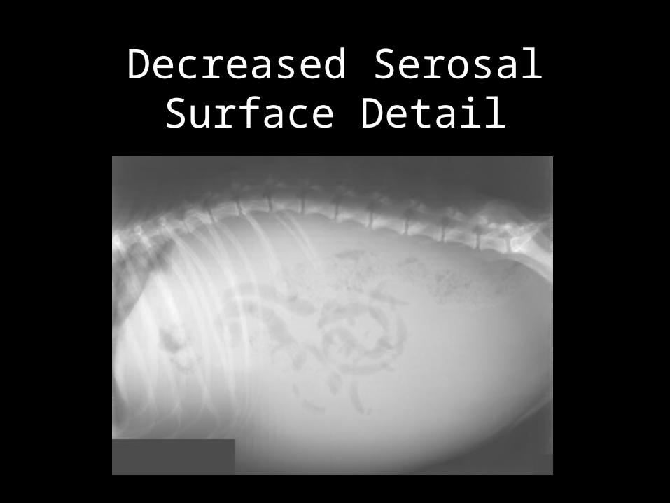
Decreased Serosal Surface Detail
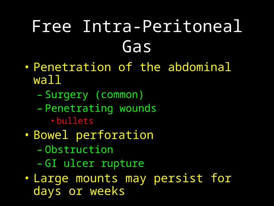
Free Intra-Peritoneal Gas
• Penetration of the abdominal wall– Surgery (common)– Penetrating wounds
• bullets
• Bowel perforation – Obstruction– GI ulcer rupture
• Large mounts may persist for days or weeks
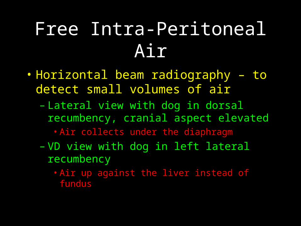
Free Intra-Peritoneal Air
• Horizontal beam radiography – to detect small volumes of air– Lateral view with dog in dorsal
recumbency, cranial aspect elevated• Air collects under the diaphragm
– VD view with dog in left lateral recumbency• Air up against the liver instead of fundus
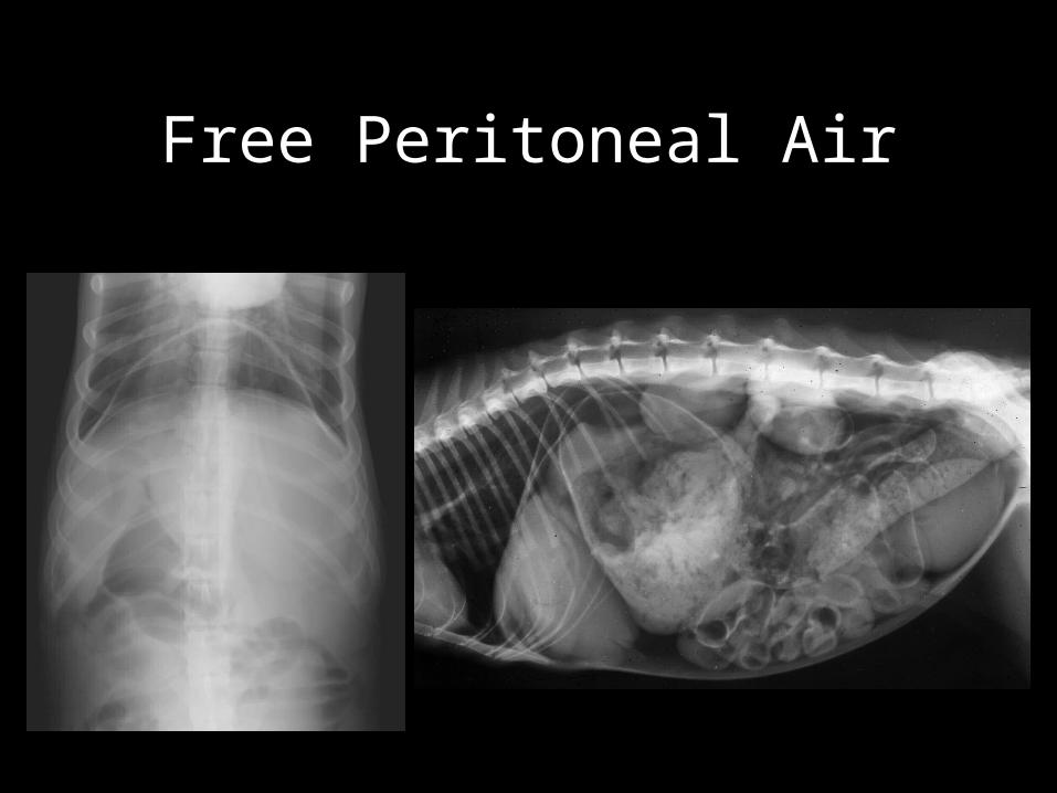
Free Peritoneal Air