Embed Size (px)
Citation preview
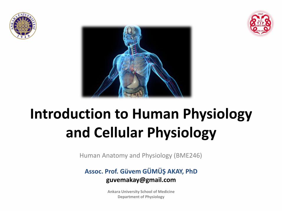
Introduction to Human Physiology and Cellular Physiology
Assoc. Prof. Güvem GÜMÜŞ AKAY, [email protected]
Ankara University School of Medicine Department of Physiology
Human Anatomy and Physiology (BME246)
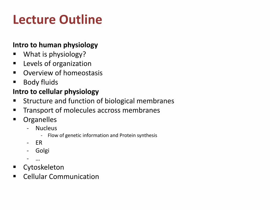
Lecture Outline
Intro to human physiology What is physiology? Levels of organization Overview of homeostasis Body fluidsIntro to cellular physiology Structure and function of biological membranes Transport of molecules accross membranes Organelles
- Nucleus- Flow of genetic information and Protein synthesis
- ER- Golgi- …
Cytoskeleton Cellular Communication

Introduction to Human Physiology
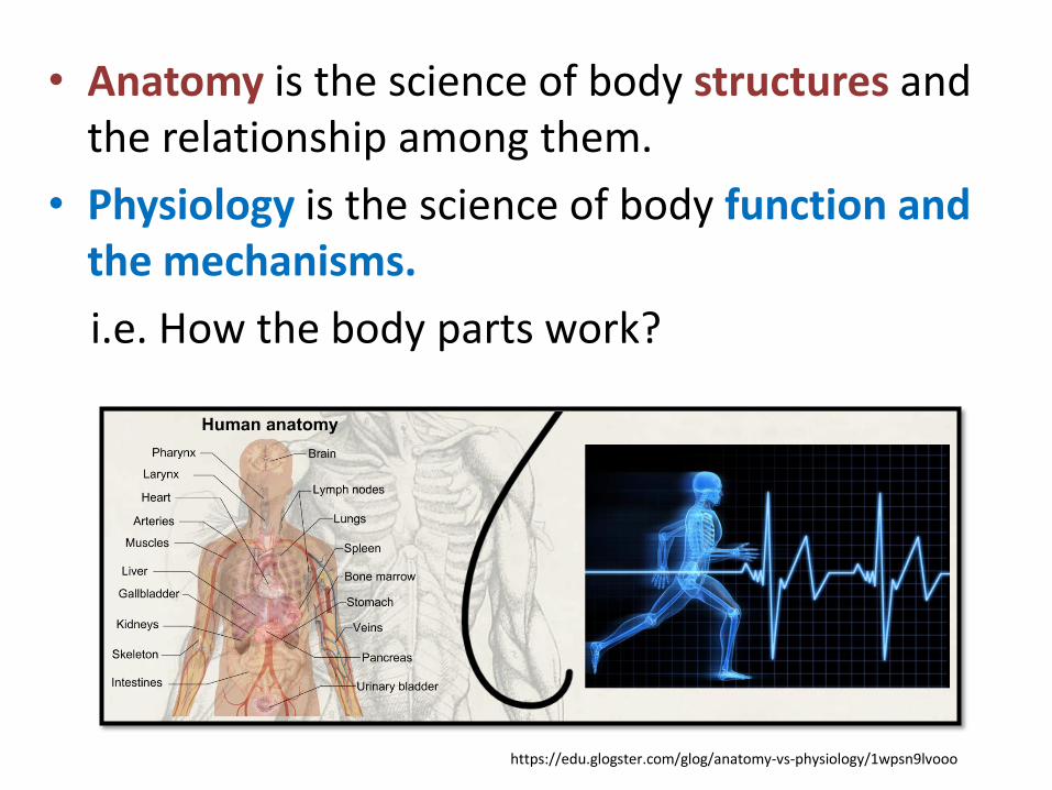
• Anatomy is the science of body structures andthe relationship among them.
• Physiology is the science of body function andthe mechanisms.
i.e. How the body parts work?
https://edu.glogster.com/glog/anatomy-vs-physiology/1wpsn9lvooo
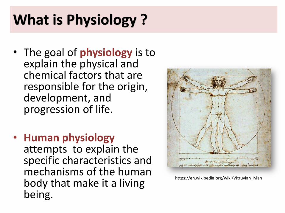
What is Physiology ?
• The goal of physiology is to explain the physical andchemical factors that are responsible for the origin,development, and progression of life.
• Human physiologyattempts to explain the specific characteristics and mechanisms of the human body that make it a living being.
https://en.wikipedia.org/wiki/Vitruvian_Man
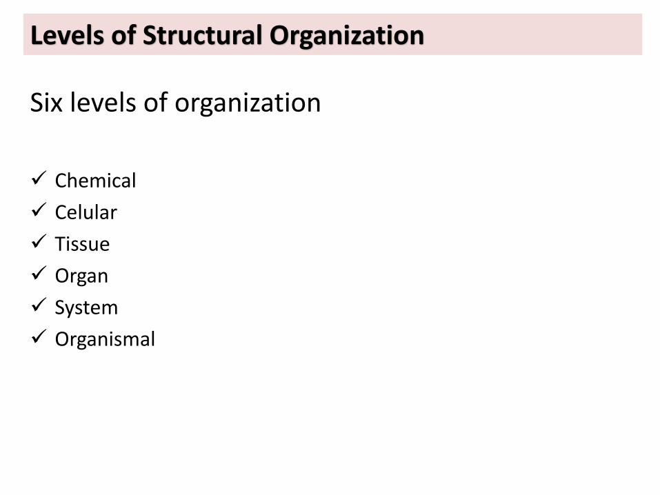
Levels of Structural Organization
Six levels of organization
Chemical
Celular
Tissue
Organ
System
Organismal
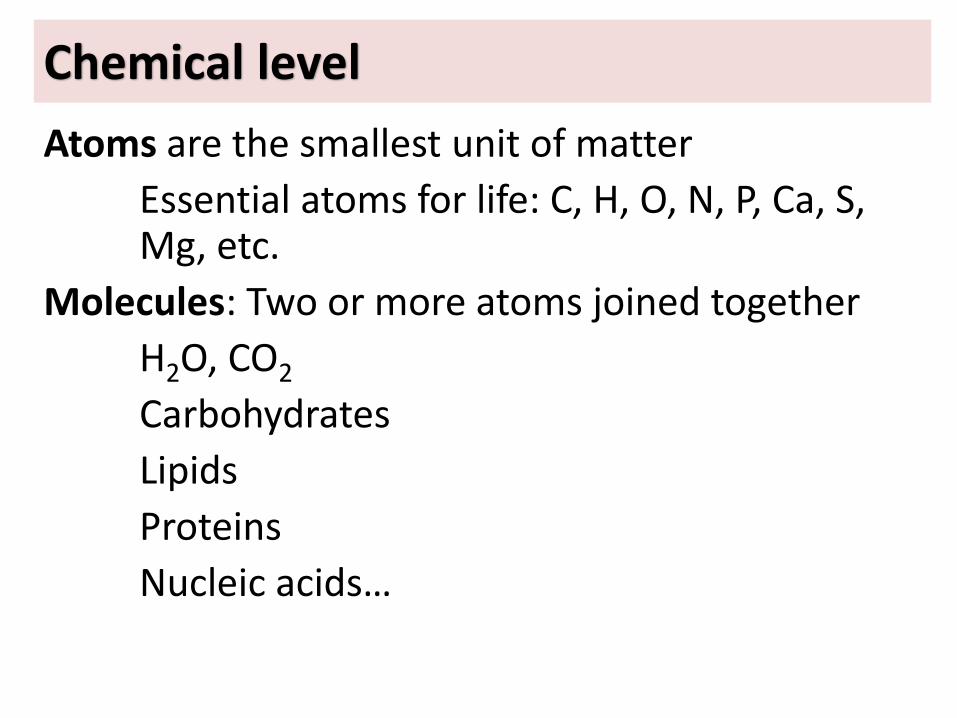
Chemical level
Atoms are the smallest unit of matter
Essential atoms for life: C, H, O, N, P, Ca, S, Mg, etc.
Molecules: Two or more atoms joined together
H2O, CO2
Carbohydrates
Lipids
Proteins
Nucleic acids…
Cellular level
Basic structural and functional units of an organism
˜200 different kinds of cells
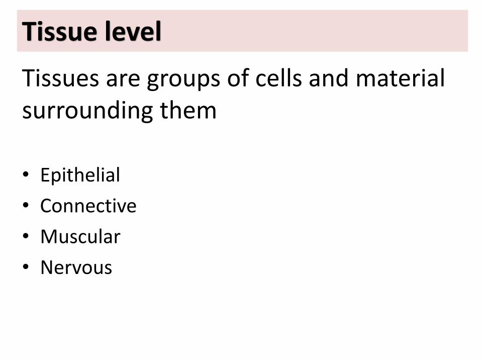
Tissue level
Tissues are groups of cells and materialsurrounding them
• Epithelial
• Connective
• Muscular
• Nervous
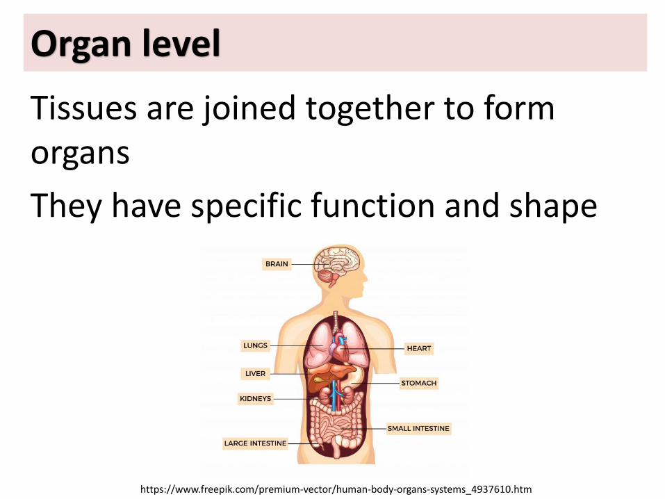
Organ level
Tissues are joined together to form organs
They have specific function and shape
https://www.freepik.com/premium-vector/human-body-organs-systems_4937610.htm
System level
A system consists of related organs witha common function
The Blood Circulation System
Respiratory System
Digestive System
Musculoskeletal System
Urinary System (Renal System)
Reproductive system
Major Functional Systems
Regulation of Body Functions
Nervous System
Endocrine System (Hormones)
14
Body Fluids
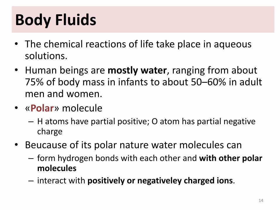
• The chemical reactions of life take place in aqueous solutions.
• Human beings are mostly water, ranging from about 75% of body mass in infants to about 50–60% in adult men and women.
• «Polar» molecule– H atoms have partial positive; O atom has partial negative
charge
• Beucause of its polar nature water molecules can– form hydrogen bonds with each other and with other polar
molecules
– interact with positively or negativeley charged ions.
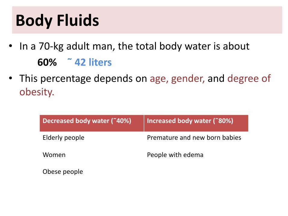
• In a 70-kg adult man, the total body water is about
60% ˜ 42 liters
• This percentage depends on age, gender, and degree of obesity.
Body Fluids
Decreased body water (˜40%) Increased body water (˜80%)
Elderly people Premature and new born babies
Women People with edema
Obese people
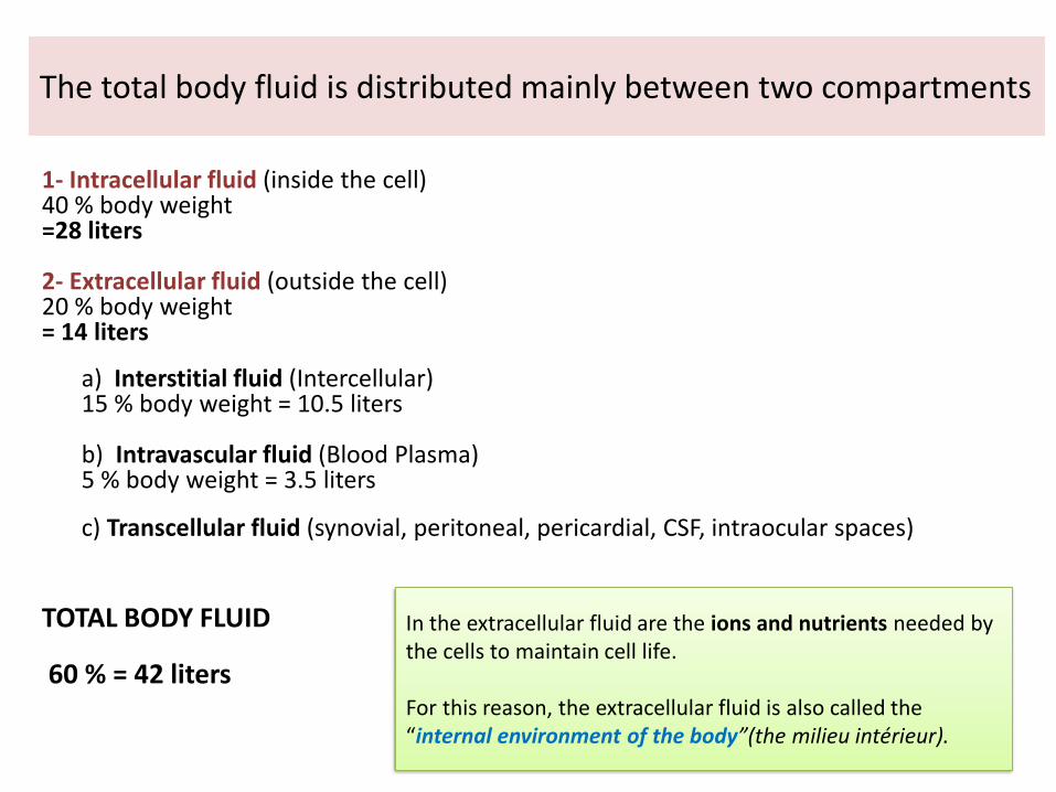
The total body fluid is distributed mainly between two compartments
1- Intracellular fluid (inside the cell)40 % body weight=28 liters
2- Extracellular fluid (outside the cell)20 % body weight= 14 liters
a) Interstitial fluid (Intercellular)15 % body weight = 10.5 liters
b) Intravascular fluid (Blood Plasma)5 % body weight = 3.5 liters
c) Transcellular fluid (synovial, peritoneal, pericardial, CSF, intraocular spaces)
TOTAL BODY FLUID
60 % = 42 liters
In the extracellular fluid are the ions and nutrients needed by the cells to maintain cell life.
For this reason, the extracellular fluid is also called the “internal environment of the body”(the milieu intérieur).

Homeostasis• The term “homeostasis” is used by physiologists
to explain maintenance of nearly constant conditions in the internal environment.
• Essentially all organs and tissues of the body perform functions that help maintain theseconstant conditions.
Characteristics of Control Systems
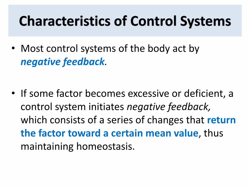
• Most control systems of the body act by negative feedback.
• If some factor becomes excessive or deficient, a control system initiates negative feedback, which consists of a series of changes that return the factor toward a certain mean value, thusmaintaining homeostasis.
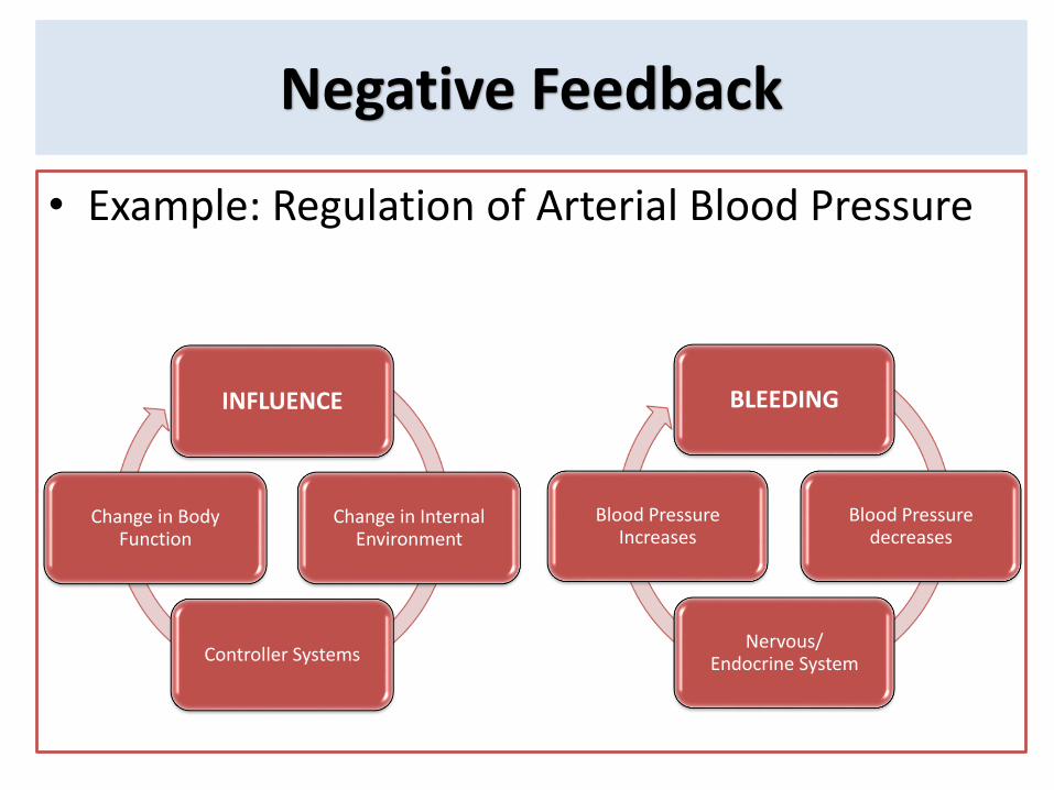
Negative Feedback
• Example: Regulation of Arterial Blood Pressure
INFLUENCE
Change in InternalEnvironment
Controller Systems
Change in Body Function
BLEEDING
Blood Pressuredecreases
Nervous/Endocrine System
Blood PressureIncreases
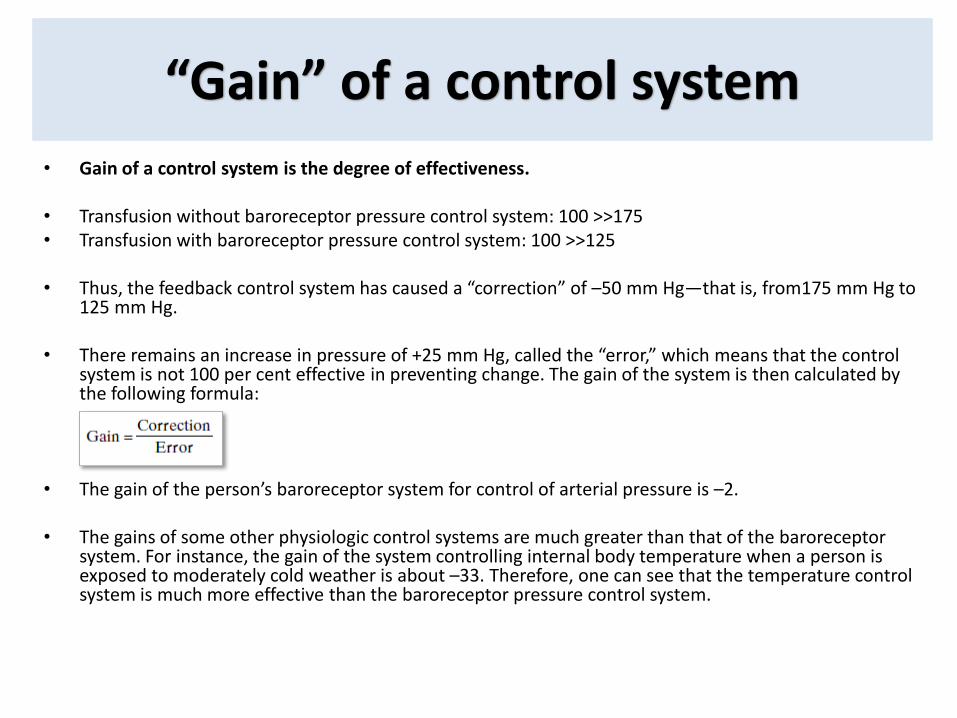
“Gain” of a control system
• Gain of a control system is the degree of effectiveness.
• Transfusion without baroreceptor pressure control system: 100 >>175• Transfusion with baroreceptor pressure control system: 100 >>125
• Thus, the feedback control system has caused a “correction” of –50 mm Hg—that is, from175 mm Hg to 125 mm Hg.
• There remains an increase in pressure of +25 mm Hg, called the “error,” which means that the control system is not 100 per cent effective in preventing change. The gain of the system is then calculated by the following formula:
• The gain of the person’s baroreceptor system for control of arterial pressure is –2.
• The gains of some other physiologic control systems are much greater than that of the baroreceptorsystem. For instance, the gain of the system controlling internal body temperature when a person is exposed to moderately cold weather is about –33. Therefore, one can see that the temperature control system is much more effective than the baroreceptor pressure control system.
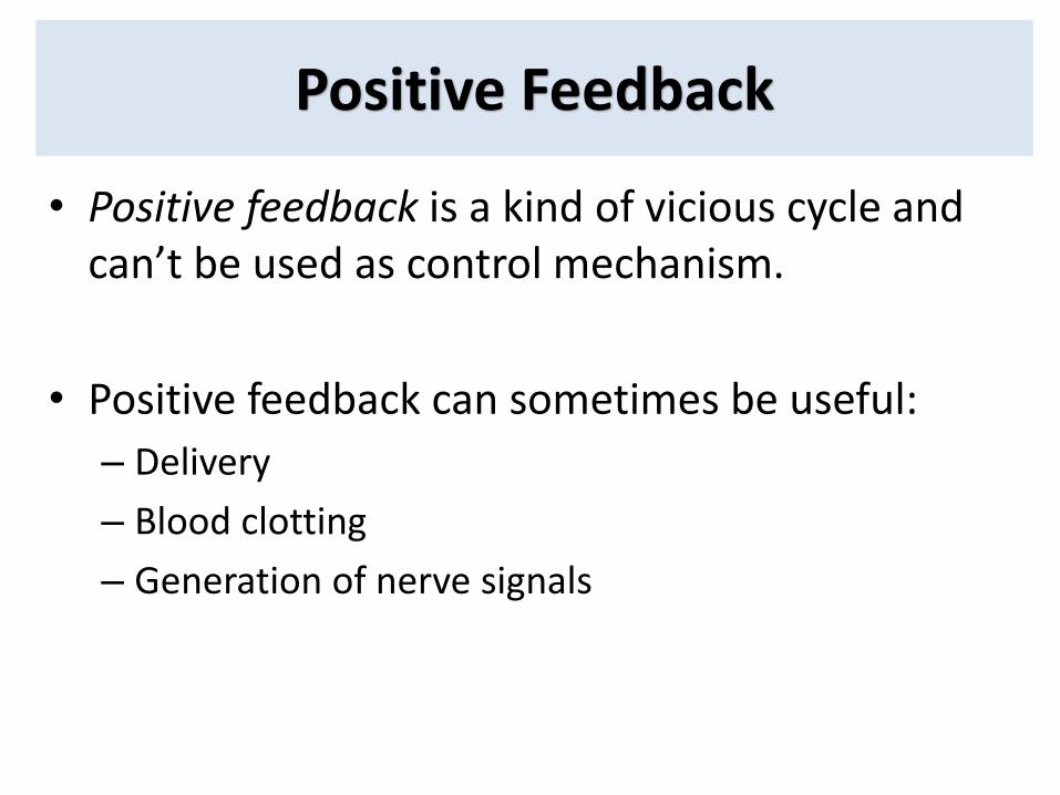
Positive Feedback
• Positive feedback is a kind of vicious cycle andcan’t be used as control mechanism.
• Positive feedback can sometimes be useful:
– Delivery
– Blood clotting
– Generation of nerve signals
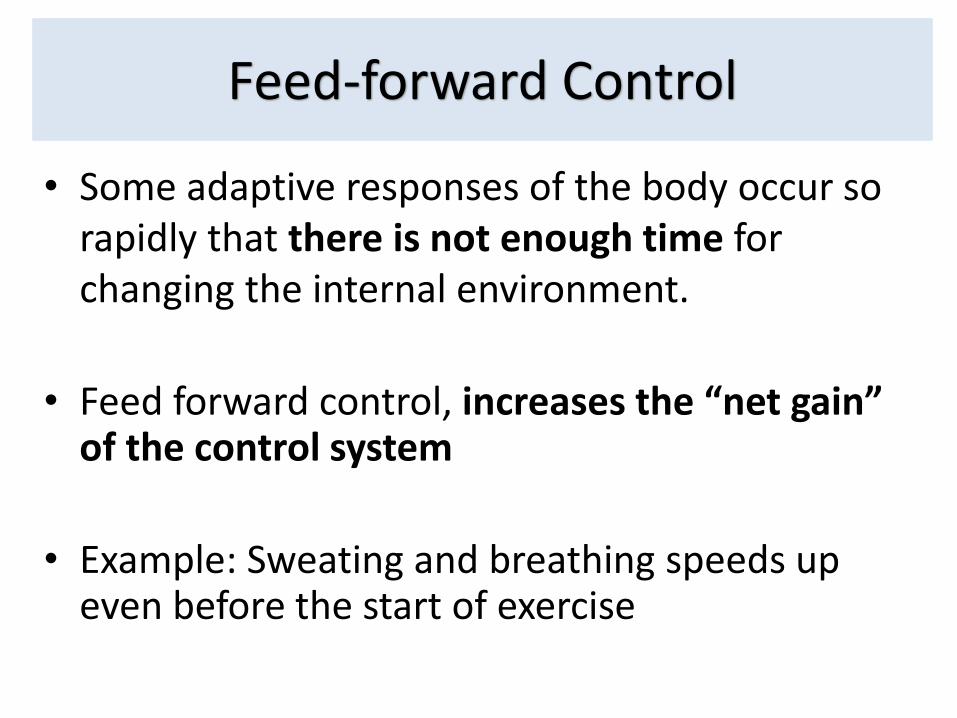
Feed-forward Control
• Some adaptive responses of the body occur so rapidly that there is not enough time forchanging the internal environment.
• Feed forward control, increases the “net gain” of the control system
• Example: Sweating and breathing speeds upeven before the start of exercise
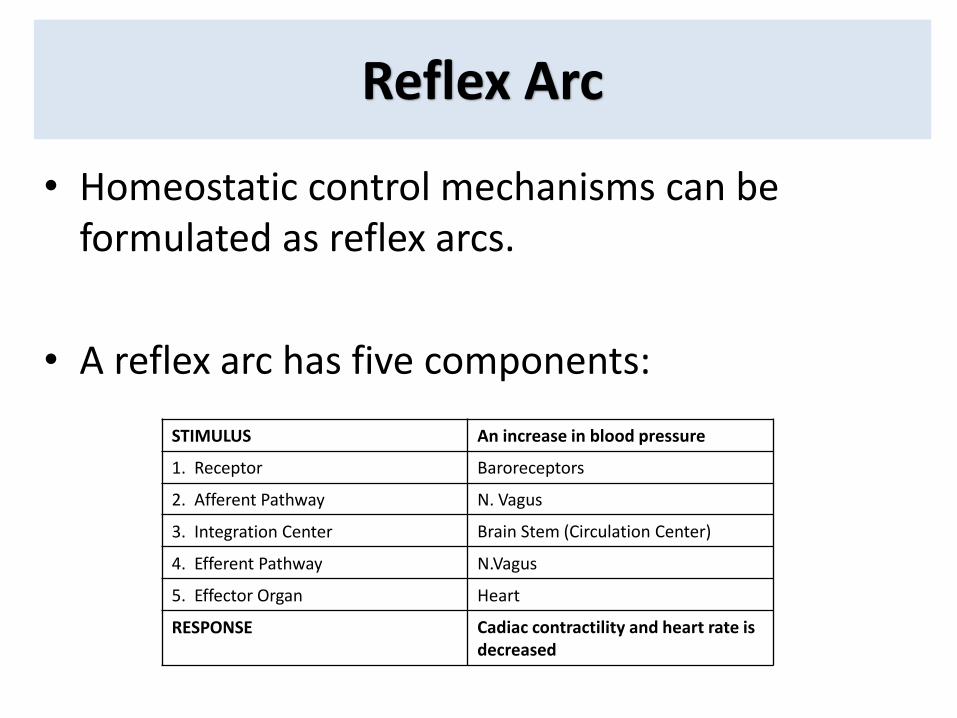
Reflex Arc
• Homeostatic control mechanisms can be formulated as reflex arcs.
• A reflex arc has five components:
STIMULUS An increase in blood pressure
1. Receptor Baroreceptors
2. Afferent Pathway N. Vagus
3. Integration Center Brain Stem (Circulation Center)
4. Efferent Pathway N.Vagus
5. Effector Organ Heart
RESPONSE Cadiac contractility and heart rate is decreased
Introduction to Cellular Physiology
Cells are the structural & functional units of all living organism
• Smallest units that perform all vital physiological functions
• Each cell maintains homeostasis at cellular level
• Homeostasis at tissue, organ, organ system, and organismlevel due to coordinated action of many cells
Biological Membranes
• The structure and functions of the cells are highly dependent on the membranes. The cell membrane (aka. the plasma membrane) is a biological
membrane that separates the interior of the cell (cytosol) from the outside environment (the extracellular space).
Create internal compartments and surround various organelles (e.g. Nucleus, ER, Golgi, …): organellar membranes
• They are impermeable barrier for most water-soluble molecules: Semi-permiable
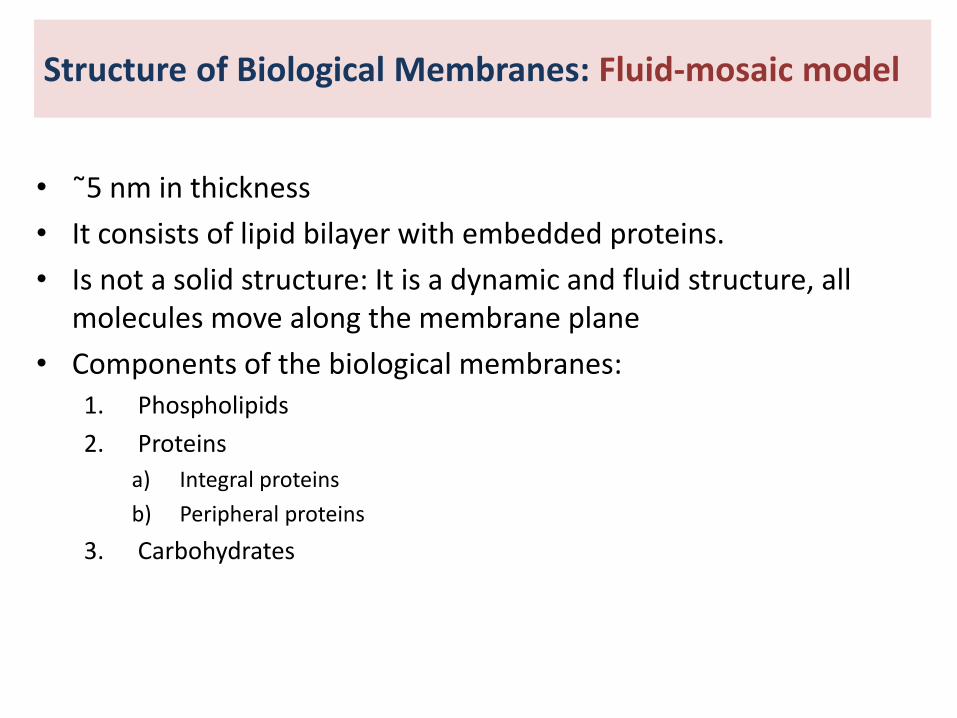
• ˜5 nm in thickness
• It consists of lipid bilayer with embedded proteins.
• Is not a solid structure: It is a dynamic and fluid structure, all molecules move along the membrane plane
• Components of the biological membranes:1. Phospholipids
2. Proteins
a) Integral proteins
b) Peripheral proteins
3. Carbohydrates
Structure of Biological Membranes: Fluid-mosaic model
Structure of Biological Membranes: Fluid-mosaic model
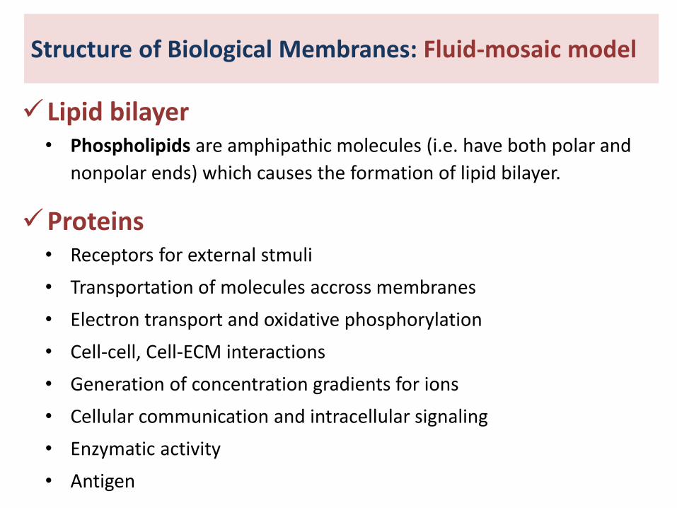
Lipid bilayer• Phospholipids are amphipathic molecules (i.e. have both polar and
nonpolar ends) which causes the formation of lipid bilayer.
Proteins• Receptors for external stmuli
• Transportation of molecules accross membranes
• Electron transport and oxidative phosphorylation
• Cell-cell, Cell-ECM interactions
• Generation of concentration gradients for ions
• Cellular communication and intracellular signaling
• Enzymatic activity
• Antigen
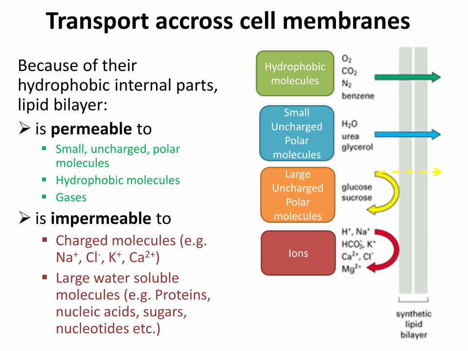
Because of theirhydrophobic internal parts, lipid bilayer:
is permeable to Small, uncharged, polar
molecules
Hydrophobic molecules
Gases
is impermeable to Charged molecules (e.g.
Na+, Cl-, K+, Ca2+)
Large water solublemolecules (e.g. Proteins, nucleic acids, sugars, nucleotides etc.)
Hydrophobicmolecules
SmallUncharged
Polar molecules
LargeUncharged
Polar molecules
Ions
Transport accross cell membranes
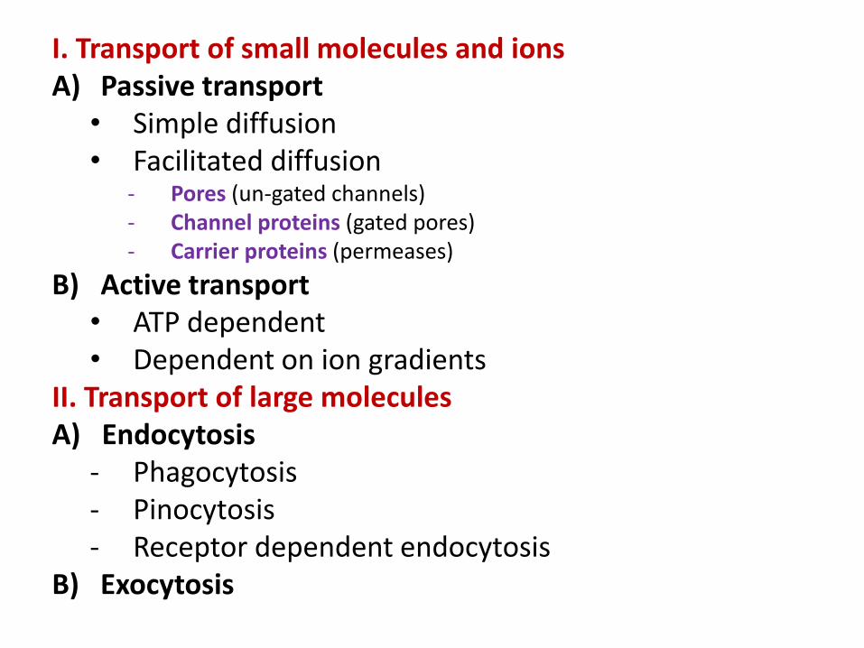
I. Transport of small molecules and ionsA) Passive transport
• Simple diffusion• Facilitated diffusion
- Pores (un-gated channels) - Channel proteins (gated pores)- Carrier proteins (permeases)
B) Active transport• ATP dependent• Dependent on ion gradients
II. Transport of large moleculesA) Endocytosis
- Phagocytosis- Pinocytosis- Receptor dependent endocytosis
B) Exocytosis
Transport of Small Molecules
1) Simple diffusion
2) Facilitated diffusion
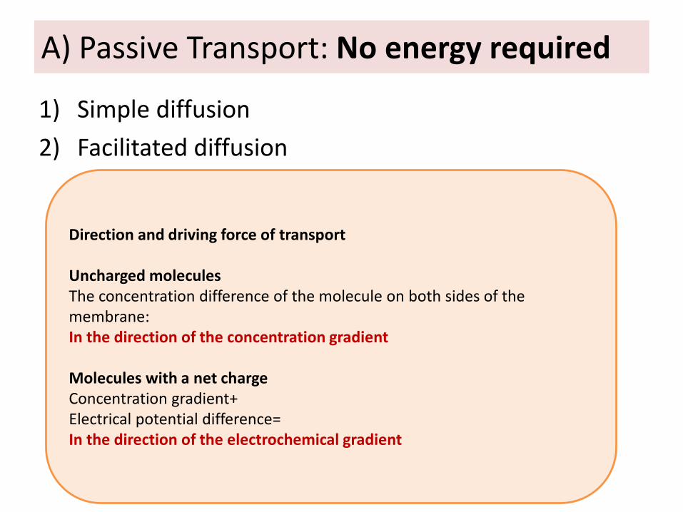
A) Passive Transport: No energy required
Direction and driving force of transport
Uncharged moleculesThe concentration difference of the molecule on both sides of the membrane:In the direction of the concentration gradient
Molecules with a net chargeConcentration gradient+Electrical potential difference= In the direction of the electrochemical gradient
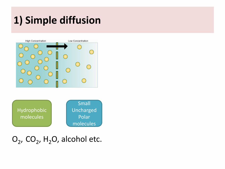
1) Simple diffusion
O2, CO2, H2O, alcohol etc.
Hydrophobicmolecules
SmallUncharged
Polar molecules
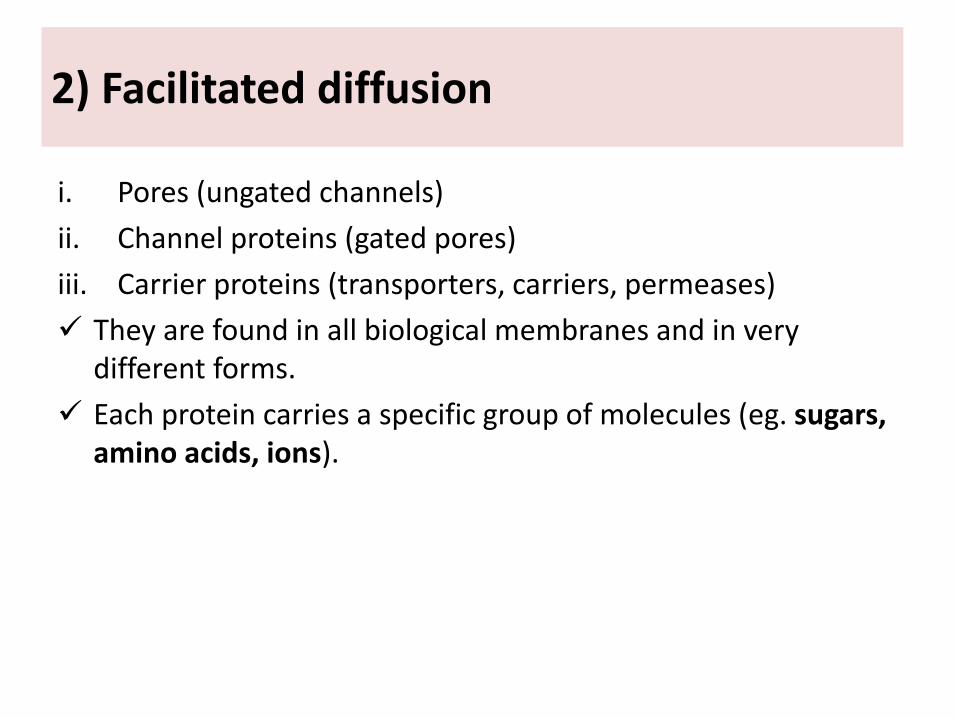
2) Facilitated diffusion
i. Pores (ungated channels)
ii. Channel proteins (gated pores)
iii. Carrier proteins (transporters, carriers, permeases)
They are found in all biological membranes and in very different forms.
Each protein carries a specific group of molecules (eg. sugars, amino acids, ions).
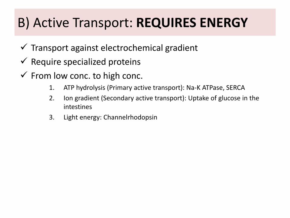
Transport against electrochemical gradient
Require specialized proteins
From low conc. to high conc.1. ATP hydrolysis (Primary active transport): Na-K ATPase, SERCA
2. Ion gradient (Secondary active transport): Uptake of glucose in theintestines
3. Light energy: Channelrhodopsin
B) Active Transport: REQUIRES ENERGY
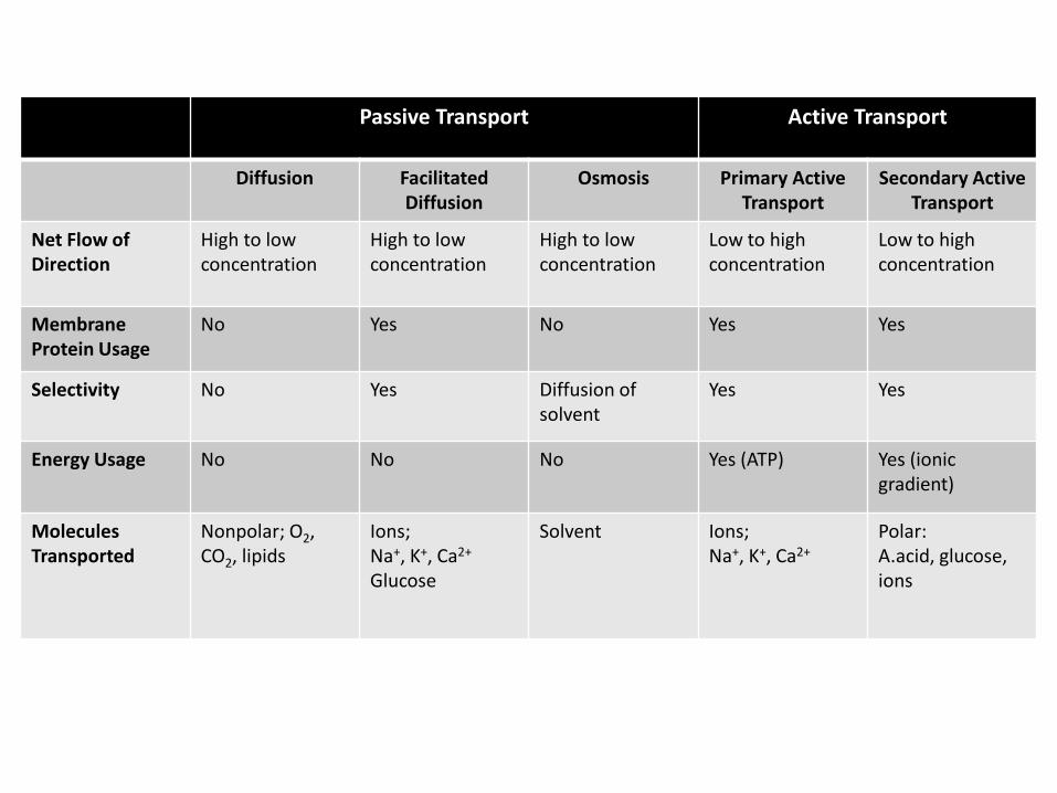
Passive Transport Active Transport
Diffusion Facilitated Diffusion
Osmosis Primary ActiveTransport
Secondary ActiveTransport
Net Flow of Direction
High to lowconcentration
High to lowconcentration
High to lowconcentration
Low to highconcentration
Low to highconcentration
MembraneProtein Usage
No Yes No Yes Yes
Selectivity No Yes Diffusion of solvent
Yes Yes
Energy Usage No No No Yes (ATP) Yes (ionicgradient)
MoleculesTransported
Nonpolar; O2, CO2, lipids
Ions;Na+, K+, Ca2+
Glucose
Solvent Ions;Na+, K+, Ca2+
Polar: A.acid, glucose,ions

Transport of Large Molecules
Endocytosis
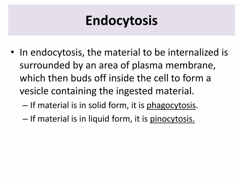
• In endocytosis, the material to be internalized is surrounded by an area of plasma membrane, which then buds off inside the cell to form a vesicle containing the ingested material.
– If material is in solid form, it is phagocytosis.
– If material is in liquid form, it is pinocytosis.
Exocytosis
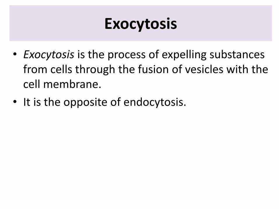
• Exocytosis is the process of expelling substances from cells through the fusion of vesicles with the cell membrane.
• It is the opposite of endocytosis.
Cytoplasm vs. Cytosol
• The cytosol, aka. intracellular fluid (ICF) or cytoplasmic matrix, is the liquid found inside cells.
• Cytosol + Organelles = Cytoplasm
Membrane bound organelles
Cells can concentrate and isolate enzymes and reactants in a smaller volume, thereby increasing the rate and efficiency of chemical reactions.
Cells can confine potentially harmful proteins and molecules in membrane-bound organelles, protecting the rest of the cells from their harmful effects.
Nucleus
• Contains cell's genetic material, organized as multiple long linear DNA molecules in complex with histoneproteins
• Enclosed by nuclear envelope: double membrane that isolates nuclear contents from the cellular cytoplasm.
• Because the nuclear membrane is impermeable to large molecules, nuclear pores are required to regulate nuclear transport of molecules between nucleus andcytoplasm
• Replication of DNA, transcription and processing of mRNAs take place within the nucleus.
• It plays a central role in cell proliferation.• It guides all the metabolic activities of the living cell.
Cytoplasm
Nucleus
DNA
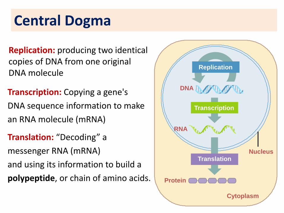
Central Dogma
RNA
Protein
Replication
Replication: producing two identical copies of DNA from one original DNA molecule
Transcription
Transcription: Copying a gene's
DNA sequence information to make
an RNA molecule (mRNA)
Translation
Translation: “Decoding” a
messenger RNA (mRNA)
and using its information to build a
polypeptide, or chain of amino acids.
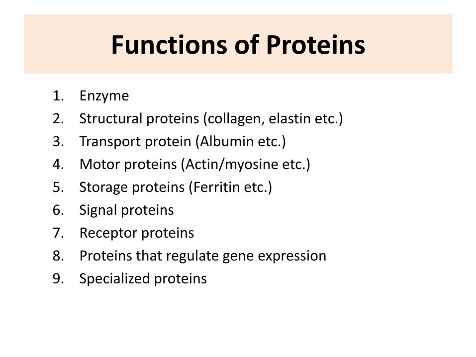
Functions of Proteins
1. Enzyme
2. Structural proteins (collagen, elastin etc.)
3. Transport protein (Albumin etc.)
4. Motor proteins (Actin/myosine etc.)
5. Storage proteins (Ferritin etc.)
6. Signal proteins
7. Receptor proteins
8. Proteins that regulate gene expression
9. Specialized proteins
Proteins
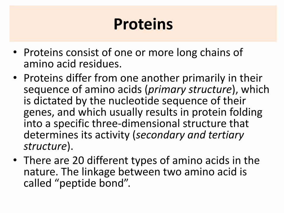
• Proteins consist of one or more long chains of amino acid residues.
• Proteins differ from one another primarily in their sequence of amino acids (primary structure), which is dictated by the nucleotide sequence of their genes, and which usually results in protein folding into a specific three-dimensional structure that determines its activity (secondary and tertiarystructure).
• There are 20 different types of amino acids in thenature. The linkage between two amino acid is called “peptide bond”.
Protein Synthesis
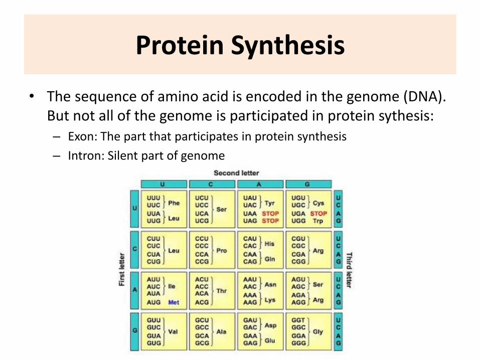
• The sequence of amino acid is encoded in the genome (DNA). But not all of the genome is participated in protein sythesis:– Exon: The part that participates in protein synthesis
– Intron: Silent part of genome
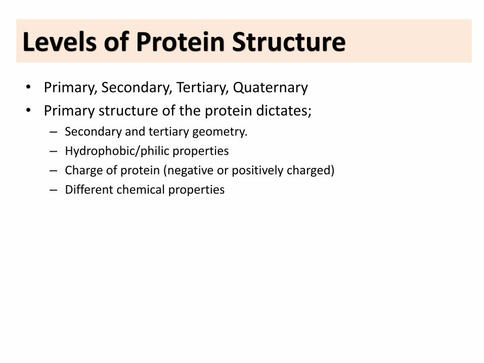
Levels of Protein Structure
• Primary, Secondary, Tertiary, Quaternary
• Primary structure of the protein dictates;– Secondary and tertiary geometry.
– Hydrophobic/philic properties
– Charge of protein (negative or positively charged)
– Different chemical properties
• Reticulum: Network
• ER is a continuous membrane system that forms a series of flattened sacs within the cytoplasm of eukaryotic cells– Granular (Smooth) ER: GER
– Agranular (Rough) ER: AGER
• Serves multiple functions
Endoplasmic Reticulum
• GER: Protein synthesis and sorting (membraneproteins, organellar proteins and secretedproteins)
• AGER: Lipid synthesis (fatty acids andphospholipids)
• Enzymes of AGER in liver cells play a role in detoxification of toxic substances (barbutals, amphetamines, morphins).
• Sarcoplasmic reticulum (Ca2+ storage and release)
• ER makes the cell more resistant to mechanical effects.
Endoplasmic Reticulum
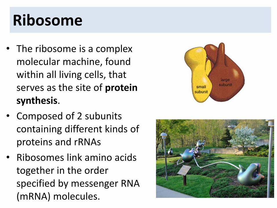
• The ribosome is a complex molecular machine, found within all living cells, that serves as the site of protein synthesis.
• Composed of 2 subunitscontaining different kinds of proteins and rRNAs
• Ribosomes link amino acids together in the orderspecified by messenger RNA (mRNA) molecules.
Ribosome
• The Golgi apparatus (aka. Golgi complex, Golgi body), is closely related to the ER.
• It usually is composed of four or more stacked layers of thin, flat, enclosed vesicles lying near one side of the nucleus.
• This apparatus is prominent in secretory cells, where it is located on the side of the cell from which the secretory substances are extruded.
• Golgi makes terminal modifications of proteins(“packages the proteins”).
Golgi apparatus
Lysosome
The lysosomes provide an intracellular digestive system that allows the cell to digest
1. damaged cellular structures (otophagy)2. food particles that have been ingested by the cell3. unwanted matter such as bacteria
The lysosomes contain hydrolytic enzymes that can break down many kinds of biomolecules and low pH inside (4.5–5.0).
If lysosomal digestive enzymes are released into cytoplasm thecell digests itself. This process called “autolysis”.
• Peroxisomes, contain oxidases. Several of the oxidases are capable of combining oxygen with hydrogen ions derived from different intracellularchemicals to form hydrogen peroxide (H2O2).
• Hydrogen peroxide is a highly oxidizing substance and is used in association with catalase, another oxidase enzyme present in large quantities in peroxisomes, to oxidize many substances that might otherwise be poisonous to the cell.
• For instance, about half of the alcohol a person drinks is detoxified by the peroxisomes of the liver cells in this manner.
Peroxisome
• Mitochondria generate most of the cell's supply of ATP, used as a source of chemical energy via oxidative phosphorylation (=respiration).
• Mitochondria are present in all areas of each cell’s cytoplasm, but the total number per cell varies from less than a hundred up to several thousand, depending on the amount of energy required by the cell.
• Mitochondrion is composed mainly of two lipid bilayer–protein membranes
• Mitochondria are self-replicative, which means that one mitochondrion can form a second one, a third one, and so on, whenever there is a need in the cell for increased amounts of ATP.
• Mitochondria contain DNA similar to that found in the cell nucleus. The DNA of the mitochondrion plays a similar role, controlling replication of the mitochondrion itself.
Mitochondrion
CYTOSKELETON
Cytoskelton
• Gives the cell its unique Providing mechanical support to the cell
• Cell movement
• Movement of chromosomes into daughter cells during cell division
• Intracellular transport of organelles and vesicles
Basic components of the cytoskeleton
• Actin filaments (microfilaments)
• Intermediate filaments
• Microtubules
Actin Filaments(Microfilaments)
• Diameter: 7nm
• The most abundant cytoskelton protein is actin in most of thecell types
• Actin polymerization Actin filaments
•Cellular contraction, migration, widening of the surface area, cytokinesis
• Cell membrane protrusions and extensions
– Motion
– Phagocytosis
– Absorption
– Nutrition
Actin Filaments(Microfilaments)
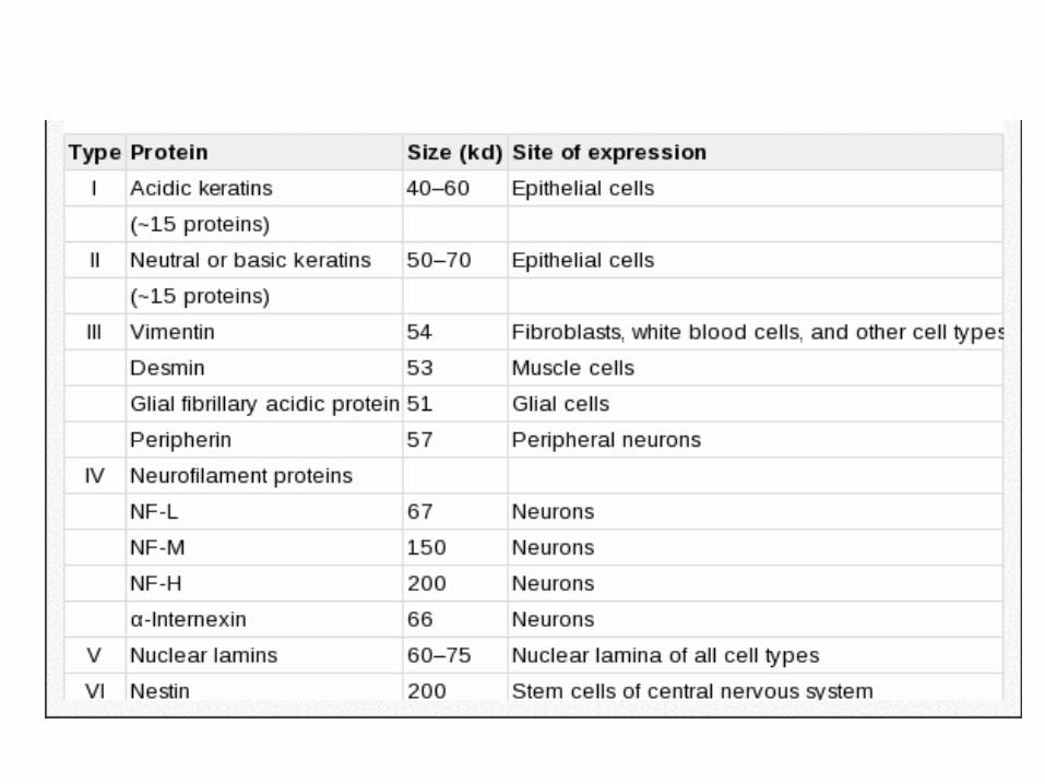
Intermediate Filaments
• Diameter: 10 nm
• Structural support
• Intermediate filaments are most extensively developed in regions of cells that are subject to mechanical stress (for example, in association with desmosomes)
Microtubules
• Diameter: 25 nm
• Organelle and vesicle transport within the cell
• Seperation of chromosomes during cell division
• Cell movement (cilia and flagella)

CELLULAR COMMUNICATION
While every cell in our body functions as a single lifeunit, they need to communicate with each other tomaintain homeostasis and perform specializedfunctions.
External signals
–Chemical messengers: Odorants, metabolites, ions, hormones, growth factors, neurotransmitters
– Light, mechanical, thermal stimuli aretransduced into chemical messengers
–Most chemical messengers interact with specific cell-surface receptors, triggering a series of intracellular reactions that mediate the cell's response to that stimulus.
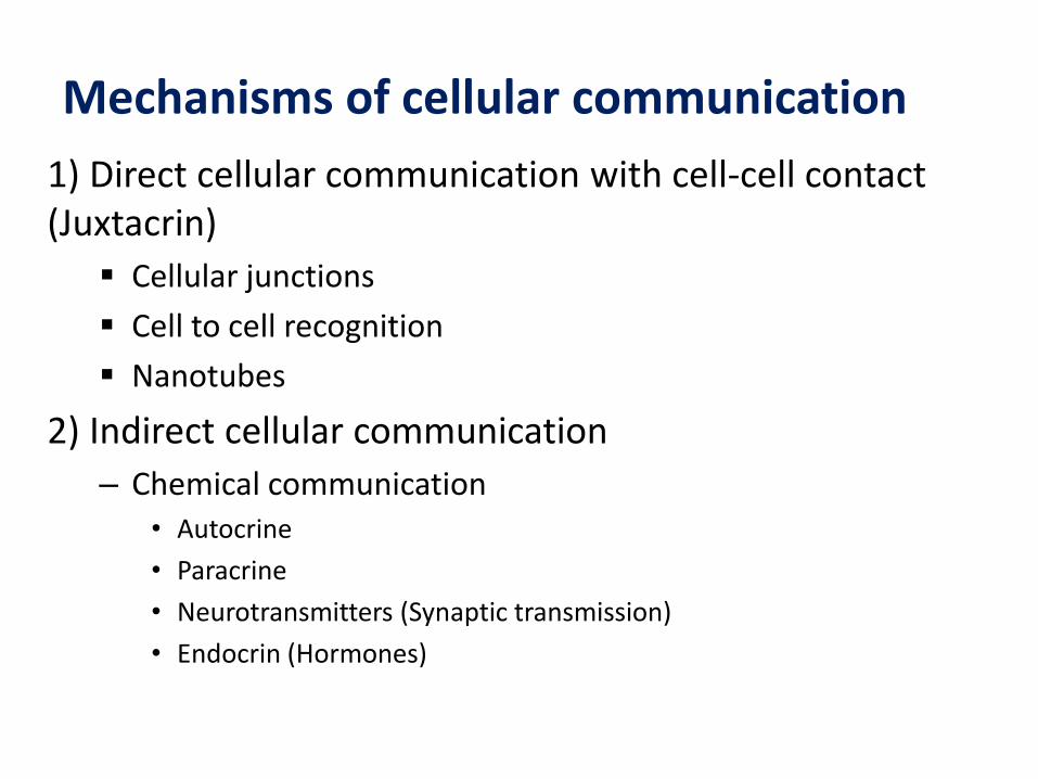
Mechanisms of cellular communication
1) Direct cellular communication with cell-cell contact(Juxtacrin)
Cellular junctions
Cell to cell recognition
Nanotubes
2) Indirect cellular communication
– Chemical communication• Autocrine
• Paracrine
• Neurotransmitters (Synaptic transmission)
• Endocrin (Hormones)
1. Direct cellular communication requiring physical cell-cell contact (Juxtacrine)
Intercellular junctions• Cells can be connected directly to each other with
specialized structures
• Desmosomes are cell structures specialized for cell-to-cell adhesion. Prevents excessive stretching of the tissues
• Tight junctions are the areas where two cells are almost stuck together. They form tight barriers that prevent leakage between cells
• Gap junctions allows direct communication between two cells. Cytoplasms of the two cells are interconnected by tunnels made up of connexonproteins.
Cell to Cell recognition
• The cell membrane contains surface carbohydrate molecules(glycolipids, glycoproteins) that generate signals for othercells
• Example: Recognition of self and non-self cells in immunesystem
Nanotubes Filamentous membrane extensions
Ions Organelles
Embryonic development, maintenance of homeostasis, spread of infectious agents, drugresistance, etc.
2. Indirect cellular communication that does not require cell-cell contact
Communication via chemical messengers
A cell can communicate with another cell locally or at a distance via effective chemical signaling molecules.
Chemicals are secreted from one cell and target another cell.
It mostly requires ligand-receptor interaction.
• Ligand: Extracellular chemicals that act as a signal transduction molecule.
• Receptor: Proteins in the target cell membrane that specifically bind ligand.
1. Receptor ligand interaction is highly specific.2. Only cell A has the appropriate receptor for this
chemical messenger and, therefore, it is the only one among the group that is a target cell for themessenger.
3. The amount of receptors can also be modifieda. Decrement of the receptor: downregulationb. Increment of the receptor: upregulation
Chemical Communication
• Local chemical communication
– Autocrine
– Paracrine
– Neurotransmitters (Synaptic transmission)
• Long-distance chemical communication
– Hormones
– Neurohormones
Paracrine and Autocrine Communication
• Paracrine signals: After secretion, the ligand spreads by diffusion and acts on the target cells in the close vicinity.
• Autocrine signals: Ligand affects the cell that secretes its own.
Neurotransmitters (Synaptic transmission)
• Special form of paracrine communication.
• After secreted by neuronal cells, NTs diffuses tothe synaptic cleft and binds to their receptor onnearby neuron / gland / muscle cell.
Hormones and Neurohormones
• Hormones: Chemical messengers secreted from the endocrine glands. They act on target cells at long distances through blood circulation.
• Neurohormones: Chemicals secreted from neuron cells act on target cells at long distances through blood circulation.
Signaling Molecules: Ligands
Ligands bind to receptors in target cells act as chemical signals (first messenger).
Second messengers are intracellular signaling molecules released by the cell to trigger physiological changes such as proliferation, differentiation, migration, survival, and apoptosis(cAMP, cGMP, IP3, DAG, and Ca+2).
The cell releases second messenger molecules in response to exposure to extracellular signalingmolecules—the first messengers.
Secondary messengers multiply the signal during intracellular signal transduction: Signal amplification
Cellular responses
Activation of receptor may cause;
1. Changes plasma membrane permeability, transport function or electrical status
2. Changes in cellular metabolism
3. Changes cellular secretory activity
4. Changes proliferation and differentiation rate of thecell
5. Cellular contractility
6. Changes in gene expression
References
John E. Hall - Guyton and Hall textbook of medical physiology-Saunders (2015)
Eric Widmaier, Hershel Raff, Kevin Strang - Vander’s Human Physiology-McGraw-Hill Education (2018)
Walter F. Boron and Emile L. Boulpaep. Medical Physiology: ACellular and Molecular Approach. (2017). Saunders Elsevier
Cooper, Geoffrey M., and Robert E. Hausman. The Cell: A Molecular Approach. (2009). Washington, D.C.: ASM Press