Embed Size (px)
Citation preview
Introduction to strabismus Strabismus is a symptom
Raoul de Haller
Swiss Eye Week - 2017 Neuchâtel
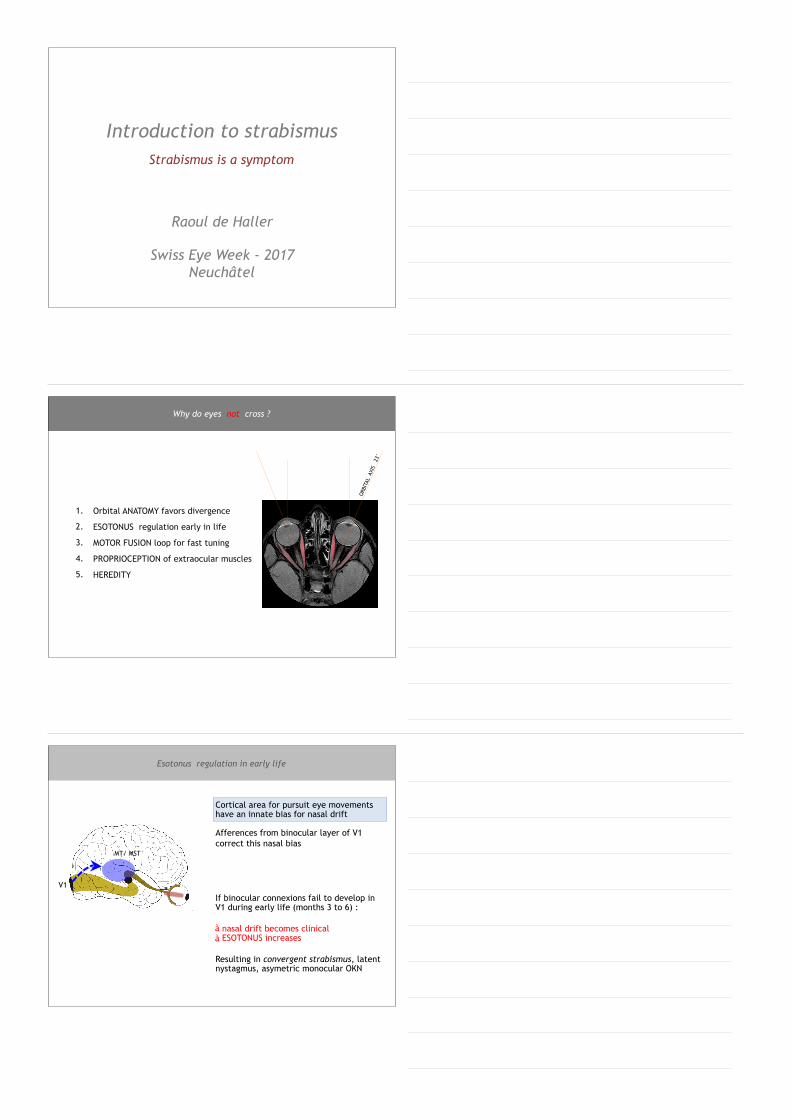
1. Orbital ANATOMY favors divergence
2. ESOTONUS regulation early in life
3. MOTOR FUSION loop for fast tuning
4. PROPRIOCEPTION of extraocular muscles
5. HEREDITY
ORB
ITAL
AXI
S 2
3°
Why do eyes not cross ?
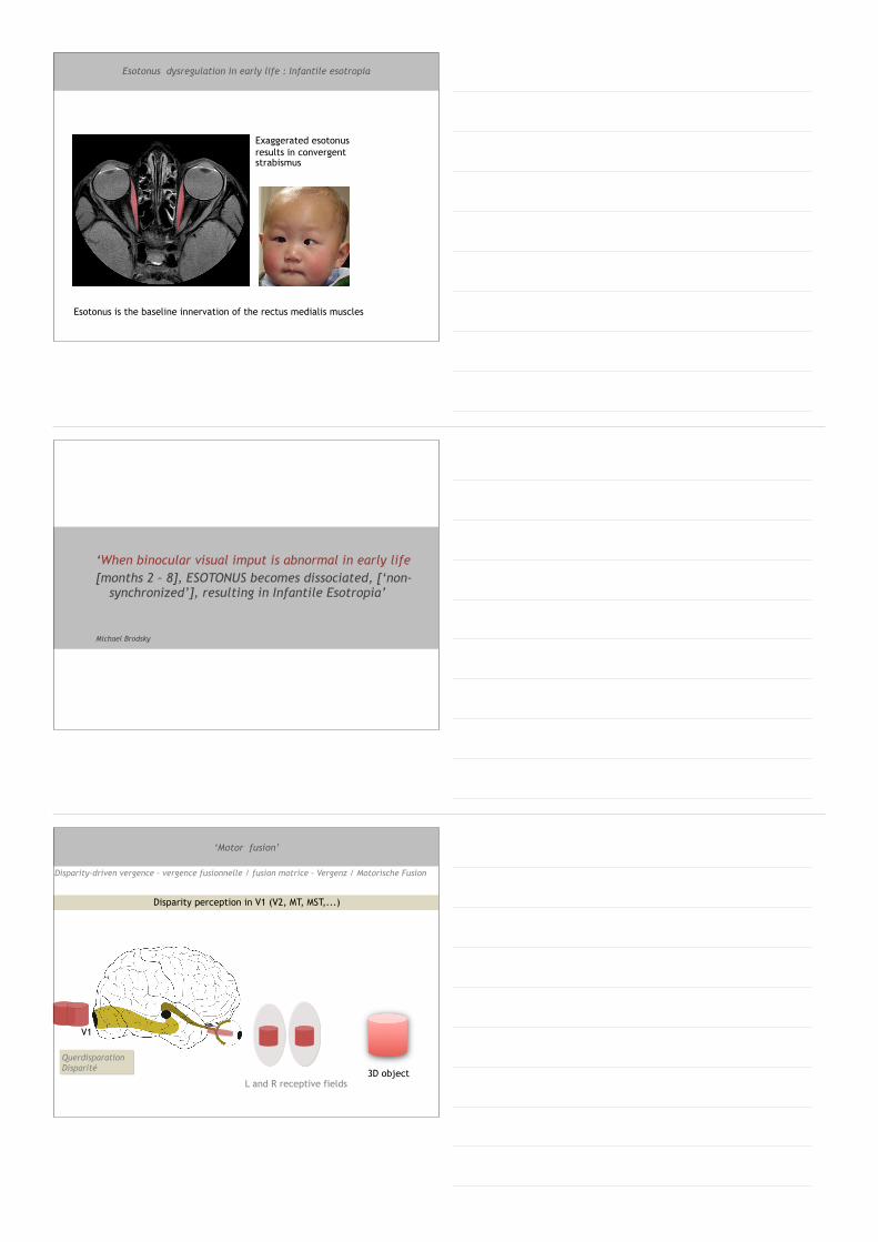
MT/ MST
Cortical area for pursuit eye movements have an innate bias for nasal drift
Afferences from binocular layer of V1 correct this nasal bias
If binocular connexions fail to develop in V1 during early life (months 3 to 6) :
à nasal drift becomes clinical à ESOTONUS increases
Resulting in convergent strabismus, latent nystagmus, asymetric monocular OKN
V1
Esotonus regulation in early life
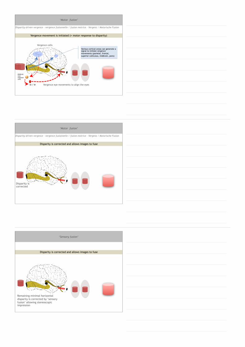
Exaggerated esotonus results in convergent strabismus
Esotonus is the baseline innervation of the rectus medialis muscles
Esotonus dysregulation in early life : Infantile esotropia
‘When binocular visual imput is abnormal in early life [months 2 – 8], ESOTONUS becomes dissociated, [‘non-
synchronized’], resulting in Infantile Esotropia’
Michael Brodsky
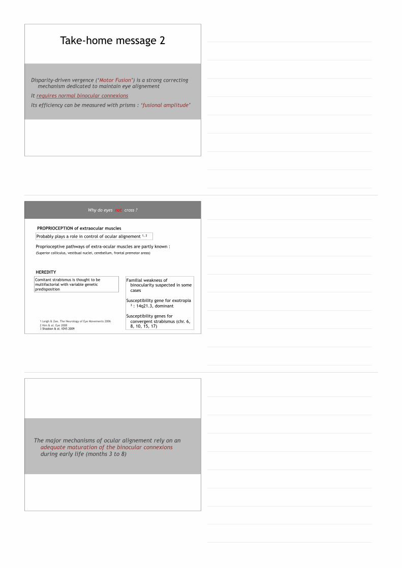
V1
Querdisparation Disparité
Disparity perception in V1 (V2, MT, MST,...)
‘Motor fusion’
Disparity-driven vergence – vergence fusionnelle / fusion motrice – Vergenz / Motorische Fusion
L and R receptive fields3D object
Midbrain near response cells
III / VI
Vergence cells
Vergence eye movements to align the eyes
Vergence movement is initiated (= motor response to disparity)
Various cortical areas can generate a signal to initiate vergence movements (parietal, frontal, superior colliculus, midbrain, pons)
‘Motor fusion’
Disparity-driven vergence – vergence fusionnelle / fusion motrice – Vergenz / Motorische Fusion
Disparity is corrected
Disparity is corrected and allows images to fuse
‘Motor fusion’
Disparity-driven vergence – vergence fusionnelle / fusion motrice – Vergenz / Motorische Fusion
Remaining minimal horizontal disparity is corrected by ‘sensory fusion’ allowing stereoscopic impression
Disparity is corrected and allows images to fuse
‘Sensory fusion’
Take-home message 2
Disparity-driven vergence (‘Motor Fusion’) is a strong correcting mechanism dedicated to maintain eye alignement
It requires normal binocular connexions
Its efficiency can be measured with prisms : ‘fusional amplitude’
PROPRIOCEPTION of extraocular muscles
Proprioceptive pathways of extra-ocular muscles are partly known :
(Superior colliculus, vestibual nuclei, cerebellum, frontal premotor areas)
1 Leigh & Zee. The Neurology of Eye Movements 2006 2 Kim & al. Eye 2008 3 Shaaban & al. IOVS 2009
Probably plays a role in control of ocular alignement 1, 2
Why do eyes not cross ?
Familial weakness of binocularity suspected in some cases
Susceptibility gene for exotropia 3 : 14q21.3, dominant
Susceptibility genes for convergent strabismus (chr. 6, 8, 10, 15, 17)
HEREDITY
Comitant strabismus is thought to be multifactorial with variable genetic predisposition
The major mechanisms of ocular alignement rely on an adequate maturation of the binocular connexions during early life (months 3 to 8)
[%]
3M 6M 12M 2y 3y 4y
Conditions for the development of binocularity : • adequate visual acuity developpment • intact visual cortex • ocular alignement during critical period • normal chiasmatic and callosal decussations
At 6-8 mo : 100% of children have acquired a stereoscopic vision. Those who have not will not develop this binocular competence
Stereoscopy
*
Visual acuity
Maturation of binocularity
*
[%]
3M 6M 12M 2y 3y 4y
Clinical signs of abnormal binocularity : • Esotonus ! (convergent strabismus) • Absent motor fusion, absent stereoscopy
• Latent nystagmus
• Dissociated deviations
• Abnormal retinal correspondanceStereoscopy
Consequences of very early strabismus
• Definitive abnormal binocularity • If strabismus is unilateral : deep amblyopia
‘Latent nystagmus’ Nystagmus latens
‘Manifest latent nystagmus’
Manifester Nystagmus von Latenstyp
Nystagmus latent manifeste
Clinical signs of binocular immaturityClinical signs of binocular immaturity : latent nystagmus
Latent nystagmus is defined clinically as nystagmus which appears on covering one eye and beats in the direction of the uncovered, fixating eye. When covering the other eye, the nystagmus will change direction.
The slow phase typically shows decreasing speed.
This very particular form of nystagmus is pathognomonic of binocular immaturity.
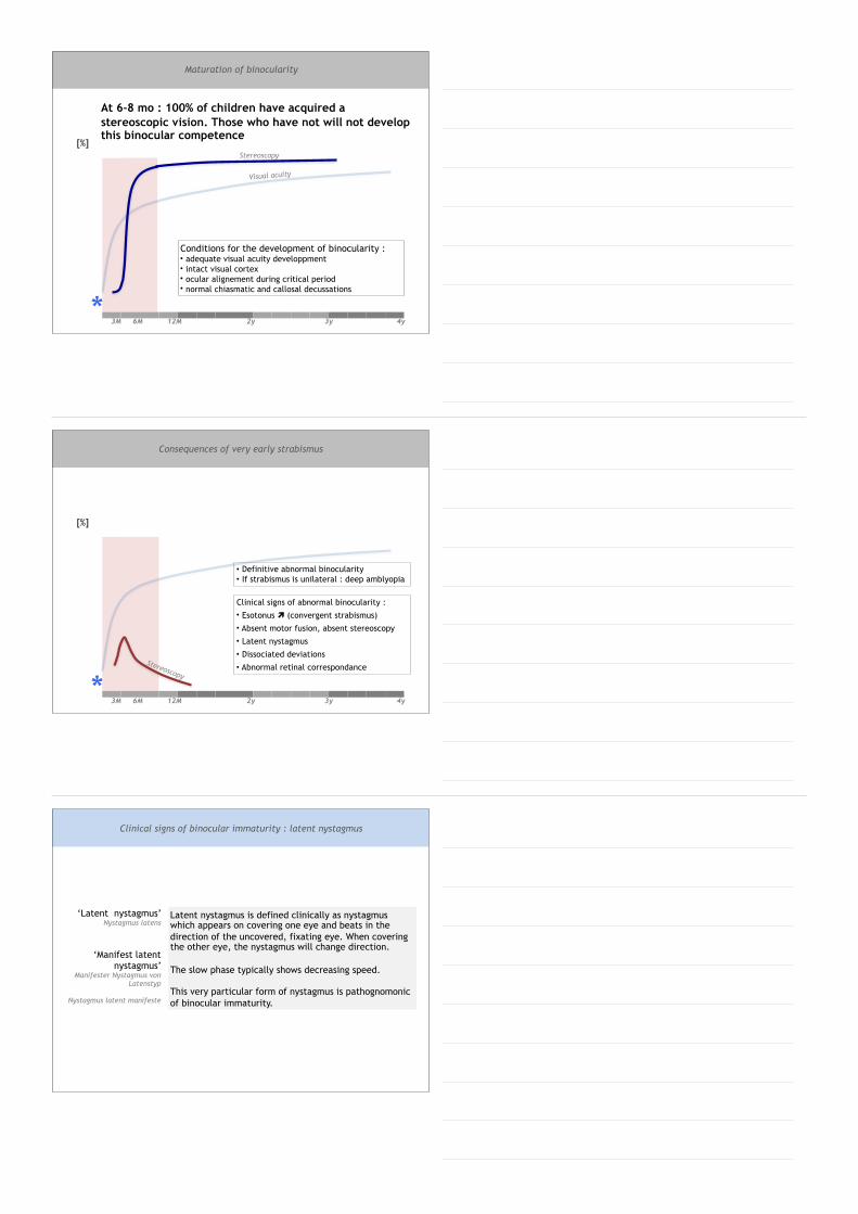
Dissociated vertical deviation (DVD)
Dissozierte vertikal Deviation
Déviation verticale dissociée
Clinical signs of binocular immaturity : dissociated deviations
In patients with an early-onset defect of binocular function, the occlusion of one eye may induce an elevation of the occluded eye, secondary to an imbalance in vestibular system. The eye elevates slowly and extorts. The phenomenon is generally bilateral although asymmetric.
*
[%]
3M 6M 12M 2y 3y 4y
Stereoscopy
Visual acuity
Binocularity is not consolidated
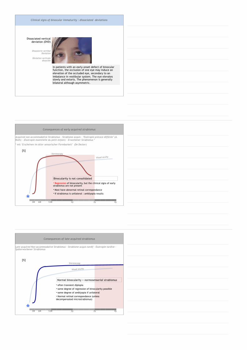
Acquired non accommodative Strabismus – Strabisme acquis – ‘Esotropie précoce différée’ (A. Roth) – (Esotropie essentielle du petit enfant) – Erworbener Strabismus *
* mit ‘Erscheinen im Alter sensorischer Formbarkeit’ (De Decker)
• Regression of binocularity, but the clinical signs of early strabismus are not present
• Most have abnormal retinal correspondance • If strabismus is unilateral : amblyopia results
Consequences of early acquired strabismus
Normal binocularity = normosensorial strabismus
*
[%]
3M 6M 12M 2y 3y 4y
• often transient diplopia • some degree of regression of binocularity possible • some degree of amblyopia if unilateral
• Normal retinal correspondance (unless decompensated microstrabismus)
Stereoscopy
Visual acuity
Late acquired Non-accommodative Strabismus – Strabisme acquis tardif – Esotropie tardive – Späterworbener Strabismus
Consequences of late acquired strabismus
Take home message 4
Abnormal retinal correspondance is a ‘sensorial abnormality’
It is a cortical adaptive process to prolonged eye deviation in early infancy
Clinical Entities Klinische Bilder von Bedeutung Tableaux cliniques significatifs
! Refer for control if • Deviation more than seconds – minutes per day • always the same eye deviates • No improvement during M2 • Persistance after M3
Neonatal ocular misalignement is a physiological, transient instability of eye alignement during the first few weeks of life
Eye position stabilized at ± 5 w (46 WGA)
Neonatal ocular misalignement
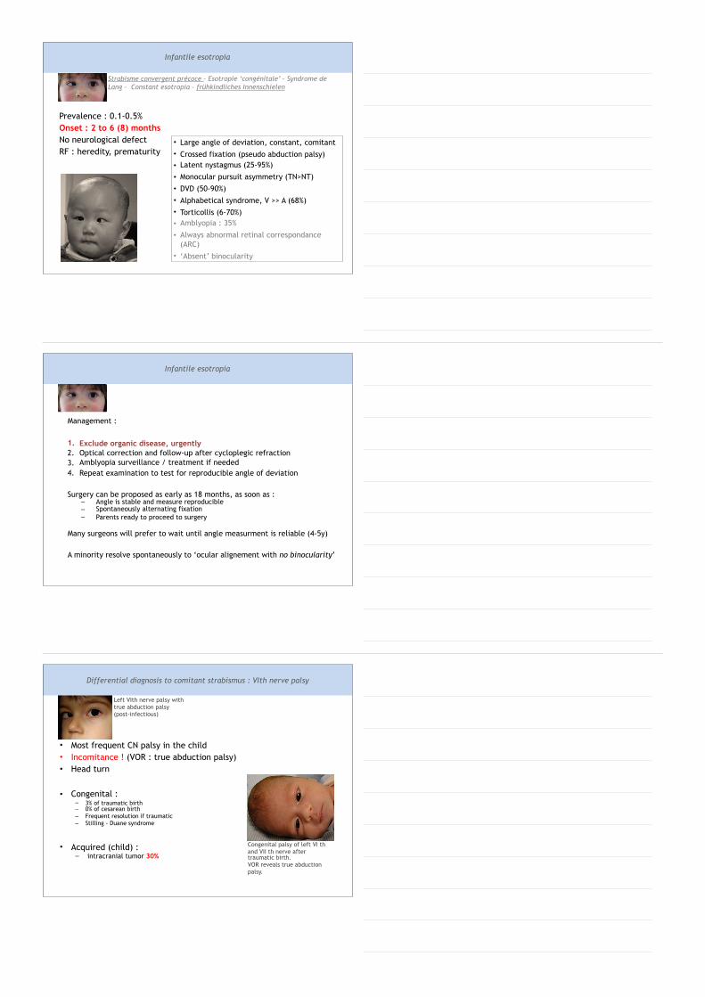
Prevalence : 0.1-0.5% Onset : 2 to 6 (8) months No neurological defect RF : heredity, prematurity
• Large angle of deviation, constant, comitant • Crossed fixation (pseudo abduction palsy) • Latent nystagmus (25-95%)
• Monocular pursuit asymmetry (TN>NT)
• DVD (50-90%) • Alphabetical syndrome, V >> A (68%) • Torticollis (6-70%) • Amblyopia : 35%
• Always abnormal retinal correspondance (ARC)
• ‘Absent’ binocularity
Strabisme convergent précoce – Esotropie ‘congénitale’ – Syndrome de Lang – Constant esotropia – frühkindliches Innenschielen
Infantile esotropia
Management :
1. Exclude organic disease, urgently 2. Optical correction and follow-up after cycloplegic refraction 3. Amblyopia surveillance / treatment if needed 4. Repeat examination to test for reproducible angle of deviation
Surgery can be proposed as early as 18 months, as soon as : – Angle is stable and measure reproducible – Spontaneously alternating fixation – Parents ready to proceed to surgery
Many surgeons will prefer to wait until angle measurment is reliable (4-5y)
A minority resolve spontaneously to ‘ocular alignement with no binocularity’
Infantile esotropia
• Most frequent CN palsy in the child • Incomitance ! (VOR : true abduction palsy) • Head turn
• Congenital : – 3% of traumatic birth – 0% of cesarean birth – Frequent resolution if traumatic – Stilling - Duane syndrome
• Acquired (child) : – intracranial tumor 30%
Congenital palsy of left VI th and VII th nerve after traumatic birth. VOR reveals true abduction palsy.
Left VIth nerve palsy with true abduction palsy (post-infectious)
Differential diagnosis to comitant strabismus : VIth nerve palsy
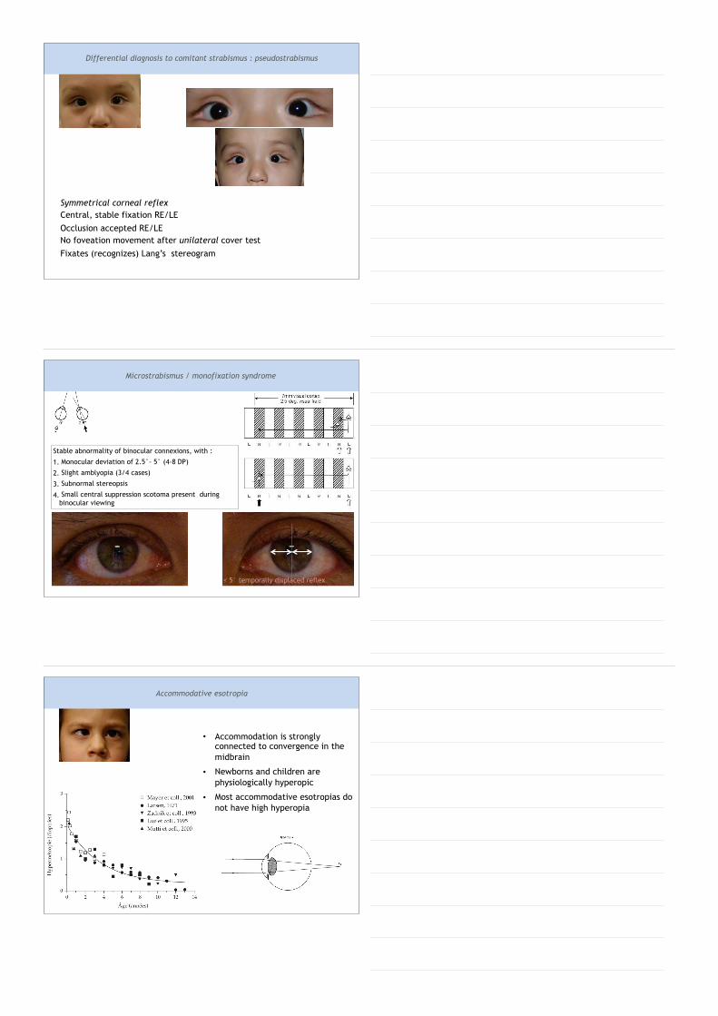
Symmetrical corneal reflex Central, stable fixation RE/LE
Occlusion accepted RE/LE No foveation movement after unilateral cover test
Fixates (recognizes) Lang’s stereogram
Differential diagnosis to comitant strabismus : pseudostrabismus
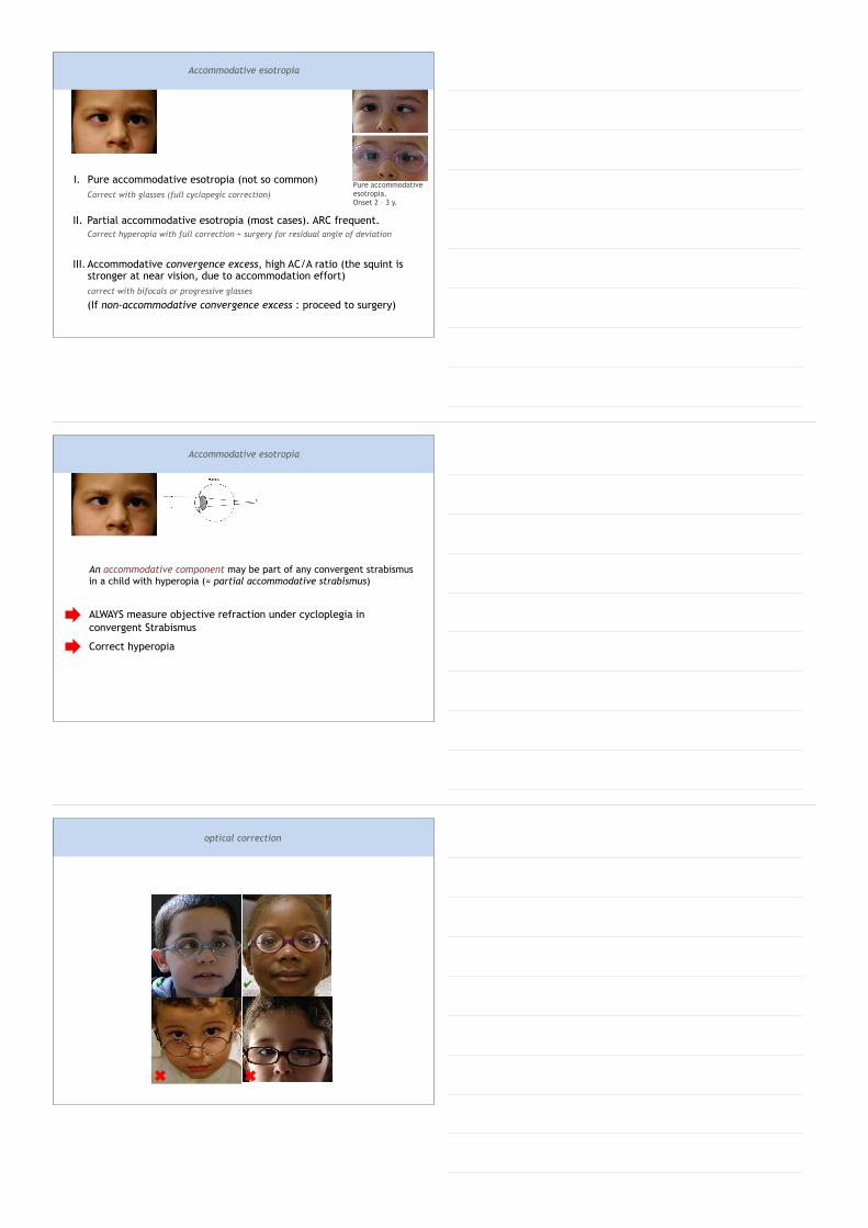
Stable abnormality of binocular connexions, with :
1. Monocular deviation of 2.5°- 5° (4-8 DP)
2. Slight amblyopia (3/4 cases)
3. Subnormal stereopsis
4. Small central suppression scotoma present during binocular viewing
< 5° temporally displaced reflex
Microstrabismus / monofixation syndrome
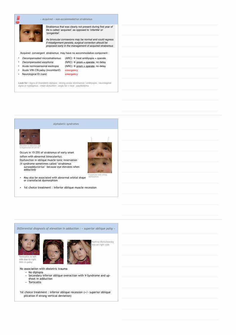
• Accommodation is strongly connected to convergence in the midbrain
• Newborns and children are physiologically hyperopic
• Most accommodative esotropias do not have high hyperopia
Accommodative esotropia
I. Pure accommodative esotropia (not so common) Correct with glasses (full cyclopegic correction)
II. Partial accommodative esotropia (most cases). ARC frequent. Correct hyperopia with full correction + surgery for residual angle of deviation
III. Accommodative convergence excess, high AC/A ratio (the squint is stronger at near vision, due to accommodation effort)
correct with bifocals or progressive glasses (If non-accommodative convergence excess : proceed to surgery)
Accommodative esotropia
Pure accommodative esotropia. Onset 2 – 3 y.
An accommodative component may be part of any convergent strabismus in a child with hyperopia (= partial accommodative strabismus)
ALWAYS measure objective refraction under cycloplegia in convergent Strabismus
Correct hyperopia
Accommodative esotropia
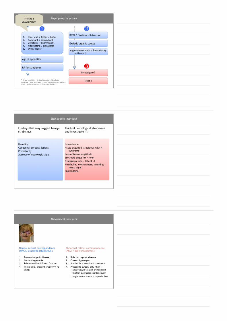
optical correction
✔ ✔
Acquired convergent strabismus may have no accommodative component :
• Decompensated microstrabismus (ARC) " treat amblyopia + operate • Decompensated esophoria (NRC) " prism + operate, no delay• Acute normosensorial esotropia (NRC) " prism + operate, no delay• Acute VIth CN palsy (incomitant!) emergency • Neurological Et (rare) emergency
Look for : signs of (transient) diplopia ; strong ocular dominance / amblyopia ; neurological signs or nystagmus ; intact abduction ; angle far > near ; papilledema
« Acquired » non-accommodative strabismus
Strabismus that was clearly not present during first year of life is called ‘acquired’, as opposed to ‘infantile’ or ‘congenital’.
As binocular connexions may be normal and could regress if misalignment persists, surgical correction should be proposed early in the management of acquired strabismus
Occurs in 15-25% of strabismus of early onset (often with abnormal binocularity). Dysfunction in oblique muscle tonic innervation (V syndrome sometimes called ‘strabismus
sursoadductorius’ because eye elevates when adducted)
• May also be associated with abnormal orbital shape or craniofacial dysmorphism
Elevation in adduction due to hyperaction of Left IO
V syndrome with orbital dysmorphism
Alphabetic syndromes
• 1st choice treatment : inferior oblique muscle recession
No association with obstetric trauma – No diplopia – Secondary inferior oblique overaction with V-Syndrome and up-
shoot in adduction – Torticollis
Torticollis to left side due to right IVth cn palsy
1st choice treatment : inferior oblique recession (+/- superior oblique plication if strong vertical deviation)
Positive Bielschowsky test on right side
Differential diagnosis of elevation in adduction : « superior oblique palsy »

• Incomitant exotropia (III rd nerve palsy; Thyroid Orbitopathy ; myasthenia ; Duane Type II ; CFEOM)
• Neurological exotropia : – Diffuse cerebral lesions (leukomalacia ; encephalopathies ; perinatal ventricular hemorrage) – Pontine Xt – Bilateral INO (WEBINO)
In a child with constant exotropia, probability of finding an associated systemic or ocular disease is higher than with a constant esotropia.
(Hunter. Ophthalmology 1999)
Divergent strabismus
• Intermittent exotropia (80% of cases) • Sensory exotropia (due to lost afference) • Primary exotropia (rare)
• Alternance between – normal binocular vision (straight) – Monocular vision and suppression
(divergent)
• Loss of alignement far > near • No diplopia • No amblyopia • Generally o measurable binocular
abnormality
Operate only if :
1. Eye tends to remain divergent 2. X(t) decompensates also at near 3. Angle of deviation ! 4. Social / professional handicap
Recurrence rate : 20-60%
Intermittent exotropia
Strabisme sensoriel (secondaire) / sekundärer Strabismus
Secondary strabismus
Any organic disease that causes visual impairment can cause strabismus
RF for strabismus
BCVA / Fixation + Refraction1. Eso / exo / hyper / hypo 2. Comitant / incomitant 3. Constant / intermittent 4. Alternating / unilateral 5. Other signs*
Age of apparition
Exclude organic causes
Investigate ?
Treat ?
❶ ❷
Step-by-step approach
❸
1st step : DESCRIPTION
Angle measurment / binocularity (orthoptics)
* Angle variability – Vertical deviation (Alphabetic syndrome ; DVD ; CN palsy) – latent nystagmus – torticollis – ptosis – globe retraction – afferent pupil defect
Heredity Congenital cerebral lesions Prematurity Absence of neurologic signs
Incomitance Acute acquired strabismus with A
syndrome Loss of fusion amplitude Esotropia angle far > near Nystagmus (non « latent ») Headache, awkwardness, vomiting,
neuro signs Papilledema
Findings that may suggest benign strabismus
Think of neurological strabismus and investigate if :
Step-by-step approach
1. Rule out organic disease 2. Correct hyperopia 3. Prisms to allow bifoveal fixation 4. In the child, proceed to surgery, no
delay
Normal retinal correspondance (NRC) / acquired strabismus :
1. Rule out organic disease 2. Correct hyperopia 3. Amblyopia prevention / treatment 4. Proceed to surgery only when :
• amblyopia is treated or stabilized • fixation alternates spontaneously • angle measurement is reproducible
Abnormal retinal correspondance (ARC) / early strabismus :
Management principles
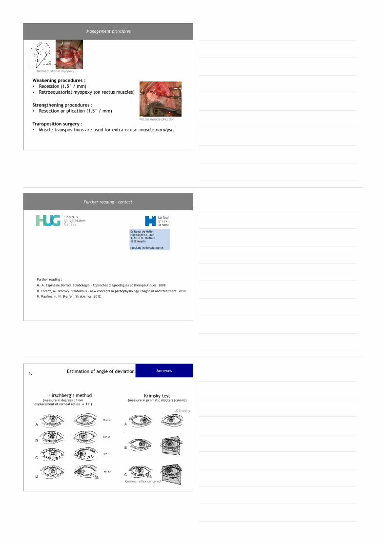
Weakening procedures : • Recession (1.5° / mm) • Retroequatorial myopexy (on rectus muscles)
Strengthening procedures : • Resection or plication (1.5° / mm)
Transposition surgery : • Muscle transpositions are used for extra-ocular muscle paralysis
Retroequatorial myopexy
Rectus muscle plication
Management principles
Dr Raoul de Haller Hôpital de La Tour 3, Av. J. D. Maillard 1217 Meyrin
H. Kaufmann, H. Steffen. Strabismus. 2012
B. Lorenz, M. Brodsky. Strabismus – new concepts in pathophysiology, Diagnosis and treatment. 2010
Further reading - contact
M.-A. Espinasse Berrod. Strabologie – Approches diagnostiques et thérapeutiques. 2008
Further reading :
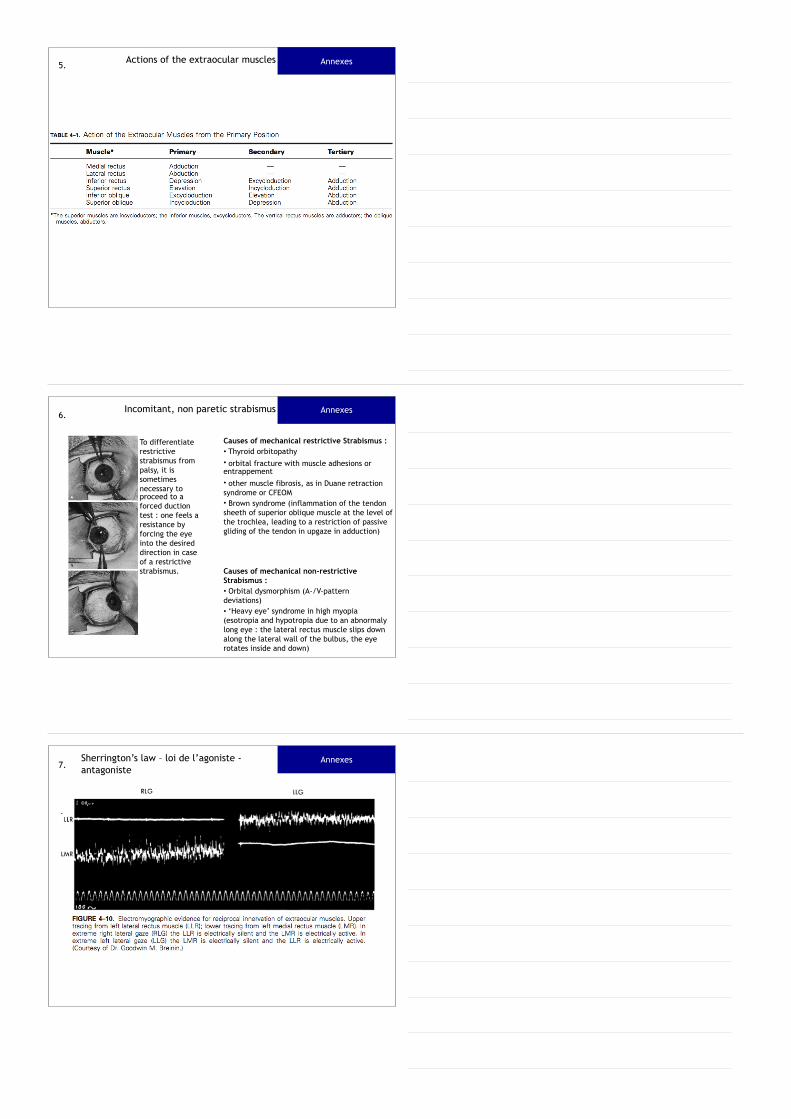
Estimation of angle of deviation
Hirschberg’s method (measure in degrees : 1mm
displacement of corneal reflex ≅ 11°)
Krimsky test (measure in prismatic diopters [cm/m])
LE fixating
Corneal reflex centered
Annexes1.
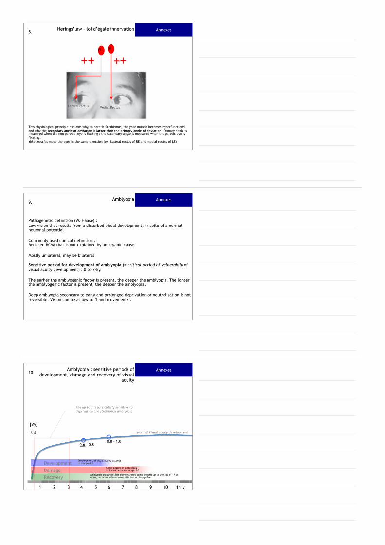
AnnexesClassification of Esotropia according to von Noorden 2.
Don’t forget : ocular myasthenia can mimic any oculomotor paresis
AnnexesExtraocular muscles of the right eye3.
Lid levator (III)
Superior rectus (III)
Lateral rectus (VI)
Inferior oblique (III)
Inferior rectus (III)
Medial rectus (III)
Superior oblique (IV)
AnnexesActions of the extraocular muscles4.
Superior oblique muscle, from above Inferior oblique muscle, from below
AnnexesActions of the extraocular muscles5.
To differentiate restrictive strabismus from palsy, it is sometimes necessary to proceed to a forced duction test : one feels a resistance by forcing the eye into the desired direction in case of a restrictive strabismus.
AnnexesIncomitant, non paretic strabismus6.
Causes of mechanical restrictive Strabismus : • Thyroid orbitopathy • orbital fracture with muscle adhesions or entrappement • other muscle fibrosis, as in Duane retraction syndrome or CFEOM • Brown syndrome (inflammation of the tendon sheeth of superior oblique muscle at the level of the trochlea, leading to a restriction of passive gliding of the tendon in upgaze in adduction)
Causes of mechanical non-restrictive Strabismus : • Orbital dysmorphism (A-/V-pattern deviations) • ‘Heavy eye’ syndrome in high myopia (esotropia and hypotropia due to an abnormaly long eye : the lateral rectus muscle slips down along the lateral wall of the bulbus, the eye rotates inside and down)
AnnexesSherrington’s law – loi de l’agoniste - antagoniste7.
AnnexesHerings’law – loi d’égale innervation8.
IIIVI
Lateral rectus Medial Rectus
This physiological principle explains why, in paretic Strabismus, the yoke muscle becomes hyperfunctional, and why the secondary angle of deviation is larger than the primary angle of deviation. Primary angle is measured when the non paretic eye is fixating ; the secondary angle is measured when the paretic eye is fixating. Yoke muscles move the eyes in the same direction (ex. Lateral rectus of RE and medial rectus of LE)
AnnexesAmblyopia9.
Pathogenetic definition (W. Haase) : Low vision that results from a disturbed visual development, in spite of a normal neuronal potential
Commonly used clinical definition : Reduced BCVA that is not explained by an organic cause
Mostly unilateral, may be bilateral
Sensitive period for development of amblyopia (= critical period of vulnerabily of visual acuity development) : 0 to 7-8y.
The earlier the amblyogenic factor is present, the deeper the amblyopia. The longer the amblyogenic factor is present, the deeper the amblyopia.
Deep amblyopia secondary to early and prolonged deprivation or neutralisation is not reversible. Vision can be as low as ‘hand movements’.
[VA]
1.0
1 2 3 4 5 6 7 8 9 10 11 y
Normal Visual acuity development
Development
Damage
Recovery
0.6 – 0.80.8 – 1.0
Development of visual acuity extends to this period
Some degree of amblyopia still may occur up to age 8-9
Amblyopia treatment has demonstrated some benefit up to the age of 17 or more, but is considered most efficient up to age 3-4.
Age up to 3 is particularly sensitive to deprivation and strabismus amblyopia
AnnexesAmblyopia : sensitive periods of development, damage and recovery of visual
acuity
10.

Causes of amblyopia Annexes11.
AnnexesAmblyopia treatment12.
Pre-requisite : best adapted optical correction
Occlusion therapy by patching of the better eye (Lancet 2006; Cochrane 2009) • Some recommend initiating treatment by full-time occlusion and control after a few days or weeks to monitor visual improvement. We initiate treatment with half wake-time occlusion, then adapt according to age and importance of amblyopia.
• As a general rule : occlusion 1h/day pro year of age (ex. : 2y old will have 2h occlusion/ day, 7/7 until next control).
• Atropine 2x/week in the better eye as penalisation is efficient for moderate amblyopia as an alternative if patching is not tolerated. CAVE risk of inverse amblyopia if uncontrolled. • The major part of improvement occurs within 3 months of treatment begin.
• A treatment attempt is sometimes recommended up to age 17 if never treated before, although treatment after 6 years seldom results in significant improvement of VA. Early begin of treatment is the key to success.