Embed Size (px)
Citation preview
PARALYTIC AND PARETIC STRABISMUS GENERAL FEATURES, IMPORTANCE OF HERINGS LAW, INVESTIGATIONS OF PARETC STRABISMUS
01/03/151
Presenter Pabita Dhungel B.optometryInstitute of Medicine
References
1) BINOCULAR VISION & STRABISMUS –GK VON NOORDEN
2) CLINICAL MANGEMENT OF STRABISMUS- ELEZABETH E.CALAROSSA & MICHAEL W. ROUSE
3) AAO- SECTION: PEDIATRIC OPHTHALMOLOGY & STRABISMUS
4) STRABISMUS SIMPLIFIED- PRADEEP SHARMA5) PRACTICAL ORTHOPTICS IN THE TREATMENT OF
SQUINT- LYLE AND JACKSON’S
01/03/152
PRESENTATION LAYOUTIntroduction to extra ocular musclesFew related terms• Introduction to paralytic and paretic strabismusClassificationClinical characteristicsImportance of Herings lawInvestigations of paretic strabismusSummary
01/03/153
Introduction
There are 6 extraocular muscles – 4 rectus muscles, 2 oblique muscles
5 muscles arise from the apex of the orbit, the inferior oblique arises form the inferonasal angle of the orbit
The 4 recti muscles originate form the apex of the orbit at the level of the Annulus of Zinn
01/03/15 4
Contd....The recti muscles are inserted in front of the ocular equator, the
obliques are inserted behindMovements occur about 3 primary axes around the centre of
rotation – the vertical, horizontal and saggital axesThe action of a muscle depends on the angle of its plane and
the anterio-posterior axis of the eye. It follows that the action of the muscle may vary according the
positions of the globe in the orbit.
01/03/15 5
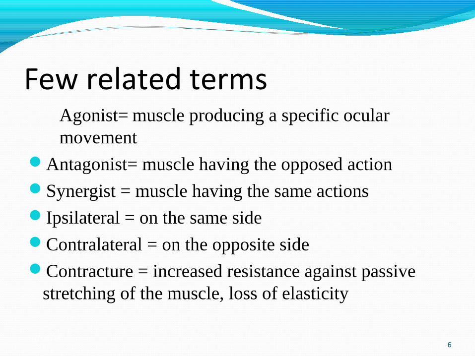
Few related termsAgonist= muscle producing a specific ocular movement
Antagonist= muscle having the opposed actionSynergist = muscle having the same actionsIpsilateral = on the same sideContralateral = on the opposite sideContracture = increased resistance against passive
stretching of the muscle, loss of elasticity
01/03/156
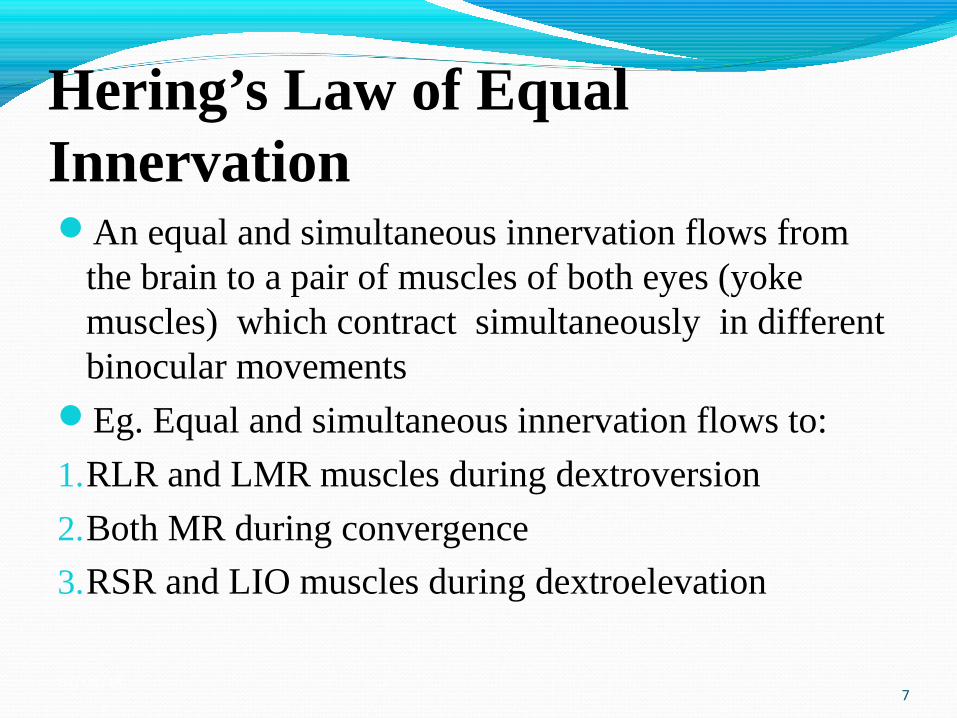
Hering’s Law of Equal Innervation An equal and simultaneous innervation flows from
the brain to a pair of muscles of both eyes (yoke muscles) which contract simultaneously in different binocular movements
Eg. Equal and simultaneous innervation flows to:
1.RLR and LMR muscles during dextroversion
2.Both MR during convergence
3.RSR and LIO muscles during dextroelevation
01/03/157
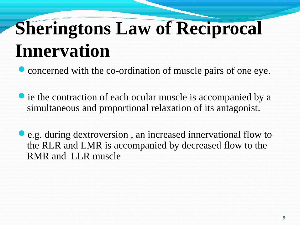
Sheringtons Law of Reciprocal Innervation concerned with the co-ordination of muscle pairs of one eye.
ie the contraction of each ocular muscle is accompanied by a simultaneous and proportional relaxation of its antagonist.
e.g. during dextroversion , an increased innervational flow to the RLR and LMR is accompanied by decreased flow to the RMR and LLR muscle
01/03/158
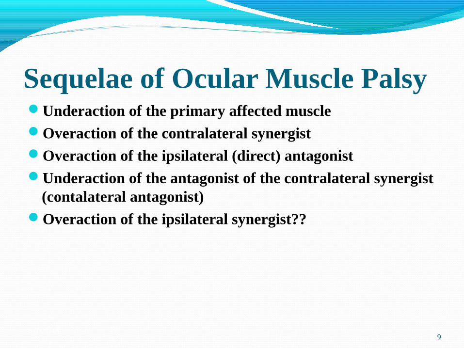
Sequelae of Ocular Muscle PalsyUnderaction of the primary affected muscleOveraction of the contralateral synergistOveraction of the ipsilateral (direct) antagonistUnderaction of the antagonist of the contralateral synergist
(contalateral antagonist)Overaction of the ipsilateral synergist??
01/03/159
Overaction of the contralateral synergist always present. This overaction occurs when the affected eye is fixing as
a result of increased innervation being required to rotate the affected muscle into its field of action.
Due to Herings Law an overstimulation of the contralateral synergist follows
This is always the largest overaction in the sequelae.
01/03/1510
Overaction of the ipsilateral (direct) antagonist can lead to a permanent contracture of the muscle and a
loss of elasticityIf the patient fixes with the non-involved eye within a few
days a contracture will develop in the direct antagonist muscle
because the normal contracture of the direct antagonist is unopposed by the weak muscle.
01/03/1511
Underaction of the antagonist of the contralateral synergist (contalateral
antagonist) with the involved eye fixing, the movement of the eye
into the field of action of the weak muscles antagonist requires less innervation than normal due to the contracture.
Therefore less innervation is supplied to the contralateral antagonist which under-acts
01/03/1512
For example in paralysis of the right superior rectusunderaction of right superior rectusoveraction of the left inferior obliqueoveraction of the right inferior rectusunderaction of the left superior oblique(overaction of the right inferior oblique)
01/03/1513
Introduction to paralytic strabismus
Terms paretic and paralytic often are used interchangeably in clinical ophthalmology
Paralytic strabismus is an incomitant strabismus due to motor deficiency of one or a group of extra ocular muscles
Incomplete paralysis is called paresis and complete deficiency is called paralysis, while palsy is used for both without specifying
01/03/1514
ClassificationA). Neurogenici) supranuclear ii) nucleariii) internucleariv) infranuclear- fascicular ( Main nerve trunk or subdivision) oculomotor nerve (III CN)Trochlear nerve (IV CN)Abducens nerve (VI CN)
01/03/1515
Contd...B. Myogenic i) nerve muscle junction lesion (myasthenia)ii) muscle(a) Congenital Absence ,hypoplasia mal insertion or musculofascial
anomalies(b) Traumatic laceration, disinertion(c) inflammatory (myositis)(d) Orbito- myopathies(e) Dystrophy01/03/15
16
Causes of neurogenic lesions1. congenital2. traumatic3. inflammatory4. neoplastic5. ischemic6. toxic7.Demyelinating disease8. idiopathic
01/03/1517
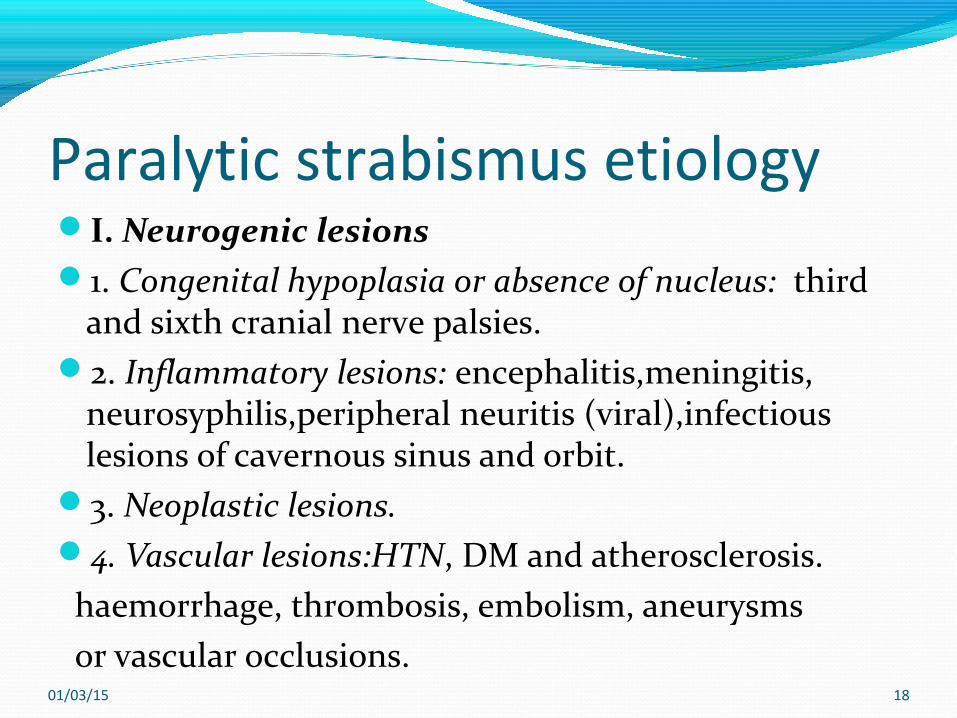
Paralytic strabismus etiologyI. Neurogenic lesions1. Congenital hypoplasia or absence of nucleus: third
and sixth cranial nerve palsies.2. Inflammatory lesions: encephalitis,meningitis,
neurosyphilis,peripheral neuritis (viral),infectious lesions of cavernous sinus and orbit.
3. Neoplastic lesions. 4. Vascular lesions:HTN, DM and atherosclerosis. haemorrhage, thrombosis, embolism, aneurysms or vascular occlusions.
01/03/15 18
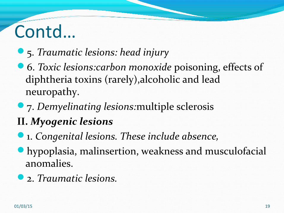
Contd…5. Traumatic lesions: head injury6. Toxic lesions:carbon monoxide poisoning, effects of
diphtheria toxins (rarely),alcoholic and lead neuropathy.
7. Demyelinating lesions:multiple sclerosis II. Myogenic lesions1. Congenital lesions. These include absence,hypoplasia, malinsertion, weakness and musculofacial
anomalies.2. Traumatic lesions.
01/03/15 19

Contd…3. Inflammatory lesions: Myositis (viral) , influenza,
measles.4. Myopathies:These include thyroid
myopathy,carcinomatous myopathy,Progressive external ophthalmoplegia
III. Neuromuscular junction lesionIt includes myasthenia gravis.
01/03/15 20
Paralytic strabismusSYMPTOMS:
1. LIMITATION OF OCULAR MOVEMENTS2. SUDDEN ONSET OCULAR DEVIATION3. DIPLOPIA4.CONFUSION5. NAUSEA , VERTIGO
01/03/15 21
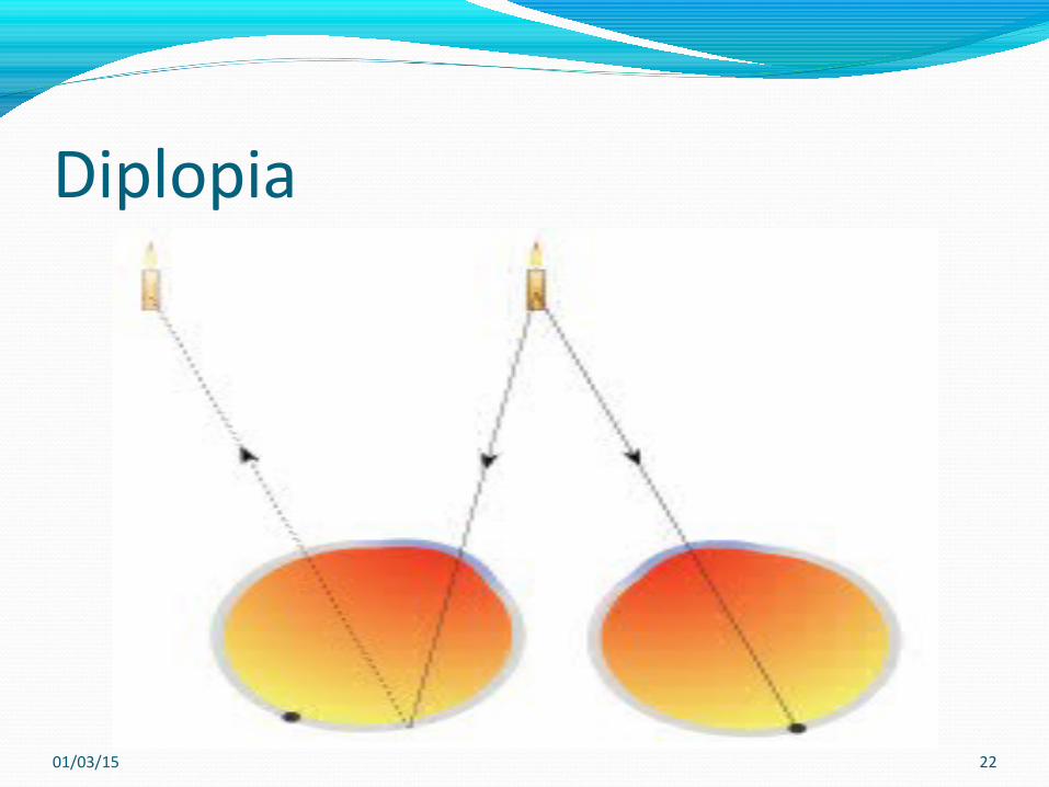
Diplopia
01/03/15 22
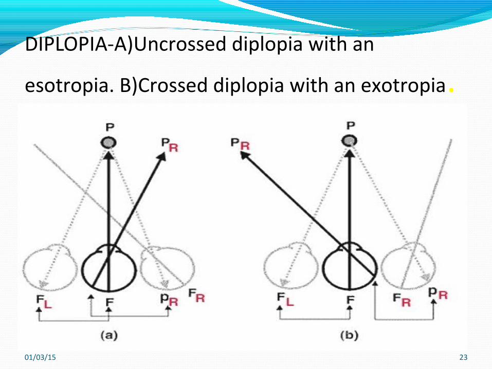
DIPLOPIA-A)Uncrossed diplopia with an
esotropia. B)Crossed diplopia with an exotropia.
01/03/15 23
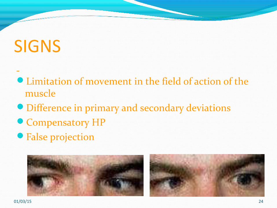
SIGNS Limitation of movement in the field of action of the
muscleDifference in primary and secondary deviationsCompensatory HPFalse projection
01/03/15 24
Clinical characteristicsi) incomitance variable ocular deviation in different position
which is maximum in the field of action of the muscle (eg. In a LLR palsy esotropia is maximal in the abduction of LE)
ii)Limitation of movement of the eye in the field of action of the EOM( in
LLR palsy the abduction of the LE is deficient or limited)
01/03/1525
Contd...iii) difference in primary and secondary deviation deviation of the squinting eye with normal eye fixing
is called primary deviationDeviation of the normal eye with the paretic eye
fixing is called secondary deviationIn paralytic strabismus secondary deviation is greater
than primary deviation
01/03/1526
Contd...This is because the paretic eye requires more effort
to straighten (come to the primary position to take up fixation) and this extra effort is passed on to the contralateral synergist, which is normal, increasing the ocular deviation
The reverse is true for spastic strabismus i.e primary deviation is greater than the secondary deviation
01/03/1527
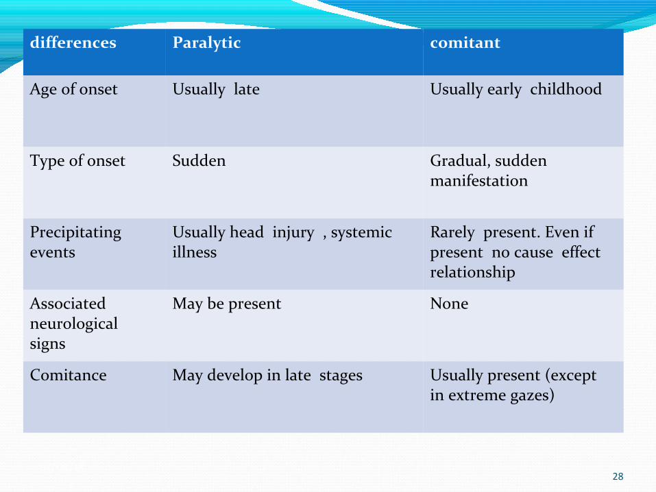
differences Paralytic comitant
Age of onset Usually late Usually early childhood
Type of onset Sudden Gradual, sudden manifestation
Precipitating events
Usually head injury , systemic illness
Rarely present. Even if present no cause effect relationship
Associated neurological signs
May be present None
Comitance May develop in late stages Usually present (except in extreme gazes)
01/03/1528
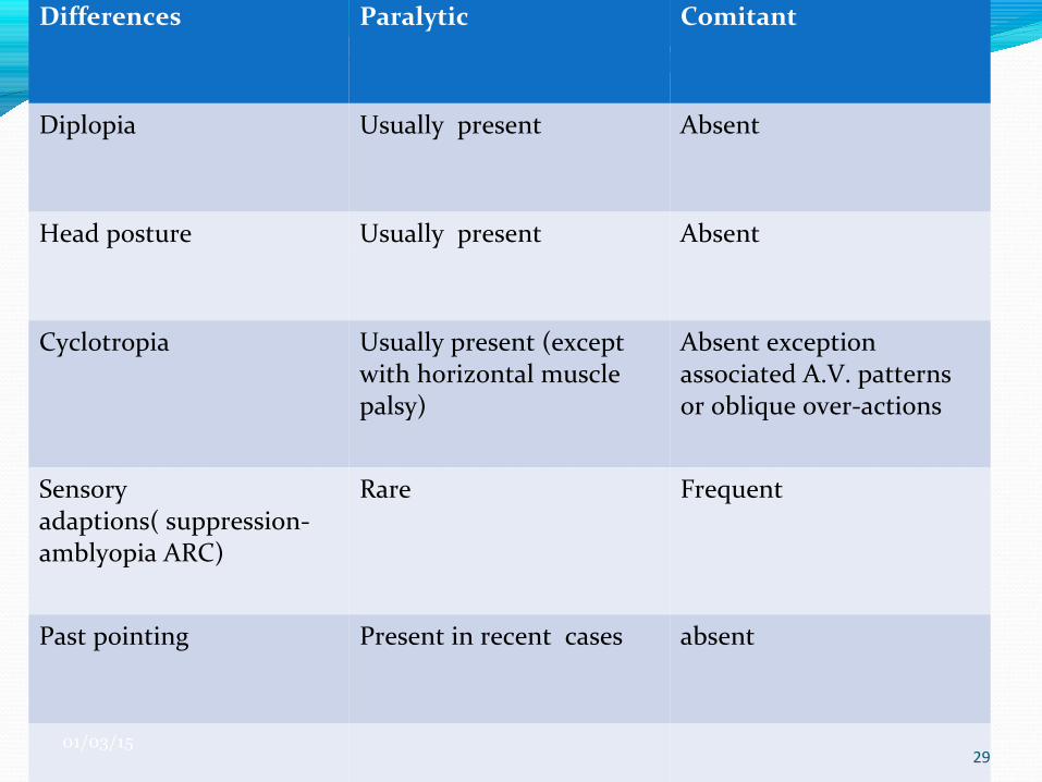
Differences Paralytic Comitant
Diplopia Usually present Absent
Head posture Usually present Absent
Cyclotropia Usually present (except with horizontal muscle palsy)
Absent exception associated A.V. patterns or oblique over-actions
Sensory adaptions( suppression- amblyopia ARC)
Rare Frequent
Past pointing Present in recent cases absent
01/03/1529
Stages of paralytic squintUndergoes three stages1. Paresis of the particular muscle2. Overaction of the ipsilateral antagonist3. Underaction of the antagonist of the
contralateral synergist
01/03/1530
1. First stageThe maximal deviation is in the field of action of
the paretic muscle for e.g. In a case left LR palsy, the deviation is maximum in levoversion
01/03/1531
2. Second stageAs the overaction of the ipsilateral antagonist
occurs the deviation overacting, the duration increases in the dextroversion
01/03/1532
3. Third stageThe underaction of the contralateral synergist
occurs.this is known as inhibitional palsy
01/03/1533
Importance of hering’s law1. Secondary deviation( fixating with squinting eye) is more than primary deviation in paralytic strabismusThis is based in hering’s law because when the patient
fixates with the squinting eye an excess innervation is required to the paralysed muscle to fixate and the
concomitant excess supply to the yoke muscle from the normal eye causes excess contraction leading to more, the so-called secondary deviation
01/03/1534
Contd…2. Inhibitional palsy of the contralateral antagonist muscle developing in patients with paralytic squint is based on Hering’s law for e.g if a patient has RSO muscle paresis and fixates with RE on object located on patient’s left, less innervation of RIO is required to move the eye in this gaze, because it doesn’t have to overcome the normal antagonistic effect of RSO muscle
01/03/1535
Third nerve palsyThe III nerve divides into two branches. The superior branch supplies the LPS and SRThe inferior branch supplies the MR, IR and IO
muscles. A complete lesion of the III nerve involving both
branches will result in a deficit of elevation, adduction and depression in abduction. There will be an accompanying ptosis and pupil
dilatation
01/03/1536
Contd...AetiologyLocalisation of lesionNuclearNuclear III often produces bilateral defects of ocular
motility and lid function. The levator palpebral superioris muscles share a
common central nucleus that producesClinical Orthoptics, Third Edition. Fiona J. Rowe.C 2012 John Wiley & Sons, Ltd. Published 2012 by Blackwell Publishing Ltd.
01/03/1537
Neurogenic disorder A nuclear lesion may also result in the following: Unilateral III with bilateral ptosis Unilateral III with contralateral superior rectus
underaction Isolated extraocular muscle palsy of inferior rectus,
inferior oblique or medialrectus muscles (Brown 1957) Bilateral III with spared levator function (Biousse & Newman 2000, Saeki et al. 2000).
01/03/1538
Internuclear
Internuclear ophthalmoplegia Weber’s syndrome: III and contralateral hemiplegia
due to lesion of corticospinal tract Benedikt’s syndrome: III and contralateral ataxia and
intension tremor Claude’s syndrome: Lesion of the red nucleus and III
nucleus producing anipsilateral III nerve palsy and contralateral ataxia
(Broadley et al. 2001)
01/03/1539
Infranuclear
Third nerve palsy may be central, sparing the pupil or peripheral with pupil involvement.
If the pupil is spared, the cause is most likely vascular.
When the pupil is involved, the cause is likely to be an
aneurysm (Goldstein&Cogan 1960). However, pupil sparing in children (unlike inadults) may not be helpful in differentiating the causes
of the palsy and third nerve01/03/15
40
InvestigationVisual acuity It is necessary to lift the ptotic lid to evaluate visual
acuity.May be reduced due to mydriasis, particularly for near
visual acuityCover test An exo- and hypo-deviation is present
01/03/1541
Ocular motilityThere will be limited elevation, depression and
adduction,which may be complete or partial limitations, depending on the extent of paresis/palsy.
The examiner should always check for the presence of IV nerve function by asking the patient to attempt to look down and outwards and observe for the presence of incyclotorsion during this movement.
01/03/1542
Contd…In case of presence of ptosis the upper lid will have
to be raised by the examiner (or an assistant) in order to
perform the ocular motility assessment. It is important to check for unilateral versus bilateral
signs and extent of limitations (ductions) versus version movements.
01/03/1543
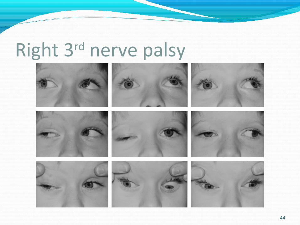
Right 3rd nerve palsy
01/03/1544
Investigation Hess chart The affected eye will show a markedly constricted
field whereas the other eye demonstrates overaction of its
muscles
Diplopia There will be constant diplopia unless complete
ptosis is present and blocks the vision of the affected eye.
01/03/15 45

Hess chart of right 3rd nerve palsy
01/03/1546
Convergence This will be absent if the medial rectus muscle is
paralysed.Binocular functionThis is usually absent unless the III nerve paresis is
mild and partial.Accommodation If the underlying cause of the lesion has resulted in
pupillary dilatation, then fibres to the ciliary body are also likely to be involved so that accommodation will be defective.
01/03/1547
Aberrant regenerationchange in the actions of muscles supplied by the third
nerve due to regrowth of damaged nerve fibres following complete or severe third nerve palsy
(Shuttleworth et al. 1998).
It is liable to occur when either trauma or an aneurysm
has caused the lesion (Cox et al. 1979, Rossillion et al. 2001).
May occur from weeks to months after the onset of the III nerve paresis.
01/03/1548
InvestigationCover test Combined horizontal and vertical deviations are
common.Ocular motilityAbnormal movements often involve the eyelids and
pupils. There may be horizontal gaze and lid synkinesis (Chua et al. 2000)
Limited elevation and depression with globe retraction on attempted vertical movements or ipsilateral adduction on attempted elevation or depression.01/03/15
49
Contd…Ptosis The lid may rise on attempted depression, adduction
and occasionally abduction (pseudo-Graefe phenomenon).
Pupil This may constrict on attempted adduction, elevation
and depression(pseudo-Argyll Robertson pupil).Convergence This may occur on attempted elevation
01/03/1550
Cyclic oculomotor palsyThis rare condition is usually congenital and
unilateral in origin (Burian & Van Allen 1963).
It is often associated with a partial III nerve palsy with some degree of ptosis.
Acquired cyclic ocular motor palsy may occur following irradiation of the skull base and is similar to ocular neuromyotonia
(Miller & Lee 2004).
01/03/1551
Contd…It may also be due to a compressive lesion (Bateman & Saunders 1983).
The condition is described as having cyclical fluctuation in two phases:
1. Paralytic phase: There is a partial III nerve palsy.2. Miotic phase: There is convergence, lid retraction,
accommodation and pupil constriction.
01/03/1552
Single muscle palsy1. Medial rectus This produces an exo-deviation, which is greater for
near fixation.2. Inferior rectus: This produces hyper- and exo-deviation3. Superior rectus: This is often bilateral and may present with a V exo
pattern.4. Inferior oblique: This is a feature of an A eso pattern 01/03/15
53
Differential diagnosis of single muscle palsiesMR palsy : Atypical Duane’s retraction syndrome :Uni/bilateral internuclear ophthalmoplegiaIR palsy: Myogenic (myasthenia gravis) : Mechanical limitation (thyroid eye disease) : Trauma (blowout fracture)IO palsy: Brown’s syndromeSR palsy : Trauma (blowout fracture) : Mechanical limitation (thyroid eye disease)
01/03/15 54
Double elevator palsyThis often has a congenital origin and is presumed to
be caused by a supranuclear defect. The superior rectus and inferior oblique muscles of
the same eye are affected (Jampel & Fells 1968, Strachan & Innes 1987).
01/03/1555
InvestigationCover test There is a hypo-deviation in the primary position that
may be manifest or latent.Ocular motilityThere is limited elevation of one eye in both
adduction and abduction.Ptosis and/or pseudoptosis may be present. Bell’s phenomenon is usually present.Abnormal head postureThe chin is elevated to compensate for the palsy
01/03/1556
Contd…Binocular functionIf there is only a small angle of deviation in the
primary position, this may be controlled with or without an abnormal head posture, resulting in a latent hypo-deviation.
In these circumstances, binocular single vision is present.
Forced duction testThere may be full passive movement (negative result)
or it may be positive, depending on cause that is important for surgical planning
01/03/1557
Differential diagnosis double elevator palsyThe following conditions should be differentiated
from double elevator palsy as they will have a positive forced duction test:
Blowout fracture Thyroid eye disease Brown’s syndrome Congenital fibrosis of the inferior rectus muscle General fibrosis syndrome
58
IV (fourth) cranial nerveThe IV cranial nerve supplies the superior oblique
muscle only. Any lesion affecting the nerve may result in
difficulties of depression, incyclorotation and abduction of the eye
01/03/1559
AetiologyLocation of lesionFourth nerve palsy may be due to lesions in the
nucleus or fascicular lesions of the midbrain. It can be difficult to differentially diagnose nuclear
and fascicular lesions as the IV nerves decussate immediately after exiting the nuclei and exit the
dorsal midbrain after a very short intra-midbrain course.
The IV nerve is not only susceptible to damage as it exits the midbrain but also is vulnerable in the cavernous sinus and orbital apex.
01/03/1560
Contd…congenital or acquired. Acquired group may be caused by the following: Trauma: Particularly bilateral cases, as the IV cranial nerve is
the only nerve to arise from the dorsal surface of the midbrain, and therefore follows a long, winding route that renders it susceptible to injury.
Vascular, for example hypertension. Diabetes. 01/03/15
61
Contd…Space-occupying lesions.In congenital superior oblique palsy, the tendon is
usually lax and abnormally long
(Plager 1990, Plager 1992).
01/03/1562
Investigation Case history In congenital cases, the patients are often affected
bilaterally. They may present with any of the following:1. Manifest strabismus without binocular function2. Binocular function, but there may be an abnormal
head posture, depending on the extent of paresis
01/03/1563
Contd…There is often facial asymmetry in congenital superior
oblique palsy consisting of shallowing of the midfacial region between the lateral canthus and the edge of the mouth (Parks 1958).
This is found in three-fourth of patients with congenital palsy (Wilson &Hoxie 1993, Paysee et al. 1995).
In acquired cases, the paresis may be unilateral or bilateral.
Bilateral case is often associated with major closed head trauma.
01/03/1564
Contd…Symptoms of cyclovertical diplopia are experienced,
which worsen on downgaze. The presence of an abnormal head posture tends to indicate more long-standing deviations.
01/03/1565
Abnormal head postureIn unilateral IV nerve paresis, the patient may present
with chin depression with face turn and head tilt away from the affected side.
Bilateral cases demonstrate chin depression but there may well be no face turn or head tilt unless one side is affected more than the other.
Persistent abnormal head posture following surgery for IV nerve palsy should be investigated to exclude sternocleidomastoid muscle
tightness that may have developed in congenital cases (Lau et al.2009).
01/03/1566
Cover testThe test is performed with and without the abnormal
head posture for comparison. A latent deviation exists if a compensatory abnormal head posture is adopted. When the test is repeated with the head straight, the
deviation will increase and may become manifest
01/03/15 67
Contd…The affected eye shows a hyper-deviation and an
associated esodeviation.(However, an exo-deviation may be seen if this waspresent before the onset of the palsy.) It is useful to note that the vertical deviation is
commonly greater on near testing than for distance. In bilateral cases, the main deviation usually tends to
be a small-angle horizontal deviation.
01/03/1568
Ocular motilityThe primary underaction of the affected superior
oblique muscle results in a number of sequelae. There is overaction of the contralateral inferior rectus
and of the ipsilateral inferior oblique muscles. The contralateral superior rectus shows secondary
underaction.The combination of these muscle effects is variable
and often depends on whether or not the deviation is long standing
01/03/1569
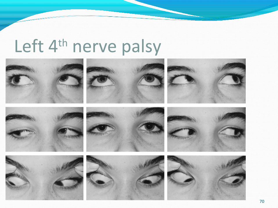
Left 4th nerve palsy
01/03/1570
Contd…In bilateral cases, a V pattern is often present. In the presence of this, one should always suspect a
bilateral case, especially where there is an excyclotorsion of greater than 7 and a tendency◦
towards reversal of any hyper-deviation or diplopic images on lateral gaze
(Hermann 1981).
01/03/1571
Contd…Convergence This may be reduced, either due to convergence
insufficiency or the vertical deviation. Differentiation is achieved by correcting the
deviation and seeing if convergence improvesField of binocular single visionThe area in which binocular single vision is retained is
displaced upwards to the affected side
01/03/1572
DiplopiaThe patient experiences a greater degree of diplopia
on near testing when looking down. The diplopia is vertical and usually uncrossed.However, if an exo-deviation is present, the
horizontal element of diplopia will be crossed
01/03/15 73
Binocular functionAssessment of binocular function is carried out with
and without the abnormal head posture, if one is present.
Binocular single vision cannot be maintained if there is a significant degree of torsion and therefore is often not present in acquired bilateral palsies.
01/03/15 74
Contd…Patients who maintain binocularity tend to have a
small angle of deviation associated with a congenital palsy.
Some patients with long-standing acquired palsies can maintain binocularity if there is a good fusional control with an extended vertical fusion range
01/03/15 75
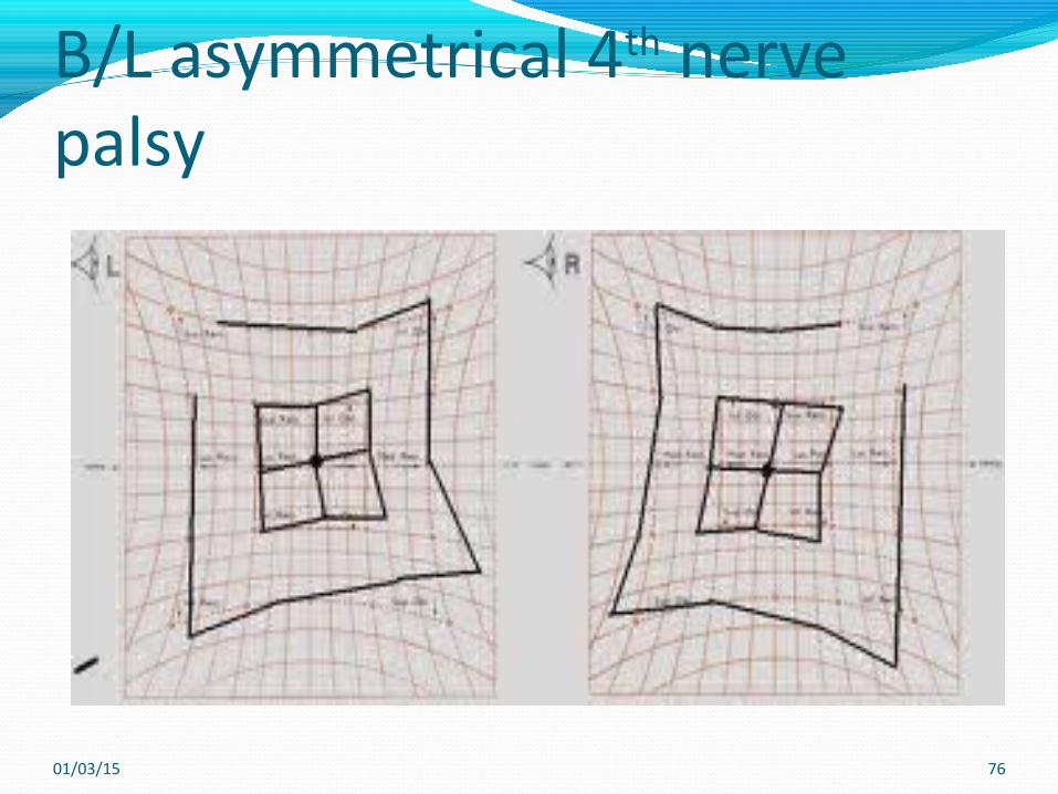
B/L asymmetrical 4th nerve palsy
01/03/15 76
Torsion Diagnostic prismsExcyclotorsion is frequently present in acquired
bilateral cases.Fresnel prisms may be used temporarily to correct the
angle of deviation. If the prism alleviates the need for an abnormal head posture, then the indication is that the IV nerve was
responsible for the abnormal head posture rather than a non-ocular cause
01/03/15 77
Differential diagnosisthyroid eye disease, ocular surgery, orbital
fracture, neurosurgery, childhood strabismus, skew deviation, third nerve palsy, myasthenia gravis and decompensated hyperphoria (Tamhankar et al. 2011).
Typically, excyclotorsion is seen in IV nerve palsy and incyclotorsion is seen in skew deviation.
Furthermore, the deviation in IV nerve palsy remains when the patient is lying down whereas skew deviation resolves on lying down (Wong 2010).
01/03/15 78
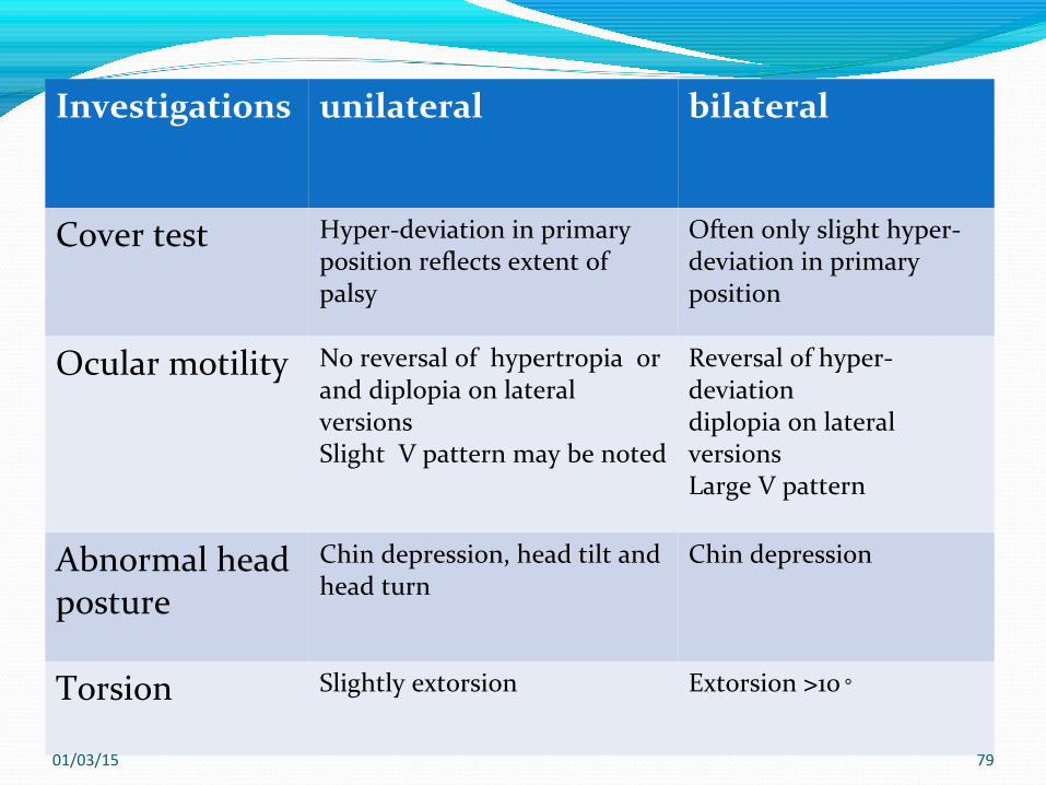
Investigations unilateral bilateral
Cover test Hyper-deviation in primaryposition reflects extent of palsy
Often only slight hyper-deviation in primary position
Ocular motility No reversal of hypertropia orand diplopia on lateral versionsSlight V pattern may be noted
Reversal of hyper-deviationdiplopia on lateral versionsLarge V pattern
Abnormal head posture
Chin depression, head tilt andhead turn
Chin depression
Torsion Slightly extorsion Extorsion >10◦
01/03/15 79

VI (sixth) cranial nerveThe VI cranial nerve supplies the lateral rectus muscle
only. A lesion affecting the nerve will result in defective
abduction of the eye.
01/03/15 80
AetiologyThe commonly recognised causes of sixth nerve palsy
include space-occupying lesions, trauma, vascular insults and inflammation.
Sixth nerve palsy secondary to raised intracranial pressure is also commonly regarded as a typical false localising sign.
Other causes have included pseudotumor cerebri (idiopathic intracranial hypertension), post-operative complications, viral infection, multiple sclerosis and otitis
01/03/15 81
Contd…Congenital Following birth trauma Hereditary Infection (maternal) Failure of lateral rectus developmentAcquiredThis differs according to age: Children (Afifi et al. 1992):– Space-occupying lesions– Infections, bacterial or viral- trauma
01/03/15 82
Contd…– Raised intracranial pressure– Decompensated esophoria– Infantile esotropia with cross-fixation– Mobius syndrome– Duane’s retraction syndrome Young adults:– Trauma– Space-occupying lesions– Post-viral inflammation
01/03/15 83
Contd…●– Multiple sclerosis– Diabetes– High myopia– Ophthalmoplegic migraineOlder adults:– Vascular– Diabetes– Space-occupying lesions– Senile lateral rectus weakness
01/03/15 84
InvestigationCase history Birth history may be significant. The patient may complain of horizontal diplopia,
which is worse in the distance and/or on lateralgaze and relatives or friends may notice the presence of
squint (esodeviation) or an abnormal head posture
01/03/15 85
Contd…Visual acuity This may be reduced if the affected eye fails to fixate
properly due to the presence of a marked deviation. Amblyopia will develop in children with unilateral
strabismus.Abnormal head postureThe face is turned towards the affected side.
01/03/15 86
Contd…Cover test An eso-deviation is present that is often greater on
distance testing.The test should be carried out with and without an
abnormal head posture, if one is present. The deviation increases without the head posture but
does not necessarily become manifest
01/03/15 87
Contd…Ocular motilityThe primary underaction of the affected lateral rectus
results in limited abduction and is followed by a number of sequelae.
There is overaction of both the contralateral and ipsilateral medial recti.
There is also secondary underaction of the contralateral lateral rectus
01/03/15 88
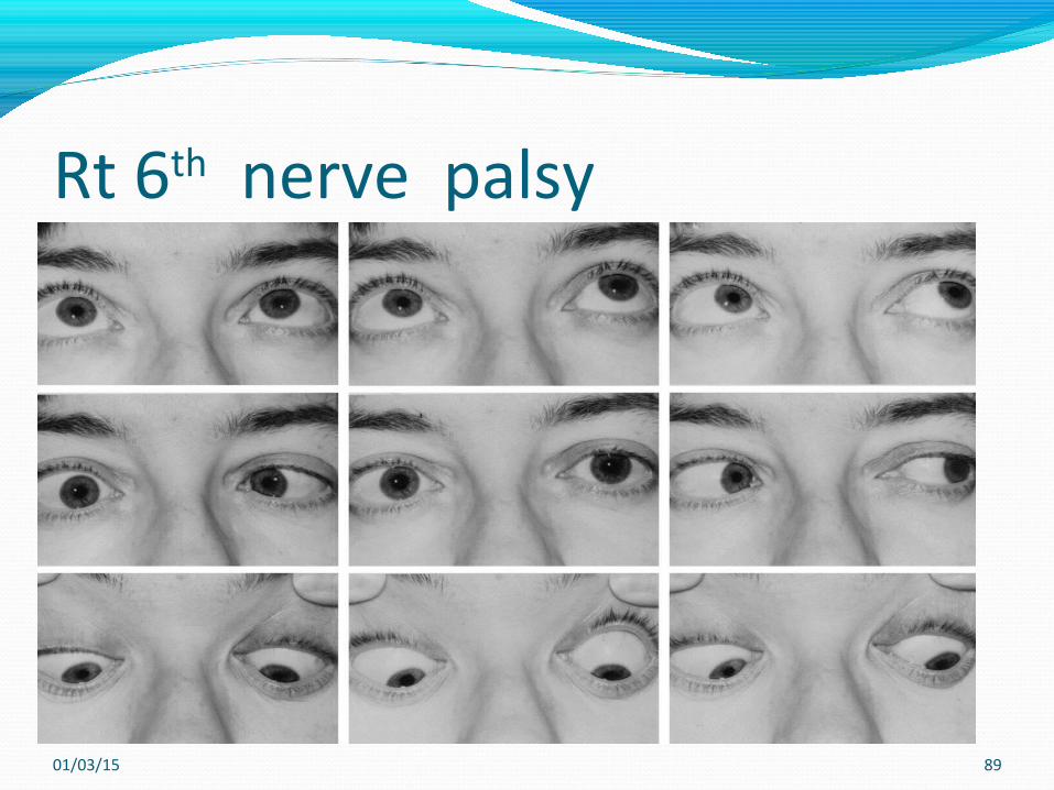
Rt 6th nerve palsy
01/03/15 89
Contd…Binocular functionThis is often retained in the presence of an abnormal
head posture.As the angle of deviation is often smaller for near
fixation, binocular function is usually present on near fixation.
In cases of head trauma, fusion may have been lost.
01/03/15 90
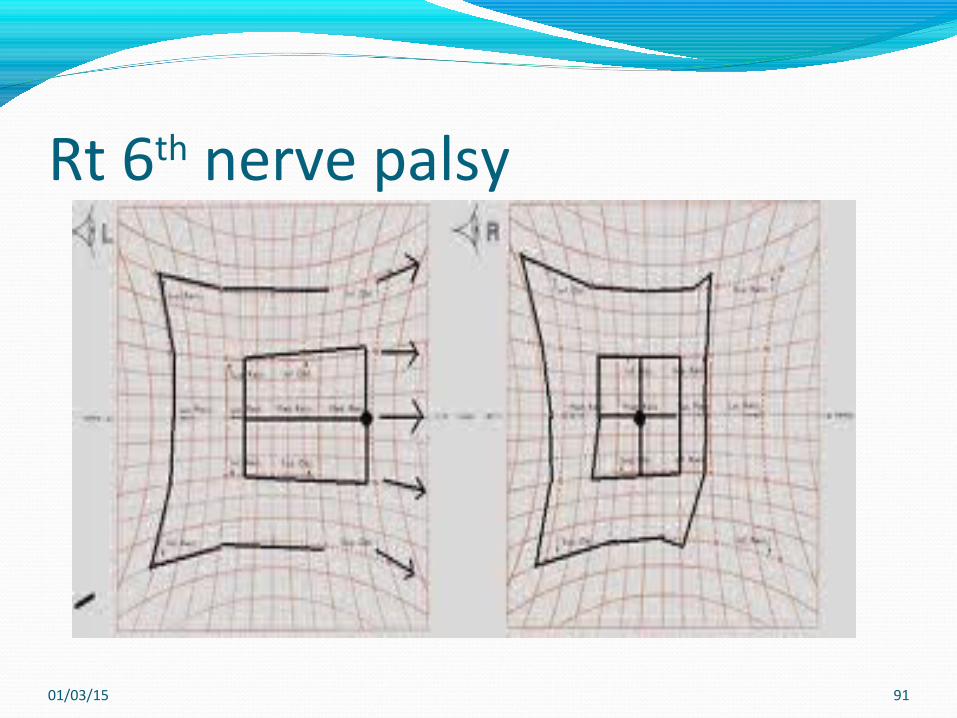
Rt 6th nerve palsy
01/03/15 91
Differential diagnosisDuane’s retraction syndrome, Infantile esotropia, Nystagmus block syndrome Medial wall fracture
01/03/15 92

01/03/15 93
Thank you !!!