Embed Size (px)
Citation preview
Inverse association between programmed death ligand 1 (PDL1) and genes in the
vascular endothelial growth factor (VEGF) pathway in primary clear cell renal cell
(ccRCC).
Richard W. Joseph1, Mansi Parasramka2, Jeanette E. Eckel-Passow3, Dan Serie4, Kevin Wu5,
Jennifer Jiang5, Krishna Kalari3, R. Houston Thompson6, Thai Ho7, Erik Castle8, John Cheville9,
Eugene Kwon6, E. Aubrey Thompson2, Alexander Parker10
1. Department of Hematology and Oncology, Mayo Clinic Florida
2. Department of Cancer Biology, Mayo Clinic Florida
3. Department of Health Sciences Research, Mayo Clinic Rochester
4. Department of Health Sciences Research, Mayo Clinic Florida
5. Department of Pathology, Mayo Clinic Florida
6. Department of Urology, Mayo Clinic Rochester
7. Department of Hematology and Oncology, Mayo Clinic Arizona
8. Department of Urology, Mayo Clinic Arizona
9. Department of Pathology, Mayo Clinic Rochester
10. Department of Urology and Epidemiology, Mayo Clinic Florida
Running title: Inverse relationship between angiogenesis and immune evasion in ccRCC
Keywords: PDL1, VEGF, ccRCC, immunotherapy, immune evasion, angiogenesis
Conflicts of Interest: The authors have nothing to declare.
*Corresponding author: Richard W. Joseph, M. D.
Assistant Professor
Mayo Clinic Florida
4500 San Pablo Rd.
Jacksonville, FL 32224
TEL: 904–953–7290
e-mail: [email protected]
on June 14, 2018. © 2013 American Association for Cancer Research. cancerimmunolres.aacrjournals.org Downloaded from
Author manuscripts have been peer reviewed and accepted for publication but have not yet been edited. Author Manuscript Published OnlineFirst on August 29, 2013; DOI: 10.1158/2326-6066.CIR-13-0042

Abstract
Increased angiogenesis and tumor induced immune evasion are two mechanisms by which
clear cell renal cell carcinoma (ccRCC) proliferate and metastasize; however the relationship
between these pathways in human ccRCC tumor samples is poorly understood. We conducted
a nested case control study by identifying PDL1 positive by immunohistochemistry (IHC)
primary ccRCC tumors that we matched on the Mayo SSIGN score with PDL1 negative primary
ccRCC tumors. From these tumors we extracted RNA from FFPE slides and quantified gene
expression of VEGFA, VEGFR1, VEGFR2, and PDL1. In addition, we also assessed tumor-
infiltrating lymphocytes by IHC for CD3. We then analyzed the relationship between VEGFA,
VEGFR1, VEGFR2, CD3, and PDL1. When analyzed as a continuous variable, PDL1 protein
expression by IHC inversely correlates with the expression of the three VEGF related genes:
VEGFA (r=-0.23, p=0.01), VEGFR1 (r=-0.34, p<0.001), and VEGFR2 (r=-0.23, p=0.01). When
dichotomized, the PDL1 positive cohort trended towards a lower expression of VEGFA (fold
change=0.72, p=0.056) and VEGFR1 (fold change=0.69, p=0.057). In addition, there was a
significant and positive relationship between TIL as assessed by IHC for CD3 and PDL1 by IHC
(r=0.25, p=0.015), and there was a trend towards an inverse relationship between TIL and
VEGFA gene expression (r=-0.18, p=0.089). In conclusion, we are the first demonstrate an
inverse association between the angiogenesis and PDL1 in primary ccRCC human tumor
samples, and that this relationship may be related to the immune suppressive effects of VEGF
signaling.
on June 14, 2018. © 2013 American Association for Cancer Research. cancerimmunolres.aacrjournals.org Downloaded from
Author manuscripts have been peer reviewed and accepted for publication but have not yet been edited. Author Manuscript Published OnlineFirst on August 29, 2013; DOI: 10.1158/2326-6066.CIR-13-0042

Introduction:
Despite the approval of multiple agents to treat metastatic clear cell renal cell carcinoma
(ccRCC), only a very small minority of patients remains alive at 5 years.(1) An improved
understanding of the molecular underpinnings that support the pathogenesis of ccRCC will lead to
improved clinical outcomes. Related to this, two proposed mechanisms by which ccRCC tumors
proliferate and metastasize are through increased angiogenesis and tumor induced immune
evasion. While much is known about the individual roles of the angiogenesis and tumor immune
evasion in ccRCC pathogenesis, the potential relationship between these two pathways in human
ccRCC samples is poorly understood.
In ccRCC, tumor angiogenesis is triggered by activation of the vascular endothelial growth
factor (VEGF) pathway resulting in improved blood supply and enriched source of nutrients for the
tumor. (2-5) VEGF acts through binding VEGF receptors 1 and 2 (VEGFR1 and VEGFR2,
respectively) with a net effect of increasing angiogenesis, cellular proliferation, survival, and
migration. (6) Anti-VEGF therapies revolutionized the care for patients with metastatic ccRCC by
inducing response rates of 30-40% and improving overall survival.(7, 8) Despite the success of anti-
VEGF agents, resistance develops in virtually all patients, and strategies to improve both the rate
and duration of response of anti-VEGF therapies are clearly needed.
In addition to angiogenesis, ccRCC also proliferate through evading immune surveillance.
One mechanism by which ccRCC evade immune surveillance is through tumor expression of
programmed death ligand one (PDL1). PDL1 negatively regulates the immune system through
binding PD-1 expressed on activated T and B cells.(9, 10) Our group and others have reported a
strong association between higher tumor-based PDL1 expression and adverse clinical outcomes for
patients with ccRCC.(11-13) Of interest, blockade of the PDL1/PD-1 interaction has demonstrated
durable responses in ~30% of patients with ccRCC with responses appearing to be related to tumor
PDL1 expression.(14, 15) Tumor PDL1 expression is at least partly driven as an adaptive response
to immune cell infiltrates as a means to evade immune detection.(16)
on June 14, 2018. © 2013 American Association for Cancer Research. cancerimmunolres.aacrjournals.org Downloaded from
Author manuscripts have been peer reviewed and accepted for publication but have not yet been edited. Author Manuscript Published OnlineFirst on August 29, 2013; DOI: 10.1158/2326-6066.CIR-13-0042

While VEGF is primarily linked with increased angiogenesis, there is mounting evidence that
VEGF has immunosuppressive properties and that augmenting tumor vasculature will increase the
efficacy of immunotherapies.(17) With this in mind, we hypothesized that tumors with increased
VEGF expression would possess an immunosuppressive tumor microenvironment, decreased
immune cell infiltrates, and therefore less PDL1 expression. In order to test whether an association
between PDL1 and VEGF exists, we analyzed Affymetrix-based gene expression of a set of 30
primary and patient-matched metastatic ccRCC samples and noted an inverse association of PDL1
expression and multiple genes involved in the VEGF pathway. As such, we leveraged the data and
tissue resources available in the Mayo Clinic Nephrectomy Registry to conduct a nested case-
control study to directly explore the association of VEGF-related genes in PDL1 positive and
negative ccRCC tumors. Our primary endpoint was to study the association of PDL1 protein by IHC
with gene expression of VEGFA, VEGFR1, and VEGFR2 in primary human ccRCC tumor samples.
Secondary endpoints included the following: association between tumor-infiltrating lymphocytes as
assessed by IHC for CD3 and PDL1 and genes in the VEGF pathway; the association of PDL1
protein by IHC and PDL1 gene expression; the association of VEGFA with VEGFR1 and VEGFR2
with each other. Herein, we report the first evidence of an inverse association of PDL1 expression
and expression of VEGFA, VEGFR1 and VEGFR2 in human primary ccRCC samples.
on June 14, 2018. © 2013 American Association for Cancer Research. cancerimmunolres.aacrjournals.org Downloaded from
Author manuscripts have been peer reviewed and accepted for publication but have not yet been edited. Author Manuscript Published OnlineFirst on August 29, 2013; DOI: 10.1158/2326-6066.CIR-13-0042
METHODS:
Patient Selection
Under an Institutional Review Board approved protocol, we queried our Nephrectomy
Registry to identify all patients undergoing nephrectomy to treat sporadic, unilateral, non-cystic
ccRCC between 1990 and 2006. Immunohistochemical (IHC) staining for PDL1 expression on
tumor samples from these patients was available from a previous investigation.(13) Using these
existing data, we selected 50 patients with highest level of IHC-based PDL1 (PDL1 positive
patients). When the blocks of the 50 PDL1 positive tumors were reviewed, a pathologist
identified that two specimens were from metastatic lymph nodes that were replaced by ccRCC
and these samples were removed from the analysis. We then identified 50 PDL1 negative
ccRCC tumors that were matched on Mayo SSIGN score with the PDL1 positive patients.(18,
19)
We abstracted the following demographic and clinicopathologic features from the
Nephrectomy Registry: age at surgery, sex, symptoms at presentation, Eastern Co-operative
Oncology Group (ECOG) performance status, histologic subtype, 2002 AJCC primary tumor
classification, regional lymph node involvement, distant metastases, 2002 AJCC TNM stage
groupings, tumor size, nuclear grade, and presence of coagulative tumor necrosis.
Quantifying Tumor PDL1 and CD3 Expression by Immunohistochemistry (IHC)
Patients for this study all had data available for IHC-based expression of PDL1 from a previous
investigation.(13) The membranous staining pattern of PDL1 was quantified as the percentage
of positive tumor cells in 5% increments as determined by a genitourinary pathologist (JCC). In
order to quantify the expression of both the tumor and microenvironment, we did not perform a
micro-dissection. We also utilized IHC-based expression of CD3 to quantify tumor T cell
infiltrates. Two pathologists (KW and JJ) quantified CD3 expression as the percentage of
positive cells within the tumor in 5% increments.
on June 14, 2018. © 2013 American Association for Cancer Research. cancerimmunolres.aacrjournals.org Downloaded from
Author manuscripts have been peer reviewed and accepted for publication but have not yet been edited. Author Manuscript Published OnlineFirst on August 29, 2013; DOI: 10.1158/2326-6066.CIR-13-0042

RNA isolation and generation of cDNA from FFPE samples
We isolated total RNA from tumor rich areas of FFPE tissue blocks using the Qiagen
Allprep DNA/NRA FFPE Kit reagents (Qiagen, Valencia CA) following vendor's standard
protocols. Three slides per sample at 10 microns each were used for RNA extraction. Isolated
FFPE RNA was treated with DNase (Qiagen) and RNA concentration and purity were
determined spectrophotometrically (OD260/280 > 1.9) (Nano Drop 2000 spectrophotometer,
Thermo Scientific, Waltham MA). 1 µg of FFPE tissue RNA was reverse transcribed using ABI
High capacity RNA to cDNA kit (Applied Biosystems, Carlsbad, CA) as described in the
manufacturer's instructions and resulting cDNA was diluted 1:15 in molecular grade
RNase/DNase free H2O and stored at − 20 °C prior to use.
FFPE RNA quantification via RT-PCR
FFPE compatible primer sets were designed with amplicon sizes <80 base pairs. All
qPCR reactions were assembled in triplicate using ABI TaqMan Fast PCR reagents kit (Applied
Biosystems, Carlsbad, CA) and run on ABI 7900HT Fast Real-Time PCR System and
quantification performed using Sequence Detection Systems software v2.4 (Applied
Biosystems) to quantify gene expression of PDL1 (Hs01125301), VEGFA (Hs00900054),
VEGFR1 (Hs00176573), VEGFR2 (Hs00911708) and POLR2a (Hs00172187). All primer
probes were ordered from Life Technologies, Carlsbad, California. The standard replicates for
each qPCR assay were examined for amplification efficiencies between 90–105% and all
standards and sample replicate data were analyzed for product specific melt curves. If sample
or standard replicates did not conform to these parameters we tested a different dilution, and
the cDNA was diluted with a range of 1:10 to 1:15. We assessed the RNA quality within each
group post reverse-transcriptionally through POLR2a expression.
Statistical Analysis
on June 14, 2018. © 2013 American Association for Cancer Research. cancerimmunolres.aacrjournals.org Downloaded from
Author manuscripts have been peer reviewed and accepted for publication but have not yet been edited. Author Manuscript Published OnlineFirst on August 29, 2013; DOI: 10.1158/2326-6066.CIR-13-0042

We employed Fisher’s exact tests and Chi-Square tests, as appropriate, to compare
demographic and clinic-pathologic characteristics between the PDL1 positive tumors and PDL1
negative tumors. Gene expression from the RT-PCR data was normalized to the control gene
POLR2a. Specifically, the CT values for each gene were averaged across the triplicate
measures and subsequently, the average CT value for POLR2a was subtracted from the
average CT value for each gene of interest for each sample. We used a Spearman rank
correlation to evaluate the continuous association of PDL1 protein expression with the gene
expression of PDL1 and VEGF related genes, to evaluate the continuous association of CD3
protein expression with the gene expression of VEGF related genes, and to evaluate the
continuous association of VEGF related genes with each other. We employed a linear mixed
effects model to compare normalized gene expression of VEGF-A, VEGFR1 and VEGFR2
between PDL1 positive and PDL1 negative tumors.
on June 14, 2018. © 2013 American Association for Cancer Research. cancerimmunolres.aacrjournals.org Downloaded from
Author manuscripts have been peer reviewed and accepted for publication but have not yet been edited. Author Manuscript Published OnlineFirst on August 29, 2013; DOI: 10.1158/2326-6066.CIR-13-0042

RESULTS:
Clinical and Pathologic Characteristics
Of the 50 PDL1 positive tumors we selected from the Mayo Clinic Renal Registry, two tumor
samples came from blocks that were metastatic lymph nodes and were therefore excluded from
the study. The clinical and pathologic characteristics are summarized in Table 1. PDL1 positive
tumors (n=48) had the following distribution of PDL1 positivity: n=18 with 5%, n=10 with 10%,
n=11 with 15-50%, and n=9 with >50%. Expectedly, there were no differences in key pathologic
characteristics between PDL1 positive and PDL1 negative tumors since by design they were
matched on the Mayo SSIGN score. The majority of patients were male (58%) with a median
age at surgery of 67.1 years, and a median follow up of 10.3 years. In the total cohort, 33 (34%)
patients developed metastatic disease and 32/33 (97%) of these patients died from RCC.
Association of the gene expression of VEGFA, VEGFR1, VEGFR2, and PDL1 with protein
Expression of PDL1 by IHC
We could quantify transcripts of VEGFA in 100%, VEGFR1 in 99%, and VEGFR2 in 98% of
tumor samples. We could quantify PDL1 gene expression in 64/98 (65%) all samples with 36/48
(75%) in the PDL1 positive tumors and 23/50 (46%) of the PDL1 negative tumors. When we
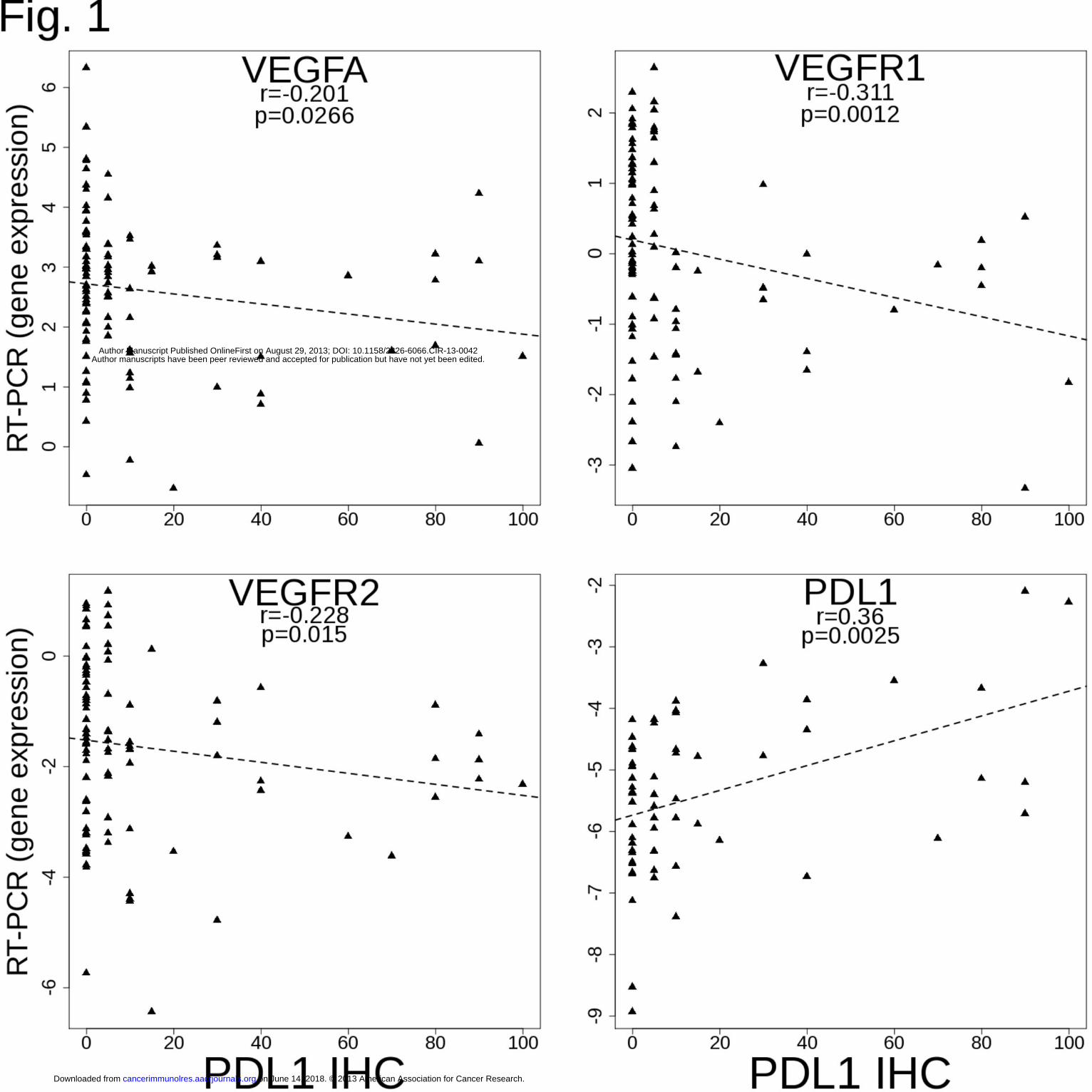
analyzed PDL1 IHC as a continuous variable we observed a significant inverse correlation
between the expression of PDL1 and VEGF related genes as follows (Figure 1): VEGFA (r=-
0.201, p=0.027), VEGFR1 (r=-0.31, p=0.001), and VEGFR2 (r=-0.23, p=0.015). Predictably,
there was also strong positive correlation between PDL1 protein and RNA expression (0.36,
p=0.003).
Next, taking into account the experimental design of this study, we compared RNA expression
of VEGFA, VEGFR1, and VEGFR2 (Figure 2A) between the 50 PDL1 negative (0% by IHC)
and 48 PDL1 positive tumors (≥5% by IHC). In the PDL1 negative cohort, there was a trend of
on June 14, 2018. © 2013 American Association for Cancer Research. cancerimmunolres.aacrjournals.org Downloaded from
Author manuscripts have been peer reviewed and accepted for publication but have not yet been edited. Author Manuscript Published OnlineFirst on August 29, 2013; DOI: 10.1158/2326-6066.CIR-13-0042

higher RNA expression of VEGFA (fold change 0.72, p=0.06) and VEGFR1 (fold change 0.67,
p=0.06) while there was no observed difference in VEGFR2 expression between the two
cohorts (fold change 0.73, p=0.16).
Lastly, with the rationale that PDL1 positivity has been defined as >0% cells(11) as well as >5%
cells (12), as an exploratory analysis we also compared VEGF related genes between subjects
with PDL1 values 0-5% (n=68) versus PDL1>5% (n=30) by IHC (Figure 2B). Using this cutoff
for PDL1, all VEGF related genes were significantly higher in the PDL1 0-5% cohort versus
PLD1 >5% cohort as follows: VEGFA (fold change=1.82, p<0.001), VEGFR1 (FC=2.63,
p<0.001), and VEGFR2 (FC=2.13, p=0.001).
Association of tumor infiltrating lymphocytes (TIL) with VEGFA, VEGFR1, VEGFR2, and
PDL1
In order to determine the relationship between TIL, angiogenesis, and PDL1 expression we
assessed all tumors for CD3 by IHC and analyzed expression as a continuous variable versus
the gene expression of VEGFA, VEGFR1, VEGFR2, and PDL1 protein expression by IHC
(Figure 3). We found a trend of an inverse relationship between CD3 and VEGFA (r=-0.18,
p=0.089) and a significant positive association between CD3 and PDL1 (r=0.25, p=0.015).
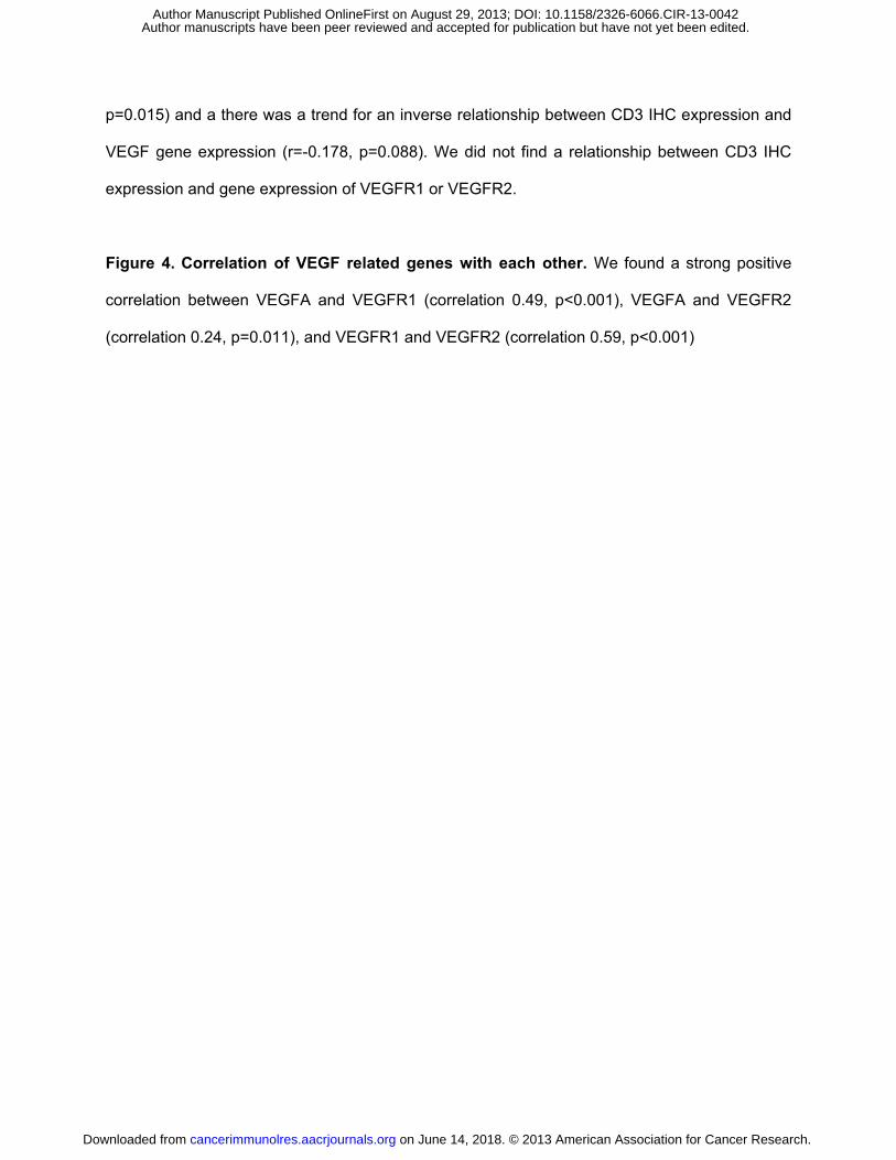
Association of VEGF related genes with each other
Since we found that VEGFA, VEGFR1, and VEGFR2 all inversely correlated with PDL1 by IHC,
we compared the association of all three VEGF related genes to each other (Figure 4). We
found a strong positive correlation between VEGFA and VEGFR1 (correlation 0.49, p<0.001),
between VEGFA and VEGFR2 (correlation 0.24, p=0.011), and between VEGFR1 and VEGFR2
(correlation 0.59, p<0.001).
on June 14, 2018. © 2013 American Association for Cancer Research. cancerimmunolres.aacrjournals.org Downloaded from
Author manuscripts have been peer reviewed and accepted for publication but have not yet been edited. Author Manuscript Published OnlineFirst on August 29, 2013; DOI: 10.1158/2326-6066.CIR-13-0042

DISCUSSION:
Given the existing clinical benefits and limitations of anti-VEGF therapies, the prognostic
significance of PDL1 expression, and the promising results of PDL1/PD-1 blockade, a logical next
step to advance the field and help inform the next generation of clinical studies for ccRCC is to
enhance our understanding of the relationship between VEGF and PDL1 expression in ccRCC
samples. Indeed, while the individual roles of angiogenesis and immune evasion in ccRCC
development and progression are well described, to date very little is known about how these
pathways are associated in clinical samples. To our knowledge, this is the first demonstration of
an inverse relationship between angiogenesis, TIL, and PDL1 expression in human ccRCC
clinical samples.
While activation of the VEGF pathway is most commonly associated with increased
angiogenesis, there is emerging evidence that increased angiogenesis leads to an
immunosuppressive tumor microenvironment.(17, 20, 21) Furthermore, there is growing data to
suggest that VEGF inhibition can increase tumor-infiltrating lymphocytes (TIL).(22) In our study,
we hypothesized that tumors with increased VEGF expression would have decreased immune
infiltrate and less adaptive pressure to express PDL1. Our data demonstrates a trend for an
inverse relationship between VEGF expression and TIL as assessed by CD3 expression,
however we note that the relationship has multiple outliers with some tumors having both low
TIL and VEGF expression. We acknowledge that VEGF expression and by proxy angiogenesis
is likely one of multiple mechanisms that may impact the tumor immune microenvironment.
The two leading hypotheses of why tumors express PDL1 are the innate and adaptive
models.(23) In the innate model, tumor PDL1 expression is independent of the tumor
microenvironment and is influenced by intrinsic cell signaling pathways like in glioblastomas and
ALK positive lung cancers.(24, 25) In the adaptive model of tumor PDL1 expression, tumors are
pressured to express PDL1 in the presence of an immunostimulatory microenvironment with two
examples occurring in melanoma and merkel cell carcinoma where all PDL1 positive tumors
on June 14, 2018. © 2013 American Association for Cancer Research. cancerimmunolres.aacrjournals.org Downloaded from
Author manuscripts have been peer reviewed and accepted for publication but have not yet been edited. Author Manuscript Published OnlineFirst on August 29, 2013; DOI: 10.1158/2326-6066.CIR-13-0042

had dense immune infiltrates while PDL1 negative tumors had absent immune infiltrates.(16,
26) In this current study, we demonstrate that TIL does positively associate with PDL1
expression however the relationship is not as strong as in melanoma and merkel cell carcinoma.
We also note the presence of outliers demonstrate that some ccRCC tumors express high
levels of PDL1 in the absence of TIL and conversely, some tumors do not express PDL1 even in
the presence of abundant TIL. In summary, it remains unclear if PDL1 expression in ccRCC is
an innate or adaptive response and it remains possible that both models could play a role.
While many groups quantify PDL1 as a dichotomous variable (positive or negative
variable), there is increasing data to suggest that PDL1 should be quantified as a continuous
variable. For example, not all patients with PDL1 positive tumors respond to anti-PD-1 therapies
indicating that perhaps a certain threshold of PDL1 positivity is necessary.(14) In our study,
when analyzed as a continuous variable a strong inverse association between VEGF and PDL1
existed, however, when we dichotomized tumors into PDL1 negative and positive the inverse
association between PDL1 and VEGF weakened. Given this difference, we believe future
studies should take future studies should consider analyzing PDL1 as both a continuous and
dichotomous variable.
The variability in technique for quantifying PDL1 expression by IHC has posed a great
challenge to understand the biology of PDL1 expression, and therefore alternate techniques to
assess PDL1 expression with less variability would be desirable. To our knowledge this is the
first study to both correlate PDL1 protein expression by IHC with RNA expression from FFPE
tissue as well as demonstrate a positive correlation between protein and RNA expression.
Assessing PDL1 RNA has three potential advantages over using IHC. First, RNA expression is
inherently more quantifiable than IHC. Second, commercially available reagents are used for
RNA expression rather than a proprietary antibody. Third, the sensitivity of using IHC to assess
PDL1 expression on FFPE tissues is reduced in comparison to frozen tissue. Despite the
advantages of using RNA to assess PDL1, there are several important drawbacks. First, we
on June 14, 2018. © 2013 American Association for Cancer Research. cancerimmunolres.aacrjournals.org Downloaded from
Author manuscripts have been peer reviewed and accepted for publication but have not yet been edited. Author Manuscript Published OnlineFirst on August 29, 2013; DOI: 10.1158/2326-6066.CIR-13-0042

note that many of our PDL1 negative tumors by IHC had detectable PDL1 RNA, which raises
two possibilities. The first possibility is that PDL1 negative tumors by IHC do express PDL1 at
the RNA level. The second possibility is that because we did not micro-dissect the tumor from
the stroma we are in fact detecting PDL1 RNA from the surrounding stroma rather than the
tumor itself. In summary, we believe both IHC and RNA have their unique set of advantages
and disadvantages and future studies could consider using both to complement each other.
Our case-control study was designed to evaluate the differential expression of VEGF
related genes in PDL1 positive and negative tumors, and we selected cases based on PDL1
expression, a well-known negative prognostic biomarker in ccRCC, and we matched these
cases on the Mayo SSIGN with PDL1 negative tumors. Predictably, since we initially selected
patients with an adverse feature (PDL1 positive) the cohort in this study had a higher rate of
ccRCC related death than in the total Mayo Renal Registry (34% versus 18%). However, we did
not find any statistical differences in additional clinical and pathologic features between our
cohort and the total Mayo Renal Registry similar. Related to this, the percent of PDL1 positive
tumors in this study are a higher than what is found in total Mayo Renal Registry (50% versus
30%). We acknowledge that biases of this study limit the applicability of our findings to ccRCC
as a whole, and we are currently testing whether these findings hold up in a non-selected cohort
of ccRCC tumors.
Both PDL1 and VEGF expression are associated with an adverse prognosis in
ccRCC(11, 27), and therefore the inverse relationship of these two markers is potentially
concerning, however we believe the case-control design of the study can explain this apparent
discrepancy. We selected the PDL1 positive tumors first (cases) and then matched them on
PDL1 negative tumors (controls). Both the PDL1 positive and negative cohorts have similar
outcomes and a higher rate of ccRCC related death relative to the total Mayo Renal Registry.
As the outcomes between the cohorts are similar but the PDL1 and VEGF status differ, we
hypothesize that there are at least two phenotypes that confer a poor prognosis: an immune
on June 14, 2018. © 2013 American Association for Cancer Research. cancerimmunolres.aacrjournals.org Downloaded from
Author manuscripts have been peer reviewed and accepted for publication but have not yet been edited. Author Manuscript Published OnlineFirst on August 29, 2013; DOI: 10.1158/2326-6066.CIR-13-0042
evasive phenotype (VEGFlowPDL1high) and an angiogenic phenotype (VEGFhighPDL1low). Again,
further studies in a random cohort of tumors are necessary to confirm these findings. Finally,
given that not all PDL1 high tumors were VEGF low and vice versa, we believe that these
biomarkers are not ready to guide treatment decisions at present. However, we do think that
prospective analysis of VEGF and PDL1 expression in upcoming combination trials of anti-
VEGF and anti-PDL1 therapies deserves consideration.
In summary, the mechanisms of angiogenesis and immune evasion are complex and
perhaps inversely related in primary ccRCC tumor samples. Our data is hypothesis generating
and warrants further study in larger and unselected populations of ccRCC. In addition, we
believe the relationship between VEGF and PDL1 in other tumors also warrants further
consideration. Testing the impact of VEGF inhibition on PDL1 expression could provide valuable
insights when designing a clinical trial to either combine or sequence anti-angiogenic and anti-
PDL1 therapies.
Grant Support:
This work was generously funded through the AACR Judah Folkman Career Development Award in
Anti-Angiogenesis.
on June 14, 2018. © 2013 American Association for Cancer Research. cancerimmunolres.aacrjournals.org Downloaded from
Author manuscripts have been peer reviewed and accepted for publication but have not yet been edited. Author Manuscript Published OnlineFirst on August 29, 2013; DOI: 10.1158/2326-6066.CIR-13-0042

Reference 1. Siegel R, Naishadham D, Jemal A. Cancer statistics, 2013. CA: a cancer journal for clinicians. 2013;63:11-30. 2. Folkman J, Merler E, Abernathy C, Williams G. Isolation of a tumor factor responsible for angiogenesis. The Journal of experimental medicine. 1971;133:275-88. 3. Folkman J. Tumor angiogenesis: therapeutic implications. The New England journal of medicine. 1971;285:1182-6. 4. Gnarra JR, Tory K, Weng Y, Schmidt L, Wei MH, Li H, et al. Mutations of the VHL tumour suppressor gene in renal carcinoma. Nature genetics. 1994;7:85-90. 5. Herman JG, Latif F, Weng Y, Lerman MI, Zbar B, Liu S, et al. Silencing of the VHL tumor-suppressor gene by DNA methylation in renal carcinoma. Proceedings of the National Academy of Sciences of the United States of America. 1994;91:9700-4. 6. Ferrara N, Gerber HP, LeCouter J. The biology of VEGF and its receptors. Nature medicine. 2003;9:669-76. 7. Motzer RJ, Hutson TE, Tomczak P, Michaelson MD, Bukowski RM, Rixe O, et al. Sunitinib versus interferon alfa in metastatic renal-cell carcinoma. The New England journal of medicine. 2007;356:115-24. 8. Yang JC, Haworth L, Sherry RM, Hwu P, Schwartzentruber DJ, Topalian SL, et al. A randomized trial of bevacizumab, an anti-vascular endothelial growth factor antibody, for metastatic renal cancer. The New England journal of medicine. 2003;349:427-34. 9. Freeman GJ, Long AJ, Iwai Y, Bourque K, Chernova T, Nishimura H, et al. Engagement of the PD-1 immunoinhibitory receptor by a novel B7 family member leads to negative regulation of lymphocyte activation. J Exp Med. 2000;192:1027-34. 10. Dong H, Strome SE, Salomao DR, Tamura H, Hirano F, Flies DB, et al. Tumor-associated B7-H1 promotes T-cell apoptosis: a potential mechanism of immune evasion. Nat Med. 2002;8:793-800. 11. Thompson RH, Gillett MD, Cheville JC, Lohse CM, Dong H, Webster WS, et al. Costimulatory B7-H1 in renal cell carcinoma patients: Indicator of tumor aggressiveness and potential therapeutic target. Proceedings of the National Academy of Sciences of the United States of America. 2004;101:17174-9. 12. Krambeck AE, Dong H, Thompson RH, Kuntz SM, Lohse CM, Leibovich BC, et al. Survivin and b7-h1 are collaborative predictors of survival and represent potential therapeutic targets for patients with renal cell carcinoma. Clinical cancer research : an official journal of the American Association for Cancer Research. 2007;13:1749-56. 13. Thompson RH, Kuntz SM, Leibovich BC, Dong H, Lohse CM, Webster WS, et al. Tumor B7-H1 is associated with poor prognosis in renal cell carcinoma patients with long-term follow-up. Cancer research. 2006;66:3381-5. 14. Topalian SL, Hodi FS, Brahmer JR, Gettinger SN, Smith DC, McDermott DF, et al. Safety, Activity, and Immune Correlates of Anti-PD-1 Antibody in Cancer. The New England journal of medicine. 2012. 15. Brahmer JR, Tykodi SS, Chow LQ, Hwu WJ, Topalian SL, Hwu P, et al. Safety and Activity of Anti-PD-L1 Antibody in Patients with Advanced Cancer. The New England journal of medicine. 2012.
on June 14, 2018. © 2013 American Association for Cancer Research. cancerimmunolres.aacrjournals.org Downloaded from
Author manuscripts have been peer reviewed and accepted for publication but have not yet been edited. Author Manuscript Published OnlineFirst on August 29, 2013; DOI: 10.1158/2326-6066.CIR-13-0042

16. Taube JM, Anders RA, Young GD, Xu H, Sharma R, McMiller TL, et al. Colocalization of inflammatory response with B7-h1 expression in human melanocytic lesions supports an adaptive resistance mechanism of immune escape. Sci Transl Med. 2012;4:127ra37. 17. Huang Y, Goel S, Duda DG, Fukumura D, Jain RK. Vascular normalization as an emerging strategy to enhance cancer immunotherapy. Cancer research. 2013. 18. Thompson RH, Leibovich BC, Lohse CM, Cheville JC, Zincke H, Blute ML, et al. Dynamic outcome prediction in patients with clear cell renal cell carcinoma treated with radical nephrectomy: the D-SSIGN score. The Journal of urology. 2007;177:477-80. 19. Zigeuner R, Hutterer G, Chromecki T, Imamovic A, Kampel-Kettner K, Rehak P, et al. External validation of the Mayo Clinic stage, size, grade, and necrosis (SSIGN) score for clear-cell renal cell carcinoma in a single European centre applying routine pathology. Eur Urol. 2010;57:102-9. 20. Wei J, Wu A, Kong LY, Wang Y, Fuller G, Fokt I, et al. Hypoxia potentiates glioma-mediated immunosuppression. PLoS One. 2011;6:e16195. 21. Facciabene A, Peng X, Hagemann IS, Balint K, Barchetti A, Wang LP, et al. Tumour hypoxia promotes tolerance and angiogenesis via CCL28 and T(reg) cells. Nature. 2011;475:226-30. 22. Huang Y, Yuan J, Righi E, Kamoun WS, Ancukiewicz M, Nezivar J, et al. Vascular normalizing doses of antiangiogenic treatment reprogram the immunosuppressive tumor microenvironment and enhance immunotherapy. Proceedings of the National Academy of Sciences of the United States of America. 2012;109:17561-6. 23. Pardoll DM. The blockade of immune checkpoints in cancer immunotherapy. Nature reviews Cancer. 2012;12:252-64. 24. Parsa AT, Waldron JS, Panner A, Crane CA, Parney IF, Barry JJ, et al. Loss of tumor suppressor PTEN function increases B7-H1 expression and immunoresistance in glioma. Nat Med. 2007;13:84-8. 25. Marzec M, Zhang Q, Goradia A, Raghunath PN, Liu X, Paessler M, et al. Oncogenic kinase NPM/ALK induces through STAT3 expression of immunosuppressive protein CD274 (PD-L1, B7-H1). Proceedings of the National Academy of Sciences of the United States of America. 2008;105:20852-7. 26. Lipson EJ, Vincent JG, Loyo M, Kagohara LT, Luber BS, Wang H, et al. PD-L1 Expression in the Merkel Cell Carcinoma Microenvironment: Association with Inflammation, Merkel Cell Polyomavirus, and Overall Survival. Cancer Immunology Research. 2013;1:54-63. 27. Jacobsen J, Grankvist K, Rasmuson T, Bergh A, Landberg G, Ljungberg B. Expression of vascular endothelial growth factor protein in human renal cell carcinoma. BJU Int. 2004;93:297-302.
on June 14, 2018. © 2013 American Association for Cancer Research. cancerimmunolres.aacrjournals.org Downloaded from
Author manuscripts have been peer reviewed and accepted for publication but have not yet been edited. Author Manuscript Published OnlineFirst on August 29, 2013; DOI: 10.1158/2326-6066.CIR-13-0042

Figure Legends:
Figure 1. The association of the gene expression of VEGFA, VEGFR1, and VEGFR2 with
PDL1 protein expression as assessed by IHC and as a continuous variable. Expression of
PDL1 by IHC inversely correlates with gene expression of VEGFA (r=-0.201, p=0.027),
VEGFR1 (r=-0.311, p=0.001), and VEGFR2 (r=-0.228, p=0.015). We quantified PDL1 gene
expression in 64% of all samples, in 38/50 (76%) of the cases and in 23/50 (46%) of the
controls. Predictably, there was a strong positive correlation between PDL1 protein expression
by IHC and PDL1 RNA expression (r=0.36, p=0.003).
Figure 2. Dichotomous analysis of gene expression of VEGFA, VEGFR1, and VEGFR2 by
PDL1 protein expression as assessed by IHC. A. PDL1 negative versus PDL1 positive
cohort. In the PDL1 positive cohort, RNA expression of VEGFA (fold change 0.70, p=0.036)
and VEGFR1 (fold change 0.67, p=0.037) was significantly lower than in the PDL1 absent
cohort while there was no observed difference in VEGFR2 expression in the two cohorts (fold
change 0.71, p=0.13). Solid horizontal line represents the median and the dotted horizontal
lines represent the 95% confidence interval. B. PDL1 0-5% versus PDL1 >5%. Using this
cutoff for PDL1, all VEGF related genes were significantly higher in the PLD1 0-5% cohort
versus PLD1 >5% cohort as follows: VEGFA (fold change=1.82, p<0.001), VEGFR1 (FC=2.63,
p<0.001), and VEGFR2 (FC=2.13, p=0.001). Solid horizontal line represents the median and
the dotted horizontal lines represent the 95% confidence interval.
Figure 3: The association between CD3 by IHC with gene expression of VEGFA, VEGFR1,
VEGFR2, and PDL1 protein expression by IHC. We quantified tumor infiltrating lymphocytes
with IHC by counting percent positive CD3 cells in 5% increments. Using a Spearman rank
correlation, we found a significant relationship between CD3 IHC and PDL1 IHC (r=0.25,
on June 14, 2018. © 2013 American Association for Cancer Research. cancerimmunolres.aacrjournals.org Downloaded from
Author manuscripts have been peer reviewed and accepted for publication but have not yet been edited. Author Manuscript Published OnlineFirst on August 29, 2013; DOI: 10.1158/2326-6066.CIR-13-0042
p=0.015) and a there was a trend for an inverse relationship between CD3 IHC expression and
VEGF gene expression (r=-0.178, p=0.088). We did not find a relationship between CD3 IHC
expression and gene expression of VEGFR1 or VEGFR2.
Figure 4. Correlation of VEGF related genes with each other. We found a strong positive
correlation between VEGFA and VEGFR1 (correlation 0.49, p<0.001), VEGFA and VEGFR2
(correlation 0.24, p=0.011), and VEGFR1 and VEGFR2 (correlation 0.59, p<0.001)
on June 14, 2018. © 2013 American Association for Cancer Research. cancerimmunolres.aacrjournals.org Downloaded from
Author manuscripts have been peer reviewed and accepted for publication but have not yet been edited. Author Manuscript Published OnlineFirst on August 29, 2013; DOI: 10.1158/2326-6066.CIR-13-0042

Table 1.
Table 1: Clinical and pathological information
PDL1- (N=50)
PDL1+ (N=48)
Total (N=98) p value
Male 0.5394 No 19 (38.0%) 22 (45.8%) 41 (41.8%) Yes 31 (62.0%) 26 (54.2%) 57 (58.2%) Age at surgery 0.1044 Median 65.9 68.9 67.1 Range (35.4-82.7) (26.6-85.7) (26.6-85.7) Tumor size 0.4969 Median 6.0 5.2 5.5 Range (1.5-18.0) (2.0-16.0) (1.5-18.0) Time to last follow-up 0.5109 Median 9.4 12.2 10.3 Range (0.0-22.1) (0.1-21.8) (0.0-22.1) Metastasis 1.0000 No 33 (66.0%) 32 (66.7%) 65 (66.3%) Yes 17 (34.0%) 16 (33.3%) 33 (33.7%) Time from surgery to metastasis 0.6268 Median 1.5 1.4 1.5 Range (0.1-12.3) (0.1-14.6) (0.1-14.6) Death 1.0000 No 12 (24.0%) 11 (22.9%) 23 (23.5%) Yes 38 (76.0%) 37 (77.1%) 75 (76.5%) Death from RCC 1.0000 Missing 2 2 4 No 32 (66.7%) 30 (65.2%) 62 (66.0%) Yes 16 (33.3%) 16 (34.8%) 32 (34.0%) 2010 Primary Tumor Classificiation 0.0906
on June 14, 2018. © 2013 A
merican A
ssociation for Cancer R
esearch. cancerim
munolres.aacrjournals.org
Dow
nloaded from
Author m
anuscripts have been peer reviewed and accepted for publication but have not yet been edited.
Author M
anuscript Published O
nlineFirst on A
ugust 29, 2013; DO
I: 10.1158/2326-6066.CIR
-13-0042

1 24 (48.0%) 30 (62.5%) 54 (55.1%) 2 6 (12.0%) 8 (16.7%) 14 (14.3%) 3 20 (40.0%) 9 (18.8%) 29 (29.6%) 4 0 (0.0%) 1 (2.1%) 1 (1.0%) Nuclear Grade 0.5919 1 4 (8.0%) 7 (14.6%) 11 (11.2%) 2 25 (50.0%) 23 (47.9%) 48 (49.0%) 3 18 (36.0%) 13 (27.1%) 31 (31.6%) 4 3 (6.0%) 5 (10.4%) 8 (8.2%) Coagulative Tumor Necrosis 0.4961 No 35 (70.0%) 37 (77.1%) 72 (73.5%) Yes 15 (30.0%) 11 (22.9%) 26 (26.5%) SSIGN Category 0.2265 Missing 6 8 14 0-3 25 (56.8%) 28 (70.0%) 53 (63.1%) 4-7 16 (36.4%) 10 (25.0%) 26 (31.0%) 8+ 3 (6.8%) 2 (5.0%) 5 (6.0%) PDL1 IHC (%) <0.0001 0 50 (100.0%) 0 (0.0%) 50 (51.0%) 5 0 (0.0%) 18 (37.5%) 18 (18.4%) 10 0 (0.0%) 10 (20.8%) 10 (10.2%) 15-50 0 (0.0%) 11 (22.9%) 11 (11.2%) > 50 0 (0.0%) 9 (18.8%) 9 (9.2%)
on June 14, 2018. © 2013 A
merican A
ssociation for Cancer R
esearch. cancerim
munolres.aacrjournals.org
Dow
nloaded from
Author m
anuscripts have been peer reviewed and accepted for publication but have not yet been edited.
Author M
anuscript Published O
nlineFirst on A
ugust 29, 2013; DO
I: 10.1158/2326-6066.CIR
-13-0042
on June 14, 2018. © 2013 American Association for Cancer Research. cancerimmunolres.aacrjournals.org Downloaded from
Author manuscripts have been peer reviewed and accepted for publication but have not yet been edited. Author Manuscript Published OnlineFirst on August 29, 2013; DOI: 10.1158/2326-6066.CIR-13-0042

Published OnlineFirst August 29, 2013.Cancer Immunol Res Richard W. Joseph, Mansi Parasramka, Jeanette Eckel-Passow, et al. pathway in primary clear cell renal cell carcinomaInverse association between PDL1 and genes in the VEGF
Updated version
10.1158/2326-6066.CIR-13-0042doi:
Access the most recent version of this article at:
Manuscript
Authoredited. Author manuscripts have been peer reviewed and accepted for publication but have not yet been
E-mail alerts related to this article or journal.Sign up to receive free email-alerts
Subscriptions
Reprints and
To order reprints of this article or to subscribe to the journal, contact the AACR Publications
Permissions
Rightslink site. Click on "Request Permissions" which will take you to the Copyright Clearance Center's (CCC)
.http://cancerimmunolres.aacrjournals.org/content/early/2013/08/29/2326-6066.CIR-13-0042To request permission to re-use all or part of this article, use this link
on June 14, 2018. © 2013 American Association for Cancer Research. cancerimmunolres.aacrjournals.org Downloaded from
Author manuscripts have been peer reviewed and accepted for publication but have not yet been edited. Author Manuscript Published OnlineFirst on August 29, 2013; DOI: 10.1158/2326-6066.CIR-13-0042