Embed Size (px)
Citation preview

Disease Markers 34 (2013) 387–393 387DOI 10.3233/DMA-130985IOS Press
MRP1 but not MDR1 is associated withresponse to neoadjuvant chemotherapy inbreast cancer patients
Mohsen Taheria,b,c,∗ and Frouzandeh MahjoubiaaDivision of Medical Biotechnology, National Institute of Genetic Engineering and Biotechnology (NIGEB),Tehran, IranbGenetics of Non-communicable Diseases Research Center, Zahedan University of Medical Sciences, Zahedan,IrancDepartment of Genetics, School of Medicine, Zahedan University of Medical Sciences, Zahedan, Iran
Abstract. A major problem in the treatment of breast cancer is the development of resistance to chemotherapeutic agents.Although the role of multidrug resistance 1 (MDR1) and multidrug resistance associated protein 1 (MRP1) in inducing drugresistance in many cancers has been widely investigated the clinical significance of expression of these genes in breast cancerremains unclear and the data is still controversial. We investigated the expression of MDR1 and MRP1 in breast cancer patientsas well as the possible correlation between MDR1 and MRP1 and clinical response to chemotherapy. In the present study, MDR1and MRP1 gene expression were investigated by real time reverse transcription polymerase chain reaction (RT-PCR) assay in54 breast cancer tumors and in corresponding adjacent normal tissues before neoadjuvant chemotherapy. The expression levelof MDR1 and MRP1 were significantly higher in breast tumors than normal breast tissues. Although a significant relationshipwas found between the MRP1 expression and response to treatment no association was observed between MDR1 expression andresponse to treatment. MDR1 and MRP1 expression levels have been shown to be independent of tumor size, histological gradeand the status of progesterone or estrogen receptor.
Keywords: Multidrug resistance, real time PCR, breast cancer, MDR1, MRP1
1. Introduction
Resistance to chemotherapy limits the effective-ness of anti neoplastic treatments. In multi drug re-sistance (MDR) tumors become insensitive to manydrugs with different structures and functions [1]. Eventhough breast cancer is one of the sensitive tumors tochemotherapy many of them become multidrug resis-tance [2].
∗Corresponding author: Frouzandeh Mahjoubi, Division of Med-ical Biotechnology, National Institute of Genetic Engineering andBiotechnology (NIGEB) Pajoohesh Blvd. Tehran-Karaj Highway,17th Km, Tehran, Iran. Tel.: +98 21 44580389; Fax: +98 2144580399; E-mail: [email protected].
Neoadjuvant chemotherapy is an approach in themanagement of locally advanced breast cancer. Itgives benefit in the operable breast cancers by in-creasing the chances of breast conservation. Neoadju-vant chemotherapy followed by surgery allows for as-sessment of the histopathologic response. Only about70% of patients demonstrate a clinical response andmerely 3%–27% achieve a complete histopathologicresponse (3). Therefore, roughly 30% of the patientsreceived neoadjuvant chemotherapy are no respon-sive to this treatment and suffer from the side ef-fects. The patients achieving a complete pathologic re-sponse after neoadjuvant chemotherapy have signifi-cantly longer disease-free overall and overall survivalthan nonresponders (3). Thus in any clinical practice itwould be very important and beneficial for each patient
ISSN 0278-0240/13/$27.50 c© 2013 – IOS Press and the authors. All rights reserved
388 M. Taheri and F. Mahjoubi / MDR1 and MRP1 expression in breast cancer
if the patient could be informed in advance about thechance of being responsive or resistance to the neoad-juvant therapy.
One of the mechanisms inducing drug resistance isthe efflux of drugs out of cells by increasing the ac-tivity of efflux pumps such as ATP Binding Cassettetransporters (ABC transporters) [4]. ABC transportersare a family of transmembrane proteins with the abilityto transport wide range of substrate molecules acrosscellular membranes [5].
MDR1 protein or P-gp is a transmembrane glyco-protein with molecular weight of 170 kDa, encodesby MDR gene [6]. It transports many hydrophobicsubstrates and anti-cancer drugs including etoposide,doxorubicin and Vinblastine [6,7]. The MDR1 genewith 28 exons and 1.2 kb is located on chromosome7q21.12 [5].
Like p-gp, MRP is a member of ABC transporterthat transport a wide variety of substrates, such asorganic anion, leukotriene C4 as well as a numberof chemotherapeutic agents, including doxorubicin,daunorubicin, vincristine and colchicines. Also it trans-ports glutathione conjugates and cyclic nucleotides [8].
Although the role of MDR1 and MRP1 in inducingdrug resistance in many cancers has been widely in-vestigated the data is still controversial (2, 9, 10, 11).Therefore, the aim of the present study was to analyzethe expression of MDR1 and MRP1 in breast cancerpatients to see whether or not the expression of eitherof these genes or in combination or could be used as amarker to predict of drug sensitivity.
2. Material and methods
2.1. Patients
The project was approved by the local ethics com-mittee of National Institute for Genetic Engineeringand Biotechnology (NIGEB) and written informedconsent was obtained from each case. Fifty four pa-tients with locally advanced breast cancer admitted toImam Khomeini Hospital in Tehran were enrolled inthis study. The breast cancer patients had not yet re-ceived any chemotherapy treatment.
The tumor size and the axillary lymph node statuswere measured clinically and by using ultrasonogra-phy. At this stage, fresh tissue specimens (tumor andnormal tissue adjacent to tumor) were collected by theclinicians in separated sterile tubes. Tissue sampleswere frozen and stored at −70◦C.
The patients were then received three cycles of an-thracycline based chemotherapy (FAC/FEC) regime atthree weekly intervals.
Routine and metastatic work up was done includ-ing complete blood examination (total blood count,platelet count), chest radiograph, ECG (Echocardiog-raphy when ECG had a positive finding), liver functiontests, Bone Scan.
The patients were assessed both clinically and byuntrasonography for response in the form of reductionin breast tumor size and axillary lymph node status ac-cording to UICC criteria [12].
Complete response and partial response were con-sidered when the tumor disappeared completely andwhen more than half of the tumor diameters showedregression with no local progression respectively. Noresponse was considered for the patients whose tumordiameters had less than 50% regression.
2.2. RNA extraction and cDNA synthesis
After histologic diagnosis was confirmed for allsamples (and thus cancer tissues could be distin-guished from normal tissues) RNA was isolated from100 mg tissue using Tripure Isolation Reagent (RocheApplied Sciences, Indianapolis, USA). For cDNA syn-thesis, 1 μg of total RNA from each sample was usedto synthesize first-strand cDNA according to the manu-facturer’s protocol (Frementas,Mannheim, Germany).
2.3. Real-time RT-PCR
mRNA expression of the MDR1 and MRP1 genewas quantified as described and validated previous-ly [13] by real-time RT-PCR using a lightcyclerTM
system (Roche Applied Sciences, Indianapolis, USA.)with Fast-Start DNA Master SYBR-Green I kit (RocheApplied Sciences, Indianapolis, USA.) using β-actinas housekeeping gene.
The PCR was performed in 20 μL of solution, con-sisting of 2 μL of Fast Start Master solution and0.6 μM of each primer. A total of 18 μL of this reac-tion mix was placed into glass capillaries, and 2 μL ofcDNA was added as template.
A standard Lightcycler PCR program was estab-lished for each gene. Thermal Cycling were as follows:95◦C for 10 sec followed by an amplification program55 cycles with temperature ramp rate of 20◦C/sec. Theamplification program was 95◦C for 10 sec, primerTm (Table 1) for 15 sec and 72◦C for 15 sec with asingle fluorescence acquisition at the end of the elon-
M. Taheri and F. Mahjoubi / MDR1 and MRP1 expression in breast cancer 389
Table 1Sequences of primers
Primer Amplicon Sequence Tm
sizeMRP1-Forward 294 bp 5’-CGG AAA CCA TCC
ACG ACC CTA ATC-3’64◦
MRP1-Reversed 5’-ACC TCC TCA TTCGCA TCC ACC TGG-3’
MDR1-Forward 308 bp 5’ TGA CAT TTA TTCAAA GTT AAA AGC 3’
62◦
MDR1-Reversed 5’ TAG ACA CTT TATGCA AAC ATT TCA A3’
β-actin-Forward 161 bp 5’-GAG ACC TTC AACACC CCA GCC-3’
60◦
β-actin-Reversed 5’-AGA CGC AGG ATGGCA TGG G-3’
Table 2Comparison between tumor size and response to chemotherapy
Tumor size< 5 cm 5–8 cm 8–10 cm > 10 cmN % N % N % N %
Responder 21 38.9 8 14.8 5 9.3 6 11.1Non responder 6 11.1 5 9.3 1 1.8 2 3.7
gation step. The third segment consisted of a meltingcurve program at 95◦C for 0 sec, 70◦C for 10 sec and95◦C for 0 sec with a liner temperature transition rateof 0.1◦C/sec with continuous fluorescence acquisition.Finally, a cooling program cooled the reaction mixtureto 40◦C. To ascertain that fluorescence signals wereassociated with specific products, melting curves wereconstructed for each reaction, and the PCR productswere electrophoresed on 1.5% agarose gel.
2.4. Data analysis
The raw data were analyzed using version 3.03 ofthe Lightcycler software as described. previously [13]The software calculates the relative amount of the tar-get gene and the reference gene (housekeeping gene)based on the crossing point which was defined as thecycle number at which the fitted line in the log-linearportion of the plot intersected the threshold level. Anexternal standard curve for MDR1 and β-actin wasgenerated from a serial dilution of mRNA of eachgene. For each sample, the amounts of MDR1 and thehousekeeping gene were measured. Finally, the rela-tive expression was calculated as the ratio of MDR1to β-actin in each sample. Statistical analysis was per-formed using SPSS for software V16.0 (SPSS, Inc.,Chicago, IL). Differences between groups were ana-lyzed by one-way analysis of variance (ANOVA) andTukey’s multiple comparison tests. Spearman correla-
Table 3Comparison between histological grade and response to chemother-apy
Histological gradeStage I Stage II Stage III
N % N % N %Responder 6 11.1 22 40.7 12 22.2Non responder 3 5.6 8 14.8 3 5.6
tion coefficient was used to determine the relationshipbetween expression levels of MRP1 and LRP. Asso-ciation between clinical characteristics and an expres-sion level was determined using Chi-squared test. A Pvalue less than 0.05 was considered statistically signif-icant.
3. Results
3.1. Patients’ clinical and pathological data
In total 54 breast cancer patients were studied (all fe-male). The medium age of the patients was 47.26 year(SD 13.89). Invasive ductal carcinoma and invasivelobular carcinoma were diagnosed in 85% and 11%of the patients respectively. Thirty two patients (59%)were diagnosed at postmenopause and 22 (41%) at pre-menopause status. Grade I , II, and III were diagnosedby pathology examination in 17%, 55% and 28% of thepatients respectively. Fifty percentage of the patientshad tumor size smaller than 5 cm (n = 27), about 35%(n = 19) of the patients had tumor size between 5-âAS8 cm, while 15% of the patients had tumors sizelarger than 10 cm (n = 8). Thirty four (63%) of thepatients had lymph node metastasis. Estrogen and pro-gesterone receptors positive were seen in 69% and 62%of the patients respectively.
The patients were divided into two groups accordingto the results of the neoadjuvant therapy; patients clas-sified as having partial response formed the positive re-sponse group, while patients classified as showing noresponse made up the negative chemotherapy responsegroup.
Forty patients had partial response (75%) and four-teen had no response (25%). There was no patient withcomplete response.
3.2. Association between clinicopatholgy markersand response to chemotherapy
We investigated whether or not traditional clinicalprognostic factors as stage of tumor, tumor size and
390 M. Taheri and F. Mahjoubi / MDR1 and MRP1 expression in breast cancer
Negative control (No template)
Cycle Number
Fluo
resc
ence
(F1)
Temperature (oC)
Fluo
resc
ence
-d(F
1)/d
T
Cyc
le N
umbe
r
Log Concentration
Negative control (No template)
Cycle Number
Fluo
resc
ence
(F1)
Temperature (oC)
Fluo
resc
ence
-d(F
1)/d
T
Cyc
le N
umbe
r
Log Concentration
Slope = -3.5r = -1.00
Slope = -3.4r = -1.00
(a) (b)
(c) (d)
(e) (f)
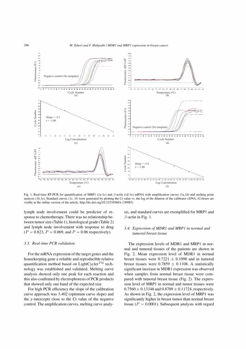
Fig. 1. Real-time RT-PCR for quantification of MRP1 (1a-1c) and β-actin (1d-1e) mRNA with amplification curves (1a,1d) and melting pointanalysis (1b,1e). Standard curves (1c, 1f) were generated by plotting the Ct value vs. the log of the dilution of the calibrator cDNA. (Colours arevisible in the online version of the article; http://dx.doi.org/10.3233/DMA-130985)
lymph node involvement could be predictor of re-sponse to chemotherapy. There was no relationship be-tween tumor size (Table 1), histological grade (Table 2)and lymph node involvement with response to drug(P = 0.823, P = 0.069, and P = 0.06 respectively).
3.3. Real-time PCR validation
For the mRNA expression of the target genes and thehousekeeping gene a reliable and reproducible relativequantification method based on LightCyclerTM tech-nology was established and validated. Melting curveanalysis showed only one peak for each reaction andthis also confirmed by electrophoresis of PCR productsthat showed only one band of the expected size.
For high PCR efficiency the slope of the calibrationcurve approach was 3.402 (optimum curve slope) andthe y-intercepts close to the Ct value of the negativecontrol. The amplification curves, melting curve analy-
sis, and standard curves are exemplified for MRP1 andβ-actin in Fig. 1.
3.4. Expression of MDR1 and MRP1 in normal andtumoral breast tissue
The expression levels of MDR1 and MRP1 in nor-mal and tumoral tissues of the patients are shown inFig. 2. Mean expression level of MDR1 in normalbreast tissues were 0.7221 ± 0.1098 and in tumoralbreast tissues were 0.7859 ± 0.1108. A statisticallysignificant increase in MDR1 expression was observedwhen samples from normal breast tissue were com-pared with tumoral breast tissue (Fig. 2). The expres-sion level of MRP1 in normal and tumor tissues were0.7560± 0.13348 and 0.8709 ± 0.11724, respectively.As shown in Fig. 2, the expression level of MRP1 wassignificantly higher in breast tumor than normal breasttissue (P = 0.0001). Subsequent analysis with regard

M. Taheri and F. Mahjoubi / MDR1 and MRP1 expression in breast cancer 391
Fig. 2. Mean expression level of MDR1 and MRP1 in normal andtumoral breast tissue.
Fig. 3. Mean expression level of MDR1 and MRP1 in responder andnon-responder.
to potential interrelationships of these drug resistancegenes revealed that the mRNA levels of MDR1 werenot related to MRP1 (P > 0.05).
3.5. Correlation between mRNA expression of MDR1and MRP1 and response to chemotherapy
A comparison between the tumors of responder ver-sus nonresponder patients with respect to the relativeexpression levels of MRP1 showed that the expres-sion level of MRP1 was significantly higher in nonre-sponder than responder patients (P < 0.05). Mean ex-pression level of MRP1 in responder and nonresponderwere 0.8422 ± 0.11331 and 0.9529 ± 0.08809. Whilethe mean expression level of MDR1 in responder andnonresponder were 0.7832 ± 0.1144 and 0.7936 ±0.1035, respectively (Fig. 3). Although this value washigher in nonresponder this difference was not statisti-cally significant.
4. Discussion
Breast cancer with 1 million new cases in everyyear is the most common malignancy in women [14].Despite the advances in therapeutic methods about40–50% of patients will eventually die from the dis-ease [14]. Therefore researchers are looking for appro-
priate therapeutic methods in order to increase the sur-vival rate. This is facilitated by finding suitable mark-ers which might predict clinical outcome (prognosticmarkers) and response to a particular type of therapy(predictive markers).
The development of multidrug resistance is a majorobstacle to successful chemotherapy in cancers. Var-ious mechanisms, such as reduced drug uptake, theefflux of intracellular drugs, the activation of DNArepair pathways, and the induction of anti-apoptoticmachineries can confer multidrug resistance in cancercells [6]. Despite the intensive studies regarding therole of these genes in inducing drug resistance in can-cer patients the data is still controversial and debated.A possible role of the MDR1 and MRP1 in clinicalbreast cancer has previously been studied but the re-sults are not conclusive [2,9,11,15].
In the present study we examined the expression ofthese two genes in breast cancer patients by real timeRT PCR. We examined whether the expression of ei-ther or combination of these two genes in breast can-cer tumor cells correlates with response to neoadjovantchemotherapy.
The expression levels of MDR1 and MRP1 wasfound to be significantly different between tumoraltissues and normal breast tissues. MDR1 and MRP1are apparently expressed at higher levels in carcinomacells than non-cancer cells. However, we found nosignificant correlation between MDR1 expression andresponse to treatment. Arnal and co workers (2000)demonstrated comparatively to normal tissues, a sig-nificant induction of MDR1 expression in untreated tu-mors but after treatment no differences were detectedbetween tumor and normal samples. Also no signif-icant relationship was found between MDR1 expres-sion and clinical response to chemotherapy neither be-fore nor after treatment [15].
Faneyte et al. [16] compared the post-chemotherapyexpression of MDR1 in tumors that had microscopicevidence of response with that did not respond. Thedifference was not statistically significant. In pre-chemotherapy samples, responders did not differ sig-nificantly from non-responders. They found no cor-relation between clinical response and MDR1 ex-pression [16]. This is similar to finding of Rudas etal. [9] that in a study on 80 patients with locally ad-vanced breast cancer demonstrated that the expres-sion of MRP1, and MDR1 increase after preoperativechemotherapy but there was no association betweentheir expression and response to chemotherapy [9]. Incontrast to these studies Burger et al. [2] reported a
392 M. Taheri and F. Mahjoubi / MDR1 and MRP1 expression in breast cancer
significant correlation between MDR1 gene expressionand response to chemotherapy. MDR1 gene expres-sion occurred in 68% of resistant patients, but in only17% of clinically responsive patients [2]. Similar tothis Atalay et al. [17] showed a strong correlation be-tween MDR1 expression and response to treatment.While the expression rate of MDR1 was 100% in clin-ically resistant patients only 5% of the clinically re-sponsive patients had MDR1 gene expression [17].
We found a significant positive correlation betweenMRP1 expression and response to treatment. Nooteret al. [18] evaluated the role of MRP1 expression inthe prognosis and response to chemotherapy of breastcancer patients. They showed the presence of MRP1protein was associated with lower response rate inpreviously untreated patients who received first-linechemotherapy [18]. Similarly Filipits and co work-ers [11] suggested that MRP1 expression plays an im-portant role in the clinical resistance to adjuvant CMFchemotherapy [11].
In contrast to the above findings Burger et al. [2]showed an inverse relation between MRP1 gene ex-pression and clinical response to chemotherapy but ex-pression of this gene was not significantly differentin clinically responsive and unresponsive patients [2].Atalay et al. [17] showed that although 80% of the clin-ically unresponsive patients had higher MRP1 gene ex-pression, but the relation between MRP1 expressionand clinical drug response was not significant [17].
In regards to relationship between the expressionlevel and clinicopathology markers such as stage of tu-mor, tumor size, hormone status and lymph node in-volvement there was no association between expres-sion of these genes and clinicopathology markers. Fur-ther studies showed similar results [2,10,19].
In conclusion, conflicting results have been obtainedfrom several studies regarding the role of these genesin inducing MDR. This may be partly due to the meth-ods applied for investigation of gene expression. Forthis reason it seems that a gold standard method shouldbe presented in order to study the expression of thesegenes and their role in drug resistance. Hence we rec-ommend Real time PCR as a good method for evalua-tion of these gene expressions at mRNA level becauseReal-time PCR is simple to use, and requires only min-imal experience and skills. It is the method that allowsquantification and provides maximum sensitivity (e.g.10,000 times more sensitive than northern blot).
Furthermore, future prospective studies on large pa-tient populations are required to further explore the im-pact of drug resistance genes on the clinical outcomeof breast cancer patients treated with chemotherapy.
Acknowledgements
This project was supported by a NIGEB grant(No:412). The authors would like to thanks all patientswho willingly participated in the study.
References
[1] Gottesman MM. Mechanisms of cancer drug resistance. AnnuRev Med 2002; 53: 615-627.
[2] Burger H, Foekens JA, Look MP, Meijer-van Gelder ME,Klijn JG et al. RNA expression of breast cancer resistanceprotein, lung resistance-related protein, multidrug resistance-associated proteins 1 and 2, and multidrug resistance gene 1in breast cancer: correlation with chemotherapeutic response.Clin Cancer Res 2003; 9(2): 827-36.
[3] Chintamani, Singhal V, Singh JP, Lyall A, Saxena S, BansalA. Is drug-induced toxicity a good predictor of response toneo-adjuvant chemotherapy in patients with breast cancer?–aprospective clinical study. BMC Cancer 2004; 13: 4-48.
[4] Kuo, MT. Roles of multidrug resistance genes in breast cancerchemoresistance. Adv Exp Med Biol 2007; 608: 23-30.
[5] Dean M. ABC Transporters, Drug Resistance, and CancerStem Cells. J Mammary Gland Biol Neoplasia 2009; 14: 3–9.
[6] Gottesman M, Fojo T, Bates SE. Multidrug resistance incancer: role of ATP-dependent transporters. Nat Rev Cancer2002; 2(1): 48-58.
[7] Van der Deen M, De Vries EG, Timens W, Scheper RJ,Timmer-Bosscha, H et al. ATP-binding cassette (ABC) trans-porters in normal and pathological lung. Respir Res 2005; 6:59.
[8] Cole SP, Bhardwaj G, Gerlach JH, Mackie JE, Grant CE et al.Overexpression of a transporter gene in a multidrug-resistanthuman lung cancer cell line. Science 1992; 258(5088): 1650-4.
[9] Rudas M, Filipits M, Taucher S, Stranzl T, Steger GG et al.Expression of MRP1, LRP and Pgp in breast carcinoma pa-tients treated with preoperative chemotherapy. Breast CancerRes Treat 2003; 81(2): 149-57.
[10] Larkin A, O’Driscoll L, Kennedy S, Purcell R, Moran E et al.Investigation of MRP-1 protein and MDR-1 P-glycoproteinexpression in invasive breast cancer: a prognostic study. Int JCance 2004; 112(2): 286-294
[11] Filipits M, Pohl G, Rudas M, Dietze O, Lax S et al. Clin-ical role of multidrug resistance protein 1 expression inchemotherapy resistance in early-stage breast cancer: the Aus-trian Breast and Colorectal Cancer Study Group. J Clin Oncol2005; 23(6): 1161-8.
[12] Hayward JL, Carbon PP, Kumaoka S, Seagloff A, RubensRD. Assessment of response to therapy in advanced breastcancer:A project of the Programme on Clinical Oncology ofthe International Union Against Cancer, Geneva, Switzerland.Cancer 1997; 39: 1289-1294.
[13] Golalipour M, Mahjoubi F, Sanati MH, Alimoghaddam K.Gene dosage is not responsible for the upregulation of MRP1gene expression in adult leukemia patients. Arch Med Res2007; 38(3): 297-304.
[14] McPherson K, Steel CM, Dixon JM. ABC of breast diseases.Breast cancer epidemiology, risk factors, and genetics. Bmj2000; 321(7261): 624-8.

M. Taheri and F. Mahjoubi / MDR1 and MRP1 expression in breast cancer 393
[15] Arnal M, Franco N, Fargeot P, Riedinger JM, Brunet-LecomteP et al. Enhancement of mdr1 gene expression in normal tis-sue adjacent to advanced breast cancer. Breast Cancer ResTreat. May 2000; 61(1): 13-20.
[16] Faneyte IF, Kristel PM, van de Vijver MJ. DeterminingMDR1/P-glycoprotein expression in breast cancer. Int J Can-cer 2001; 93(1): 114-122.
[17] Atalay C, Deliloglu Gurhan I, Irkkan C, Gunduz U. Mul-tidrug resistance in locally advanced breast cancer. Tumour
Biol 2006; 27(6): 309-18.[18] Nooter K, de la Riviere GB, Klijn J, Stoter G, Foekens J.
Multidrug resistance protein in recurrent breast cancer. Lancet1997; 349(9069): 1885-1886.
[19] Faneyte IF, Kristel PM, van de Vijver MJ. Multidrug re-sistance associated genes MRP1, MRP2 and MRP3 in pri-mary and anthracycline exposed breast cancer. Anticancer Res2004; 24(5A): 2931-2939.

Submit your manuscripts athttp://www.hindawi.com
Stem CellsInternational
Hindawi Publishing Corporationhttp://www.hindawi.com Volume 2014
Hindawi Publishing Corporationhttp://www.hindawi.com Volume 2014
MEDIATORSINFLAMMATION
of
Hindawi Publishing Corporationhttp://www.hindawi.com Volume 2014
Behavioural Neurology
EndocrinologyInternational Journal of
Hindawi Publishing Corporationhttp://www.hindawi.com Volume 2014
Hindawi Publishing Corporationhttp://www.hindawi.com Volume 2014
Disease Markers
Hindawi Publishing Corporationhttp://www.hindawi.com Volume 2014
BioMed Research International
OncologyJournal of
Hindawi Publishing Corporationhttp://www.hindawi.com Volume 2014
Hindawi Publishing Corporationhttp://www.hindawi.com Volume 2014
Oxidative Medicine and Cellular Longevity
Hindawi Publishing Corporationhttp://www.hindawi.com Volume 2014
PPAR Research
The Scientific World JournalHindawi Publishing Corporation http://www.hindawi.com Volume 2014
Immunology ResearchHindawi Publishing Corporationhttp://www.hindawi.com Volume 2014
Journal of
ObesityJournal of
Hindawi Publishing Corporationhttp://www.hindawi.com Volume 2014
Hindawi Publishing Corporationhttp://www.hindawi.com Volume 2014
Computational and Mathematical Methods in Medicine
OphthalmologyJournal of
Hindawi Publishing Corporationhttp://www.hindawi.com Volume 2014
Diabetes ResearchJournal of
Hindawi Publishing Corporationhttp://www.hindawi.com Volume 2014
Hindawi Publishing Corporationhttp://www.hindawi.com Volume 2014
Research and TreatmentAIDS
Hindawi Publishing Corporationhttp://www.hindawi.com Volume 2014
Gastroenterology Research and Practice
Hindawi Publishing Corporationhttp://www.hindawi.com Volume 2014
Parkinson’s Disease
Evidence-Based Complementary and Alternative Medicine
Volume 2014Hindawi Publishing Corporationhttp://www.hindawi.com


![Screening Of Mdr1 [Autosaved]](https://img.pdfslide.net/doc/110x75/5599ce811a28abcf4b8b482c/screening-of-mdr1-autosaved.jpg)