Embed Size (px)
Citation preview

Iron in the Normal and Pathologic Human Brain
E. Mark HaackeWayne State University
01/15/2011

Acknowledgements:
Jaladhar Neelavalli, PhDJin Tang, MS
Saifeng Liu, BS
Much of the information in the early slides on iron comes from the book:R. Lauffer. Iron and Human Disease CRC Press 1992
Outline
• Preamble: some facts about iron in the body.• Diseases related to iron.• Measuring iron with various MRI techniques.• Iterative SWIM as a means to reduce susceptibility mapping artifacts.• Susceptibility mapping of air bubbles.• Future applications.
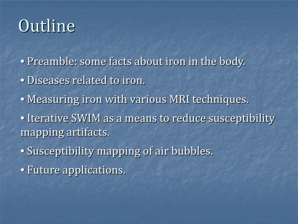
Preamble• Iron has long been considered as a key nutrient for
the body and many physiologic reactions.• The idea of iron leading to detrimental effects was
foreign to most people in medicine up until a fewshort decades ago.
• For example, the thought that high iron levelscould predispose someone for higher risks forcancer, infection and its role as a key catalyst foroxidative reactions was not considered at all untilthe late 20th century.
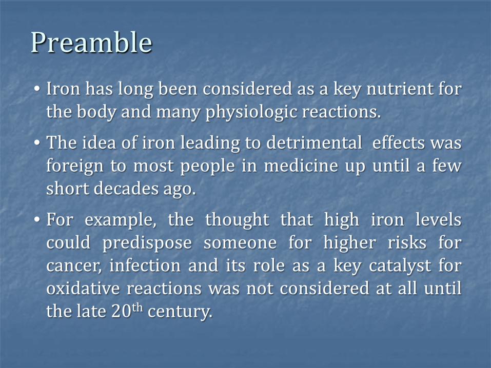
Iron• We absorb 0.5mg to 4mg per day with an average of about 1mg per day.
• Iron appears in hemoglobin, myoglobin and ferritin or hemosiderin, with small amounts in transferrin and lactoferrin.
• There is no mechanism for the excretion of iron.
• To quote Burton Drayer, “We all rust as we get older.”
• Iron can be removed by phlebotomy (blood letting).

The Storage Protein Ferritin
•Nature has developed a number of iron binding proteins for transportation and storage.
• Ferritin is similar to ferrihydrite 5Fe2O39H2O.
• Ferritin is a soluble storage protein able to hold up to 4500 iron atoms.
• Ferritin has 24 H (heavy) and L (light) subunits.

Heavy and Light Ferritin
• H-ferritin is associated with high iron utilization and low iron storage and has a ferrioxidase activity that is responsible for converting harmful Fe+2 to Fe+3.
• L-rich ferritins are more thermostable than H-rich and promote iron mineralization at the ferritin core.
• Microglia contain only L-ferritin (to scavange iron).• Neurons contain only H-ferritin.• Oligodendrocytes contain both.

Iron Mobilization from Ferritin• Iron can be mobilized from ferritin in vitro in several
ways.• Direct chelation can occur via a number of natural
chelators in the cells such as: Various sugars Thiols (dihydrolipoate and dihydrolipoamide Suntheic chelators Microbial siderophores Superoxides Flavoprotein oxidases Dehydrogenases (xanthine oxidase)
Iron and myelin
• Both a direct and indirect relationship exists between iron acquisition and myelin production.
• Iron is required as a co-factor in lipid biosynthesis and in oxidative metabolism.
Iron excess• A breakdown in the regulation of iron can occur when
there is an excessive uptake of iron. Causes can be:• Defects in iron uptake in intestinal mucosa• Hemochromatosis• Excessive breakdown of erthrocytes due to
abnormal globin synthesis enhanced by blood transfusion (beta-thalassemia)
• A defect in the heme synthesis pathway (porphyria cutanea tarda).
• Excessive iron intake from diet/chronic alcohol intake.

Serum Ferritin• Serum ferritin correlates with total iron load which
is for males roughly 750 mg versus females 250 mg
(1 μg/l corresponds to 7.5mg iron stores)
• But there is a huge variance:• 58 to 127 μg/l SFC for men• 6 to 618 μg/l SFC for women
• Another study gives:• Mean 94 range 27 to 329 μg/l SFC for men• Mean 34 range 9 to 125 μg/l SFC for women
Non-heme and heme iron• Iron is key to survival of eukaryotic/prokaryotic cells.• The active site of many cytoplasmic enzymes requires
iron – for example ribonuclease reducase uses it to reduce ribonucletides to deoxynucleotides (a step which regulates DNA synthesis during cell proliferation and development).
•Heme is formed by chelating iron into protoporphyrin IX by ferrochelatase.
• Erythrocytes make large amounts of heme iron.• Transferrin carries iron to be stored in ferritin.
Iron transport• Iron can be delivered to cells in the form of iron salts,
diferric transferrin and hemin.• Iron crosses into ferritin by an oxidizing process
where ferrous iron is transformed into the safer formof ferric iron.
• Expression of transferrin receptor levels isdecreased at the surface of cells replete with ironand vice versa: i.e., the biosynthesis of the protein isdown-regulated when there is an abundance of iron.
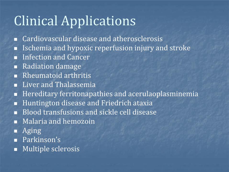
Clinical Applications Cardiovascular disease and atherosclerosis Ischemia and hypoxic reperfusion injury and stroke Infection and Cancer Radiation damage Rheumatoid arthritis Liver and Thalassemia Hereditary ferritonapathies and acerulaoplasminemia Huntington disease and Friedrich ataxia Blood transfusions and sickle cell disease Malaria and hemozoin Aging Parkinson’s Multiple sclerosis

Iron and Cardiovascular Disease• There is growing epidemiological evidence for a
relationship between iron levels and cardiovasculardisease. Some experimental data support the role ofiron in the process of lipid peroxidation, the firststep in the formation of atherosclerotic lesions.
• Macrophages and endothelial cells are involved inthis process, but the exact mechanism and the sitesof the interactions between these cells, iron, andlow-density lipoprotein are still unknown.
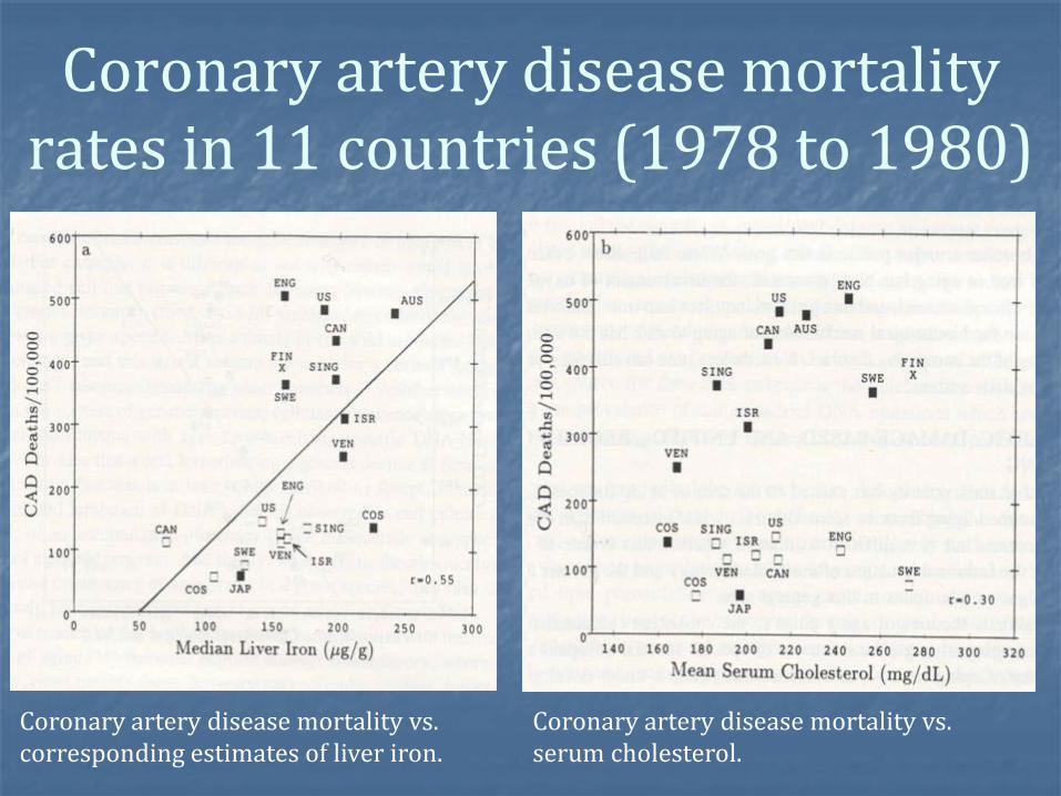
Coronary artery disease mortality rates in 11 countries (1978 to 1980)
Coronary artery disease mortality vs. corresponding estimates of liver iron.
Coronary artery disease mortality vs. serum cholesterol.
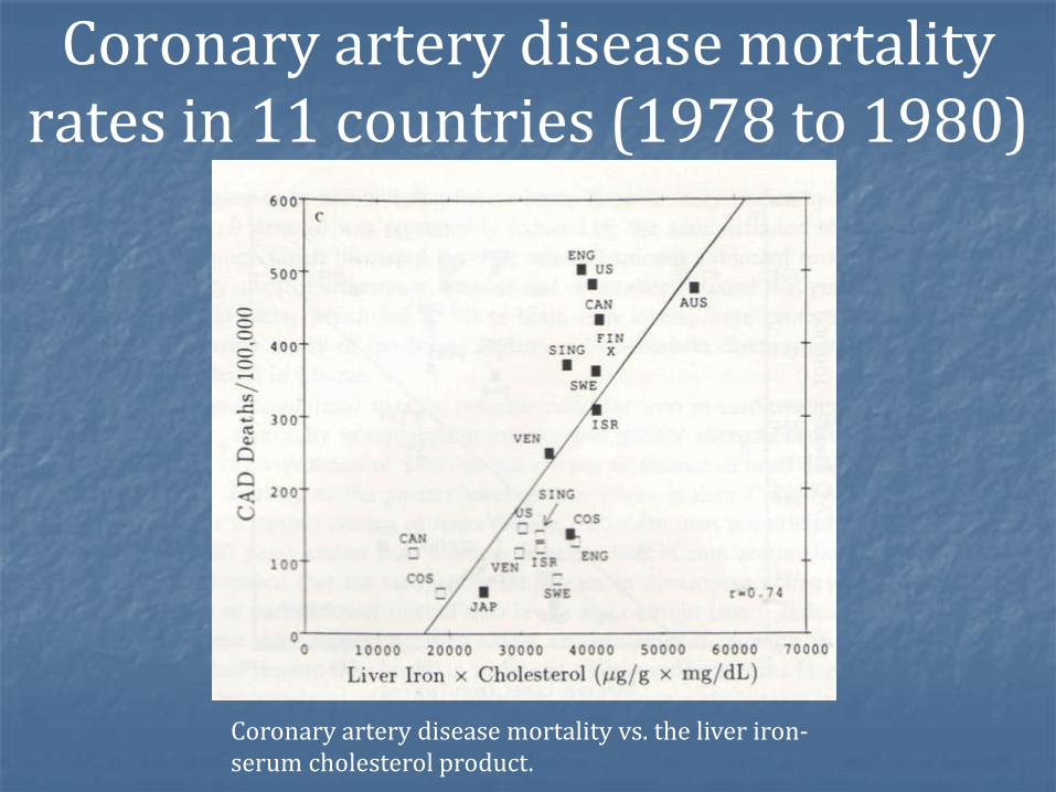
Coronary artery disease mortality rates in 11 countries (1978 to 1980)
Coronary artery disease mortality vs. the liver iron-serum cholesterol product.
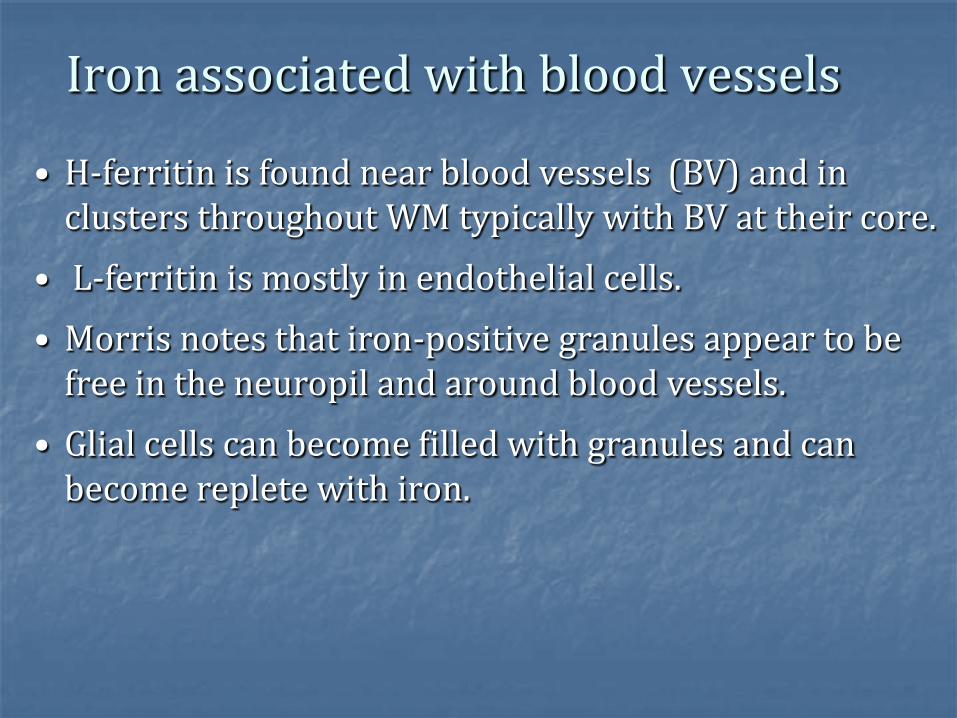
Iron associated with blood vessels
• H-ferritin is found near blood vessels (BV) and in clusters throughout WM typically with BV at their core.
• L-ferritin is mostly in endothelial cells.• Morris notes that iron-positive granules appear to be
free in the neuropil and around blood vessels.• Glial cells can become filled with granules and can
become replete with iron.
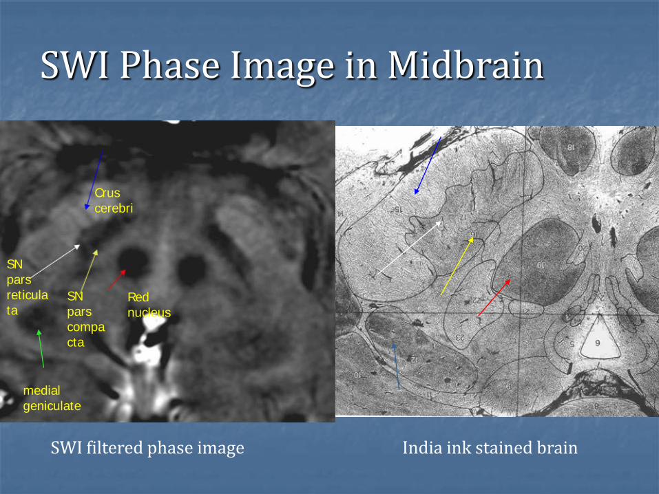
Red nucleus
Crus cerebri
medial geniculate
SN pars compacta
SN pars reticulata
SWI Phase Image in Midbrain
India ink stained brainSWI filtered phase image

Red nucleus
Crus cerebri
medial geniculate
SN pars compacta
SN pars reticulata
SWI Phase Image in Midbrain
SWIM imageSWI filtered phase image
Crus cerebri
Red nucleus
SN
medial geniculate

Iron adds to LDL oxidation• Low density lipoprotein associated cholesteral is well
known to be involved in the moncoyte/macrophage part of the early development of atherosclerotic foam cell lesions.
• The presence of iron can add to lipid peroxidation.• The cell induced oxidation is inhibited by the
presence of anti-oxidants and metal chelators. • Intra-ischemic treatment of reperfusion enhanced the
recovery of contractile function and quenched the production of oxygen free-radical generation in isolated post-ischemic rabbit hearts.
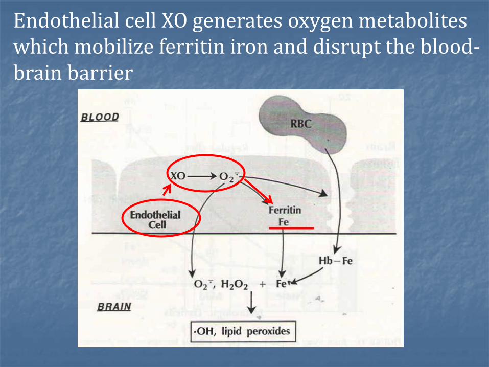
Endothelial cell XO generates oxygen metabolites which mobilize ferritin iron and disrupt the blood-brain barrier

Hemoglobin iron enters brain parenchyma and reacts with XO-derived oxygen metabolites with consequent brain injury.

StrokeIn unilateral hemispheric hypoxic-reperfusion, brain iron and XO-derived oxidants appear to have a mechanistic link with development of cerebral edema.
It is highly probable that iron and O2 metabolites interact to oxidatively modify lipids, protein, and nucleic acids, with resultant loss of blood-brain barrier function, formation of cerebral edema, and ultimately neuronal cell death.

Unilateral carotid artery occlusion: Gerbil brains had reduced serum iron levels after 6 weeks of treatment with a low-iron diet when compared with gerbils fed a standard diet.
(a) Gerbils fed an iron-deficient diet for 8 weeks showed decreased iron (p < 0.05)
(b) Serum iron levels were decreased ( p < 0.05) after 6 to 8 weeks of a low-iron diet.
(c) and (d) Blood hematocrit and brain XO + XD activity were unchanged after up to 8 weeks of an iron-deficient diet.
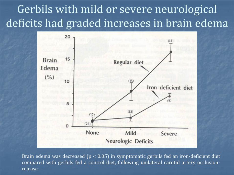
Gerbils with mild or severe neurological deficits had graded increases in brain edema
Brain edema was decreased (p < 0.05) in symptomatic gerbils fed an iron-deficient dietcompared with gerbils fed a control diet, following unilateral carotid artery occlusion-release.
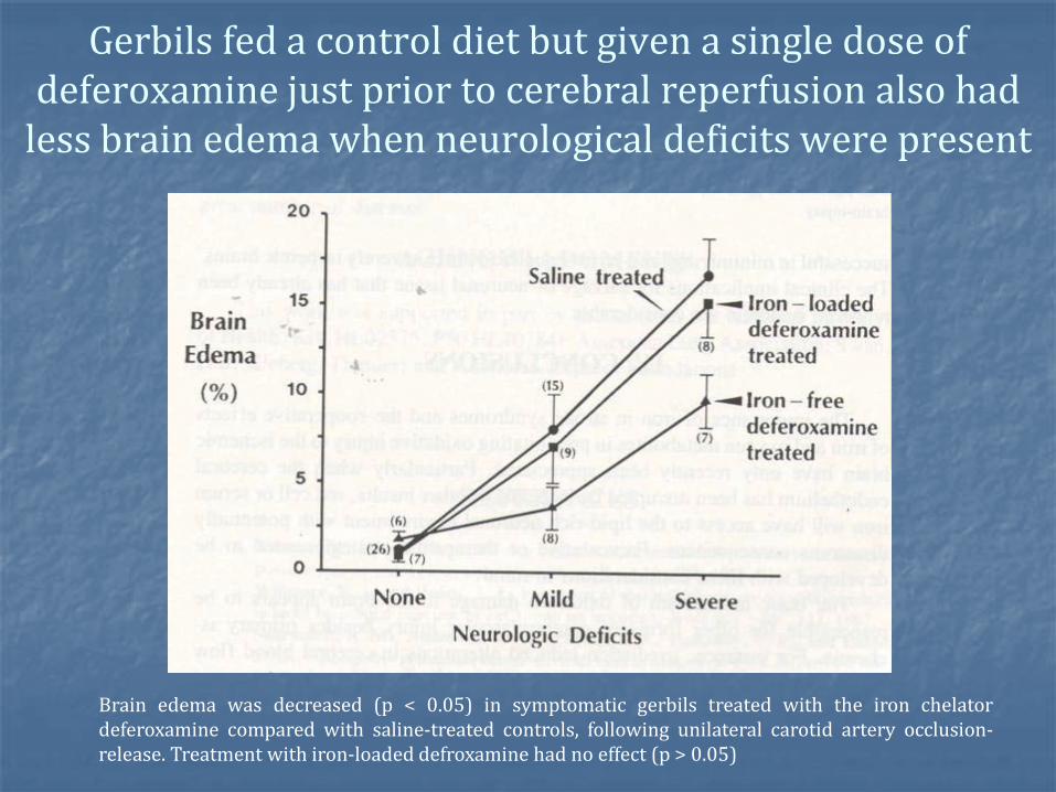
Gerbils fed a control diet but given a single dose of deferoxamine just prior to cerebral reperfusion also had
less brain edema when neurological deficits were present
Brain edema was decreased (p < 0.05) in symptomatic gerbils treated with the iron chelatordeferoxamine compared with saline-treated controls, following unilateral carotid artery occlusion-release. Treatment with iron-loaded defroxamine had no effect (p > 0.05)

Iron and free radicals
• Cultured endothelial cells loaded with chelated iron using 8-hydroxyquinoline are 50% more likely to die when presented with only 7umol/l H2O2 while those not loaded with iron survived doses as high as 2 mM more than 100 times greater.
• Surprisingly DNA can be damaged if iron or other metals are present when presented with H2O2 and may lead to mutations of DNA.

The role of hypoxic reperfusion
If the tissue is under stress and either in a hypoxic or ischemic state then during this period the endothelium may be most vulnerable.
If the tissue is re-perfused bringing with it lots of oxygen and the vessel wall has not yet returned to normal there may be a resulting hypoxic reperfusion injury.
One of the breakdown products during ischemia is xanthine oxidase which can mobilize iron from ferritin producing an environment now rich in iron and hydrogen peroxide (H2O2).

Oxidative stress (news) Findings by Dr. Syed Imam and others have shown in
the J of Neuroscience just recently that:
“After analyzing cells and post-mortem brain tissue from animals and humans, researchers noted that oxidative stress – a known culprit in neuron death –activated a protein called tyrosine kinase c-Abl in the nigra-striatum area of the brain.
Neurons in this part of the brain are particularly vulnerable to Parkinson's injury.
Oxidative stress (news) Activation of this protein led to changes in another
protein called parkin, which is known to be mutated in hereditary Parkinson's.
The altered parkin lacked the capacity to break down other proteins, leading to harmful clumps of unprocessed protein in the neuron.
The scientists believe this accumulation leads to progressive neuron death, resulting in Parkinson's symptoms that worsen over time.

Oxidative stress (news)
"When they blocked tyrosine kinase c-Abl activation, parkin function was preserved and neurons were spared,"
Dr. Imam said:
"We believe these studies provide sound rationale for moving forward with a preclinical trial of tyrosine kinase c-Abl inhibitors, with the goal of developing a potent therapeutic drug for slowing the progression of Parkinson's."
Iron, bacteria and infection• Iron chelators are present in bacteria in the form of hydroxamate siderophores.
• This may explain why there is increased risk for infection when high iron levels are present.
• Both rats with stroke and rats with cardiovascular disease followed by reperfusion take longer to recover if they have high iron levels.
• Iron chelators such as desferrioxamine (deferroxamine) help improve the outcome in CVD.
Iron and Infection• Inflammaory cytokines enhance ferritin gene expression.• Reduced levels of liver iron and serum iron lead to more resistance and less severe inflammatory response even to some bacteria.• In fact, it has been shown that within 6 hours after onset of inflammation, the body naturally tries to sequester serum iron and serum ferritin into ferritin in the liver. • Presumably this is an attempt to hide iron from the siderophores of bacteria.
Iron and Infection and Cancer• Iron deficiency is associated with reduced B-cell and T-cell production and perhaps less inflammatory response in some cases. • Iron can be used by bacteria and by neoplastic cells such as in melanoma.• Studies show that when animals are fed large amounts of iron there is an increase in:
the number of inflammatory lesions in Entamoeba histolytica and the frequency of myobacterial infection
and in infants presenting with high iron a three-fold greater risk of salmonellosis.
Iron and cell damage with x-rays• Hamster ovary cells were cultured in ferritin and exposed to 4 Gy of x-rays.• In this group roughly 50% of cells died.• In the control group with apoferritin (a protein with no iron), above 48μg/ml ferritin became toxic to the cells without radiation but not below.• In the presence of 32 μg/ml ferritin only 12% of cells survived the 4 Gy of radiation.• This ferritin was 19% iron by weight leading to 6 μg/ml of iron as the concentration high enough to lead to this type of damage.
Rhematoid arthritis
High rates of microbleeding lead to large levels of ferritin and hemosiderin in synovial cell linings.
Patients given iron dextran show exacerbation of synovial inflammation.
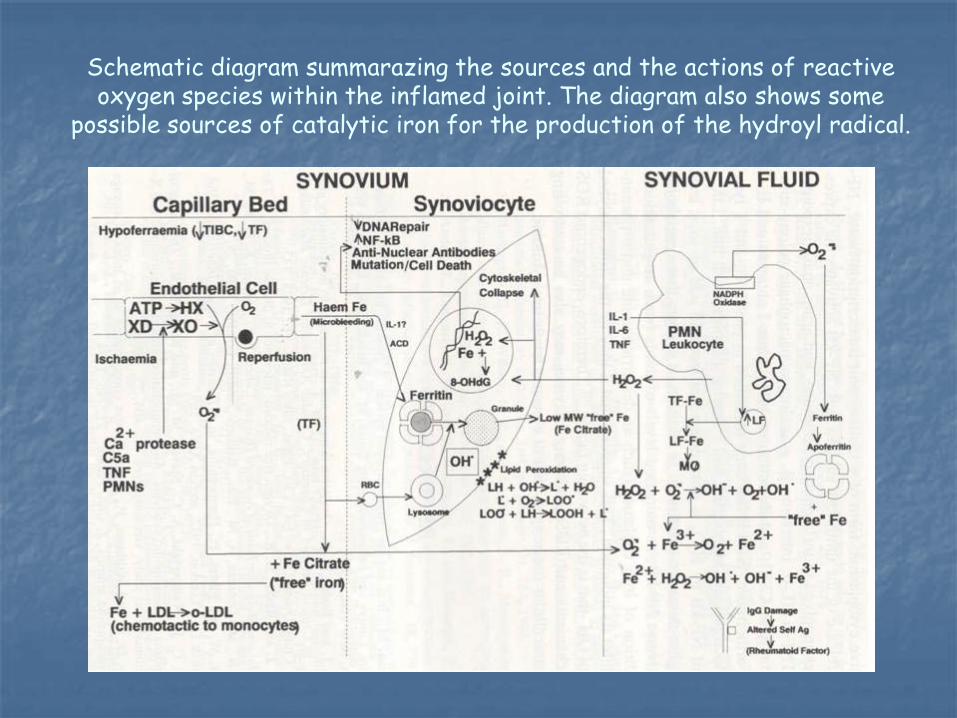
Schematic diagram summarazing the sources and the actions of reactive oxygen species within the inflamed joint. The diagram also shows some
possible sources of catalytic iron for the production of the hydroyl radical.

Iron and hemachromatosis
Increased iron is associated with increased lipid peroxidation as demonstrated in animal studies.
Increased iron is found not just in liver but also in pancreas, skin and in the pituitary gland and affects hormonal function because of it.
Fully established cirrhosis with nodules occurs later in the disease.
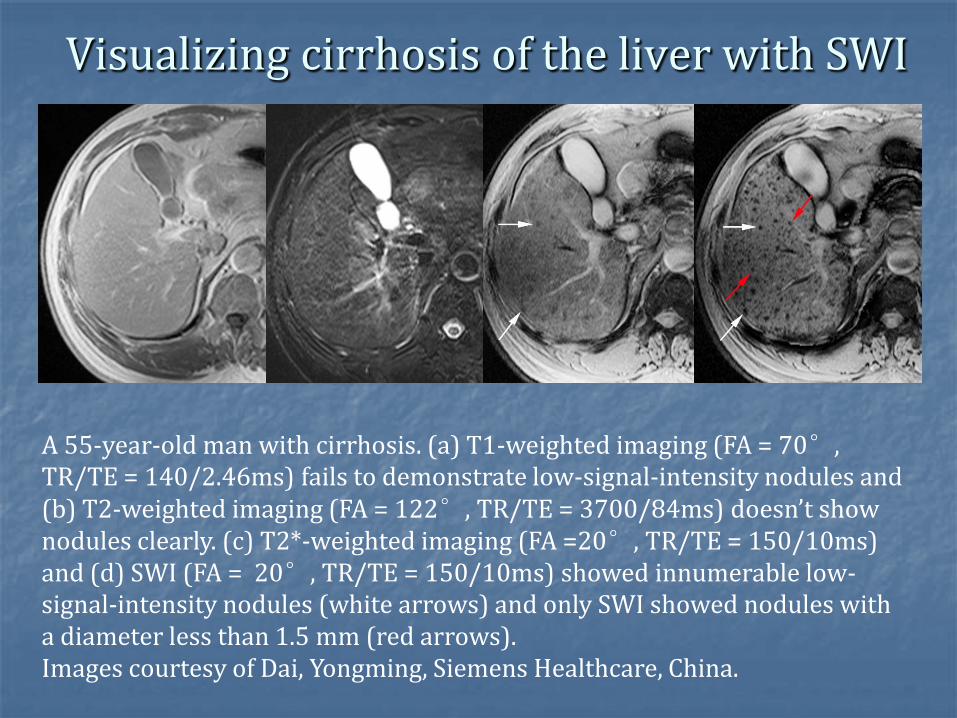
Visualizing cirrhosis of the liver with SWI
A 55-year-old man with cirrhosis. (a) T1-weighted imaging (FA = 70°, TR/TE = 140/2.46ms) fails to demonstrate low-signal-intensity nodules and (b) T2-weighted imaging (FA = 122°, TR/TE = 3700/84ms) doesn’t show nodules clearly. (c) T2*-weighted imaging (FA =20°, TR/TE = 150/10ms) and (d) SWI (FA = 20°, TR/TE = 150/10ms) showed innumerable low-signal-intensity nodules (white arrows) and only SWI showed nodules with a diameter less than 1.5 mm (red arrows).Images courtesy of Dai, Yongming, Siemens Healthcare, China.
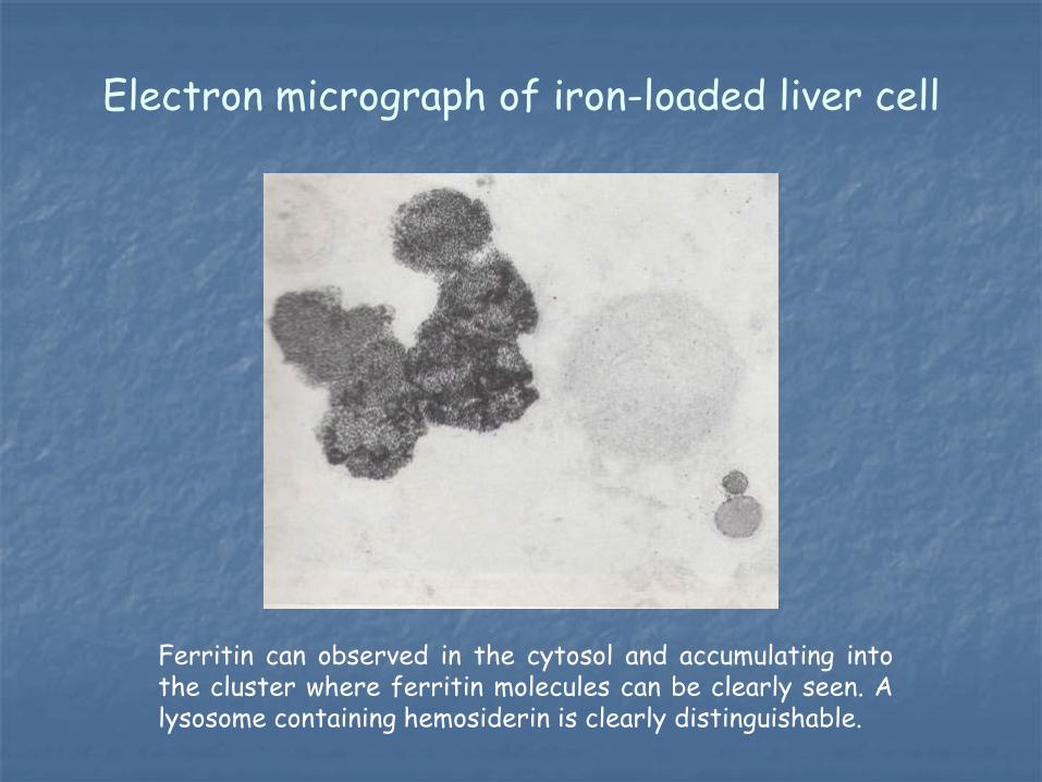
Electron micrograph of iron-loaded liver cell
Ferritin can observed in the cytosol and accumulating intothe cluster where ferritin molecules can be clearly seen. Alysosome containing hemosiderin is clearly distinguishable.
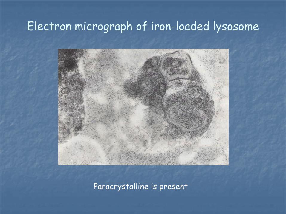
Electron micrograph of iron-loaded lysosome
Paracrystalline is present
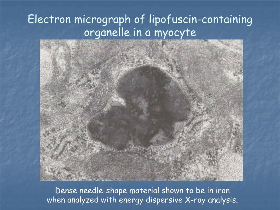
Electron micrograph of lipofuscin-containing organelle in a myocyte
Dense needle-shape material shown to be in iron when analyzed with energy dispersive X-ray analysis.

Role of mutated HFE gene The HFE gene is in association with Transferrin receptor 2
which increases cellular uptake of transferrin bound iron.
So, the mutated HFE is unable to bind to transferrin receptor 2 which might decrease the cellular uptake of transferrin bound iron and might result in iron overload.
In a mice study, deletion of HFE and Transferrin receptor 2 reduced the hepcidin level and resulted in iron overload.
D.F.Wallace et al. Combined deletion of HFE and trandferrin receptor 2 in mice leads to marked dysregulation of hepcidin and iron overload. Hapatology, vol 50. No:6.2009.
Thalassemia Thalassemia is an inherited autosomal recessive blood
disease which results in a reduced rate of synthesis or no synthesis of one of the globin chains that make up hemoglobin.
This can lead to anemia, the characteristic symptom. Hemoglobinopathies imply structural abnormalities in the
globin proteins themselves. The two conditions may overlap, however, since some conditions which cause abnormalities in globin proteins (hemoglobinopathy) also affect their production (thalassemia).
Thus, some thalassemias are hemoglobinopathies, but most are not. Either or both of these conditions may cause anemia. (from Wikepedia).
Children with thalassemia who are treated with desferrioxamine have much better survival outcomes.

Malaria Hemozoin is a disposal product formed from the digestion of blood by
some blood-feeding parasites. These hematophagous organisms such as malaria parasites digest hemoglobin and release high quantities of free heme, which is the non-protein component of hemopglobin.
A heme is a prosthetic group that consists of an iron atom contained in the center of a heterocyclic porphyrin ring.
Free heme is toxic to cells, so the parasites convert it into an insoluble crystalline form called hemozoin.
In malaria parasites, hemozoin is often called malaria pigment.
Since the formation of hemozoin is essential to the survival of these parasites, it is an attractive target drugs such as chloroquine and mefloquine which are thought to kill malaria parasites by inhibiting hemozoin biocrystalization.
From Wikepedia
Iron increases in aging
Accumulation of lipofuscin pigment is a phenomena which occurs under physiologic conditions and appears to be inevitable with age.
Electron microscopy and energy dispersive x-ray microanalysis of cells exposed to ferric iron show that iron is sequestered in the lipofuscin containing organelles.
Also high iron in cancers may make them more susceptible to radiation treatment. So seek out targeting agents with ferritin bound to them may offer a means to treat tumors.
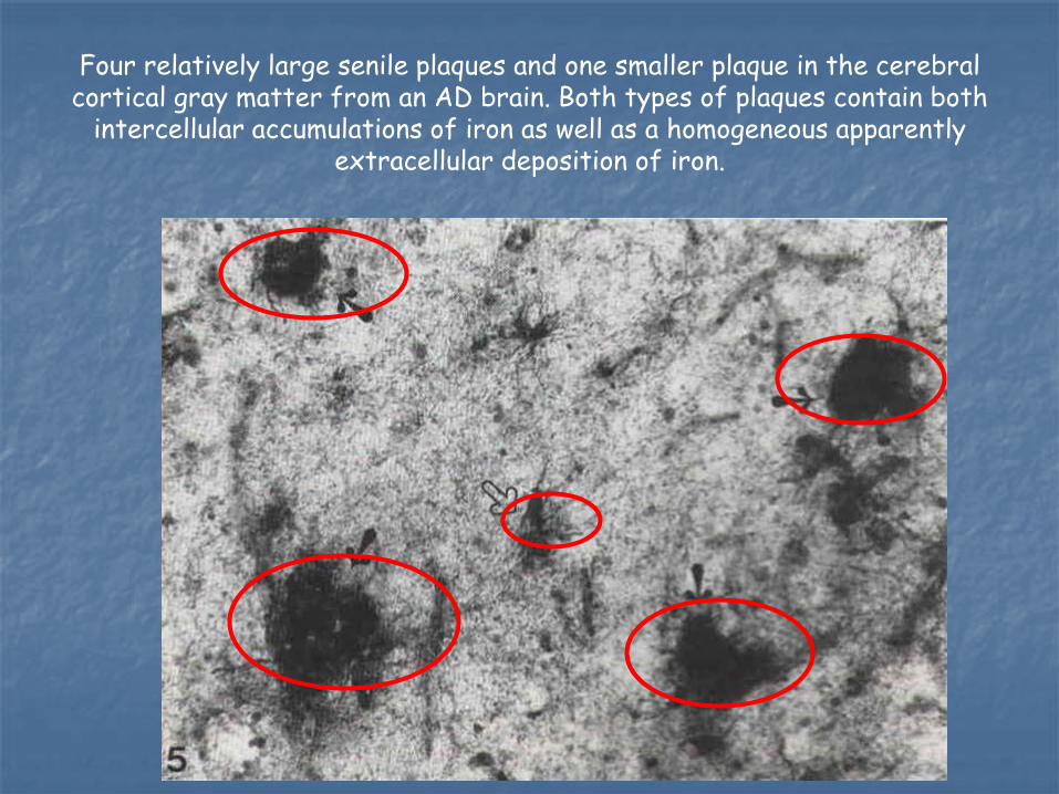
Four relatively large senile plaques and one smaller plaque in the cerebral cortical gray matter from an AD brain. Both types of plaques contain both
intercellular accumulations of iron as well as a homogeneous apparently extracellular deposition of iron.

Ferritin in senile plaques from gray matter of an AD brain. The core of one of the plaques is indicate with the red circle. The Ferritin in the plaques is diffuse and homogenous as well as intracellular (blue circle)
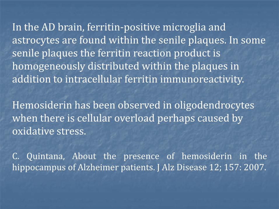
In the AD brain, ferritin-positive microglia and astrocytes are found within the senile plaques. In some senile plaques the ferritin reaction product is homogeneously distributed within the plaques in addition to intracellular ferritin immunoreactivity.
Hemosiderin has been observed in oligodendrocytes when there is cellular overload perhaps caused by oxidative stress.
C. Quintana, About the presence of hemosiderin in thehippocampus of Alzheimer patients. J Alz Disease 12; 157: 2007.

• In Alzheimer’s Disease, the characteristic hallmarkchanges in the brain are the presence of senile plaquesand neurofibrillary tangles.
• The presence of iron plaques was first discussed toour knowledge in 1953. Increased iron staining is seenin the walls of the vasculature.
• Iron is both homogeneously distributed within theplaques and within cells surrounding the plaques.
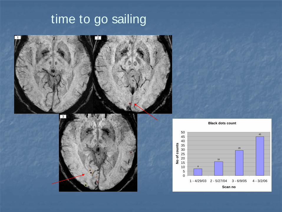
time to go sailing
Black dots count
8
16
29
45
05
101520253035404550
1 - 4/29/03 2 - 5/27/04 3 - 6/9/05 4 - 3/2/06
Scan no
No
of c
ount
s
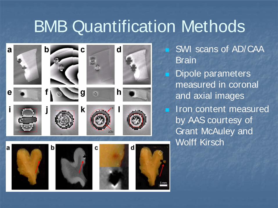
BMB Quantification Methods SWI scans of AD/CAA
Brain Dipole parameters
measured in coronal and axial images
Iron content measured by AAS courtesy of Grant McAuley and Wolff Kirsch

Quantification of punctate iron sources using magnetic resonance phase.McAuley G et al. MRM 2010 63:106-15.
See also: Iron quantification of microbleeds in postmortem brain.McAuley G et al, MRM 2011.
Spherical dipole implications

Iron in Parkinson’s disease• Neuromelanin can bind Fe+3 and create free radicals• only iron chelating agents can prevent this• Dopamine melanin is a risk factor• 6-OHDA releases iron from its binding sites in ferritin• striatal dopamine metabolism is dramatically reduced when 6-OHDA is present• adding desferrioxamine renormalizes the dopamine production supposedly by inhibiting the action of 6-OHDA (6-hydroxy-dopamine)• and what of patients symptoms with high iron in different parts of the basal ganglia?
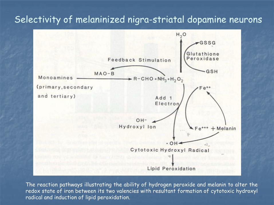
Selectivity of melaninized nigra-striatal dopamine neurons
The reaction pathways illustrating the ability of hydrogen peroxide and melanin to alter theredox state of iron between its two valencies with resultant formation of cytotoxic hydroxylradical and induction of lipid peroxidation.
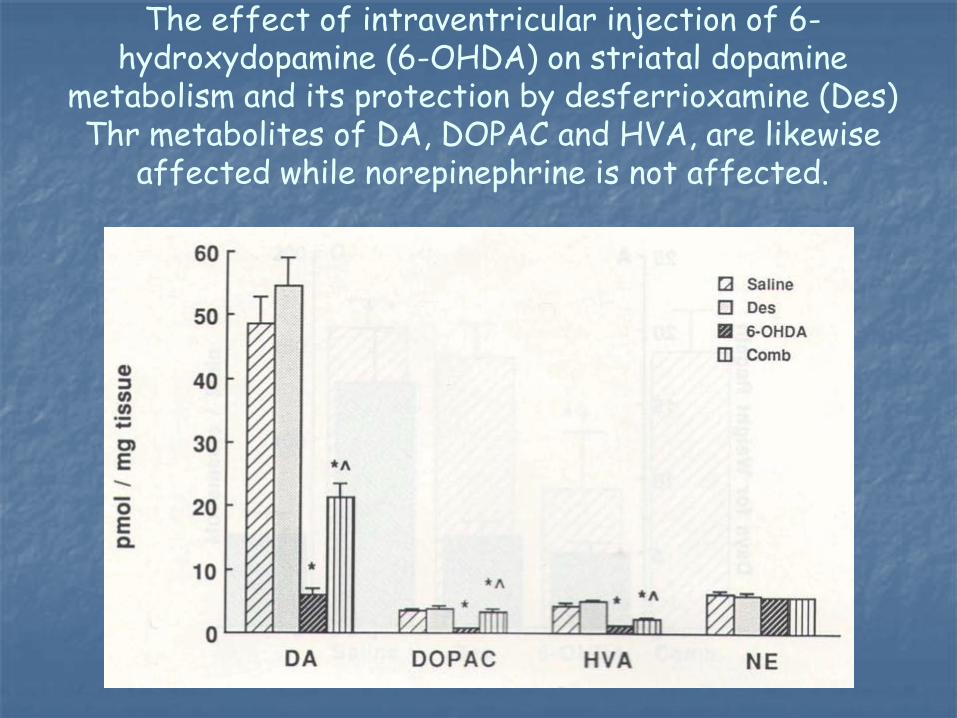
The effect of intraventricular injection of 6-hydroxydopamine (6-OHDA) on striatal dopamine
metabolism and its protection by desferrioxamine (Des)Thr metabolites of DA, DOPAC and HVA, are likewise
affected while norepinephrine is not affected.
Reduce iron by diet and exercise
Fish has 0.5mg to 1mg per serving, poultry 0.5 to 1.5mg, pork 1 to 2mg, beef 2 to 3.5mg
Wheat bread much better for you with less iron absorption if phytate is added
Phytate is an actively charged hexaorthophosphate ester of myo-inositol which avidly binds metal ions
Phytate is contained in many wheat products and a variety of beans (including soy beans) and peas
Exercise removes iron and increases iron storage
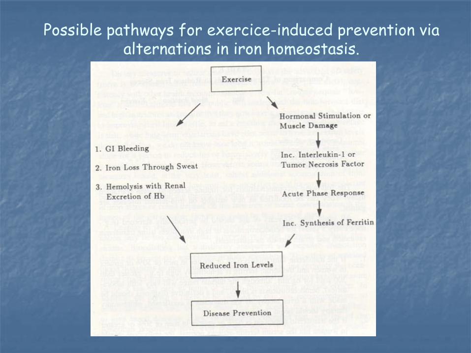
Possible pathways for exercice-induced prevention via alternations in iron homeostasis.
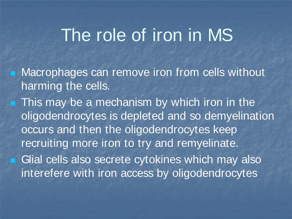
The role of iron in MS
Macrophages can remove iron from cells without harming the cells.
This may be a mechanism by which iron in the oligodendrocytes is depleted and so demyelination occurs and then the oligodendrocytes keep recruiting more iron to try and remyelinate.
Glial cells also secrete cytokines which may also interefere with iron access by oligodendrocytes
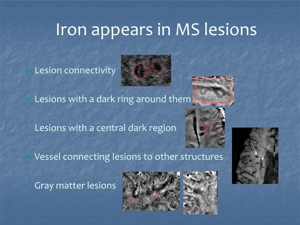
Iron appears in MS lesions
∗ Lesion connectivity
∗ Lesions with a dark ring around them
∗ Lesions with a central dark region
∗ Vessel connecting lesions to other structures
∗ Gray matter lesions

Phase images show clear rings of putative iron and iron build up in some of the lesions. This information appears to be complementary to that seen in the FLAIR images where the lesions seem rather diffuse.
63
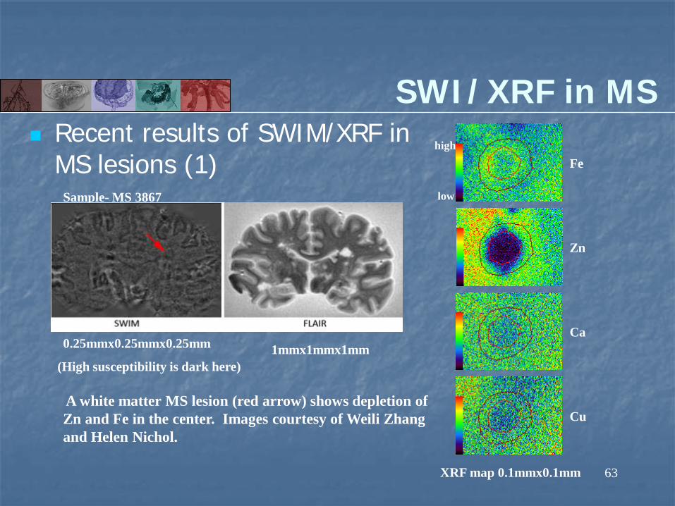
Recent results of SWIM/XRF in MS lesions (1)
SWI/XRF in MS
Fe
Zn
Ca
Cu
high
low
0.25mmx0.25mmx0.25mm 1mmx1mmx1mm
XRF map 0.1mmx0.1mm
(High susceptibility is dark here)
A white matter MS lesion (red arrow) shows depletion of Zn and Fe in the center. Images courtesy of Weili Zhang and Helen Nichol.
Sample- MS 3867

Iron in MS patients
87.6μg Fe/g tissue from XRF and 23.3/0.4 + 32.5 = 90.8 Fe μg/g tissue
Iron in MS patients
Kosma et al note that there are increased serum sTFR and ferritin levels in MS patients with active disease and this may reflect an increased iron turnover due to inflammatory and oxidative stress.
K Kosma et al, Serum ferritin, transferrin and soluble transferrin receptor levels in multiple sclerosis patients. Multiple Sclerosis 2005 11: 272.
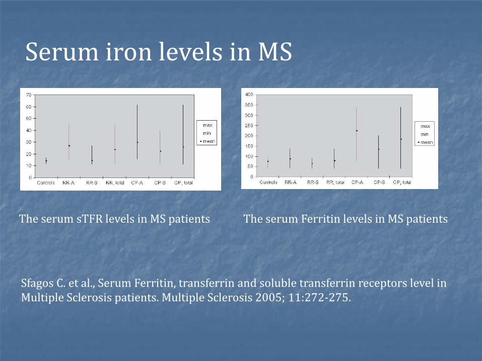
The serum Ferritin levels in MS patientsThe serum sTFR levels in MS patients
Serum iron levels in MS
Sfagos C. et al., Serum Ferritin, transferrin and soluble transferrin receptors level in Multiple Sclerosis patients. Multiple Sclerosis 2005; 11:272-275.
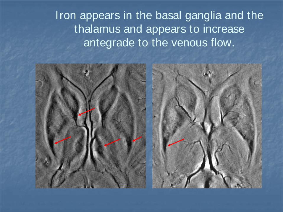
Iron appears in the basal ganglia and the thalamus and appears to increase
antegrade to the venous flow.
68
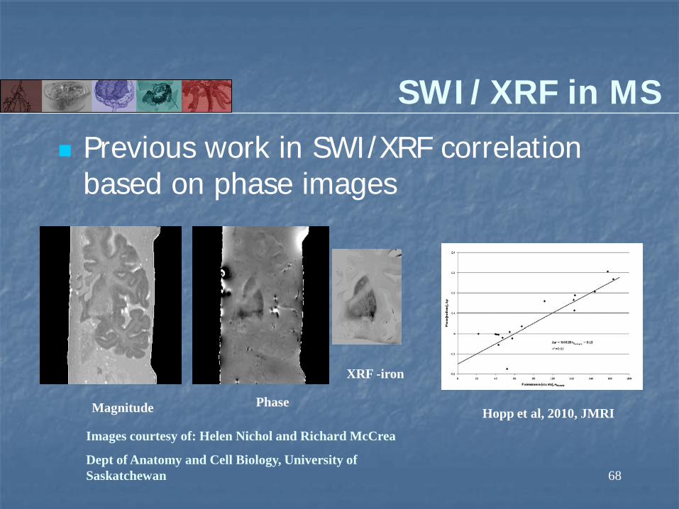
Previous work in SWI/XRF correlation based on phase images
SWI/XRF in MS
Hopp et al, 2010, JMRIMagnitude
XRF -iron
Phase
Images courtesy of: Helen Nichol and Richard McCrea
Dept of Anatomy and Cell Biology, University of Saskatchewan

Iron appears in the midbrain
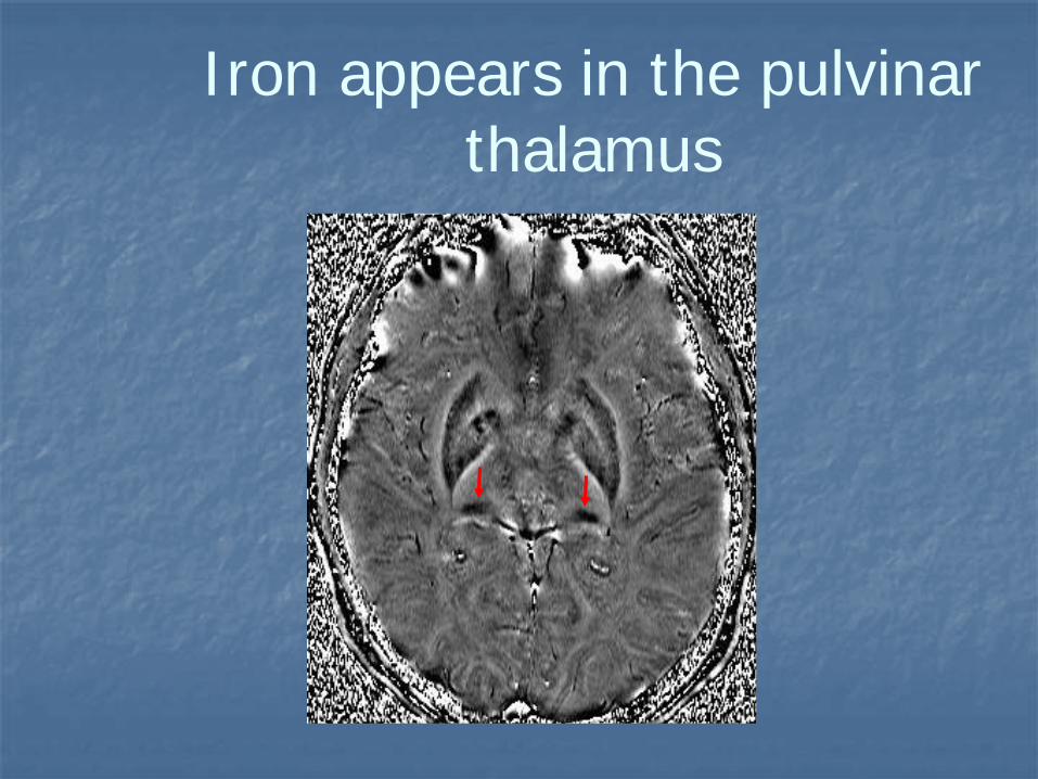
Iron appears in the pulvinar thalamus
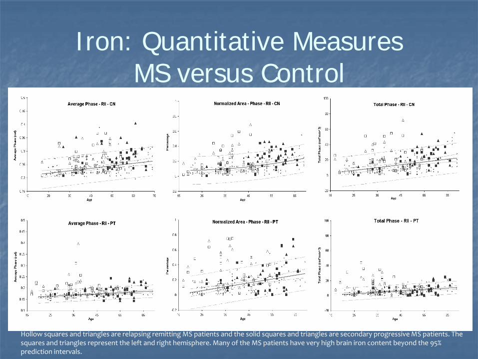
Iron: Quantitative MeasuresMS versus Control
Hollow squares and triangles are relapsing remitting MS patients and the solid squares and triangles are secondary progressive MS patients. The squares and triangles represent the left and right hemisphere. Many of the MS patients have very high brain iron content beyond the 95% prediction intervals.
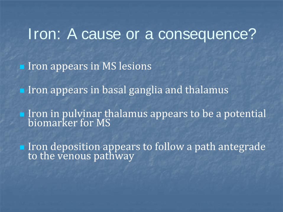
Iron appears in MS lesions
Iron appears in basal ganglia and thalamus
Iron in pulvinar thalamus appears to be a potential biomarker for MS
Iron deposition appears to follow a path antegrade to the venous pathway
Iron: A cause or a consequence?
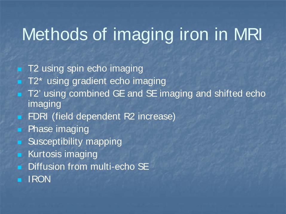
Methods of imaging iron in MRI
T2 using spin echo imaging T2* using gradient echo imaging T2’ using combined GE and SE imaging and shifted echo
imaging FDRI (field dependent R2 increase) Phase imaging Susceptibility mapping Kurtosis imaging Diffusion from multi-echo SE IRON
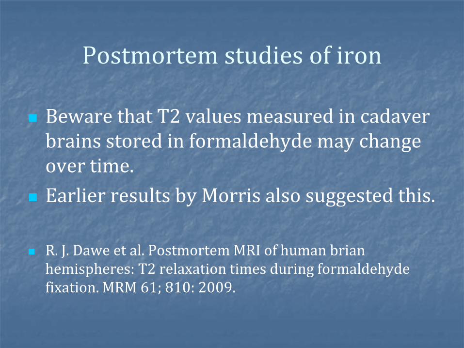
Postmortem studies of iron
Beware that T2 values measured in cadaver brains stored in formaldehyde may change over time.
Earlier results by Morris also suggested this.
R. J. Dawe et al. Postmortem MRI of human brian hemispheres: T2 relaxation times during formaldehyde fixation. MRM 61; 810: 2009.
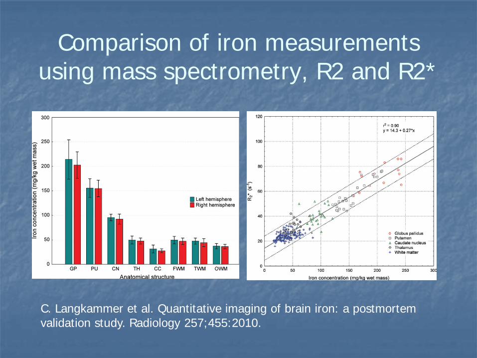
Comparison of iron measurements using mass spectrometry, R2 and R2*
C. Langkammer et al. Quantitative imaging of brain iron: a postmortem validation study. Radiology 257;455:2010.
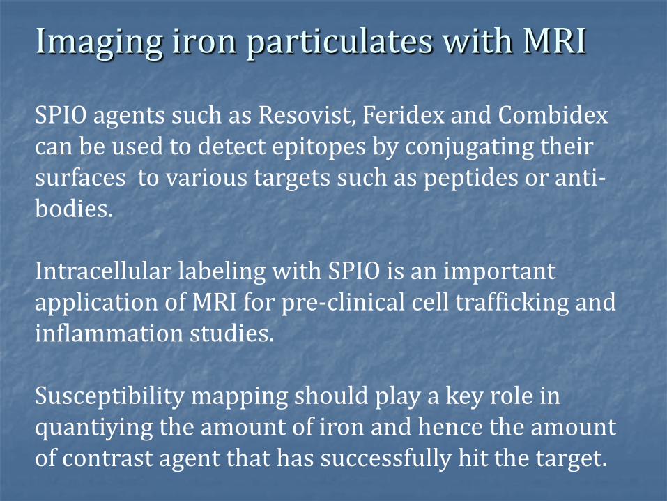
Imaging iron particulates with MRI
SPIO agents such as Resovist, Feridex and Combidex can be used to detect epitopes by conjugating their surfaces to various targets such as peptides or anti-bodies.
Intracellular labeling with SPIO is an important application of MRI for pre-clinical cell trafficking and inflammation studies.
Susceptibility mapping should play a key role in quantiying the amount of iron and hence the amount of contrast agent that has successfully hit the target.

Improving Susceptibility Mapping of Veins Using an Iterative Reconstruction Approach
77
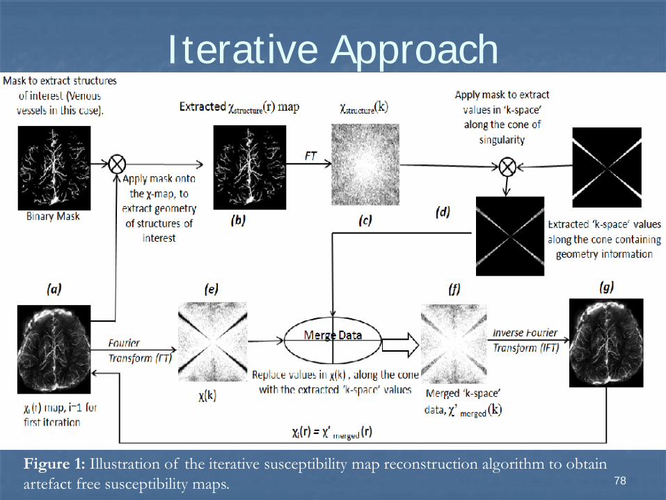
Iterative Approach
78Figure 1: Illustration of the iterative susceptibility map reconstruction algorithm to obtain artefact free susceptibility maps.
Results from Simulations
79
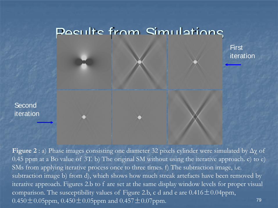
Figure 2 : a) Phase images consisting one diameter 32 pixels cylinder were simulated by Δχ of 0.45 ppm at a Bo value of 3T. b) The original SM without using the iterative approach. c) to e) SMs from applying iterative process once to three times. f) The subtraction image, i.e. subtraction image b) from d), which shows how much streak artefacts have been removed by iterative approach. Figures 2.b to f are set at the same display window levels for proper visual comparison. The susceptibility values of Figure 2.b, c d and e are 0.416±0.04ppm, 0.450±0.05ppm, 0.450±0.05ppm and 0.457±0.07ppm.
a b c
d e f
First iteration
Second iteration
Results from Simulations
80
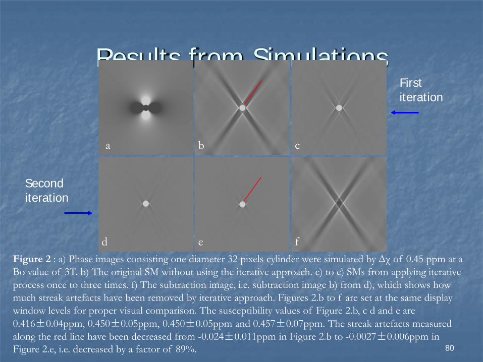
Figure 2 : a) Phase images consisting one diameter 32 pixels cylinder were simulated by Δχ of 0.45 ppm at a Bo value of 3T. b) The original SM without using the iterative approach. c) to e) SMs from applying iterative process once to three times. f) The subtraction image, i.e. subtraction image b) from d), which shows how much streak artefacts have been removed by iterative approach. Figures 2.b to f are set at the same display window levels for proper visual comparison. The susceptibility values of Figure 2.b, c d and e are 0.416±0.04ppm, 0.450±0.05ppm, 0.450±0.05ppm and 0.457±0.07ppm. The streak artefacts measured along the red line have been decreased from -0.024±0.011ppm in Figure 2.b to -0.0027±0.006ppm in Figure 2.e, i.e. decreased by a factor of 89%.
a b c
d e f
First iteration
Second iteration
Results from vivo MR Data
81
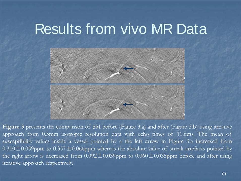
Figure 3 presents the comparison of SM before (Figure 3.a) and after (Figure 3.b) using iterativeapproach from 0.5mm isotropic resolution data with echo times of 11.6ms. The mean ofsusceptibility values inside a vessel pointed by a the left arrow in Figure 3.a increased from0.310±0.059ppm to 0.357±0.066ppm whereas the absolute value of streak artefacts pointed bythe right arrow is decreased from 0.092±0.039ppm to 0.060±0.035ppm before and after usingiterative approach respectively.
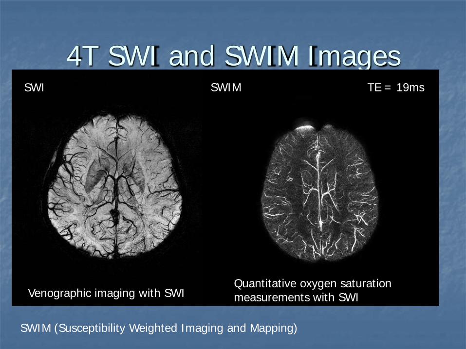
4T SWI and SWIM ImagesTE = 19msSWIMSWI
Venographic imaging with SWIQuantitative oxygen saturation measurements with SWI
SWIM (Susceptibility Weighted Imaging and Mapping)

Application to air bubble data
83
Figure 4 presents the comparison of SM before (Figure 4.a) and after (Figure 4.b) froman air-bubble phantom using iterative approach from 0.6mm isotropic resolution datawith echo times of 5.52ms. The mean of susceptibility values inside the bubble pointedby the left arrow in Figure 4.a increased 17.9% and, obviously, the streak artefacts havebeen significantly removed.

More Abroad Applications
84
Figure 4 presents the comparison of SM before (Figure 4.a) and after (Figure 4.b) froman air-bubble phantom using iterative approach from 0.6mm isotropic resolution datawith echo times of 5.52ms. The mean of susceptibility values inside the bubble pointedby the left arrow in Figure 4.a increased 17.9% and, obviously, the streak artefacts havebeen significantly removed.

Conclusions
MR offers the ability to quantify iron in both heme and non-heme applications.
A weakened endothelium may lead to hypoxic reperfusion injury with iron as a catalyst.
Of particular interest is the measurement of oxygen saturation with susceptibility mapping.
The use of iron tagged contrast agents and treatment therapies is an attractive direction.