Embed Size (px)
Citation preview
6
Iron Metabolism in Pathogenic Trypanosomes
Bruno Manta, Luciana Fleitas and Marcelo Comini Group Redox Biology of Trypanosomes,
Institut Pasteur de Montevideo, Uruguay
1. Introduction
1.1 Unique features, evolution and life cycle of trypanosomes
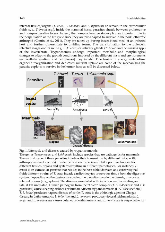
Trypanosomatids comprise a large group of flagellated unicellular protists with free-living and parasitic lifestyles. Several members of this family are widely known for being vertebrate pathogens of biomedical and veterinary importance. They belong to the order Kinetoplastida, which together with the groups Diplonemida (deep-see organisms) and Euglenida (photosynthetizing organisms) form the phylum Euglenozoa (Simpson et al., 2006; Roger & Simpson, 2009). Kinetoplastids represent one of the most ancient eukaryotic lineages that diverged after the acquisition of the mitochondrion and share unique biochemical and subcellular features including the nuclear and mitochondrial gene expression mechanism, the energy and thiol redox metabolism and the organization of the mitochondrial DNA, named kinetoplast, and the compartmentalization of the almost entire glycolytic pathway in a peroxisome-like organelle called the glycosome. Phylogenetic studies support the theory that hemoflagellate trypanosomes evolved from a free-living bodonid that subsequently became an insect parasite, which later gained capacity to adapt to mammalian hosts (Adl et al., 2005, 2007; Hamilton et al., 2004; Simpson et al., 2002). An early split of the genus Leishmania from the trypanosome taxon together with the existence of two well-defined clades within the genus Trypanosoma (the “brucei” clade and the “cruzi” clade) are strong evidences that the different lineages of pathogenic trypanosomes have evolved independently. Although they share a common ancestor, trypanosomes from the “brucei” and “cruzi” clades evolved and are geographically confined to the African and New World (South-America and Oceania) continents, respectively. In contrast to members from the “cruzi” clade, several lines of evidence indicate that African trypanosomes co-evolved with their mammalian hosts and insect vectors (Hamilton et al., 2007; Maslov et al., 1996; Stevens et al., 1998; Stevens et al., 2001). Parasitic trypanosomes are naturally transmitted by arthropods, e.g. tsetse fly for T. brucei, kissing bug for T. cruzi, and sand fly for Leishmania spp. (Fig. 1). Occasionally, bats can also act as vector agents (Dávila & Silva, 2000) and, more recently, rats and ticks are discussed as potentially involved in transmission of T. cruzi and Leishmania parasites to mammals (Herrera & Urdaneta-Morales, 2001; Colombo et al, 2011; Dantas-Torres, 2011). The natural infectious cycle of these parasites is initiated when non-dividing infective trypanosomes (metacyclic stage) are mechanically introduced in the mammal via insect bites or by involuntary deposition of infected feces from insects in the mucosa or injured dermis of mammals. Depending on the trypanosomatid species, parasites can reside in the host dermis (L. major), infect mucosa (L. brasilienzis and L. amazoniensis) or
www.intechopen.com
Iron Metabolism
148
internal tissues/organs (T. cruzi, L. donovani and L. infantum) or remain in the extracellular fluids (i. e., T. brucei ssp.). Inside the mammal hosts, parasites shuttle between proliferative and non-proliferative forms. Indeed, the non-proliferative stages play an important role in the perpetuation of the life cycle since they are pre-adapted to survive in the poikilothermic arthropod (Comini et al., 2011). They are taken up during insect blood meal of an infected host and further differentiate to dividing forms. The transformation to the quiescent infective stages occurs in the gut (T. cruzi) or salivary glands (T. brucei and Leishmania spp.) of the invertebrate. Trypanosomes undergo important metabolic and morphological changes to adapt to the growth conditions imposed by the different hosts and environments (extracellular medium and cell tissues) they inhabit. Fine tuning of energy metabolism, organelle reorganization and dedicated nutrient uptake are some of the mechanisms the parasite exploits to survive in the human host, as will be discussed below.
Insect
vector
Mammals
Nagana
Leishmania spp.T. cruziT. brucei
Parasites
HAT
tse-tse fly
Chagas’ disease
kissing bug
Leishmaniasis
sand-fly
Fig. 1. Life cycle and diseases caused by trypanosomatids. The genus Trypanosoma and Leishmania include species that are pathogenic for mammals. The natural cycle of these parasites involves their transmition by different but specific arthropods (insect vectors). Inside the host each species exhibit a peculiar tropism for different tissues, organs and systems resulting in different pathologies. For instance, T. brucei is an extracellar parasite that resides in the host´s bloodstream and cerebrospinal fluid; different strains of T. cruzi invade cardiomiocytes or nervous tissue from the digestive system; depending on the Leishmania species, the parasites invade the dermis, mucosa or internal organs (e. g., spleen). The diseases associated with infection are devastating and fatal if left untreated. Human pathogens from the ”brucei“ complex (T. b. rodhesiense and T. b. gambiense) cause sleeping sickness or human African trypanosomiasis (HAT; see section1); T. b. brucei produces nagana disease of cattle; T. cruzi is the ethiologic agent of Chagas disease in Latin-America; L. infantum and L. donovani produces visceral leishmaniasis, L. major and L. amazonensis causes cutaneous leishmaniasis, and L. brasiliensis is responsible for
www.intechopen.com
Iron Metabolism in Pathogenic Trypanosomes
149
a mucocutaneous infection. During their life cycle, the parasites undergo important biochemical and ultrastructural remodeling to adapt to the different hosts.The cycle is closed when insect feeds on infected mammals.
1.2 Trypanosomiasis: Disease, burden and treatment
Different members of the Trypanosomatida family are etiologic agents of highly disabling and often fatal diseases of humans and livestock (see Fig. 1). With respect to the parasites that are the major focus of this chapter, members from the T. brucei complex are transmitted by tsetse flies of the genus Glossina spp., which is found exclusively in equatorial Africa. The human pathogens T. b. gambiense and T. b. rodhesiense differ in their geographical distribution with the first subspecies being endemic of west and central regions while the last is mainly present in east and southern Africa (Barrett et al., 2003).
HAT present two main clinical phases with compromise of the hematolymphatic system (acute phase) and the central nervous system (CNS, chronic phase). The T. b. rhodesiense infection develops rapidly as an acute disease that is characterized by a high parasite load, severe anemia and thrombocytopenia accompanied by hypertrophy of the reticuloendothelial system. If untreated, the pathology evolves to a pancarditis with congestive heart failure, pulmonary edema and physiological collapse (Barrett et al., 2003). Due to this fast and lethal development, along with the limited sanitary conditions of endemic regions, HAT caused by T. b. rhodesiense is usually under-diagnosed explaining - at least partly - why 90% of the reported cases of African sleeping sickness are ascribed to T. b. gambiense (Birkholtz et al., 2011). In contrast, HAT produced by T. b. gambiense displays a more discrete development without characteristic symptoms in the early phase, unless the patient develops a generalized lymphadenopathy, which hinders an accurate diagnosis. The second stage of HAT starts when the parasites invade internal organs, including the CNS. For T. b. rhodesiense and T. b. gambiense this phenomena can take place within few weeks and up to years upon infection, respectively. Invasion of the CNS by the parasite is typically accompanied by intense headache, sleeping disorders and mental dysfunction, leading to a comatose state and sudden death (Barrett et al., 2003). The differential pathogenicity and clinical manifestations of both diseases have been formerly explained on the basis of mutual host-parasite adaptations that shaped pathogen virulence and host resistance during evolution (Fèvre et al., 2006). However, the hypothesis of a co-evolutionary virulence attenuation of T. b. gambiense has recently been challenged by studies on phylogenetic relationships within the T. brucei taxon (Balmer et al., 2011).
The World Health Organization (WHO) estimates that sixty million people are at risk of infection as a consequence of at least 300 separate active foci in 36 African countries (Jacobs et al., 2011), most of them in rural areas of extreme poverty. Around 300,000 people are currently infected with trypanosomes and 48,000 of them dye per year (Cavalli & Bolognesi, 2009). The lack of local human and financial resources combined with the burden of conflicts in most of the endemic countries impedes to achieve full control of HAT (Cavalli & Bolognesi, 2009). However, after continued control programs spanning vector eradication, early diagnosis and treatment, and surveillance (Barrett et al., 2003; Cavalli, 2009) the number of annual infections fell almost 5- to 7-fold in the last three decades1. Unfortunately, this progress is not
1 http://www.who.int/mediacentre/factsheets/fs259/en/
www.intechopen.com
Iron Metabolism
150
accompanied by the development of new chemotherapeutic options (see below) and is endangered by the increasing drug resistance of the naturally circulating strains of T. brucei ssp. (Cavalli & Bolognesi, 2009; Delespaux & de Koning, 2007; Jacobs et al., 2011; Matovu et al., 2001). In addition to HAT, animal trypanosomiasis represents a major problem for the agricultural and nutritional development of endemic regions. About ten million square kilometers of arable land are infested by tsetse flies (Matovu et al., 2001) capable of transmitting T. b. brucei, T. congolense and T. vivax (Nagana-disease) or T. evansi (Surra-disease) between domestic and wild (reservoirs) animals. For Africa, the total economic losses due to animal trypanosomiasis are estimated to be US$ 4.75 billion per year2. The recent detection of members from the “brucei” clade in countries from Asia, Central- and South-America should raise alarm considering the serious threat these pathogens entails for the well-developed agricultural economies of these regions (Luckins & Dwinger, 2004; Batista et al., 2007, 2009; Da Silva et al., 2011; Dávila & Silva, 2000; Mekata et al., 2009).
Confronted with the lack of prospect for vaccine development against trypanosomiasis, chemotherapy remains as the only short- and mid-term therapeutic choice for these diseases. Nevertheless, the few drugs currently available against HAT (acute phase: pentamidine and suramine, chronic phase: melarsoprol and efluornithine3) are far from optimal: most of them were originally developed for veterinary use, lacking safety compliance, and present a limited efficacy against late-stage disease (Steverding, 2010). An additional drawback associated with the inappropriate use of these drugs lies on the emergence of resistance. Unfortunately, pharmaceutical companies are less prone to engage and invest in drug discovery and development against diseases that affect the world´s poorest people (Barrett et al., 2003; Cavalli & Bolognesi, 2009; Matovu et al., 2001). However, in the last years, scientists, policy-makers and non-profit institutions (WHO, TDR, Médecins Sans Frontieres, FioCruz Institute, Drugs for Neglected Disease initiative), together with a few pharmaceutical companies (Glaxo SmithKline®, Bayer®), have joined efforts to improve this situation. As mentioned above, trypanosomes present several unique biochemical and biological features that can be exploited for the development of specific therapies. These include several organelles (glycosomes, acidocalcisomes, kinetoplast) that are absent in the mammalian host, and metabolic pathways and cellular functions that differ significantly from host counterparts, namely carbohydrate metabolism, protein and lipid modification, thiol-redox metabolism, cell cycle, programmed cell death, etc. (Naula & Burchmore, 2003). Despite the obvious indispensability of iron for pathogenic trypanosomatids, the mechanisms and components comprising the uptake, storage and usage of this metal have been poorly investigated. In the next sections will be reviewed the state-of-the-art regarding iron-homeostasis and metabolism in African trypanosomes.
2. Iron acquisition and homeostasis
Owing the extracellular and parasitic lifestyle of African trypanosomes, it deems important to comment first on the mechanisms and components controlling iron homeostasis in the human host. Dietary ferric iron (Fe3+) is reduced to its ferrous form (Fe2+) by a ferrireductase
2 Amounts in terms of agricultural Gross Domestic Product, data taken from: http://www.fao.org/ag/againfo/programmes/en/paat/disease.html 3 It must be noted that efluornithine is active only against T. b. gambiense but not T. b. rodhesiense (Cavalli & Bolognesi, 2009; Jacobs et al., 2011; Matovu et al., 2001).
www.intechopen.com
Iron Metabolism in Pathogenic Trypanosomes
151
present in the membrane of enterocytes, and then incorporated via a divalent metal transporter. Cytosolic iron is exported to the circulation from the basolateral membrane of enterocytes through ferroportin with the concomitant reoxidation to Fe3+ by the multicopper oxidase hephaestin (Kosman, 2010). Iron circulates in plasma bound to the glycoprotein transferrin (Tf), which is internalized by cells via a specific transferrin receptor (TfR1) in a clathrin-dependent mechanism. In the acidic environment of the endosome Tf, iron and TfR1 disassemble. Apo-Tf is released to circulation whereas TfR1 is recycled back to the membrane. Fe3+ is exported from the late endosomal vesicle through the concerted action of transporters and metalloreductases (Hentze et al., 2010; Kurz et al., 2011). How this “labile iron pool” (LIP, Hider & Kong, 2011; Kakhlon & Cabantchik, 2002) is trafficked within the cell remains poorly understood (Hentze et al., 2010; Anderson & Vulpe, 2009; Atanasiu et al., 2007; Hentze et al., 2010; Subramanian et al., 2011). Other major source of iron comprises the recycling of heme-iron from senescent erythrocytes, a task carried out by specialized macrophages of the reticuloendothelial system known as Kupffer cells (Anderson & Vulpe, 2009; Schultz et al., 2010). The liver is both the major site for iron storage and also the central metabolic regulator. Induced by different stimuli –from iron availability to inflammatory stresses– (Zhang & Enns, 2009), hepatocytes produce and secrete a 25-aminoacid hormone, hepcidin (Atanasiu et al., 2007), which regulates the levels of systemic iron by a negative-loop mechanism that involves ferroportin turnover. Intracellular iron homeostasis is regulated post-transcriptionally by the iron regulatory protein 1 (IRP1, see section 3.3.3.1). The vast majority of iron is dedicated to hemoglobin or myglobin synthesis, but also the biogenesis of iron-sulfur (Fe/S) proteins demands this metal. Both processes occur mostly in the mitochondria, representing the main subcellular compartment for iron utilization (Levi & Rovida, 2009). Iron is stored bound to ferritin heteropolymers, which can hold up to 4500 Fe3+ atoms (Andrews, 2010). Heme-iron is also essential in mammalian physiology (see section 4) but it poses an independent mechanism of absorption that remains mostly unsolved (Schultz et al., 2010). As envisaged, iron homeostasis is tightly controlled at several levels -from absorption to mobilization and utilization- and in an interdependent manner in humans.
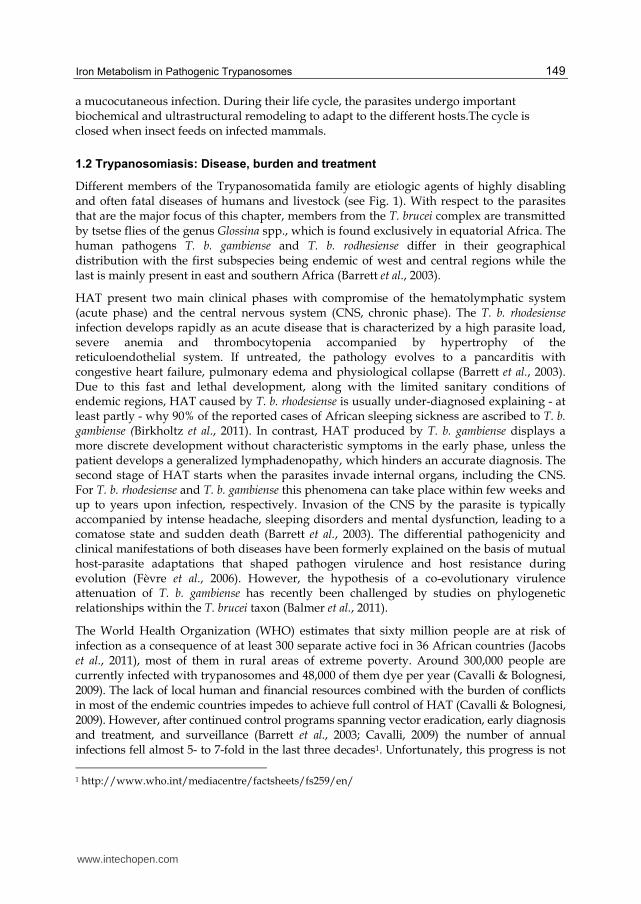
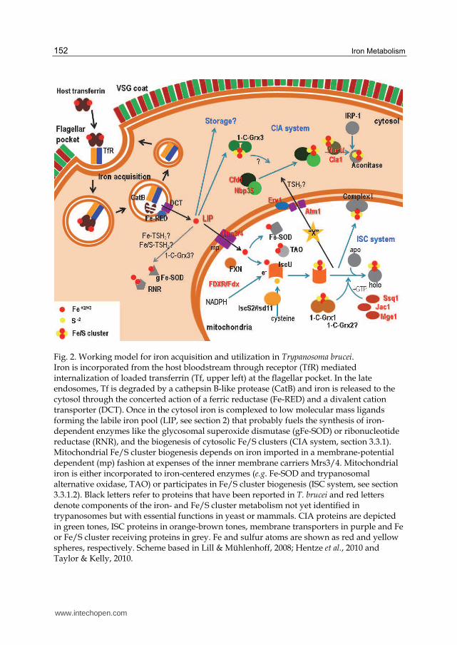
Iron is an essential element also for T. brucei, therefore the parasite has developed exceptional mechanisms to guarantee metal supply from a host that (un)intentionally limits metal availability in response to infection (see section 5). For example, iron-deficiency induced by ferritin upregulation and reduction of iron-transferrin saturation is a classical immune-based response mounted during the acute phase of the infection to limit parasite proliferation (Chisi et al., 2004). Also the chronic stage is accompanied by a notorious iron deprivation (e.g. anemia, see section 5) but, in this case, its origin is a chronic inflammatory disorder involving macrophage hyperactivation along with iron accumulation within the reticuloendothelial system (Stijlemans et al., 2008). In the bloodstream, trypanosomes acquire the metal4 via a high affinity receptor-mediated endocytosis of iron-loaded Tf (see Fig. 2; Grab et al., 1992, 1993; Steverding et al., 1995). The trypanosomal Tf receptor (TfR) was identified as a heterodimeric complex of proteins encoded by the expression-site-associated-gene (ESAG) 6 and ESAG 7 (Salmon et al., 1994; Steverding et al., 1995). ESAG 6 and 7 are truncated forms of variable surface glycoproteins (VSG) that lack the C-terminal domain, and, in the case of ESAG 7, the glycophosphatidylinositol (GPI) anchor. Under normal growth
4 The incorporation of heme-bound iron is discussed in section 4.
www.intechopen.com
Iron Metabolism
152
Fig. 2. Working model for iron acquisition and utilization in Trypanosoma brucei. Iron is incorporated from the host bloodstream through receptor (TfR) mediated internalization of loaded transferrin (Tf, upper left) at the flagellar pocket. In the late endosomes, Tf is degraded by a cathepsin B-like protease (CatB) and iron is released to the cytosol through the concerted action of a ferric reductase (Fe-RED) and a divalent cation transporter (DCT). Once in the cytosol iron is complexed to low molecular mass ligands forming the labile iron pool (LIP, see section 2) that probably fuels the synthesis of iron-dependent enzymes like the glycosomal superoxide dismutase (gFe-SOD) or ribonucleotide reductase (RNR), and the biogenesis of cytosolic Fe/S clusters (CIA system, section 3.3.1). Mitochondrial Fe/S cluster biogenesis depends on iron imported in a membrane-potential dependent (mp) fashion at expenses of the inner membrane carriers Mrs3/4. Mitochondrial iron is either incorporated to iron-centered enzymes (e.g. Fe-SOD and trypanosomal alternative oxidase, TAO) or participates in Fe/S cluster biogenesis (ISC system, see section 3.3.1.2). Black letters refer to proteins that have been reported in T. brucei and red letters denote components of the iron- and Fe/S cluster metabolism not yet identified in trypanosomes but with essential functions in yeast or mammals. CIA proteins are depicted in green tones, ISC proteins in orange-brown tones, membrane transporters in purple and Fe or Fe/S cluster receiving proteins in grey. Fe and sulfur atoms are shown as red and yellow spheres, respectively. Scheme based in Lill & Mühlenhoff, 2008; Hentze et al., 2010 and Taylor & Kelly, 2010.
www.intechopen.com
Iron Metabolism in Pathogenic Trypanosomes
153
conditions the TfR localizes at the flagellar pocket wherefrom bound-transferrin is internalized and proteolyticaly degraded in the endolysosome by a cathepsin-B like protease (O’Brien et al., 2008; Mussmann et al., 2003), which finally releases iron. The relevance of the endocytic pathway as the sole source of iron for the parasite was demonstrated using antibodies anti-TfR that inhibited growth of T. brucei in vitro (Grab et al., 1992). Expression of TfR in the parasite is regulated by iron availability and post-transcriptional control that does not involve the IRE/IRP1 system typical for mammals (Fast et al., 1999). Iron starvation induced by iron chelators or species-specific transferrins lead to a 3- to 10-fold upregulation in the expression of TfR with a concomitant redistribution of the receptor from the flagellar pocket to the entire parasite surface (Fast et al., 1999; Mussmann et al., 2004). This, together with the rapid recycling of TfR (Kabiri & Steverding, 2000) and gene-specific (in)activation events (van Luenen et al., 2004) allows trypanosomes to efficiently compete for limiting substrate and withstand iron-deprivation until a new set of higher affinity TfR is expressed. For instance, sequence polymorphisms in ESAG 6 and 7 were proposed to determine the affinities of TfR for transferrins from different mammalian species (Bitter et al., 1998) permitting for a rapid adaptation of the parasite to distinct hosts. It was also suggested that the TfR repertoire may allow the parasite to overcome anti-TfR antibody response by the host (Gerrits et al., 2002). However, estimations and studies by Steverding (1998, 2003, 2006) disproved this hypothesis and supported the view that high affinity TfRs were evolutionary selected to enable the parasite to cope with the diversity of mammalian transferrins (Steverding, 2003, 2006).
In the aerobic and buffered milieu of the cytosol iron is never free, mostly because it can readily produce highly toxic oxygen species through Fenton chemistry (see section 3.2.2), but also because the ferric form tends to form insoluble hydroxides. The cellular labile or chelatable iron pool is defined as the intracellular pool of redox-active iron that is not associated with proteins. LIP comprises ~ 5 % of the total cellular iron and consist of Fe2+ and Fe3+ associated with a diverse population of low molecular mass ligands such as organic anions (phosphate, citrate, inositol phosphate, etc.), polypeptides such as glutathione (GSH) and/or components of membranes (phospholipids, etc; Kakhlon & Cabantchik, 2002). Whether this percentage represents the situation in trypanosomes is unknown. The nature of the ligand(s) can vary between cells and among different physiological states but recent reports support the notion that intracellular GSH (2-5 mM) is the most relevant low molecular mass complexing agent in most GSH-dependent organisms (Hider & Kong, 2011; Kumar et al., 2011; Mühlenhoff et al., 2010; Overath et al., 1986). The nature of this LIP in trypanosomes was never assessed and it is worthy to speculate that trypanothione (T(SH)2, see below), and not GSH, will be the relevant physiological ligand. Beyond that T(SH)2 is the most abundant intracellular low molecular mass thiol in trypanosomes (Krauth-Siegel & Comini, 2008), our working model is supported by experimental evidences showing the formation of T(SH)2-Fe and T(SH)2-Fe/S complexes in vitro (Ceylan et al. 2010; Manta et al., unpublished, Fig. 2) and by the extensive documentation showing that most thiol-dependent functions in these organisms evolved to use T(SH)2 and not GSH as cofactor (reviewed in Irigoín et al., 2008; Krauth-Siegel and Comini, 2008). How iron enters and exits the LIP and which are the proteins involved in this process remain as open questions. Recent studies propose a link between the LIP and Fe/S protein metabolism by means of a mechanism that involves Fe/S clusters as signalling molecule and cytosolic monothiol glutaredoxins as mediators (see section 3.2.1; Kumar et al., 2011; Mühlenhoff et al., 2010). In fact, the nature of monothiol glutaredoxins (i.e. iron-sulfur proteins that use GSH as ligand
www.intechopen.com
Iron Metabolism
154
cofactor for Fe/S cluster assembly) make these proteins exceptional candidates for the integration of the cellular iron and Fe/S cluster status with thiol redox status, and as signal transducer regulating iron uptake and utilization (Mühlenhoff et al., 2010; Rodriguez-Manzaneque et al., 2002; Rouhier et al., 2010; Ye et al., 2010). Another important form of “low-molecular mass” iron species are the dinitrosyl iron complexes (DNIC, Bosworth et al., 2009) formed between LIP and low molecular mass thiols when nitric oxide is present (Vanin, 2009). This point is addressed in the next section.
3. Cellular fates of Iron
Iron is an important redox or structural cofactor of several indispensable proteins of trypanosomes. For instance, DNA synthesis, protein translation, oxidant defense and cytochrome respiration are important cellular functions that involve the utilization of this metal (Taylor & Kelly, 2010).
In this section we will review the most relevant iron-related molecules and metabolic pathways of bloodstream T. brucei. Whenever possible, the components and mechanisms employed by the parasite to incorporate different forms of iron onto target molecules will be described. Taking advantage of the recent availability of genome sequencing data for the most representative species of Trypanosoma (Berriman et al., 2005; El-Sayed et al., 2005; Ivens et al., 2005) and the current knowledge of the iron metabolism in model eukaryotes (Hentze et al., 2010), we here provide a state-of-the-art view of iron metabolism in African trypanosomes.
3.1 Dinitrosyl iron complexes
Parasites circulating in host´s bloodstream or inside the phagolysosome of activated macrophages are exposed to reactive oxygen and nitrogen species. In both cases, the second messenger nitric oxide (�NO) is produced by endothelial or immune cells, which, if not neutralized rapidly, can lead to the formation of the highly reactive oxidant peroxynitrite (Girard et al., 2005). The effect of �NO and derivatives was studied in T. brucei (Lu et al., 2011; Steverding et al., 2009; Vincendeau et al., 1992) and T. cruzi (Piacenza et al., 2007; Alvarez et al., 2011 and see papers quoted in Irigoín et al., 2008). A recent work by Bocedi et al. (2010) demonstrated the formation in vitro of a DNIC involving the parasite specific dithiol T(SH)2. Based on the high intracellular concentration of T(SH)2 the authors proposed that formation of this complex may play an important role in trapping �NO and, thus, preventing the formation of dangerous oxidants. This work raises the possibility for a new potential link between iron and low-molecular mass thiols in trypanosomes.
3.2 Mononuclear iron proteins
Mononuclear iron proteins can be classified according to their biological function, by Fe centre type, by type and number of prosthetic centres, and by sequence similarity. The iron atom is usually coordinated by thiolate groups (deprotonated form of cysteine), or the N├ atoms of histidines or the carboxylate anions of acidic residues present in the polypeptide. In these proteins the iron provides redox activity and the surrounding aminoacidic and structural environment confer the specificity for different substrates. The characteristics and functions of a number of iron-centered proteins from trypanosomatids are addressed below.
www.intechopen.com
Iron Metabolism in Pathogenic Trypanosomes
155
3.2.1 Energy metabolism: Alternative oxidase
The bloodstream form of the parasite lacks cytochrome activity yet they “respirate” at high rates (Priest & Hajduk, 1994). The molecular entity responsible for this is a plant-like mitochondrial ubiquinol oxidase (EC 1.10.3.10), known as trypanosomal alternative oxidase (TAO; Clarkson et al., 1989). The enzyme is imported into the mitochondrion of bloodstream and procyclic parasites by distinct mechanisms involving external ATP supply and inner membrane potential, respectively (Williams et al., 2008). It localizes at the outer membrane of the organelle where it transfers electrons from ubiquinol to oxygen without proton translocation or ATP generation (Chaudhuri et al., 2006), resulting in the reoxidation of NADPH produced during glycolysis. TAO genes have been identified in the genome of all the subspecies forming the “brucei” clade but are absent in related trypanosomatids such as Leishmania spp. or T. cruzi (Chaudhuri et al., 2006; Nakamura et al., 2010). In agreement with its metabolic function (NADP/NAPDH-shunt during carbohydrate catabolism), TAO is developmentally regulated (Chaudhuri et al., 2002) achieving ~100 times higher levels in bloodstream parasites than in procyclic cells (Tsuda et al., 2005; Tyler et al., 1997). The essential role of TAO in the physiology of infective T. brucei was recognized early using the iron chelator salicyl hydroxamic acid (2-hydroxybenzhydroxamic acid, SHAM) and glycerol to alter the metabolic output (Clarkson & Brohn, 1976; Grant & Sargent, 1960). On the other hand, recent studies show that TAO plays an important role in preventing oxidant-induced programmed cell death of long-slender bloodstream parasites (Tsuda et al., 2005, 2006). This antioxidant function of TAO resembles that proposed for the orthologue enzyme from plants (Maxwell et al., 1999). In trypanosomes, apoptosis is a highly regulated process deeply associated with the accumulation of quiescent parasite forms (short stumpy) in the preparation for transmission to the insect vector (Welburn et al., 2006). It is therefore tempting to speculate that, at least in bloodstream trypanosomes, TAO might be an important checkpoint connecting metabolic status (NADP+/NAPDH ratio) with programmed cell death and differentiation. In procyclic forms, TAO has been shown to compensate for a depletion of complex III or IV activities in the mitochondrial electron transfer chain (Horváth et al., 2005). Despite this backup role in respiration, the enzyme appears to fulfill a yet unknown but essential function in this parasite stage (Tsuda et al., 2005; Tyler et al., 1997).
Although no structures are available for TAO or any other related alternative oxidase, current structural models (Moore & Albury, 2008) and proteomic data (Acestor et al., 2009) indicate that it is an interfacial membrane protein that interacts with a single leaflet of the lipid bilayer, and contains a non-heme di-iron carboxylate center bounded by two highly conserved EXXH motifs (Kido et al., 2010a, 2010b). The iron-dependence of TAO was established working in vitro with chelating agents and mutant forms of the protein (Ajayi et al., 2002; Chaudhuri et al., 1998).
3.2.2 Oxidant defense: Iron-dependent superoxide dismutases
Aerobic respiration produce partly reduced oxygen intermediates that leak from several protein complexes from the electron transport chain. The most important radical species formed is the anion superoxide (O2�-), the one-electron reduction product of molecular oxygen. Superoxide can reduce or oxidize biological targets in vivo, or dismutate to the less reactive but highly diffusible oxidant hydrogen peroxide (Halliwel & Gutteridge, 1999). Despite hydrogen peroxide is biologically more toxic than O2�-, active removal of this anion
www.intechopen.com
Iron Metabolism
156
radical is necessary to prevent formation of the most reactive and harmful radical product, hydroxyl radical (OH�), which originates from a physiological reaction involving iron and known as Fenton reaction5. To accomplish this, all living organisms contain enzymes devoted to the O2�- dismutation, called superoxide dismutases (EC 1.15.1.1). There are three major families of SODs, depending on the metal cofactor. Most of the cytosolic eukaryotic SODs use a bimetallic active site with copper and zinc (CuZn-SODs) while the mitochondria harbor a bacterial-related Mg-dependent SOD. In prokaryotes and plastids most of the SODs are Fe-dependent (Abreu & Cabelli, 2010).
Trypanosomes express four different isoforms of SODs (Dufernez et al., 2006; Kabiri & Steverding, 2001; Le Trant et al., 1983; Wilkinson et al., 2006) that, in contrast to homologues from prokaryotes and eukaryotes, show a restricted metal dependency all of them being Fe-dependent enzymes (Wilkinson et al., 2006; Bachega et al., 2009). In T. brucei, SOD-A and SOD-C localize at the mitochondrion whereas SOD-B1 and SOD-B2 are mostly compartmentalized within the glycosome and less abundantly at the cytosol (Dufernez at al., 2006). A similar localization was reported for SOD-B enzymes from L. chagasi (Plewes et al., 2003). RNAi-mediated knockdown of SOD-A and SOD-C revealed that under normal growth conditions the mitochondrial isoforms are dispensable for bloodstream T. brucei (Prathalingham et al., 2007; Wilkinson et al., 2006). However, the biological importance of SOD-A was put in evidence when parasites depleted in this isoform showed a higher sensitivity towards paraquat, an O2�--generating compound (Wilkinson et al., 2006). Consistent with a role in oxidant defense for mitochondrial Fe-SOD, transgenic T. cruzi overexpressing a mitochondrial isoform was found to be more resistant to fresh human serum, a death stimuli mediated by oxidative stress (Piacenza et al., 2007). In contrast, the SOD B-type enzymes are indispensable for infective T. brucei grown under normal culture conditions (Wilkinson et al., 2006), which poses the question to the glycosomal source of O2�-
, a charged molecule that does not diffuse through lipid membranes. In this respect, unwanted O2�- might leak as byproduct from a variety of metabolic activities occurring in this organelle, such as glycolysis, oxidation of fatty acids, lipid biosynthesis, and purine salvage (Michels et al., 2006). Further dissection of the functional relevance of SOD-B isoforms was achieved by means of targeted gene replacement, which demonstrates that SOD-B1 and not SOD-B2 is critical to withstand exposure to nifurtimox and benznidazole (Prathalingham et al., 2007), two anti-trypanosomal drugs whose mechanism of action involves the generation of reactive oxygen intermediates (Maya et al., 2003 and 2007). Also the homologue isoforms present in L. chagasi and L. tropica were earlier reported to be important for parasite survival in mouse or human macrophages as well as under paraquat insult (Plewes et al., 2003; Ghosh et al., 2003). Interestingly, T. brucei SOD-B1 has been shown to be developmentally regulated with higher intracellular concentration in proliferating stages (Kabiri & Steverding, 2001). This particular expression pattern led the authors to propose that the role of SOD-B1 in dividing cells is to counteract the formation of superoxide radicals released during the generation of the iron-tyrosyl free-radical centre in the small subunit (R2) of ribonucleotide reductase, other iron-containing enzyme (see next section). Unexpectedly, a 5- to 8-fold increase in SOD-B1 activity in transgenic T. cruzi was accompanied by a significant sensitization of parasites against two pro-oxidant compounds namely gentian violet and benznidazole (Temperton et al., 1998). This striking behavior was
5 H2O2 + O2●- OH- + OH● + O2 Fe3
www.intechopen.com
Iron Metabolism in Pathogenic Trypanosomes
157
interpreted as a consequence of an imbalance in the redox homeostasis of the parasite resulting from the overexpression of Fe-SOD, a hypothesis that deserves further investigation.
In summary, trypanosomal SODs diverged from their human homologues by using iron as cofactor. They apparently evolved to protect parasites against toxic O2�- produced in the glycosomes and/or mitochondrion as a result of sudden changes in metabolism or originated from the different environment they live in (e.g., insect midgut, macrophages, epithelium, mammal bloodstream).
3.2.3 Cell proliferation: Ribonucleotide reductase
Ribonucleotide reductase (RNR, EC 1.17.4.1-2) catalyses the reduction of ribonucleotides to
deoxyribonucleotides needed for DNA synthesis. There are three different classes of RNR
being class I the most abundant in eukaryote organisms. Class I RNR are heterotetrameric
enzymes formed by the association of two related but not identical polypeptides known as
subunit R1 and R2. The large R1 subunit binds substrates and allosteric effectors, conferring
specificity and regulatory potential, while the small R2 subunit contains the catalytic center
composed of two high spin Fe3+ atoms antiferromagnetically coupled to each other through
a µ-oxo bridge, a highly conserved tyrosine residue and two cysteines (Cotruvo & Stubbe,
2011). The di-iron center generates a free radical on this catalytic tyrosine through electron
donation. In a reaction involving several intermediate states, the tyrosine radical attacks the
nucleotide resulting in reduction of the 2’-OH group of ribonucleoside and the formation of
a disulfide. Regeneration of active RNR is finally achieved via reduction of this disulfide
mainly by thioredoxin (Trx), which is subsequently reduced at expenses of NADPH
(Cotruvo & Stubbe, 2011).
The biochemical information about parasite RNR is rather limited. Both subunits of RNR
from T. brucei were cloned, expressed (Dormeyer et al., 1997; Hofer et al., 1997) and
kinetically characterized (Hofer el al., 1998). The parasite-specific thioredoxin-like
oxidoreductase tryparedoxin (TXN, Lüdemann et al., 1998) and, in contrast to all other
RNR from eukaryotes, also the low-molecular weight dithiol T(SH)2 (Dormeyer et al.,
2001) but less likely glutaredoxins (Ceylan et al., 2010) proved to be physiological
reductants of recombinant RNR. The activity of the enzyme appears to be post-
transcriptionally regulated by a redox mechanism (Dormeyer et al., 2001) and by the
selective expression of its catalytic subunit R2. For example, whereas the R1 protein is
actively expressed throughout the whole life cycle of the parasite, the R2 protein is not
detected in cell cycle-arrested short stumpy trypanosomes (Breidbach et al., 2000). How
iron is incorporated into the R2 subunit is yet elusive (Cotruvo & Stubbe, 2011). Recent
findings suggest that, at least in yeast, the di-iron non-heme incorporation into apo-
proteins is tightly related to the Fe/S cluster biogenesis machinery both from the cytosol
and mitochondria (see section 3.3) (Cotruvo & Stubbe, 2011; Mühlenhoff et al., 2010) and
in particular to the cytosolic monothiol glutaredoxins (Grx3/4), discussed later in this
review (see section 3.3.2). In trypanosomes, the only link between iron and RNR came
from experiments in which parasites treated with the iron chelator deferoxamine (DFX)
show an extremely reduced [3H]-timidine incorporation, pointing to an essential role of
iron in DNA synthesis (Breidbach et al., 2002).
www.intechopen.com
Iron Metabolism
158
3.2.4 Lipid biosynthesis: Stearoyl-CoA desaturase
Stearoyl-CoA desaturases (SCD) from eukaryotes are di-iron containing proteins responsible for the de novo synthesis of monounsaturated fatty acids from saturated fatty acids. The iron is usually coordinated by 8 histidine residues that form the active site (Man et al., 2006). They are integral membrane proteins anchored in the endoplasmic reticulum. Recent genetic and chemical validation of the orthologue enzyme from T. brucei demonstrated its indispensability for in vivo survival of bloodstream forms, as well as for the procyclic stage (Alloati et al., 2010, 2011).
3.3 Iron-sulfur cluster proteins6
The name iron-sulfur proteins refer to a broad group of proteins. A class of them contains mononuclear Fe centers coordinated directly by cysteine residues (e. g., rubredoxins and related proteins). A second class ligates a complex between iron and inorganic sulfur through side-chain atoms provided mainly by the aminoacid cysteine, or eventually histidine or aspartate. These centers are known as Fe/S clusters (Lill & Mühlenhoff, 2008). Fe/S centers are ubiquitous inorganic cofactors present in all forms of life and are probably the most ancient cofactors and catalysts in the prebiotic world. Among them, the [2Fe-2S] is the simplest and most common cluster found in vivo, whereas Fe/S clusters of higher complexity require further “maturation” (Lill & Mühlenhoff, 2008; Lill, 2009; Py & Barras, 2010). In eukaryotes, the list of Fe/S dependent proteins has nearly one hundred members (Rouault & Tong, 2005; Ye & Rouault, 2010). In general, this cofactor enables electron transfer reactions due to the propensity of the iron atoms to switch between reduced (Fe2+) and oxidized (Fe3+) states. In addition, the redox potential of the cluster can be finely tuned by the protein environment covering a wide range of potentials, from very reducing (~-500 mV) to highly oxidizing (~+300 mV) (Lill, 2009; Xu & Møller, 2011). In consequence, Fe/S clusters are essential components of the most important biological electron transport chains, i.e. photosynthesis and mitochondrial respiration. But the role Fe/S clusters can play in proteins goes far beyond their redox properties. For instance, they act as important structural or regulatory moieties of some proteins from the DNA metabolism and as cofactors in enzymes of the amino acid biosynthesis or Krebs cycle (Lill & Mühlenhoff, 2006; Lill, 2009; Netz et al., 2010; Py & Barras, 2010; Ye & Rouault, 2010a, 2010b).
3.3.1 Biogenesis of iron-sulfur clusters and proteins
Despite the chemical simplicity of these cofactors, their biosynthesis and insertion into
apoproteins within the cell requires devoted machineries that are are highly conserved from
bacteria to humans (Lill & Mühlenhoff, 2006, 2008; Xu & Møller, 2011). In bacteria, there are
three different systems for the biogenesis of Fe/S proteins, all encoded in specific operons
and tightly regulated. The “nitrogen fixation” (NIF) system was the first Fe/S cluster
biogenesis mechanism described and is exclusively dedicated to the maturation of
nitrogenase enzymes from certain bacterias. On the contrary, the “iron-sulfur cluster” (ISC)
6 Due to the high amount of literature on this topic, this section contains mainly quotations to the most recent reviews, facilitating further reading for interested lectors. We therefore apologize to the authors of the original contributions.
www.intechopen.com
Iron Metabolism in Pathogenic Trypanosomes
159
and “sulfur utilization factor” (SUF) systems are widely distributed among bacteria and are
responsible for the biosynthesis of Fe/S proteins in basal and stressed conditions,
respectively (Xu & Møller, 2011). The mitochondria and plastids of eukaryotes have
inherited the ISC and SUF systems, respectively, from ancient symbionts (Balk & Lobréaux,
2005; Balk & Pilon, 2011; Xu & Møller, 2011). Additionally, maturation of Fe/S cluster
proteins in eukaryotes can be accomplished in the cytosol by a specific set of proteins that
constitute the cytosolic iron sulfur cluster assembly (CIA) machinery. In this section we will
summarize the most relevant aspects of the mitochondrial machinery related to the bacterial
ISC system, and will introduce the limited information available for the CIA system. Most of
the reports published stem from studies with the budding yeast Saccharomyces cerevisiae, and
we will refer to them before describing what is known for Fe/S protein biogenesis in
trypanosomes (Section 3.3.2).
The building blocks for the biosynthesis of Fe/S clusters are: iron atoms (in the reduced form, Fe2+), sulfide (S2-), reducing power and proteins, which act as scaffolds to assemble the Fe/S cluster that is being formed (Py & Barras, 2010; Lill & Mühlenhoff, 2006). Sulfide is generated from cysteine by the pyridoxal-5-phosphate-dependent cysteine desulfurase (eukaryotic Nfs1 or the bacterial NifS, IscS and SufS). This reaction produces alanine and leaves a protein-bound cysteine persulfide intermediate, which is presumably reduced by the ferredoxin system (NADPH-dependent ferredoxin reductase and ferredoxin) leading to release of S2-. Ferredoxin is a Fe/S protein that in mammals is also known as adrenodoxin, an essential component of the thyroid hormone production. Recent studies identified an accessory protein, Isd11, that is essential for in vivo Fe/S biogenesis in eukaryotes (Adam et
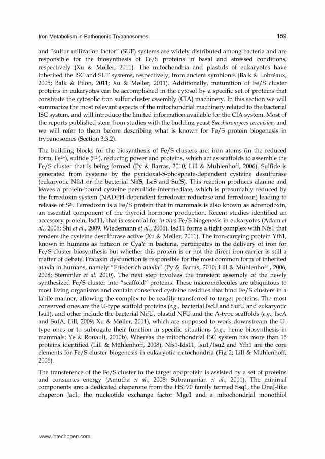
al., 2006; Shi et al., 2009; Wiedemann et al., 2006). Isd11 forms a tight complex with Nfs1 that renders the cysteine desulfurase active (Xu & Møller, 2011). The iron-carrying protein Yfh1, known in humans as frataxin or CyaY in bacteria, participates in the delivery of iron for Fe/S cluster biosynthesis but whether this protein is or not the direct iron-carrier is still a matter of debate. Frataxin dysfunction is responsible for the most common form of inherited ataxia in humans, namely “Friederich ataxia” (Py & Barras, 2010; Lill & Mühlenhoff., 2006, 2008; Stemmler et al. 2010). The next step involves the transient assembly of the newly synthesized Fe/S cluster into “scaffold” proteins. These macromolecules are ubiquitous to most living organisms and contain conserved cysteine residues that bind Fe/S clusters in a labile manner, allowing the complex to be readily transferred to target proteins. The most conserved ones are the U-type scaffold proteins (e.g., bacterial IscU and SufU and eukaryotic Isu1), and other include the bacterial NifU, plastid NFU and the A-type scaffolds (e.g., IscA and SufA; Lill, 2009; Xu & Møller, 2011), which are supposed to work downstream the U-type ones or to subrogate their function in specific situations (e.g., heme biosynthesis in mammals; Ye & Rouault, 2010b). Whereas the mitochondrial ISC system has more than 15 proteins identified (Lill & Mühlenhoff, 2008), Nfs1-Ids11, Isu1/Isu2 and Yfh1 are the core elements for Fe/S cluster biogenesis in eukaryotic mitochondria (Fig 2; Lill & Mühlenhoff, 2006).
The transference of the Fe/S cluster to the target apoprotein is assisted by a set of proteins and consumes energy (Amutha et al., 2008; Subramanian et al., 2011). The minimal components are: a dedicated chaperone from the HSP70 family termed Ssq1, the DnaJ-like chaperon Jac1, the nucleotide exchange factor Mge1 and a mitochondrial monothiol
www.intechopen.com
Iron Metabolism
160
glutaredoxin (Grx5 in humans and yeast; Fig. 2). The requirement for a glutaredoxin-like protein, formerly considered an oxidoreductase (Lillig et al., 2008; Rouhier et al., 2010), in Fe/S cluster biogenesis was initially suggested by studies in a yeast Grx5 deletion mutant. These cells displayed deficient cluster assembly for at least two Fe/S proteins (aconitase and succinate dehydrogenase), leading to impaired respiratory growth and increased sensitivity to oxidative stress with accumulation of free iron in the cell (Rodríguez-Manzaneque et al., 1999 and 2002). Although the precise function of monothiol glutarredoxins within the iron metabolism remain to be established, they are critical components of the ISC biosynthetic pathway in distantly related organisms such as bacteria (Fernandes et al., 2005), protists (Comini et al., 2008) and vertebrates (Xu & Møller, 2011; Ye et al., 2010; Ye & Rouault, 2010b).
The CIA machinery was first discovered in yeast when seeking for cytosolic proteins essential for Fe/S cluster assembly on aconitase (Fig. 2). Nowadays, several components are recognized: i) two P-loop NTPases (Cfd1 and Nbp35) that form a stable heterotetrameric complex with a [4Fe-4S] cluster bound to the C-terminal region, suggesting a scaffold role for cluster assembly; ii) Nar1, a Fe/S-dependent protein related to bacterial iron-containing hydrogenases, iii) WD40 ┚-propeller protein Cia1, which preferentially localizes in the nucleus and assists cluster delivery to Nbp35 (Lill & Mühlenhoff, 2008; Sharma et al., 2010; Xu & Møller, 2011), and iv) redox components of the CIA machinery (Tah18, Dre2 and monothiol glutaredoxins 3 and 4). In yeast, intracellular iron level is sensed by the transcription factor Aft1 and requires the regulatory proteins Fra1-Fra2 that interact with the cytosolic-nuclear monothiol glutaredoxins Grx3 or Grx4 through the formation of heterodimers linked by a Fe/S cluster (Kumánovics et al., 2008; Mühlenhoff et al., 2010; Pujol-Carrion et al., 2006). Interestingly, besides the existence of specific and compartmentalized machineries, Fe/S cluster biogenesis in the cytosol has been confirmed to depend on the mitochondrial system. A still unknown essential compound (noted as “X” in Fig. 2) is exported from mitochondria via the inner mitochondrial membrane ATP-binding cassette (ABC) transporter Atm1 and the intermembrane space protein Erv1, in a process that requires GSH (Netz et al., 2010; Sharma et al., 2010; Xu & Møller, 2011). As pointed by Ye and Rouault (2010a, 2010b) the next frontier in Fe/S cluster biogenesis will consist in unraveling how the monothiol glutaredoxin 5 and ABCB7 transporter are involved in the mitochondrial synthesis and export to the cytosol/nucleus of a still unknown factor that connects these processes in both compartments. Moreover, it remains to be studied how the system operates as a whole (Mühlenhoff et al., 2010; Netz et al., 2010; Zhang et al., 2008).
3.3.2 Biogenesis of iron-sulfur clusters in trypanosomes
The characterization of the Fe/S biogenesis in trypanosomes was driven by the completion of the genome sequencing of three trypanosomatid species (Berriman et al., 2005; El Sayed et al., 2005; Ivens et al., 2005). As noted previously, the mitochondrion is a key organelle in Fe/S cluster biosynthesis that undergoes important metabolic and morphological changes in trypanosomatids, especially in African trypanosomes, during life cycle. For instance, several mitochondrial respiratory-chain proteins that depend on these cofactors are developmentally synthesized in insect stage parasites (Alfonzo & Lukeš, 2011; Tyler et al, 1997). Based on sequence comparison with identified Fe/S cluster biogenesis proteins from yeast, plants and humans, a number of members of the ISC system are conserved in
www.intechopen.com
Iron Metabolism in Pathogenic Trypanosomes
161
trypanosomatids (see Fig. 2). Smíd et al. (2006) characterized two major components of this machinery present in T. brucei: a cysteine desulfurase (TbIscS2, related to Nfs1) and one U-type scaffold protein (TbIscU, related to Isu1). Both proteins proved to be essential for the mitochondrial biogenesis of Fe/S proteins (Smíd et al., 2006). In agreement with phenotypes observed in yeast mutants, down-regulation of TbIscS2 in procyclic parasites by RNAi results in inhibition of Fe/S cluster dependent processes both in the mitochondrion and cytosol, with a concomitant impairment in ATP production, cellular respiration and growth (Smíd et al., 2006; Paris et al., 2009). The occurrence of this protein in the rudimentary mitochondrion of bloodstream parasites was still not addressed but appears reasonable considering that this stage relies on important Fe/S protein activities (Comini et al., 2008). As mentioned above, a stable tricomponent system between Nfs1, Isd11 and IscU have to be formed to enable the formation of Fe/S cluster on IscU. An Isd11 homologue is actively expressed by both life stages of T. brucei and, as expected, is critical for cytosolic and mitochondrial Fe/S cluster biosynthesis in trypanosomes (Paris et al., 2010). A second Nfs-like gene was identified in T. brucei (Smíd et al., 2006) and characterized as a selenocysteine lyase (SLC; Poliak et al., 2010), an enzyme that cleaves selenocysteine into alanine and selenium during selenoprotein metabolization. Supporting the experimental evidences on the dispensability of the selenoproteome for this parasite (Aeby et al, 2009), down-regulation of SLC is not detrimental for T. brucei, at least under cultivation (Poliak et al., 2010). A single-copy gene with a significant sequence similarity to eukaryotic frataxin was identified in T. brucei genome (TbFXN; Long et al., 2008b). Abrogation of frataxin function in the mitochondrion of procyclic cells induces a growth-retardation phenotype with a marked inhibition of Fe/S-dependent processes, e.g., aconitase activity (Long et al., 2008a, 2008b). Interestingly, TbFXN was also identified in the bloodstream form of the parasite but its biological relevance was not addressed (Long et al., 2008b). Works with the fission yeast indicate that thiolation of tRNA depends on components from the mitochondrial and cytosolic ISC systems (Lill & Mühlenhoff, 2006, 2008). A similar link between both pathways has recently been disclosed for trypanosomes (Alfonzo & Lukeš, 2011; Bruske et al., 2009). More recently, two A-type scaffold proteins from T. brucei were characterized and their function rated as essential for the growth and Fe/S cluster metabolism of procyclic but not of bloodstream parasites (Long et al., 2011), suggesting specificity for Fe/S protein acceptors that are developmentally regulated. The final step in mitochondrial Fe/S cluster biogenesis consists in the delivery of the cluster to acceptor proteins, in an ATP-depedent process assisted by mitochondrial HSP70 and monothiol glutaredoxins, interestingly, the last being Fe/S proteins (Comini et al., 2008; Manta et al., unpublished). Three sequences for putative monothiol glutaredoxins, named 1-C-Grx1 to 3, are present in the genome of different trypanosomatids (Berriman et al., 2005; El Sayed et al., 2005; Ivens et al., 2005). The proteins from T. b. brucei are the best studied so far. 1-C-Grx1 and 1-C-Grx2 localizes at the parasite mitochondrion (Filser et al., 2007; Comini et al., 2008; Manta et al. unpublished), whereas 1-C-Grx3, a hybrid protein containing an N-terminal Trx domain, is probably cytosolic. 1-C-Grx1 and 1-C-Grx3, but not 1-C-Grx2, are abundant proteins in bloodstream parasites and reach maximum levels in stationary phase cultures (Comini et al., 2008). Strikingly, targeting these proteins to the mitochondria of S. cerevisiae defective in Grx5 did not rescue the mutant phenotype, suggesting the existence of structural or biochemical differences between the trypanosomal and eukaryote/prokaryotes orthologues (Filser et al., 2007). Indeed, in vitro all three proteins are capable to coordinate an Fe/S cluster at expenses of a protein thiol and low molecular mass thiols that are parasite-specific, namely T(SH)2 and
www.intechopen.com
Iron Metabolism
162
monoglutathionilspermidine (Comini et al., 2008; Manta et al. unpublished). We observed a similar behavior for T. cruzi 1-C-Grx1 (Fleitas et al., unpublished). 1-C-Grx1 is indispensable for infective T. brucei with an important function in parasite iron homeostasis and no role in protection against oxidants (Comini et al., 2008; Manta et al., unpublished), as previously proposed for yeast Grx5 (Rodriguez-Manzaneque et al., 1999). Overexpression of a functional apo-mutant of 1-C-Grx1 impairs parasite survival inside an animal host (Manta et al., unpublished). Also overexpression of a wildtype form of 1-C-Grx1 was detrimental for parasite survival either in vitro (under iron deprivation or oxidative stress conditions) and in vivo (mice), indicating that iron homeostasis in African trypanosomes is tightly controlled with this protein playing an important role. Altogether, this provides the first evidence for a critical physiological role of iron and Fe/S cluster metabolism for T. brucei survival during infection. The information concerning Fe/S cluster biogenesis in the cytosol of trypanosomes is very scarce. So far, only a putative orthologue of ABCB7 was identified in T. brucei genome (Sauvage et al., 2009) and, as outlined above, the occurrence of a putative cytosolic monothiol glutaredoxin orthologue of yeast Grx3/Grx4 was described (Smíd et al., 2006; Filser et al., 2007; Comini et al., 2008). Further investigations are required to establish whether trypanosomal 1-C-Grx3 shares a regulatory function on iron- and iron-sulfur-homeostasis as their yeast counterparts.
As noted by Long et al. (2011), following the identification of several components of the ISC
system in T. brucei and owing to the high degree of evolutionary conservation for this
important pathway it is reasonable to assume a similar complexity for the Fe/S cluster
assembly machinery in this unicellular eukaryote with respect to that of yeast or even
human. Nevertheless, certain structural or biochemical specialization in some of it molecular
components may be envisaged (Filser et al., 2007; Comini et al., 2008; Manta et al.,
unpublished). The regulation of Fe/S biogenesis in trypanosomes is equally unexplored yet.
Considering the Fe/S metabolic repertoire of each developmental stage of the parasite, it
makes sense to consider multiple and inter-dependent mechanisms controlling substrate
supply, Fe/S-biosynthesis and turnover, all important issues that await elucidation. Another
challenging question raised by Smíd et al. (2006) deals with the identification of the factor(s)
triggering Fe/S-cluster and -protein synthesis during differentiation.
3.3.3 Examples of iron-sulfur proteins in trypanosomes
3.3.3.1 Aconitase
The enzyme aconitase (EC 4.2.1.3) is an essential component of the mitochondrial
tricarboxylic acid cycle that catalyzes the reversible conversion of citrate to isocitrate
through the intermediary formation of the tricarboxylic acid cis-aconitate. Aconitase
contains an [2Fe-2S] center that acts both in substrate binding and catalytic addition or
removal of H2O, but not in electron transfer reactions. In addition to this enzymatic activity,
cytosolic isoforms of apo-aconitase participate in regulation of cellular iron homeostasis,
therefore, receiving also the name of iron-regulatory proteins (IRP). In its apo-form (non-
cluster bound) aconitase undergoes a conformational change that confers the protein with
affinity to bind the 3’- or 5’- untranslated regions, known as IRE (iron-response elements),
present in the mRNA encoding for several proteins related to iron uptake and
metabolization. Under iron starvation the ratio apo:holo aconitase increases and, hence, IRP
www.intechopen.com

Iron Metabolism in Pathogenic Trypanosomes
163
activation takes over the post-translational response that triggers iron mobilization, uptake
and utilization (Hentze et al., 2010).
As mentioned previously, the bloodstream form of T. brucei possess a rudimentary
mitochondrion with most enzymes from the Krebs cycle repressed (Saas et al., 2000). In this
parasite, aconitase is encoded by a single gene whose sequence resembles that of the
cytosolic isoform of mammalian aconitase (Fast et al., 1999; Saas et al., 2000). The protein
localizes within both the cytosol and mitochondrion (Saas et al., 2000). While the mRNA
abundance is almost unaffected during development, the protein content increases several
times during transformation from dividing to arrested bloodstream form (Overath et al.,
1986), in concordance with the morphological and biochemical events that occur in the
preparation of the parasite to differentiate to the insect stage (Saas et al., 2000). The protein is
not essential for infective or procyclic parasites (van Weelden et al., 2003) neither involved in
the regulation of the transferrin receptor (Fast et al., 1999), as occurs in mammals. Moreover,
genome survey indicated the absence of sequences with homology to IREs in trypanosomes
(Berriman et al., 2005; Ivens et al., 2005; El-Sayed et al., 2005), which in principle agrees with
the low aconitase activity detected in bloodstream parasites and its dispensability for
procyclic forms. The discovery that insect stage T. brucei does not rely on Krebs cycle for
pyruvate metabolisation has set a new metabolic paradigm for this parasite species. Despite
T. brucei aconitase has never been shown in vitro to be a Fe/S-dependent protein, its high
sequence conservation (Saas et al., 2000) and several biological data supports its Fe/S nature.
For instance, knockdown of IscS, IscU (Smíd et al., 2006) and Isd11 (Paris et al., 2010) was
followed by down-regulation of aconitase activity both in the mitochondria and cystosol.
The final answer for the physiological (in)dispensability of aconitase in infective parasites
will have to await experiments in animal infection models.
3.3.3.2 Fumarate hydratases
Fumarases, also called fumarate hydratases (FHs, E.C. 4.2.1.2), are ubiquitous enzymes that
catalyze the stereospecific reversible hydration of fumarate to malate. Most prokaryotes and
eukaryotes express two isoforms of fumarases. In eukaryotes, the mitochondrial isoform is a
component of the tricarboxylic acid cycle and, hence, central to aerobic respiration. The
cytosolic isoform is probably involved in the metabolism of fumarate that stem from a
number of reactions occurring in the cytosol. Two distinct classes of fumarases, class I and
class II, have been identified so far. The proteins lack sequence homology but are
functionally related. Class II fumarases (or fumC enzymes) are iron-independent enzymes.
They occur in several bacteria and in eukaryotes, such as fungi, mammals, and higher
plants. In contrast, class I fumarases are Fe/S-containing enzymes. Bacteria but also
unicellular eukaryotes contain class I fumarases. The characterization of two class I
fumarases in T. brucei revealed that two single copy genes code for mitochondrial and
cytosolic isoforms that are expressed in procyclic but not in bloodstream parasites (Coustou
et al., 2006). Simultaneous downregulation of both transcripts was deleterious for the
viability of procyclic trypanosomes, a phenotype that was counteracted by the addition of
extracellular fumarate, highlighting the essential and stage-specific role of this metabolite
(Coustou et al., 2006). Taking into account that secretion of succinate has been described for
the infective stage of Leishmania spp. and T. cruzi grown in glucose-supplemented medium
www.intechopen.com
Iron Metabolism
164
(Cazzulo, 1992), the authors proposed the occurrence of FHs in these trypanosomatids.
Nevertheless, if any, their biochemical importance awaits elucidation.
4. Heme uptake and utilization
Heme is an essential cofactor for various proteins. It can perform diverse functions such as
oxygen transport and storage (hemoglobin and myoglobin), mitochondrial electron
transport (Complex II–IV), xenobiotic detoxification and steroid metabolism (cytochromes),
signal transduction (nitric oxide synthases, soluble guanylate cyclases), and regulation of
antioxidant defense enzymes, since many enzymes like peroxidases, catalases, and the large
group of cytochrome P450 (CYP450) rely on heme as a prosthetic group. Heme-proteins
have also been implicated in microRNA processing and microbicide defense (Mochizuki et
al., 2010; Roberts & Montfort, 2007).
Heme consists of a cyclic tetrapyrrole ring structure (protoporphyrin IX) that coordinates an
iron atom which can adopt Fe+3 or Fe+2 oxidation states. Different substitutions over the
pyrrolic moieties originate the different types of heme (a, b and c; Severance & Hamza, 2009;
Tripodi et al., 2011). As stated earlier, African trypanosomes are extracellular parasites that
depend on heme uptake to face their metabolic needs (Fig. 3), thus, the biosynthetic
pathway operating in the host will be briefly outlined. The complete heme biosynthetic
pathway involves eight-steps. Although the synthesis of ├-aminolevulinic acid (ALA), the
first heme precursor, may differ between organisms the remaining seven steps of the
pathway (from ALA to heme b) are strictly conserved. In most heterotrophic eukaryotes the
biosynthesis alternates between the mitochondria and the cytosol while in photosynthetic
organisms heme is exclusively produced in the chloroplasts (Korený et al., 2010). Despite its
essentiality and versatility, iron and porphyrins are highly toxic to cells due to iron-induced
pro-oxidant effect on DNA, proteins and membrane lipids. Therefore, its biosynthesis and
storage is tightly regulated in order to reduce the level of free heme inside the cell.
Certain organisms lack some components of the biosynthetic pathway from ALA to
protoporphyrin IX and have developed strategies to obtain heme from other sources. The
tick Boophilus microplus (Braz et al., 1999), the filarial nematode Brugia malayi (Wo et al., 2009),
the free-living nematode Caenorhabditis elegans (Rao et al., 2005) and most kinetoplastid
parasites are examples of eukaryotes that are partially or completely unable to synthesize
heme despite some of their developmental stages depend on oxidative fosforilation (i.e.,
cytochromes) to obtain energy. The order Kinetoplastida contains species where the
complete biosynthetic pathway is absent (e.g. most members of the genus Trypanosoma), and
others, such as Leishmania spp. or Crithidia spp., able to perform only the last three
biosynthetic steps (Chang et al., 1975; Salzman et al., 1982). Although it is very likely that a
kinetoplastid’s ancestor harbored a complete set of eukaryotic genes for heme synthesis,
during evolution these genes have been either lost (e.g. Trypanosoma genus) or subsequently
rescued from a ┛-proteobacterium endosymbiont by horizontal gene transfer, as proposed
for the non-Trypanosoma trypanosomatids (Korený et al., 2010). As a general consequence,
trypanosomatids must scavenge heme or precursors from their hosts. At this point, it is
important to note that there are several species of trypanosomatids that parasitize insects,
including Herpetomonas roitmani, Crithidia deanei, C. desouzai, C. oncopelti and Blastocrithidia
www.intechopen.com
Iron Metabolism in Pathogenic Trypanosomes
165
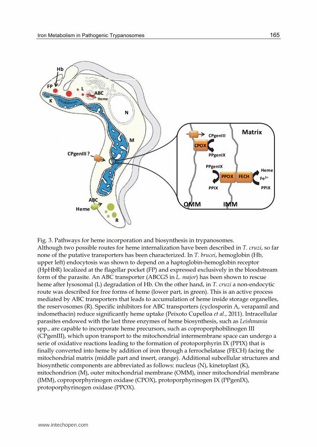
Heme
CPgenIII ?
Heme
Hb
L
N
K
M
FP
R
ABC
ABC
Matrix
CPOX
CPgenIII
PPgenIX
PPgenIX
PPIX
FECHPPOX
OMM IMM
PPIX
Heme
Fe2+
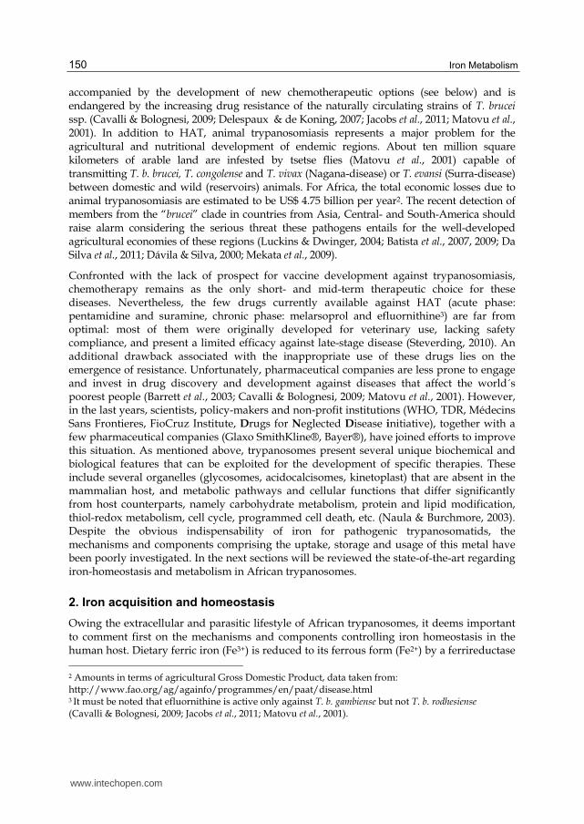
Fig. 3. Pathways for heme incorporation and biosynthesis in trypanosomes. Although two possible routes for heme internalization have been described in T. cruzi, so far none of the putative transporters has been characterized. In T. brucei, hemoglobin (Hb, upper left) endocytosis was shown to depend on a haptoglobin-hemoglobin receptor (HpHbR) localized at the flagellar pocket (FP) and expressed exclusively in the bloodstream form of the parasite. An ABC transporter (ABCG5 in L. major) has been shown to rescue heme after lysosomal (L) degradation of Hb. On the other hand, in T. cruzi a non-endocytic route was described for free forms of heme (lower part, in green). This is an active process mediated by ABC transporters that leads to accumulation of heme inside storage organelles, the reservosomes (R). Specific inhibitors for ABC transporters (cyclosporin A, verapamil and indomethacin) reduce significantly heme uptake (Peixoto Cupelloa et al., 2011). Intracellular parasites endowed with the last three enzymes of heme biosynthesis, such as Leishmania spp., are capable to incorporate heme precursors, such as coproporphobilinogen III (CPgenIII), which upon transport to the mitochondrial intermembrane space can undergo a serie of oxidative reactions leading to the formation of protoporphyrin IX (PPIX) that is finally converted into heme by addition of iron through a ferrochelatase (FECH) facing the mitochondrial matrix (middle part and insert, orange). Additional subcellular structures and biosynthetic components are abbreviated as follows: nucleus (N), kinetoplast (K), mitochondrion (M), outer mitochondrial membrane (OMM), inner mitochondrial membrane (IMM), coproporphyrinogen oxidase (CPOX), protoporphyrinogen IX (PPgenIX), protoporphyrinogen oxidase (PPOX).
www.intechopen.com
Iron Metabolism
166
culicis, which have been found to contain symbiotic bacteria that may supply the protists with heme and other essential nutrients (Chang & Trager, 1974). The bacterial symbiotes would be subject of regulation by their hosts, hence, functioning as cell organelles rather than as independent living entities (Chang et al., 1975).
Heme’s transport and distribution in trypanosomatids remain elusive. Diffusion across the lipid bilayer is hampered for heme due to its anionic carboxylate side-chains. Instead, high affinity heme-binding proteins in the cell surface of T. cruzi (Lara et al., 2007), procyclic forms of T. brucei (Vanhollebeke et al., 2008) and L. donovani (Campos-Salinas et al., 2011) allows for active heme uptake. In T. cruzi epimastigotes, both endocytotic and non-endocytotic mechanisms of heme internalization have been confirmed. L. donovani can also incorporate heme by receptor-mediated endocytosis followed by lysosomal degradation of hemoglobin. An ABC transporter localized in membranes of multivesicular structures is probably related to intracellular heme trafficking after hemoglobin degradation. Porphyrin trafficking in trypanosomatids is yet an uncovered subject but intracellular transport appears to involve membrane-bound vesicles as carriers (Severance & Hamza, 1982; Tripodi et al., 2011).
In summary, pathogenic trypanosomatids are auxotrophic for heme and have to adapt their heme-dependent metabolic pathways (i.e. biosynthesis of sterols and polyunsaturated fatty acids, respiration, oxidative stress response and detoxification) to fluctuations in nutrient availability across their life cycle. The most relevant cellular functions requiring heme in trypanosomatids are discussed in the next sections.
4.1 Heme in oxidative defense
Trypanosomes lack catalase and selenium-dependent glutathione peroxidases but instead express two types of trypanothione/tryparedoxin dependent peroxidases to decompose endogenous and exogenously produced oxidants (rewieved in Schlecker et al., 2007 and Krauth-Siegel & Comini, 2008) and some species have an additional plant-like ascorbate-dependent hemoperoxidase (APX; Adak & Datta, 2005; Wilkinson et al., 2002a). Interestingly, APX is present in intracellular trypanosomatids (T. cruzi and Leishmania spp.) but absent in parasites with an extracellular life style in the mammal host, namely T. brucei. This feature has been attributed to the major demand for anti-oxidant capacity that intracellular parasites need during infection (Wilkinson et al., 2005; Dolai et al., 2009). APX localizes to the endoplasmic reticulum (ER; Wilkinson et al., 2002a) and cell membrane (M. Hugo et al., personal communication) of T. cruzi and in the intermembrane space of the mitochondrial inner membrane in the case of L. major (Dolai et al., 2008). Both enzymes are reduced by ascorbate or, alternatively, cytochrome c (Wilkinson et al., 2002a; Dolai et al., 2008; M. Hugo et al., personal communication). The regeneration of reduced ascorbate occurs upon spontaneous reaction of dehydroascorbate with T(SH)2 (Krauth-Siegel & Lüdemann, 1996). With respect to the source of ascorbate, in contrast to humans, African and American trypanosomes can synthesize this metabolite de novo, a process that takes place in the glycosome (Wilkinson et al., 2005). T. cruzi cannot take up ascorbate from the environment, as T. brucei does, and therefore relies entirely on its biosynthesis to fuel other unknown cellular functions, in addition to oxidant detoxication (Logan et al., 2007).
The biological role of APX is consistently related to oxidant defense. Overexpression of APX in T. cruzi and L. major enhances tolerance against oxidative stress induced by exogenous
www.intechopen.com
Iron Metabolism in Pathogenic Trypanosomes
167
hydrogen peroxide (Wilkinson et al., 2002a and 2005) and mitochondrial cardiolipin oxidation, providing thus protection towards programmed cell death and protein damage (Dolai et al., 2008, 2009). This function appears to be redundant with that performed by the ER glutathione-like trypanothione-dependent peroxidase of T. cruzi (GPxII; Wilkinson et al., 2002a) and the mitochondrial trypanothione-dependent peroxiredoxin (mTXNPx) from Leishmania spp. However, an explanation for this may lie on different substrate specificities (e.g., GpxII reduces fatty acid and phospholipid hydroperoxides; Wilkinson et al., 2002b) or the macromolecular targets these enzymes have.
4.2 Heme in polyunsaturated fatty acids biogenesis
Fatty acid biosynthesis in trypanosomatids seems to be an essential pathway for the parasite life cycle. As stated earlier, differentiation involves dramatic morphological changes in the cell and its organelles that certainly require membrane fluidity. The biosynthesis of unsaturated fatty acids is another plant-like pathway inherited by trypanosomatids. In this respect, lipid desaturation occurs at the methyl end of the molecule, in contrast to mammals, in which a double bond is generated at the carboxy end of the molecule.
T. cruzi and both life stages of T. brucei are able to synthesize fatty acids, although T. brucei bloodstream form was formerly thought to depend on lipid uptake from their host. Short and medium chain fatty acids are usually synthesized by means of the soluble fatty-acid synthetase system and subsequently elongated by the elongase system that resides in the ER. T. brucei can elongate fatty acids up to stearate (C18:0). T. cruzi can further elongate C18 to C24 and C26 fatty acids required in the synthesis of anchors for surface macromolecules. The stearate is mainly converted into linoleate (C18:2), which represents near 30% and 40% of total fatty acids in T. brucei and T. cruzi, respectively. Moreover, unsaturated fatty acids can represent up to 70% of total fatty acid content depending on the parasite species and life cycle stage (Alloatti et al., 2010).
T. brucei oleate desaturase shares high similarity with ∆12 desaturases and, to a lesser extent, with ω3 desaturases although the parasite enzyme lacks ω3 desaturase activity. Fatty acid desaturases require an electron donor, that can be either ferredoxin in plastids and bacteria or cytochrome b5 (cytb5) in the ER, as it is the case for the orthologue from T. brucei (Uttaro, 2006). Since T. brucei oleate desaturase lacks a consensus sequence for the covalent binding of cytb, it has been suggested that electron donation occurs via complex formation with another desaturase capable to bind cytb5 (Petrini et al., 2004). The drastic growth defect observed upon partial ablation of enzyme activity (even an 8% reduction) by RNAi is a strong indication of the essentiality of this pathway for infective parasites (see also section 3.2.4; Alloatti et al., 2010)
4.3 Trypanosomes contain unusual single-cys cytochromes
Cytochromes are heme-containing complexes that participate in many electron transfer reactions including oxidative phosphorylation in the respiratory chain (cytochromes type c, cytc and cytochromes type c1, cytc1), desaturation of fatty acids or xenobiotic detoxification (Comini et al., 2011). In cytc heme is covalently attached through thioether bonds between the vinyl groups of the heme and the thiols of two cysteine residues in a conserved CXXCH motif, in which the histidine functions as an axial ligand for heme iron. Mitochondrial cyt c
www.intechopen.com
Iron Metabolism
168
is the best known example of this type of protein. It is located in the intermembrane space and transfers electrons from the cytochrome bc1 complex to the cytochrome aa3 oxidase. Cytochrome c1 of the bc1 complex (cytochrome reductase) is also a c-type cytochrome. Heme coordination by cytc and cytc1 from euglenoids (e.g. the green flagellated algae Euglena gracilis) and trypanosomatids occurs through a single thioether bond involving the single thiol group from their binding motifs AAQCH and FAPCH, respectively (Priest & Hajduk, 1994). As another example of a metabolic trait that trypanosomatids share with higher plants, plastids contain b6f-type cytochromes with heme covalently attached through a single thioether bond (Allen et al., 2004; Tripodi et al., 2011). The biogenesis of holo cytochrome c is a catalyzed process that involves different mechanisms and enzymatic components that can be grouped into five major systems (Tripodi et al., 2011). The cytochrome c maturation machinery employed by kinetoplatids remains unidentified and is possibly unique to this and the entire euglenid taxon (Allen et al., 2004; Tripodi et al., 2011). Based on the metabolic repertoire of each developmental stage, the relevance of cytochrome activity for energy production appears to be restricted to the procyclic form of the parasite.
4.4 Heme in sterol biogenesis
Cholesterol, ergosterol and sitosterol are essential structural components of plasma membranes from mammals, fungi and plants, respectively. They stabilize membranes, determine their fluidity and permeability, and modulate the activity of membrane-bound enzymes and ion channels. T. cruzi and T. brucei encode all sterol biosynthetic enzymes, including sterol 14-┙-demethylase (CYP51), a member of the CYP450 superfamily that operates at the initial phase (post-squalene stage) of the pathway that drives the oxidative removal of the 14-┙-methyl group from the newly cyclized sterol precursors. Azole-based compounds, common in antifungal therapies (e.g., posaconazole) proved to be potent inhibitors of CYP51 by interacting with the enzyme active site and its prosthetic group (Chen et al., 2010). Inhibition of CYP51 leads to accumulation of 14┙-methylated sterols, which are unable to replace ergosterol in the membrane because of steric hindrance, followed by growth arrest and cell death (Lepsheva et al., 2007; Vanden Bosschef et al., 1995). In contrast to T. cruzi and Leishmania spp., ergosterol has been thought to be dispensable for T. brucei. However, the observation that antifungals are also detrimental for this parasite lead to the proposal that sterol derivatives may additionally serve as precursors for bioactive molecules that act as regulators of cell cycle and development (Lepsheva et al., 2007).
Other CYP450s possess a monooxygenase activity that, upon oxygen activation, catalyzes the addition of an oxygen atom into an organic substrate and the reduction of the second one to water. After each catalytic cycle, CYPs must be restored to their reduced state by a CYP450 reductase (CPR), a NADPH-dependant diflavin (FAD/FMN) enzyme that couple a two-electron donor (NADPH) with one-electron acceptors, supplying the electrons one at a time. CYPs and CPR are involved in the metabolism of a wide range of endogenous compounds and xenobiotics including fatty acids, steroids, drugs, alkanes, polycyclic hydrocarbons, insecticides and other environmental contaminants (Murataliev et al., 2004). In mammals, there is only one CPR gene. However, T. cruzi and L. major encode three different CPR genes (CPR-A, CPR-B and CPR-C), while T. brucei possesses four sequences, all of them sharing a high sequence identity. Similar to some higher plant species
www.intechopen.com

Iron Metabolism in Pathogenic Trypanosomes
169
(Koopmann & Hahlbrock, 1997; Ro et al., 2002; Urban et al., 1997), T. cruzi CPRs are encoded by a small gene family, with differential regulation and subcellular localization (Portal et al., 2008). TcCPR-B is localized mainly in reservosomes, TcCPR-C is mostly found in the ER while TcCPR-A displays a more ubiquitous distribution pattern (De Vas et al., 2011). The presence of multiple isoforms argues for a well-supported P450-mediated molecular system. Portal et al. (2008) showed that CPR-B overexpression in T. cruzi confers increased resistance to nifurtimox and, more significantly, to benznidazole. In addition, increased levels of CPRs are observed in trypomastigotes, a stage that is particularly exposed to stress during cell invasion supporting a role for CPRs in oxidative stress defense (Portal et al., 2008). Moreover, CPR-B and CPR-C overexpression increases the synthesis of ergosterol, with the concomitant augmentation in membrane rigidity and decrease in endocytic activity. Modification in cell membrane fluidity could result in alteration of drug uptake and, as a consequence, in the observed drug resistance (De Vas et al., 2011).
5. Iron and infection
As noted in the introductory section, African trypanosomiasis is a parasitic disease of medical and veterinary importance that has adverse impact in the health and economical development of the sub-Saharian region of Africa (Maudlin, 2006; Stijlemans et al., 2008, 2010a). Since African trypanosomes multiply predominantly in the bloodstream as extra-cellular parasites, they are continuously dependent on host nutrient supply and exposed to host immune attack. As will be addressed next, the response mounted by the mammalian host towards infection includes the activation of both arms of the immune system.
Beyond the aforementioned clinical outcomes for HAT (Section 1), the most prominent immunopathological disease-related feature of trypanosomiasis is anemia. In fact, anemia is the major cause of death in bovine trypanosomiasis and an important debilitating symptom in HAT (Stijlemans et al., 2008). Anemia of inflammation, also termed anemia of chronic disease (ACD), is frequent among patients suffering chronic inflammatory disorders (e. g., autoimmune diseases) or chronic infections (Camaschella & Strati, 2010). ACD is the consequence of a persistent activated inflammatory immune response and is considered to be a defense mechanism where the host establishes an iron “withholding” strategy to limit the availability of essential iron for the pathogens (Lalonde & Holbein, 1984; Stijlemans et al., 2008; Sutak et al., 2008). Stijlemans and coworkers (Stijlemans et al., 2008, 2010a, 2010b; Vankrunkelsven et al., 2010) studied the immunological and hematological development of T. brucei infection using animal models (cattle and mice). Despite mouse models show limitations and artifacts, like the extremely high parasitemia levels reached compared to infection in the natural host, they have contributed significantly to our current understanding of trypanosomiasis patho-physiology (Magez & Caljon, 2011). The molecular mechanism underlying the development of ACD during trypanosomiasis resembles that observed in other infectious diseases. Briefly, during the early stage of the infection trypanosomes stimulate T-cells to secrete interferon-┛ resulting in the activation of macrophages (type I cellular immune response) that liberate pro-inflammatory mediators like tumor necrosis factor, interleukin (IL) -1, IL-6 and �NO (Stijlemans et al., 2010b). This response leads to parasite clearance but also enhances erythrophagocytosis by activated macrophages within the spleen and liver (Kupffer cells). At this point it is important to recall that under physiological conditions Kupffer cells recycle iron from engulfed senescent
www.intechopen.com
Iron Metabolism
170
erythrocytes (see Section 2; Hentze et al., 2010). Thus, enhanced removal of red blood cells from systemic fluids by the reticuloendothelial system will contribute to deprive host of iron. In addition, hepcidin (Section 2) is secreted in response to pro-inflammatory mediators, blocking iron release from macrophages and iron intake by enterocytes (Nishimura et al., 2011), which decreases plasma iron levels and limits host and pathogen accessibility to the metal (Taylor & Kelly, 2010). The upregulation of several iron-related molecules (e.g. hemooxygenase 1, ferritin and ferroportin) provides additional evidence for the strong type I immune response, augmented liver iron-metabolism and accelerated senescence of erythrocytes that takes place during trypanosomal infection (Nishimura et al., 2011; Omotainse & Anosa, 1992; Stijlemans et al., 2010a; see Section 2). The class of anemia originated during the acute phase of trypanosomiasis is a typical non-haematopoietic anemia. However, if at the long term the type I response persists, then iron will be sequestered intracellularly, resulting in a marked iron deprivation with compromise of erythropoiesis and development of ACD (Mwangi et al., 1995; Omotainse & Anosa, 1992; Stijlemans et al., 2010b). Despite its adverse effects, if sustained on time, limited iron bioavailability can be considered a reasonable host response to control parasitemia (Ganz, 2009). In the last years, several evidences point to the possibility that the parasite deliberately manipulates the mammalian host immune response. Activation of a strong type I immune response in early stages of the infection promotes erythrophagocytosis (Igbokwe et al., 1994), a strategy that yields abundant iron and lipids (e.g. myristic acid), the last needed for GPI biosynthesis and VSG coating (Stijlemans et al., 2010a, 2010b). It is important to stress that the trypanosome body is totally covered by a dense GPI-anchored coat of VSGs that plays an important protective (physical and immunological barrier) function in bloodstream parasites (Comini et al., 2011).
A large number of microbial pathogens have been reported to affect the morbidity and mortality of patients with high iron content, a physiopathological state characterized by the presence of non-transferrin bound iron in plasma. Moreover, iron overload due to genetic predisposition, therapeutic intervention or nutritional status is known to increase the risk of several infections (Kontoghiorghes et al., 2010). It is worth to note that human iron overload is common in rural populations inhabiting sub-Saharan regions of Africa, the “tsetse belt”, with a high incidence of HAT (Gordeuk, 2002; McNamara et al., 1999). Originally, this chronic condition was supposed to be an exclusive consequence of an iron-rich diet based on the large consumption of homebrewed beers, but recent studies of pedigrees suggest the presence of genetic components (Gordeuk, 2002; Moyo et al., 1998). It remains to be investigated whether there is a co-evolutionary development of iron overload and HAT, as it is the case for glucose-6-phosphate dehydrogenase deficiency and malaria.
Considering the well-known role of iron in T. brucei infection and the fact that in vitro iron deprivation with chelating agents cause growth retardation of bloodstream T. brucei (Fast et
al., 1999; Breidbach et al., 2002; Comini et al., 2008), it is surprising the absence of works studying the therapeutic potential of iron chelation against T. brucei (Tayor & Kelly, 2010). This is not the case for T. cruzi, for which the chelating agent deferoxamine (DFX) proved its efficacy to halt replication of intra-macrophage amastigotes (Arantes et al., 2011; Lalonde & Holbein, 1984; Jones et al., 1996), and to decrease parasitemia and mortality of infected mice (Loo & Lalonde, 1984). However, in contrast to extracellular T. brucei, the possible success of chelating therapies against T. cruzi infection are hampered by the intracellular nature of the
www.intechopen.com
Iron Metabolism in Pathogenic Trypanosomes
171
infective stage of this parasite, which, if not specifically targeted to the pathogen, will imply a high dosage or a long-term treatment, both deleterious for the host. Based on the demonstration that iron metabolism is crucial for parasite survival in the host (Manta et al., unpublished; see section 3.3.2), our group has recently evaluated the therapeutic effect of intraperitoneal (i.p.) and oral administration of DFX to T. brucei infected mice. Preliminary results show a marked increase in life span of the animal group receiving i.p. treatment (200 mg/Kg for ~10 days) and almost no side effects (red blood cell and reticulocyte counts) on animals treated but non-infected (Manta et al., unpublished). However, DFX displayed a cytostatic effect against acute infection by T. brucei, which implies that a successful therapeutic regimen will require drug association.
6. Summary and outlook
As any other living organism on earth, trypanosomes depend on iron to fulfill critical cellular functions (DNA and lipid biosynthesis, oxidant protection and energy generation). Their complex life cycle has added an additional problem in the struggle for the supply of (micro)nutrients. However, these organisms have developed different strategies and mechanisms in order to ensure a physiological iron status within the distinct hosts. Moreover, host-parasite co-evolution has resulted in the development of well-balanced growth regulation systems, allowing the parasite to survive sufficiently long without killing its mammalian host and ensuring an efficient transmission cycle. Interruption of this cycle is necessary to abolish the devastating disease these parasites produce to humans and livestock. In this respect, the iron metabolism of trypanosomatids should be considered as a promising target for effective antiparasitic treatments. Although metal-chelating compounds are, obviously, not innocuous to the host, in general they are less toxic than the drugs actually used for the treatment of HAT and their side effect is fully reversible upon dietary supplementation. In addition, several chelating agents are approved for the treatment of human pathologies, some of them are relatively cheap and available for oral administration (Sharma & Pancholi, 2010). The risk/benefit assessment for the use of iron chelating drugs as antiparasitic therapeutic depends on many factors not addressed yet.
Moreover, beyond the relevance of iron homeostasis for parasite survival, some components of this metabolism and/or iron-, Fe/S cluster- or heme-proteins offer excellent opportunities for the design of more specific and efficacious drugs. For instance, the enzymes involved in fatty acids biogenesis (oleate desaturase and stearoyl-CoA-desaturase) and sterol biosynthesis (CYP51); in defense against oxidants (the plant-like ascorbate peroxidase) and xenobiotics (CYP450 reductases); in cell proliferation (ribonucleotide reductase); and in Fe/S cluster biogenesis. Alternatively, chemical downregulation of the functions these proteins fulfill in the cell can be complemented by the action of first line drugs currently used in chemotherapy. A combined therapy would have the advantage of reducing dosage, shortening treatment and loweing the possibility for development of drug resistance. The specificity of the transferrin receptor is another target to be exploited as an efficient mechanism to deliver toxic molecules inside the parasite
The future prospects look promising in terms of finding novel therapeutic options to fight trypanosomiasis. However as depicted in Fig. 2 and Fig. 3, there are still several open questions that deserve to be investigated in detail to disclose atomic and functional divergences within the components of the parasite and host iron metabolism that can lead to the development of safer and efficacious drugs.
www.intechopen.com
Iron Metabolism
172
7. References
Abreu IA & Cabelli DE. (2010) Superoxide dismutases-a review of the metal-associated mechanistic variations. Biochim. Biophys. Acta. Vol. 1804, No. 2, pp. 263–274.
Acestor N, Panigrahi AK, Ogata Y, Anupama A & Stuart KD. (2009) Protein composition of Trypanosoma brucei mitochondrial membranes. Proteomics. Vol. 9, No. 24, pp. 5497–5508.
Adak S & Datta AK. (2005) Leishmania major encodes an unusual peroxidase that is a close homologue of plant ascorbate peroxidase: a novel role of the transmembrane domain. Biochem. J. Vol. 390, pp. 465-474.
Adam AC, Bornhövd C, Prokisch H, Neupert W & Hell K. (2006) The Nfs1 interacting protein Isd11 has an essential role in Fe/S cluster biogenesis in mitochondria. EMBO J. Vol. 25, No. 1, pp. 174–183.
Adl SM, Simpson AGB, Farmer MA, Andersen RA, Anderson OR, Barta JR, Bowser SS, Brugerolle G, Fensome RA, Fredericq S, James TY, Karpov S, Kugrens P, Krug J, Lane CE, Lewis LA, Lodge J, Lynn DH, Mann DG, McCourt RM, Mendoza L, Moestrup O, Mozley-Standridge SE, Nerad TA, Shearer CA, Smirnov AV, Spiegel FW & Taylor MF. (2005) The new higher level classification of eukaryotes with emphasis on the taxonomy of protists. J. Eukaryot. Microbiol. Vol. 52, No. 5, pp. 399–451.
Adl SM, Leander BS, Simpson AGB, Archibald JM, Anderson OR, Bass D, Bowser SS, Brugerolle G, Farmer MA, Karpov S, Kolisko M, Lane CE, Lodge DJ, Mann DG, Meisterfeld R, Mendoza L, Moestrup Ø, Mozley-Standridge SE, Smirnov AV & Spiegel F. (2007) Diversity, nomenclature, and taxonomy of protists. Syst. Biol. Vol. 56, No 4, pp. 684–689.
Aeby E, Seidel V & Schneider A. (2009) The selenoproteome is dispensable in bloodstream forms of Trypanosoma brucei. Mol. Biochem. Parasitol. Vol. 168, No. 2, pp. 191–193.
Ajayi WU, Chaudhuri M & Hill GC. (2002) Site-directed mutagenesis reveals the essentiality of the conserved residues in the putative diiron active site of the trypanosome alternative oxidase. J. Biol. Chem. Vol. 277, No. 10, pp. 8187–8193.
Alfonzo JD & Lukeš J. (2011) Assembling Fe/S-clusters and modifying tRNAs: ancient co-factors meet ancient adaptors. Trends Parasitol. Vol. 27, No. 6, pp. 235–238.
Allen JWA, Gringer ML & Ferguson SJ. (2004) Maturation of the unusual single-cysteine (XXXCH) mitochondrial c-type cytochromes found in trypanosomatids must occur through a novel biogenesis pathway. Biochem. J. Vol. 383, pp. 537-542.
Alloatti A, Gupta S, Gualdrón-López M, Igoillo-Esteve M, Nguewa PA, Deumer G, Wallemacq P, Altabe SG, Michels PAM & Uttaro AD. (2010) Genetic and chemical evaluation of Trypanosoma brucei oleate desaturase as a candidate drug target. PLoS ONE Vol. 5, No. 12, pp. 1-10, e14239
Alloatti A, Gupta S, Gualdrón-López M, Nguewa PA, Altabe SG, Deumer G, Wallemacq P, Michels PA & Uttaro AD. (2011) Stearoyl-CoA desaturase is an essential enzyme for the parasitic protist Trypanosoma brucei. Biochem. Biophys. Res. Commun. Vol. 412, No. 2, pp. 286-290.
Alvarez MN, Peluffo G, Piacenza L & Radi R. (2011) Intraphagosomal peroxynitrite as a macrophage-derived cytotoxin against internalized Trypanosoma cruzi: consequences for oxidative killing and role of microbial peroxiredoxins in infectivity. J. Biol. Chem. Vol. 286, No. 8, pp. 6627–6640.
www.intechopen.com
Iron Metabolism in Pathogenic Trypanosomes
173
Amutha B, Gordon DM, Gu Y, Lyver ER, Dancis A & Pain D. (2008) GTP is required for iron-sulfur cluster biogenesis in mitochondria. J. Biol. Chem. Vol. 283, No. 3, pp. 1362–1371.
Anderson GJ & Vulpe CD. (2009) Mammalian iron transport. Cell. Mol. Life Sci. Vol. 66, No. 20, pp. 3241–3261.
Andrews SC. (2010) The Ferritin-like superfamily: Evolution of the biological iron storeman from a rubrerythrin-like ancestor. Biochim. Biophys. Acta. Vol. 1800, No. 8, pp. 691–705.
Arantes JM, Francisco AF, de Abreu Vieira PM, Silva M, Araújo MSS, de Carvalho AT, Pedrosa ML, Carneiro CM, Tafuri WL, Martins-Filho AO & Elói-Santos SM. (2011) Trypanosoma cruzi: desferrioxamine decreases mortality and parasitemia in infected mice through a trypanostatic effect. Exp. Parasitol. Vol. 128, No. 4, pp. 401–408.
Atanasiu V, Manolescu B & Stoian I. (2007) Hepcidin: central regulator of iron metabolism. Eur. J. Haematol. Vol. 78, No. 1, pp. 1–10.
Bachega JFR, Navarro MVAS, Bleicher L, Bortoleto-Bugs RK, Dive D, Hoffmann P, Viscogliosi E & Garratt RC. (2009) Systematic structural studies of iron superoxide dismutases from human parasites and a statistical coupling analysis of metal binding specificity. Proteins. Vol. 77, No. 1, pp. 26–37.
Balk J & Lobréaux S. (2005) Biogenesis of iron-sulfur proteins in plants. Trends Plant Sci. Vol. 10, No. 7, pp. 324–331.
Balk J & Pilon M. (2011) Ancient and essential: the assembly of iron-sulfur clusters in plants. Trends Plant Sci. Vol. 16, No. 4, pp. 218–226.
Balmer O, Beadell JS, Gibson W, & Caccone, A. (2011). Phylogeography and taxonomy of Trypanosoma brucei. PLoS Negl. Trop. Dis. Vol. 5, e961.
Barrett MP, Burchmore RJS, Stich A, Lazzari JO, Frasch AC, Cazzulo JJ, & Krishna S. (2003) The trypanosomiases. Lancet. Vol. 362, No. 9394, pp. 1469–1480.
Batista JS, Riet-Correa F, Teixeira MMG, Madruga CR, Simões SDV & Maia TF. (2007) Trypanosomiasis by Trypanosoma vivax in cattle in the Brazilian semiarid: Description of an outbreak and lesions in the nervous system. Vet. Parasitol. Vol. 143, No. 2, pp.174–181.
Batista JS, Oliveira AF, Rodrigues CMF, Damasceno CAR, Oliveira IRS, Alves HM, Paiva ES, Brito PD, Medeiros JM, Rodrigues AC & Teixeira MM. (2009) Infection by Trypanosoma vivax in goats and sheep in the Brazilian semiarid region: from acute disease outbreak to chronic cryptic infection. Vet. Parasitol. Vol. 165, No. 1-2, pp.131–135.
Berriman, M., Ghedin, E., Hertz-Fowler, C., Blandin, G., Lennard, N.J., Bartholomeu, D., Renauld, H.J., Caler, E., Hamlin, N., Haas, B., Harris, B.R., Hannick, L., Barrell, B., Donelson, J., Hall, N., Fraser, C.M., Melville, S.E., El-Sayed, N., Böhme, U.C., Shallom, J., Aslett, M., Hou, L., Atkin, B., Barron, A.J., Bringaud, F., Brooks, K., Cherevach, I., Chillingworth, T., Churcher, C., Clark, L.N., Corton, C.H., Cronin, A., Davies, R., Doggett, J., Djikeng, A., Feldblyum, T., Fraser, A., Goodhead, I., Hance, Z., Harper, A.D., Hauser, H., Hostetler, J., Jagels, K., Johnson, D., Johnson, J., Jones, C., Kerhornou, A., Koo, H., Larke, N., Larkin, C., Leech, V., Line, A., MacLeod, A., Mooney, P., Moule, S., Mungall, K., Norbertczak, H., Ormond, D., Pai, G., Peterson, J., Quail, M.A., Rajandream, M.A., Reitter, C., Sanders, M., Schobel, S., Sharp, S., Simmonds, M., Simpson, A.J., Tallon, L., Turner, C.M., Tait, A., Tivey, A., Van Aken, S., Walker, D., Wanless, D., White, B., White, O., Whitehead, S., Wortman, J., Barry, J.D., Fairlamb, A.H., Field, M.C., Gull, K., Landfear, S., Marcello, L., Martin, D.M., Opperdoes, F., Ullu, E., Whickstead, B.,
www.intechopen.com

Iron Metabolism
174
Alsmark, C., Arrowsmith, C., Carrington, M., Embley, T.M., Ivens, A., Lord, A., Morgan, G.M., Peacock, C.S., Rabbinowitsch, E., Salzberg, S., Wang, S., Woodward, J., Adams, M.D., Embley, T.M., Gull, K., Ullu, E., Barry, J.D., Fairlamb, A.H., Opperdoes, F., Barrell, B.G., Donelson, J.E., Hall, N., Fraser, C.M., Melville, S.E. & El-Sayed, N.M. (2005). The genome of the African trypanosome, Trypanosoma brucei. Science. Vol. 309, No. 5733, pp. 416-422.
Birkholtz L-M, Williams M, Niemand J, Louw AI, Persson L & Heby O. (2011) Polyamine homoeostasis as a drug target in pathogenic protozoa: peculiarities and possibilities. Biochem. J. Vol. 438, No. 2, pp. 229–244.
Bitter W, Gerrits H, Kieft R & Borst P. (1998) The role of transferrinreceptor variation in the host range of Trypanosoma brucei. Nature. Vol. 391, No. 6666 pp. 499-502.
Bocedi A, Dawood KF, Fabrini R, Federici G, Gradoni L, Pedersen JZ & Ricci G. (2010) Trypanothione efficiently intercepts nitric oxide as a harmless iron complex in trypanosomatid parasites. FASEB J. Vol. 24, No. 4, pp. 1035–1042.
Bosworth CA, Toledo JC Jr, Zmijewski JW, Li Q & Lancaster JR Jr. (2009) Dinitrosyliron complexes and the mechanism(s) of cellular protein nitrosothiol formation from nitric oxide. Proc. Natl. Acad. Sci. USA. Vol. 106, No. 12, pp. 4671-4676.
Braz, GRC, Coelho HSL, Masuda H & Oliveira PL. (1999) A missing metabolic pathway in the cattle tick Boophilus microplus. Curr. Biol. Vol. 9, No. 13, pp. 703-706.
Breidbach T, Krauth-Siegel RL & Steverding D. (2000) Ribonucleotide reductase is regulated via the R2 subunit during the life cycle of Trypanosoma brucei. FEBS Lett. Vol. 473, No. 2, pp. 212–216.
Breidbach T, Scory S, Krauth-Siegel RL & Steverding D. (2002) Growth inhibition of bloodstream forms of Trypanosoma brucei by the iron chelator deferoxamine. Int. J. Parasitol. Vol 32, No. 4, pp. 473–479.
Bruske EI, Sendfeld F & Schneider A. (2009) Thiolated tRNAs of Trypanosoma brucei are imported into mitochondria and dethiolated after import. J. Biol. Chem. Vol. 284, No. 52, pp. 36491–36499.
Camaschella C & Strati P. (2010) Recent advances in iron metabolism and related disorders Intern. Emerg. Med. Vol. 5, No. 5, pp. 393–400.
Campos-Salinas J, Cabello-Donayre M, García-Hernández R, Pérez-Victoria I, Castanys S, Gamarro F & Pérez-Victoria JM. (2011) A new ATP-binding cassette protein is involved in intracellular haem trafficking in Leishmania. Molec. Microbiol. Vol. 79, No. 6, pp. 1430-1444.
Cavalli A & Bolognesi ML. (2009) Neglected tropical diseases: multi-target-directed ligands in the search for novel lead candidates against Trypanosoma and Leishmania. J. Med. Chem. Vol. 52, No. 23, pp. 7339–7359.
Cazzulo JJ. (1992) Aerobic fermentation of glucose by trypanosomatids. FASEB J. Vol. 6, pp- 3153-3161.
Ceylan S, Seidel V, Ziebart N, Berndt C, Dirdjaja N & Krauth-Siegel RL. (2010) The dithiol glutaredoxins of african trypanosomes have distinct roles and are closely linked to the unique trypanothione metabolism. J. Biol. Chem. Vol. 285, No. 45, pp. 35224–35237.
Chang KP & Trager W. (1974) Nutritional Significance of Symbiotic Bacteria in Two Species of Hemoflagellates. Science Vol. 183, pp. 531-532.
Chang KP, Chang CS & Sassa S. (1975) Heme biosynthesis in bacterium-protozoon symbioses: enzymic defects in host hemoflagellates and complemental role of their intracellular symbiotes. Proc. Nat. Acad. Sci. Vol. 72, No. 8, pp. 2979-2983.
www.intechopen.com
Iron Metabolism in Pathogenic Trypanosomes
175
Chaudhuri M, Ajayi W & Hill GC. (1998) Biochemical and molecular properties of the Trypanosoma brucei alternative oxidase. Mol. Biochem. Parasitol. Vol. 95, No. 1, pp. 53–68.
Chaudhuri M, Sharan R & Hill GC. (2002) Trypanosome alternative oxidase is regulated post-transcriptionally at the level of RNA stability. J. Eukaryot. Microbiol. Vol. 49, No. 4, pp. 263–269.
Chaudhuri M, Ott RD & Hill GC. (2006) Trypanosome alternative oxidase: from molecule to function. Trends Parasitol. Vol. 22, No. 10, pp. 484–491.
Chisi JE, Misiri H, Zverev Y, Nkhoma A & Sternberg JM (2004). Anaemia in human African trypanosomiasis caused by Trypanosoma brucei rhodesiense. East Afr. Med. J. Vol. 81, No. 10, pp. 505-508.
Chen CK, Leung SSF, Guilbert C, Jacobson MP, Mckerrow JH & Podust LM. (2010) Structural characterization of CYP51 from Trypanosoma cruzi and Trypanosoma brucei bound to the antifungal drugs posaconazole and fluconazole. PLoS Neglected Tropical Diseases, 4(4): e651.
Clarkson AB Jr & Brohn FH. (1976) Trypanosomiasis: an approach to chemotherapy by the inhibition of carbohydrate catabolism. Science. Vol. 194, No. 4261, pp. 204–206.
Clarkson AB Jr, Bienen EJ, Pollakis G & Grady RW. (1989) Respiration of bloodstream forms of the parasite Trypanosoma brucei brucei is dependent on a plant-like alternative oxidase. J. Biol. Chem. Vol. 264, No. 30, pp. 17770–17776.
Colombo FA, Odorizzi RM, Laurenti MD, Galati EA, Canavez F & Pereira-Chioccola VL. (2011) Detection of Leishmania (Leishmania) infantum RNA in fleas and tickscollected from naturally infected dogs. Parasitol. Res. Vol. 109, No. 2, pp. 267-274.
Comini MA, Rettig J, Dirdjaja N, Hanschmann E-M, Berndt C & Krauth-Siegel RL. (2008) Monothiol glutaredoxin-1 is an essential iron-sulfur protein in the mitochondrion of African trypanosomes. J. Biol. Chem. Vol. 283, No. 41, pp. 27785–27798.
Comini MA, Medeiros AM & Manta B. (2011) Stress response in the infective stage of Trypanosoma brucei. In: Stress response in microbiology. Ed. José M. Requena. Horizon Scientific Press, Norwich, UK. In press
Coustou V, Biran M, Besteiro S, Rivière L, Baltz T, Franconi JM & Bringaud F. (2006) Fumarate is an essential intermediary metabolite produced by the procyclic Trypanosoma brucei. J. Biol. Chem. Vol. 281, No. 37, pp. 26832-26846.
Cotruvo JA & Stubbe J. (2011) Class I ribonucleotide reductases: metallocofactor assembly and repair in vitro and in vivo. Annu. Rev. Biochem. Vol. 80, pp. 733–767.
Dantas-Torres F. (2011) Ticks as vectors of Leishmania parasites. Trends Parasitol. Vol. 27, No. 4, pp.155-159.
Da Silva AS, Garcia Perez HA, Costa MM, França RT, De Gasperi D, Zanette RA, Amado JA, Lopes ST, Teixeira MM & Monteiro SG. (2011) Horses naturally infected by Trypanosoma vivax in southern Brazil. Parasitol. Res. Vol. 108, No. 1, pp. 23–30.
Dávila AM & Silva RA. (2000) Animal trypanosomiasis in South America. Current status, partnership, and information technology. Ann. N. Y. Acad. Sci. Vol. 2000, No. 916, pp. 199–212.
De Vas MG, Portal P, Alonso GD, Schleinger M, Flawiá MM, Torres HN, Fenrández Villamil S & Paveto C. (2011) The NADPH–cytochrome P450 reductase family in Trypanosoma cruzi is involved in the sterol biosynthesis pathway. Int. J. Parasitol. Vol. 41, pp. 99-108.
Delespaux V & de Koning HP. (2007) Drugs and drug resistance in African trypanosomiasis. Drug Resist. Updat. Vol 10, No. 1-2, pp. 30–50.
www.intechopen.com
Iron Metabolism
176
Dolai S, Yadav RK, Pal S & Adak S. (2008) Leishmania major ascorbate peroxidase overexpression protects cells against reactive oxigen species-mediated cardiolipin oxidation. Free Rad. Biol. Med. Vol. 45, pp. 1520-1529.
Dolai S., Yadav R. K., Pal S. & Adak S. (2009) Overexpression of mitochondrial Leishmania major ascorbate peroxidase enhances tolerance to oxidative stress-induced programmed cell death and protein damage. Eukaryot. Cell. Vol. 8. No. 11, pp. 1721-1731.
Dormeyer M, Schöneck R, Dittmar GA & Krauth-Siegel RL. (1997) Cloning, sequencing and expression of ribonucleotide reductase R2 from Trypanosoma brucei. FEBS Lett. Vol. 414, No. 2, pp. 449–453.
Dormeyer M, Reckenfelderbäumer N, Ludemann H & Krauth-Siegel RL. (2001) Trypanothione-dependent synthesis of deoxyribonucleotides by Trypanosoma brucei ribonucleotide reductase. J. Biol. Chem. Vol. 276, No. 14, pp. 10602–10606.
Dufernez F, Yernaux C, Gerbod D, Noël C, Chauvenet M, Wintjens R, Edgcomb VP, Capron M, Opperdoes FR & Viscogliosi E. (2006) The presence of four iron-containing superoxide dismutase isozymes in trypanosomatidae: characterization, subcellular localization, and phylogenetic origin in Trypanosoma brucei. Free Radic. Biol. Med. Vol. 40, No. 2, pp. 210–225.
El-Sayed NM, Myler PJ, Bartholomeu DC, Nilsson D, Aggarwal G, Tran AN, Ghedin E, Worthey EA, Delcher AL, Blandin G, Westenberger SJ, Caler E, Cerqueira GC, Branche C, Haas B, Anupama A, Arner E, Aslund L, Attipoe P, Bontempi E, Bringaud F, Burton P, Cadag E, Campbell DA, Carrington M, Crabtree J, Darban H, da Silveira JF, de Jong P, Edwards K, Englund PT, Fazelina G, Feldblyum T, Ferella M, Frasch AC, Gull K, Horn D, Hou L, Huang Y, Kindlund E, Klingbeil M, Kluge S, Koo H, Lacerda D, Levin MJ, Lorenzi H, Louie T, Machado CR, McCulloch R, McKenna A, Mizuno Y, Mottram JC, Nelson S, Ochaya S, Osoegawa K, Pai G, Parsons M, Pentony M, Pettersson U, Pop M, Ramirez JL, Rinta J, Robertson L, Salzberg SL, Sanchez DO, Seyler A, Sharma R, Shetty J, Simpson AJ, Sisk E, Tammi MT, Tarleton R, Teixeira S, Van Aken S, Vogt C, Ward PN, Wickstead B, Wortman J, White O, Fraser CM, Stuart KD & Andersson B. (2005) The genome sequence of Trypanosoma cruzi, etiologic agent of Chagas disease. Science. Vol. 309, No. 5733, pp. 409-415.
Fast B, Kremp K, Boshart M & Steverding D. (1999) Iron-dependent regulation of transferrin receptor expression in Trypanosoma brucei. Biochem. J. Vol. 342, Pt 3, pp. 691–696.
Fernandes AP, Fladvad M, Berndt C, Andrésen C, Lillig CH, Neubauer P, Sunnerhagen M, Holmgren A & Vlamis-Gardikas A. (2005) A novel monothiol glutaredoxin (Grx4) from Escherichia coli can serve as a substrate for thioredoxin reductase. J. Biol. Chem. Vol. 280, No. 26, pp. 24544–24552.
Fèvre EM, Picozzi K, Jannin J, Welburn SC & Maudlin I. (2006) Human African trypanosomiasis: Epidemiology and control. Adv. Parasitol. Vol. 61, pp. 167–221.
Filser M, Comini MA, Molina-Navarro MM, Dirdjaja N, Herrero E & Krauth-Siegel RL. (2008) Cloning, functional analysis, and mitochondrial localization of Trypanosoma brucei monothiol glutaredoxin-1. Biol. Chem. Vol. 389, No. 1, pp. 21–32.
Ganz T. (2009) Iron in innate immunity: starve the invaders. Curr. Opin. Immunol. Vol. 21, No. 1, pp. 63–67.
Gerrits H, Mussmann R, Bitter W, Kieft R & Borst P. (2002). The physiological significance of transferrin receptor variations in Trypanosoma brucei. Mol. Biochem. Parasitol. Vol. 119, No. 2, pp. 237-247.
www.intechopen.com
Iron Metabolism in Pathogenic Trypanosomes
177
Girard M, Giraud S, Courtioux B, Jauberteau-Marchan M-O & Bouteille B.(2005) Endothelial cell activation in the presence of African trypanosomes. Mol. Biochem. Parasitol. Vol. 139, No. 1, pp. 41–49.
Gordeuk VR. (2002) African iron overload. Semin. Hematol. Vol. 39, No. 4, pp. 263–269. Ghosh S, Goswami S & Adhya S. (2003) Role of superoxide dismutase in survival of
Leishmania within the macrophage. Biochem. J. Vol. 369, Pt. 3, pp. 447–452. Grab DJ, Wells CW, Shaw MK, Webster P & Russo DC. (1992). Endocytosed transferrin in
African trypanosomes is delivered to lysosomes and may not be recycled. Eur. J. Cell Biol. Vol. 59, No. 2, pp. 398-404.
Grab DJ, Shaw MK, Wells CW, Verjee Y, Russo DC, Webster P, Naessens J, & Fish, W.R. (1993). The transferrin receptor in African trypanosomes: identification, partial characterization and subcellular localization. Eur. J. Cell Biol. Vol 62, No. 1, pp. 114-126.
Grant PT & Sargent JR. (1960) Properties of L-alpha-glycerophosphate oxidase and its role in the respiration of Trypanosoma rhodesiense. Biochem. J. Vol. 76, pp. 229–237.
Halliwel B & Gutteridge J. (1999) Free Radical in Biology and Medicine. Oxford University Press.ISBN 978-0198500445.
Hamilton PB, Stevens JR, Gaunt MW, Gidley J & Gibson WC. (2004). Trypanosomes are monophyletic: evidence from genes for glyceraldehyde phosphate dehydrogenase and small subunit ribosomal RNA. Int. J. Parasitol. Vol. 34, No. 12, pp. 1393-1404.
Hamilton PB, Gibson WC & Stevens JR (2007). Patterns of co-evolution between trypanosomes and their hosts deduced from ribosomal RNA and protein-coding gene phylogenies. Mol. Phylogenetics Evol. Vol. 44, No. 1, pp.15–25.
Hentze MW, Muckenthaler MU, Galy B & Camaschella C. (2010) Two to tango: regulation of Mammalian iron metabolism. Cell. Vol 142, No. 1, pp. 24–38.
Herrera L & Urdaneta-Morales S. (2001) Experimental transmission of Trypanosoma cruzi through the genitalia of albino mice. Mem Inst Oswaldo Cruz. Vol. 96, No. 5, pp. 713-717.
Hider RC & Kong XL. (2011) Glutathione: a key component of the cytoplasmic labile iron pool. Biometals. DOI: 10.1007/s10534-011-9476-8
Hofer A, Schmidt PP, Gräslund A & Thelander L. (1997) Cloning and characterization of the R1 and R2 subunits of ribonucleotide reductase from Trypanosoma brucei. Proc. Natl. Acad. Sci. U.S.A. Vol. 94, No. 13, pp. 6959–6964.
Hofer A, Ekanem JT & Thelander L. (1998) Allosteric regulation of Trypanosoma brucei ribonucleotide reductase studied in vitro and in vivo. J. Biol. Chem. Vol. 273, No. 51, pp. 34098–34104.
Horváth A, Horáková E, Dunajcíková P, Verner Z, Pravdová E, Slapetová I, Cuninková L & Lukes J. (2005) Downregulation of the nuclear-encoded subunits of the complexes III and IV disrupts their respective complexes but not complex I in procyclic Trypanosoma brucei. Mol. Microbiol. Vol. 58, No. 1, pp. 116-130.
Igbokwe IO, Esievo KA, Saror DI & Obagaiye OK. (1994) Increased susceptibility of erythrocytes to in vitro peroxidation in acute Trypanosoma brucei infection of mice. Vet. Parasitol. Vol. 55, No. 4, pp. 279–286.
Irigoín F, Cibils L, Comini MA, Wilkinson SR, Flohé L & Radi R.(2008) Insights into the redox biology of Trypanosoma cruzi: Trypanothione metabolism and oxidant detoxification. Free Radic. Biol. Med. Vol. 45, No. 6, pp. 733–742.
Ivens AC, Peacock CS, Worthey EA, Murphy L, Aggarwal G, Berriman M, Sisk E, Rajandream MA, Adlem E, Aert R, Anupama A, Apostolou Z, Attipoe P, Bason N, Bauser C, Beck A, Beverley SM, Bianchettin G, Borzym K, Bothe G, Bruschi CV,
www.intechopen.com
Iron Metabolism
178
Collins M, Cadag E, Ciarloni L, Clayton C, Coulson RM, Cronin A, Cruz AK, Davies RM, De Gaudenzi J, Dobson DE, Duesterhoeft A, Fazelina G, Fosker N, Frasch AC, Fraser A, Fuchs M, Gabel C, Goble A, Goffeau A, Harris D, Hertz-Fowler C, Hilbert H, Horn D, Huang Y, Klages S, Knights A, Kube M, Larke N, Litvin L, Lord A, Louie T, Marra M, Masuy D, Matthews K, Michaeli S, Mottram JC, Müller-Auer S, Munden H, Nelson S, Norbertczak H, Oliver K, O'neil S, Pentony M, Pohl TM, Price C, Purnelle B, Quail MA, Rabbinowitsch E, Reinhardt R, Rieger M, Rinta J, Robben J, Robertson L, Ruiz JC, Rutter S, Saunders D, Schäfer M, Schein J, Schwartz DC, Seeger K, Seyler A, Sharp S, Shin H, Sivam D, Squares R, Squares S, Tosato V, Vogt C, Volckaert G, Wambutt R, Warren T, Wedler H, Woodward J, Zhou S, Zimmermann W, Smith DF, Blackwell JM, Stuart KD, Barrell B &, Myler PJ. (2005) The genome of the kinetoplastid parasite, Leishmania major. Science Vol. 309, No. 5733, pp.:436-42.
Jacobs RT, Nare B & Phillips MA. (2011) State of the art in African trypanosome drug discovery. Curr Top Med Chem. Vol. 11, No. 10, pp. 1255–1274.
Jones MM, Singh PK, Lane JE, Rodrigues RR, Nesset A, Suarez CC, Bogitsh BJ & Carter CE. (1996) Inhibition of Trypanosoma cruzi epimastigotes in vitro by iron chelating agents. Arzneimittelforschung. Vol. 46, No. 12, pp. 1158–1162.
Kakhlon O & Cabantchik ZI. (2002) The labile iron pool: characterization, measurement, and participation in cellular processes. Free Radic. Biol. Med. Vol. 33, No. 8, pp. 1037–1046.
Kabiri M & Steverding D. (2000). Studies on the recycling of the transferrin receptor in Trypanosoma brucei using an inducible gene expression system. Eur. J. Biochem. Vol. 267, No. 11, pp. 3309-3314.
Kabiri M & Steverding D. (2001) Identification of a developmentally regulated iron superoxide dismutase of Trypanosoma brucei. Biochem. J. Vol. 360, Pt 1, pp. 173–177.
Kido Y, Shiba T, Inaoka DK, Sakamoto K, Nara T, Aoki T, Honma T, Tanaka A, Inoue M, Matsuoka S, Moore A, Harada S & Kita K. (2010a) Crystallization and preliminary crystallographic analysis of cyanide-insensitive alternative oxidase from Trypanosoma brucei brucei. Acta Crystallogr. Sect. F Struct. Biol. Cryst. Commun. Vol. 66, Pt 3, pp. 275–278.
Kido Y, Sakamoto K, Nakamura K, Harada M, Suzuki T, Yabu Y, Saimoto H, Yamakura F, Ohmori D, Moore A, Harada S & Kita K. (2010b) Purification and kinetic characterization of recombinant alternative oxidase from Trypanosoma brucei brucei. Biochim. Biophys. Acta. Vol. 1797, No. 4, pp. 443–450.
Kontoghiorghes GJ, Kolnagou A, Skiada A & Petrikkos G. (2010) The role of iron and chelators on infections in iron overload and non iron loaded conditions: prospects for the design of new antimicrobial therapies. Hemoglobin. Vol. 34, No. 3, pp. 227–239.
Koopmann E & Hahlbrock K. (1997) Differentially regulated NADPH:cytochrome P450 oxidoreductases in parsley. Proc. Natl. Acad. Sci. Vol. 94, pp. 14954-14959.
Korený L, Lukes, J & Oborník M. (2010) Evolution of the haem synthetic pathway in kinetoplastid flagellates: an essential pathway that is not essential after all? International Journal for Parasitol. Vol. 40, pp. 149-156.
Kosman DJ. (2010) Redox cycling in iron uptake, efflux, and trafficking. J. Biol. Chem. Vol. 285, No. 35, pp. 26729–26735.
Krauth-Siegel RL & Comini MA. (2008) Redox control in trypanosomatids, parasitic protozoa with trypanothione-based thiol metabolism. Biochim. Biophys. Acta. Vol. 1780, No. 11, pp. 1236–1248.
www.intechopen.com
Iron Metabolism in Pathogenic Trypanosomes
179
Krauth-Siegel RL & Lüdemann H. (1996) Reduction of dehydroascorbate by trypanothione. Mol. Biochem. Parasitol. Vol. 80, No. 2, pp. 203-208.
Kumánovics A, Chen OS, Li L, Bagley D, Adkins EM, Lin H, Dingra NN, Outten CE, Keller G, Winge D & Ward DM & Kaplan J. (2008) Identification of FRA1 and FRA2 as genes involved in regulating the yeast iron regulon in response to decreased mitochondrial iron-sulfur cluster synthesis. J. Biol. Chem. Vol. 283, No. 16, pp. 10276–10286.
Kumar C, Igbaria A, D’Autreaux B, Planson A-G, Junot C, Godat E, Bachhawat AK, Delaunay-Moisan A & Toledano MB. (2011) Glutathione revisited: a vital function in iron metabolism and ancillary role in thiol-redox control. EMBO J. Vol. 30, No.,10, pp-.2044–2056.
Kurz T, Eaton JW & Brunk UT. (2011) The role of lysosomes in iron metabolism and recycling. Int. J. Biochem. Cell. Biol., Vol. 43, No. 12, pp. 1686-1697.
Lalonde RG & Holbein BE. (1984) Role of iron in Trypanosoma cruzi infection of mice. J. Clin. Invest. Vol. 73, No. 2, pp. 470–476.
Lara FA, Sant'Anna C, Femos D, Laranja GAT, Coelho MGP, Reis Salles I, Michael A, Oliveira PL, Cunha-e-Silva N, Salmon D & Paes MC. (2007) Heme requirement and intracellular trafficking in Trypanosoma cruzi epimastigotes. Biochem. Biophys. Res. Comm. Vol. 355, pp. 16-22.
Le Trant N, Meshnick SR, Kitchener K, Eaton JW & Cerami A. (1983) Iron-containing superoxide dismutase from Crithidia fasciculata. Purification, characterization, and similarity to Leishmanial and trypanosomal enzymes. J. Biol. Chem. Vol. 258, No. 1, pp. 125–30.
Lepesheva GI, Ott RD, Hargrove TY, Kleshchenko YY, Schuster I, Nes WD, Hill GC, Villalta F & Waterman MR. (2007) Sterol 14alpha-demethylase as a potential target for antitrypanosomal therapy: enzyme inhibition and parasite cell growth. Chem. Biol. Vol. 14, No. 11, pp1283-1293.
Levi S & Rovida E. (2009) The role of iron in mitochondrial function. Biochim. Biophys. Acta. Vol. 1790, No. 7, pp. 629–636.
Lill R & Mühlenhoff U. (2006) Iron-sulfur protein biogenesis in eukaryotes: components and mechanisms. Annu. Rev. Cell Dev. Biol. Vol. 22, pp. 457–486.
Lill R & Mühlenhoff U. (2008) Maturation of iron-sulfur proteins in eukaryotes: mechanisms, connected processes, and diseases. Annu. Rev. Biochem. Vol. 77, pp. 669–700.
Lill R. (2009) Function and biogenesis of iron-sulphur proteins. Nature. Vol. 460, No. 7257, pp. 831–838.
Lillig CH, Berndt C & Holmgren A. (2008) Glutaredoxin systems. Biochim. Biophys. Acta. Vol. 1780, No. 11, pp. 1304–1317.
Logan FJ, Taylor MC, Wilkinson SR, Kaur H & Kelly JM. (2007) The terminal step in vitamin C biosynthesis in Trypanosoma cruzi is mediated by a FMN-dependent galactonolactone oxidase. Biochem. J. Vol. 407, No. 3, pp. 419-426.
Long S, Jirku M, Ayala FJ & Lukes J. (2008a) Mitochondrial localization of human frataxin is necessary but processing is not for rescuing frataxin deficiency in Trypanosoma brucei. Proc. Natl. Acad. Sci. U.S.A. Vol. 105, No. 36, pp. 13468–13473.
Long S, Jirků M, Mach J, Ginger ML, Sutak R, Richardson D, Tachezy J & Lukes J. (2008b) Ancestral roles of eukaryotic frataxin: mitochondrial frataxin function and heterologous expression of hydrogenosomal Trichomonas homologues in trypanosomes. Mol. Microbiol. Vol. 69, No. 1, pp. 94–109.
www.intechopen.com
Iron Metabolism
180
Long S, Changmai P, Tsaousis AD, Skalický T, Verner Z, Wen Y-Z, Roger AJ, & Lukeš J. (2011) Stage-specific requirement for Isa1 and Isa2 proteins in the mitochondrion of Trypanosoma brucei and heterologous rescue by human and Blastocystis orthologues. Mol. Microbiol. Vol. 81, No. 6, pp. 1403–1418.
Loo VG & Lalonde RG.(1984) Role of iron in intracellular growth of Trypanosoma cruzi. (1984) Infect. Immun.Vol. 45, No. 3, pp.726–730.
Lu W, Wei G, Pan W & Tabel H. (2011) Trypanosoma congolense infections: induced nitric oxide inhibits parasite growth in vivo. J. Parasitol. Res.,:316067.
Lüdemann H, Dormeyer M, Sticherling C, Stallmann D, Follmann H & Krauth-Siegel RL. (1998) Trypanosoma brucei tryparedoxin, a thioredoxin-like protein in African trypanosomes. FEBS Lett. Vol. 431, No. 3, pp. 381–385.
Luckins AG & Dwinger RH. (2004). Non-tsetse-transmitted trypanosomiasis. In The Trypanosomiases, I. Maudlin, P.H. Holmes and M.A. Miles, eds. (Wallingford, UK: CA International Press), pp. 269-281.
Magez S & Caljon G. (2011) Mouse models for pathogenic African trypanosomes: unravelling the immunology of host-parasite-vector interactions. Parasite Immunol. Vol. 33, No. 8, pp. 423–429.
Man WC, Miyazaki M, Chu K & Ntambi JM. (2006) Membrane topology of mouse stearoyl-CoA desaturase 1. J. Biol. Chem. Vol. 281, No. 2, pp. 1251-1260.
Maslov DA, Lukes J, Jirku M & Simpson L. (1996). Phylogeny of trypanosomes as inferred from the small and large subunit rRNAs: implications for the evolution of parasitism in the trypanosomatid protozoa. Mol. Biochem. Parasitol. Vol. 75, No. 2, pp. 197-205.
Matovu E, Seebeck T, Enyaru JC & Kaminsky R. (2001) Drug resistance in Trypanosoma brucei spp., the causative agents of sleeping sickness in man and nagana in cattle. Microbes Infect. Vol. 3, No. 9, pp. 763–770.
Maudlin I. (2006) African trypanosomiasis. Ann Trop Med Parasitol. Vol. 100, No. 8, pp. 679–701.
Maxwell DP, Wang Y & McIntosh L. (1999) The alternative oxidase lowers mitochondrial reactive oxygen production in plant cells. Proc. Natl. Acad. Sci. USA. Vol. 96, No. 14, pp. 8271-8276.
Maya JD, Bollo S, Nuñez-VergaraLJ, Squella JA, Repetto Y, Morello A, Périé J & Chauvière G. (2003) Trypanosoma cruzi: effect and mode of action of nitroimidazole and nitrofuran derivatives. Biochem. Pharmacol. Vol. 65, No. 6, pp. 999-1006.
Maya JD, Cassels BK, Iturriaga-Vásquez P, Ferreira J, Faúndez M, Galanti N, Ferreira A & Morello A. (2007) Mode of action of natural and synthetic drugs against Trypanosoma cruzi and their interaction with the mammalian host. Comp. Biochem. Physiol. A Mol Integr Physiol. Vol. 146, No. 4, pp. 601–620.
McNamara L, MacPhail AP, Mandishona E, Bloom P, Paterson AC, Rouault TA & Gordeuk VR. (1999) Non-transferrin-bound iron and hepatic dysfunction in African dietary iron overload. J. Gastroenterol. Hepatol. Vol. 14, No. 2, pp. 126–132.
Mekata H, Konnai S, Witola WH, Inoue N, Onuma M & Ohashi K. (2009) Molecular detection of trypanosomes in cattle in South America and genetic diversity of Trypanosoma evansi based on expression-site-associated gene 6. Infect. Genet. Evol. Vol. 9, No. 6, pp. 1301–1305.
Michels PA, Bringaud F, Herman M & Hannaert V. (2006) Metabolic functions of glycosomes in trypanosomatids. Biochim Biophys Acta. Vol. 1763, No. 12, pp. 1463-1477.
www.intechopen.com
Iron Metabolism in Pathogenic Trypanosomes
181
Mochizuki N, Tanaka R, Grimm B, Masuda T, Moulin M, Smith AG, Tanaka A & Terry MJ. (2010) The cell biology of tetrapyrroles: a life and death struggle.Trends Plant Sci. Vol. 15, No. 9, pp. 488-498.
Moore AL & Albury MS. (2008) Further insights into the structure of the alternative oxidase: from plants to parasites. Biochem. Soc. Trans. Vol. 36, Pt 5, pp. 1022–1026.
Moyo VM, Mandishona E, Hasstedt SJ, Gangaidzo IT, Gomo ZA, Khumalo H, Saungweme T, Kiire CF, Paterson AC, Bloom P, MacPhail AP, Rouault T & Gordeuk VR. (1998) Evidence of genetic transmission in African iron overload. Blood. Vol. 91, No. 3, pp- 1076–1082.
Mühlenhoff U, Molik S, Godoy JR, Uzarska MA, Richter N, Seubert A, Zhang Y, Stubbe J, Pierrel F, Herrero E, Lillig CH & Lill R. (2010) Cytosolic monothiol glutaredoxins function in intracellular iron sensing and trafficking via their bound iron-sulfur cluster. Cell Metab. Vol. 12, No. 4, pp. 373–385.
Murataliev MB, Feyereisen R & Walker FA. (2004) Electron transfer by diflavin reductases. Biochim. Biophys. Acta Vol. 1698, pp. 1-26.
Mussmann R, Janssen H, Calafat J, Engstler M, Ansorge I, Clayton C & Borst, P. (2003). The expression level determines the surface distribution of the transferrin receptor in Trypanosoma brucei. Mol. Microbiol. Vol. 47, No. 1, pp. 23-35.
Mussmann R, Engstler M, Gerrits H, Kieft R, Toaldo CB, Onderwater J, Koerten H, van Luenen HG & Borst, P. (2004). Factors affecting the level and localization of the transferrin receptor in Trypanosoma brucei. J. Biol. Chem. Vol. 279, No. 39, pp. 40690-40698.
Mwangi SM, McOdimba F & Logan-Henfrey L. (1995) The effect of Trypanosoma brucei brucei infection on rabbit plasma iron and zinc concentrations. Acta Trop. Vol. 59, No. 4, pp. 283–291.
Nakamura K, Fujioka S, Fukumoto S, Inoue N, Sakamoto K, Hirata H, Kido Y, Yabu Y, Suzuki T, Watanabe Y, Saimoto H, Akiyama H & Kita K. (2010) Trypanosome alternative oxidase, a potential therapeutic target for sleeping sickness, is conserved among Trypanosoma brucei subspecies. Parasitol. Int. Vol. 59, No. 4, pp. 560–564.
Naula C & Burchmore R. (2003) A plethora of targets, a paucity of drugs: progress towards the development of novel chemotherapies for human African trypanosomiasis. Expert Rev. Anti Infect. Ther. Vol 1, No. 1, pp. 157-165.
Netz DJA, Stümpfig M, Doré C, Mühlenhoff U, Pierik AJ & Lill R. (2010) Tah18 transfers electrons to Dre2 in cytosolic iron-sulfur protein biogenesis. Nat. Chem. Biol. Vol. 6, No. 10, pp. 758–765.
Nishimura K, Nakaya H, Nakagawa H, Matsuo S, Ohnishi Y & Yamasaki S. (2011) Effect of Trypanosoma brucei brucei on erythropoiesis in infected rats. J. Parasitol. Vol. 97, No. 1, pp. 88–93.
O'Brien TC, Mackey ZB, Fetter RD, Choe Y, O'Donoghue AJ, Zhou M, Craik CS, Caffrey CR & McKerrow JH. (2008) A parasite cysteine protease is key to host protein degradation and iron acquisition. J. Biol Chem. Vol. 283, No. 43, pp. 28934-28943.
Omotainse SO & Anosa VO. (1992) Erythrocyte response to Trypanosoma brucei in experimentally infected dogs. Rev Elev Med Vet Pays Trop. Vol. 45, No. 3-4, pp. 279–283.
Overath P, Czichos J & Haas C. (1986) The effect of citrate/cis-aconitate on oxidative metabolism during transformation of Trypanosoma brucei. Eur. J. Biochem. Vol. 160, No. 1, pp. 175–182
www.intechopen.com
Iron Metabolism
182
Paris Z, Rubio MAT, Lukes J & Alfonzo JD. (2009) Mitochondrial tRNA import in Trypanosoma brucei is independent of thiolation and the Rieske protein. RNA. Vol. 15, No. 7, pp. 1398–1406.
Paris Z, Changmai P, Rubio MAT, Zíková A, Stuart KD, Alfonzo JD & Lukes J. (2010) The Fe/S cluster assembly protein Isd11 is essential for tRNA thiolation in Trypanosoma brucei. J. Biol. Chem. Vol. 285, No. 29, pp. 22394–22402.
Peixoto Cupelloa M, Fernandes de Souzaa C, Buchenskyb C, Baptista Rocha Corrêa Soaresc, Augusto Travassos Laranjaa JG, Garcia Pinto Coelhoa M, Criccob JA & Paes MC. (2011) The heme uptake process in Trypanosoma cruzi epimastigotes is inhibited by heme analogues and by inhibitors of ABC transporters. Acta Tropica. Vol. 120, No. 3, pp. 211-218.
Petrini G, Altabe SG & Uttaro AD. (2004) Trypanosoma brucei oleate desaturase may use a cytochrome b5-like domain in another desaturase as an electron donor. Eur. J. Biochem. Vol. 271, pp.1079-1086.
Piacenza L, Irigoín F, Alvarez MN, Peluffo G, Taylor MC, Kelly JM, Wilkinson SR & Radi R. (2007) Mitochondrial superoxide radicals mediate programmed cell death in Trypanosoma cruzi: cytoprotective action of mitochondrial iron superoxide dismutase overexpression. Biochem. J. Vol. 403, No. 2, pp. 323–334.
Plewes KA, Barr SD, & Gedamu L. (2003) Iron superoxide dismutases targeted to the glycosomes of Leishmania chagasi are important for survival. Infect. Imm. Vol. 71, No. 1, pp. 5910–5920.
Poliak P, Van Hoewyk D, Oborník M, Zíková A, Stuart KD, Tachezy J, Pilon M & Lukes J. (2010) Functions and cellular localization of cysteine desulfurase and selenocysteine lyase in Trypanosoma brucei. FEBS J. Vol. 277, No. 2, pp. 383–393.
Portal P, Fernández Villamil S, Alonso GD, De Vas MG, Flawiá MM, Torres HN & Paveto C. (2008) Multiple NADPH–cytochrome P450 reductases from Trypanosoma cruzi. Suggested role on drug resistance. Molec. Biochem. Parasitol. Vol. 160, pp. 42-51
Prathalingham SR, Wilkinson SR, Horn D & Kelly JM. (2007) Deletion of the Trypanosoma brucei superoxide dismutase gene sodb1 increases sensitivity to nifurtimox and benznidazole. Antimicrob. Agents Chemother. Vol. 51, No. 2, pp. 755–758.
Priest JW & Hajduk SL. (1994) Developmental regulation of mitochondrial biogenesis in Trypanosoma brucei. J. Bioenerg. Biomembr. Vol. 26, No. 2, pp. 179–191.
Pujol-Carrion N, Belli G, Herrero E, Nogues A & de la Torre-Ruiz MA. (2006) Glutaredoxins Grx3 and Grx4 regulate nuclear localisation of Aft1 and the oxidative stress response in Saccharomyces cerevisiae. J. Cell. Sci. Vol. 119, Pt 21, pp. 4554–4564.
Py B & Barras F. (2010) Building Fe-S proteins: bacterial strategies. Nat. Rev. Microbiol. Vol. 8, No. 6, pp. 436–446.
Rao AU, Carta LK., Lesuisse E & Hamza I. Lack of heme synthesis in a free-living eukaryote. (2005) Proc. Nat. Acad. Sci. USA. Vol. 102, No. 12, pp. 4270-4275.
Ro D-K., Ehlting J & Douglas CJ. (2002) Cloning, functional expression, and subcellular localization of multiple NADPH-cytochrome P450 reductases from hybrid poplar. Plant Physiol. Vol. 130, pp. 1837-1851.
Roberts SA & Monfort WR. (2007) Haem proteins. In: Encyclopedia of life science. doi: 10.1002/9780470015902.a0003054
Rodríguez-Manzaneque MT, Ros J, Cabiscol E, Sorribas A & Herrero E. (1999) Grx5 glutaredoxin plays a central role in protection against protein oxidative damage in Saccharomyces cerevisiae. Mol. Cell. Biol. Vol. 19, No. 12, pp. 8180–8190.
www.intechopen.com
Iron Metabolism in Pathogenic Trypanosomes
183
Rodríguez-Manzaneque MT, Tamarit J, Bellí G, Ros J & Herrero E. (2002) Grx5 is a mitochondrial glutaredoxin required for the activity of iron/sulfur enzymes. Mol. Biol. Cell. Vol. 13, No. 4, pp. 1109–1121.
Roger AJ & Simpson AGB. (2009) Evolution: revisiting the root of the eukaryote tree. Curr. Biol. Vol. 19, No. 4, pp. 165–167.
Rouault TA & Tong W-H. (2005) Iron-sulphur cluster biogenesis and mitochondrial iron homeostasis. Nat. Rev. Mol. Cell Biol. Vol 6, No. 4, pp. 345–351.
Rouhier N, Couturier J, Johnson MK & Jacquot J-P. (2010) Glutaredoxins: roles in iron homeostasis. Trends Biochem. Sci. Vol. 35, No. 1, pp. 43–52.
Saas J, Ziegelbauer K, von Haeseler A, Fast B & Boshart M. (2000) A developmentally regulated aconitase related to iron-regulatory protein-1 is localized in the cytoplasm and in the mitochondrion of Trypanosoma brucei. J. Biol. Chem. Vol. 275, No. 4, pp. 2745–2755.
Salmon D, Geuskens M, Hanocq F, Hanocq-Quertier J, Nolan D, Ruben L & Pays E. (1994). A novel heterodimeric transferrin receptor encoded by a pair of VSG expression site-associated genes in T. brucei. Cell. Vol. 78, No. 1, pp. 75-86.
Salzman TA, Stella AM., Wider de Xifra EA., Del Batlle AM, Docampo R & Stoppani AOM. (1982) Porphyrin biosynthesis in parasitic hemoflagellates: functional and defective enzymes in Trypanosoma cruzi. Comp. Biochem. Physiol. Vol. 72B, No. 4, pp. 663-667.
Sauvage V, Aubert D, Escotte-Binet S & Villena I. (2009) The role of ATP-binding cassette (ABC) proteins in protozoan parasites. Mol. Biochem. Parasitol. Vol. 167, No. 2, pp. 81–94.
Schlecker T, Comini MA, & Krauth-Siegel RL. (2007) Chapter 11: The trypanothione system, pp. 231-252. In: Peroxiredoxin systems: Structures and functions. Eds. Leopold Flohé and Robin J. Harris. Springer-Verlag, Berlin, Germany. ISBN 9781402060502
Schultz IJ, Chen C, Paw BH & Hamza I. (2010) Iron and porphyrin trafficking in heme biogenesis. J. Biol. Chem. Vol. 285, No. 35, pp. 26753–26759.
Severance S & Hamza, I. Trafficking of heme and porphyrins in metazoa. (2009) Chem. Rev. Vol. 109, pp. 4596-4616.
Sharma AK, Pallesen LJ, Spang RJ & Walden WE. (2010) Cytosolic iron-sulfur cluster assembly (CIA) system: factors, mechanism, and relevance to cellular iron regulation. J. Biol. Chem. Vol. 285, No. 35, pp. 26745–26751.
Sharma RN & Pancholi SS. (2010) Oral Iron Chelators: A New Avenue for the Management of Thalassemia Major. J. Curr. Pharm. Res. Vol. 1, pp. 1-7.
Shi Y, Ghosh MC, Tong W-H & Rouault TA. (2009) Human ISD11 is essential for both iron-sulfur cluster assembly and maintenance of normal cellular iron homeostasis. Hum. Mol. Genet. Vol. 18, No. 16, pp. 3014–3025.
Simpson AG, Lukes, J & Roger, AJ (2002) The evolutionary history of kinetoplastids and their kinetoplasts. Mol. Biol. Evol. Vol. 19, No. 12, pp. 2071-2083.
Simpson AGB, Stevens JR & Lukes J. (2006) The evolution and diversity of kinetoplastid flagellates. Trends Parasitol. Vol. 22, No. 4, pp. 168–174.
Smíd O, Horáková E, Vilímová V, Hrdy I, Cammack R, Horváth A, Lukes J & Tachezy J. (2006) Knock-downs of iron-sulfur cluster assembly proteins IscS and IscU down-regulate the active mitochondrion of procyclic Trypanosoma brucei. J. Biol. Chem. Vol. 281, No. 39, pp. 28679–28686.
Stemmler TL, Lesuisse E, Pain D & Dancis A. (2010) Frataxin and mitochondrial FeS cluster biogenesis. J. Biol. Chem. Vol. 285, No. 35, pp. 26737–26743.
Stevens JR, Noyes H & Gibson W. (1998) The evolution of trypanosomes infecting humans and primates. Mem. Inst. Oswaldo Cruz. Vol. 93, No. 5, pp. 669-676.
www.intechopen.com
Iron Metabolism
184
Stevens JR, Noyes HA, Schofield CJ & Gibson W. (2001) The molecular evolution of Trypanosomatidae. Adv. Parasitol. Vol. 48, pp. 1-56.
Steverding D, Stierhof YD, Fuchs H, Tauber R & Overath P. (1995) Transferrin-binding protein complex is the receptor for transferrin uptake in Trypanosoma brucei. J. Cell Biol. Vol. 131, No. 5, pp. 1173-1182.
Steverding D. (1998). Bloodstream forms of Trypanosoma brucei require only small amounts of iron for growth. Parasitol. Res. Vol. 84, No. 1, pp, 59-62.
Steverding D. (2003). The significance of transferrin receptor variation in Trypanosoma brucei. Trends Parasitol. Vol. 19, No. 3, pp. 125-127.
Steverding D. (2006). On the significance of host antibody response to the Trypanosoma brucei transferrin receptor during chronic infection. Microbes Infect. Vol. 8, pp. 2777-2782.
Steverding D, Wang X & Sexton DW. (2009) The trypanocidal effect of NO-releasing agents is not due to inhibition of the major cysteine proteinase in Trypanosoma brucei. Parasitol. Res. Vol. 105, No. 5, pp. 1333–1338.
Steverding D. (2010) The development of drugs for treatment of sleeping sickness: a historical review. Parasit. Vectors. Vol. 3, No. 1, pp. 15.
Stijlemans B, Vankrunkelsven A, Brys L, Magez S & De Baetselier P. (2008) Role of iron homeostasis in trypanosomiasis-associated anemia. Immunobiology. Vol. 213, No. 9-10, pp. 823–835.
Stijlemans B, Vankrunkelsven A, Brys L, Raes G, Magez S & De Baetselier P. (2010a) Scrutinizing the mechanisms underlying the induction of anemia of inflammation through GPI-mediated modulation of macrophage activation in a model of African trypanosomiasis. Microbes Infect. Vol. 12, No. 5, pp. 389–399.
Stijlemans B, Vankrunkelsven A, Caljon G, Bockstal V, Guilliams M, Bosschaerts T, Beschin A, Raes G, Magez S & De Baetselier P. (2010b) The central role of macrophages in trypanosomiasis-associated anemia: rationale for therapeutical approaches. Endocr. Metab. Immune Disord. Drug Targets. Vol. 10, No. 1, pp. 71–82.
Subramanian P, Rodrigues AV, Ghimire-Rijal S & Stemmler TL. (2011) Iron chaperones for mitochondrial Fe-S cluster biosynthesis and ferritin iron storage. Curr Opin Chem Biol. Vol. 15, No. 2, pp. 312–318.
Sutak R, Lesuisse E, Tachezy J & Richardson DR. (2008) Crusade for iron: iron uptake in unicellular eukaryotes and its significance for virulence. Trends Microbiol. Vol. 16, No. 6, pp. 261–268.
Taylor MC & Kelly JM. (2010) Iron metabolism in trypanosomatids, and its crucial role in infection. Parasitology. Vol. 137, No. 6, pp. 899–917.
Temperton NJ, Wilkinson SR, Meyer DJ & Kelly JM. (1998) Overexpression of superoxide dismutase in Trypanosoma cruzi results in increased sensitivity to the trypanocidal agents gentian violet and benznidazole. Mol. Biochem. Parasitol. Vol. 96, No. 1-2, pp. 67–76.
Tripodi KE, Menendez Bravo SM, & Cricco JA. (2011) Role of heme and heme-proteins in trypanosomatid essential metabolic pathways. Enzyme Res. 873230.
Tsuda A, Witola WH, Ohashi K & Onuma M. (2005) Expression of alternative oxidase inhibits programmed cell death-like phenomenon in bloodstream form of Trypanosoma brucei rhodesiense. Parasitol. Int. Vol. 54, No. 4, pp. 243–251.
Tsuda A, Witola WH, Konnai S, Ohashi K & Onuma M. (2006) The effect of TAO expression on PCD-like phenomenon development and drug resistance in Trypanosoma brucei. Parasitol. Int. Vol. 55, No. 2, pp. 135–142.
www.intechopen.com
Iron Metabolism in Pathogenic Trypanosomes
185
Tyler KM, Matthews KR & Gull K. (1997) The bloodstream differentiation-division of Trypanosoma brucei studied using mitochondrial markers. Proc. Biol. Sci. Vol. 264, No. 1387, pp. 1481–1490.
Urban P, Mignotte C, Kazmaier MI, Delorme F & Pompon D. (1997) Cloning, yeast expression, and characterization of the coupling of two distantly related Arabidopsis thaliana NADPH-Cytochrome P450 reductases with P450 CYP73A5. J. Biol. Chem. Vol. 272, No. 31, pp. 19176-19186.
Uttaro AD (2006) Biosynthesis of polyunsaturated fatty acids in lower eukaryotes. IUBMB Life, Vol. 58, No. 10, pp. 563–571.
van Luenen HG, Kieft R, Mussmann R, Engstler M, ter Riet B & Borst P. (2004). Trypanosomes change their transferrin receptor expression to allow effective uptake of host transferrin. Mol. Microbiol. Vol. 58, No. 1, pp. 151-165.
van Weelden SWH, Fast B, Vogt A, van der Meer P, Saas J, van Hellemond JJ, Tielens AGM & Boshart M. (2003) Procyclic Trypanosoma brucei Do Not Use Krebs Cycle Activity for Energy Generation. J. Biol. Chem. Vol. 278, No. 15, pp. 12854–12863.
Vanden Bosschef H, Koymans L & Moereels H. (1995) P450 inhibitors of use in medical treatment: focus on mechanisms of action. Pharmac. Ther. Vol. 67, No. 1, pp. 79-100.
Vanhollebeke B, De Muylder G, Nielsen MJ, Pays A, Tebabi P, Dieu M, Raes M, Moestrup SK & Pays E. (2008) A haptoglobin-hemoglobin receptor conveys innate immunity to Trypanosoma brucei in humans. Science. Vol. 320, pp. 677-671.
Vanin AF. (2009) Dinitrosyl iron complexes with thiolate ligands: physico-chemistry, biochemistry and physiology. Nitric Oxide. Vol. 21, No. 1, pp. 1–13.
Vankrunkelsven A, De Ceulaer K, Hsu D, Liu F-T, De Baetselier P & Stijlemans B. (2010) Lack of galectin-3 alleviates trypanosomiasis-associated anemia of inflammation. Immunobiology. Vol. 215, No. 9-10, pp. 833–841.
Vincendeau P, Daulouède S, Veyret B, Darde ML, Bouteille B & Lemesre JL. (1992) Nitric oxide-mediated cytostatic activity on Trypanosoma brucei gambiense and Trypanosoma brucei brucei. Exp. Parasitol. Vol. 75, No. 3, pp. 353–360.
Welburn SC, Macleod E, Figarella K & Duzensko M. (2006) Programmed cell death in African trypanosomes. Parasitology. Vol. 132, Suppl: S7–18.
Wiedemann N, Urzica E, Guiard B, Müller H, Lohaus C, Meyer HE, Ryan MT, Meisinger C, Mühlenhoff U, Lill R & Pfanner N. (2006) Essential role of Isd11 in mitochondrial iron-sulfur cluster synthesis on Isu scaffold proteins. EMBO J. Vol. 25, No. 1, pp. 184–195.
Wilkinson SR, Obado SO, Mauricio IL & Kelly JM. (2002) Trypanosoma cruzi exhibits a plant-like ascorbate-dependant hemoperoxidase localized to the endoplasmic reticulum. Proc. Nat. Acad. Sci. USA Vol. 99, No. 21, pp. 13453-13458.
Wilkinson SR, Taylor MC, Touitha S, Mauricio IL, Meyer DJ & Kelly JM. (2002) TcGPXII, a glutathione-dependent Trypanosoma cruzi peroxidase with substrate specificity restricted to fatty acid and phospholipid hydroperoxides, is localized to the endoplasmic reticulum. Biochem. J. Vol. 364, pp. 787-794.
Wilkinson SR, Prathalingam SR, Taylor MC, Horn D & Kelly JM. (2005) Vitamin C biosynthesis in trypanosomes: A role for the glycosome. Proc. Nat. Acad. Sci. USA Vol. 102, No. 33, pp. 11645-11650.
Wilkinson SR, Prathalingam SR, Taylor MC, Ahmed A, Horn D & Kelly JM. (2006) Functional characterisation of the iron superoxide dismutase gene repertoire in Trypanosoma brucei. Free Radic. Biol. Med. Vol. 40, No. 2, pp. 198–209.
www.intechopen.com
Iron Metabolism
186
Williams S, Saha L, Singha UK, & Chaudhuri M. (2008) Trypanosoma brucei. Differential requirement of membrane potential for import of proteins into mitochondria in two developmental stages. Exp Parasitol. Vol. 118, No. 3, pp. 420–433.
Wo B, Novelli J, Foster J, Vaisvila R, Conway L, Ingram J, Ganatra M, Rao AU, Hamza I & Slatko B. (2009) The heme biosynthetic pathway of the obligate Wolbachia endosymbiont of Brugia malayi. PLoS Negl. Trop. Dis. Vol. 3, No. 7, doi:10.1371/journal.pntd.0000475.
Xu XM & Møller SG. (2011) Iron-sulfur clusters: biogenesis, molecular mechanisms, and their functional significance. Antioxid. Redox Signal. Vol. 15, No. 1, pp. 271–307.
Ye H & Rouault TA. (2010) Human iron-sulfur cluster assembly, cellular iron homeostasis, and disease. Biochemistry. Vol. 49, No. 24, pp. 4945–4956.
Ye H, Jeong SY, Ghosh MC, Kovtunovych G, Silvestri L, Ortillo D, Uchida N, Tisdale J, Camaschella C & Rouault TA. (2010) Glutaredoxin 5 deficiency causes sideroblastic anemia by specifically impairing heme biosynthesis and depleting cytosolic iron in human erythroblasts. J. Clin. Invest. Vol. 120, No. 5, pp. 1749–1761.
Ye H & Rouault TA. Erythropoiesis and iron sulfur cluster biogenesis. (2010) Adv. Hematol. 329394.
Zhang A-S & Enns CA. (2009) Iron homeostasis: recently identified proteins provide insight into novel control mechanisms. J. Biol. Chem. Vol. 284, No. 2, pp. 711–715.
Zhang Y, Lyver ER, Nakamaru-Ogiso E, Yoon H, Amutha B, Lee D-W, Bi E, Ohnishi T, Daldal F, Pain D & Dancis A. (2008) Dre2, a conserved eukaryotic Fe/S cluster protein, functions in cytosolic Fe/S protein biogenesis. Mol. Cell. Biol. Vol. 28, No. 18, pp. 5569–5582.
www.intechopen.com
Iron MetabolismEdited by Dr. Sarika Arora
ISBN 978-953-51-0605-0Hard cover, 186 pagesPublisher InTechPublished online 29, June, 2012Published in print edition June, 2012
InTech EuropeUniversity Campus STeP Ri Slavka Krautzeka 83/A 51000 Rijeka, Croatia Phone: +385 (51) 770 447 Fax: +385 (51) 686 166www.intechopen.com
InTech ChinaUnit 405, Office Block, Hotel Equatorial Shanghai No.65, Yan An Road (West), Shanghai, 200040, China
Phone: +86-21-62489820 Fax: +86-21-62489821
Iron has various functions in the body, including the metabolism of oxygen in a variety of biochemicalprocesses. Iron, as either heme or in its "nonheme" form, plays an important role in key reactions of DNAsynthesis and energy production. However, low solubility of iron in body fluids and the ability to form toxichydroxyl radicals in presence of oxygen make iron uptake, use and storage a serious challenge. The discoveryof new metal transporters, receptors and peptides and as well as the discovery of new cross-interactionsbetween known proteins are now leading to a breakthrough in the understanding of systemic iron metabolism.The objective of this book is to review and summarize recent developments in our understanding of irontransport and storage in living systems.
How to referenceIn order to correctly reference this scholarly work, feel free to copy and paste the following:
Bruno Manta, Luciana Fleitas and Marcelo Comini (2012). Iron Metabolism in Pathogenic Trypanosomes, IronMetabolism, Dr. Sarika Arora (Ed.), ISBN: 978-953-51-0605-0, InTech, Available from:http://www.intechopen.com/books/iron-metabolism/iron-metabolism-in-pathogenic-trypanosomes

© 2012 The Author(s). Licensee IntechOpen. This is an open access articledistributed under the terms of the Creative Commons Attribution 3.0License, which permits unrestricted use, distribution, and reproduction inany medium, provided the original work is properly cited.