Embed Size (px)
Citation preview

1Fender EA, et al. Heart 2017;0:1–9. doi:10.1136/heartjnl-2017-311586
Isolated tricuspid regurgitation: outcomes and therapeutic interventionsErin A Fender,1 Chad J Zack,1,2 Rick A Nishimura1
AbstrActIsolated tricuspid regurgitation (TR) can be caused by primary valvular abnormalities such as flail leaflet or secondary annular dilation as is seen in atrial fibrillation, pulmonary hypertension and left heart disease. There is an increasing recognition of a subgroup of patients with isolated TR in the absence of other associated cardiac abnormalities. Left untreated isolated TR significantly worsens survival. Stand-alone surgery for isolated TR is rarely performed due to an average operative mortality of 8%–10% and a paucity of data demonstrating improved survival. When surgery is performed, valve repair may be preferred over replacement; however, there is a risk of significant recurrent regurgitation after repair. Existing society guidelines do not fully address the management of isolated TR. We propose that patients at low operative risk with symptomatic severe isolated TR and no reversible cause undergo surgery prior to the onset of right ventricular dysfunction and end-organ damage. For patients at increased surgical risk novel percutaneous interventions may offer an alternative treatment but further research is needed. Significant knowledge gaps remain and future research is needed to define operative outcomes and provide comparative data for medical and surgical therapy.
IntroductIonMore than 1.6 million Americans have at least moderate to severe tricuspid regurgitation (TR), yet fewer than 8000 tricuspid valve (TV) operations are performed annually in the USA.1–3 The aetiology of TR is divided into primary and secondary causes. Historically, primary TR was thought to be limited to patients with congenital heart disease and rarely observed in adults. Secondary TR is associated with pulmonary hypertension (PH), left-sided valvulop-athy or myocardial disease. However, there is an emerging population of adult patients without left-sided heart disease, PH or congenital abnormalities who nonetheless develop symptomatic isolated TR.
The incidence of isolated TR appears to be rising along with the prevalence of atrial fibrillation (AF) and intracardiac devices.4–6 This has prompted interest in corrective therapies; however, existing guidelines do not fully address management. In this article, we examine the natural history, presen-tation and evaluation of isolated TR. Next, we review surgical guidelines, evaluate postoperative outcomes and propose a management algorithm for isolated disease. Finally, we discuss percutaneous devices and address areas for future research.
AetIology And pAthophysIologyPrimary valvular disease accounts for 10% of cases of TR in adults. Patients with congenital disease
may have primary TV disease such as in Ebstein’s anomaly, atrioventricular defects and myxomatous prolapse. Acquired primary conditions include endocarditis, rheumatic disease, carcinoid or flail leaflet caused by trauma. There is an increasing population of patients with isolated primary TR caused by endomyocardial biopsy or intracardiac leads.7–9 Intracardiac leads can perforate or adhere to leaflets, entangle the chordal apparatus or impinge leaflets. In a series of patients undergoing de novo device implantation, the development of ≥2+ TR was reported in 38% of patients.9
Secondary TR results from annular dilation and leaflet tethering leading to malcoaptation (figure 1, online supplementary video 1). Secondary TR commonly develops in response to right ventric-ular (RV) remodelling due to PH, which is the final common pathway of both intrinsic pulmo-nary vascular disease and left-sided myocardial or valvular disease. The resultant pressure overload causes ventricular enlargement, papillary muscle displacement, leaflet tethering, and annular flat-tening and dilation (figure 2). A similar pattern of secondary TR occurs in diseases of the myocar-dium where RV dilation is the dominate mech-anism, such as with dilated cardiomyopathies, RV infarction and arrhythmogenic RV dysplasia. Chronic volume overload of the RV can also cause secondary TR, as it occurs with intracardiac shunts or high-output states. In about 10% of secondary TR no cause of the regurgitation can be identified, which has been termed ‘idiopathic’ TR.5 8 Patients with idiopathic disease tend to be older with a high prevalence of AF.8 10 Echocardiographic studies of idiopathic TR in patients with AF have identified extreme annular dilation as the driving mecha-nism, suggesting AF may be a cause rather than a consequence of the TR.10 Figure 3 outlines the aeti-ologies of primary and secondary TR. We discuss herein isolated TR in the absence of other aetiolog-ical factors.
presentAtIon And evAluAtIonThe presentation of isolated TR is characterised by the physical finding of an elevated venous pressure with systolic pulsatility indicating a high right atrial (RA) pressure and large C-V wave. This is followed by development of right heart failure (neck fullness, hepatomegaly, oedema and ascites) and low cardiac output (fatigue). Exertional dyspnoea results from enhanced ventricular interaction with the dilated RV causing poor effective operative compliance of the left ventricle (LV).11 Hepatic and renal failure results from venous congestion and low perfusion pressure.12 13 Patients with isolated severe TR will present with right heart failure despite normal LV
review
to cite: Fender EA, Zack CJ, Nishimura RA. Heart Published Online First: [please include Day Month Year]. doi:10.1136/heartjnl-2017-311586
► Additional material is published online only. To view please visit the journal online (http:// dx. doi. org/ 10. 1136/ heartjnl- 2017- 311586).
1Department of Cardiovascular Disease, Mayo Clinic, Rochester, Minnesota, USA2Division of Cardiology, Duke University, Durham, NC, USA
correspondence toDr Rick A Nishimura, Mayo Clinic; rnishimura@ mayo. edu
Received 1 June 2017Revised 2 November 2017Accepted 15 November 2017
Heart Online First, published on December 11, 2017 as 10.1136/heartjnl-2017-311586
Copyright Article author (or their employer) 2017. Produced by BMJ Publishing Group Ltd (& BCS) under licence.
on March 20, 2020 by guest. P
rotected by copyright.http://heart.bm
j.com/
Heart: first published as 10.1136/heartjnl-2017-311586 on 11 D
ecember 2017. D
ownloaded from

2 Fender EA, et al. Heart 2017;0:1–9. doi:10.1136/heartjnl-2017-311586
review
systolic function, left-sided valves and pulmonary pressures. Similar findings occur in constrictive pericarditis and restrictive cardiomyopathy, which must be considered in the differential diagnosis. A history of long-standing AF or a prior intracardiac device should raise the suspicion of severe isolated TR.
echocArdIogrAphIc AssessmentA comprehensive two-dimensional and Doppler echocardio-gram should be performed in all patients in whom severe TR is suspected to confirm the diagnosis of severe TR as well as elucidate its aetiology. The morphology of the TV, annular size, and RV size and function should be assessed. The echocardio-graphic criteria for severe TR include a central jet area >10 cm2, proximal isovelocity surface area >0.9 cm, vena contracta diam-eter >0.7 cm, effective regurgitant orifice area ≥40 mm2 and regurgitant volume ≥45 mL.14 15 Additional criteria include a dense TR continuous wave Doppler signal with a ‘dagger-shape’ due to rapid pressure equalisation between the RA and RV, a dilated inferior vena cava and systolic reversals in the hepatic veins (figures 1 and 4). It may be difficult to determine the mech-anism of TR, particularly if device leads cause acoustic shad-owing.16 Three-dimensional views can elucidate the relationship between the valve and device leads, localise prolapse or flail and aid in measurement of annular dimensions.10 16 17
nAturAl hIstoryThe clinical impact of TR was first established in patients with significant left-sided heart disease where the presence of TR worsened survival.18 The importance of isolated TR was first described in 2004.4 In a cohort study of 5223 patients, the presence of severe TR was associated with decreased survival after adjustment for PH and ejection fraction (HR 1.31, 95% CI 1.05 to 1.66). In a series of 60 patients with isolated flail, an excess mortality of 3.8% per year was observed (P=0.02).19 In an observational study of patients with new TR following cardiac device implantation, ≥2+ TR was independently asso-ciated with all-cause mortality (HR 1.75, 95% CI 1.01 to 3.02, P=0.047).9 Similarly, a 2014 study of 68 subjects with isolated severe secondary TR demonstrated worse long-term survival (HR 2.67, 95% CI 1.66 to 4.23).5 These series indicate that even in the absence of significant cardiopulmonary comorbidities, isolated TR adversely impacts survival.
current guIdelIne recommendAtIons for treAtmentMost patients with symptomatic TR are managed with therapies targeted towards the underlying disease process and diuretics to address volume overload.20 While diuretics may temporise symp-toms, it is unclear if they alter disease progression, particularly in patients with primary valve disease. Therefore, TV surgery is the definitive therapy for severe symptoms. Current guidelines for surgery are reviewed in table 1.20 21 Most recommendations are targeted at patients undergoing concomitant aortic or mitral operations. These recommendations are based on the findings that the size and configuration of the TV will not reliably return to baseline after relief of RV overload.20 Furthermore, reopera-tion for isolated TR after previous sternotomy is associated with significant in-hospital mortality.22 There is a paucity of recom-mendations for surgery in patients with isolated TR. Existing recommendations are based on limited data in young otherwise healthy patients with primary TV flail.19 However, patients with flail are fundamentally different from those with isolated TR due to a device lead or idiopathic secondary TR associated with AF. These latter patients tend to be older with more comorbidities.
figure 1 (Online video): Echocardiographic evaluation of severe tricuspid regurgitation. Panel (A) demonstrates right ventricular and atrial enlargement with subsequent leaflet malcoaptation. Panel (B) shows a broad-based regurgitant jet across the tricuspid valve by colour-flow Doppler. Panel (C) highlights the classic ‘dagger-shaped’ continuous wave Doppler pattern of the regurgitant jet which results from rapid pressure equalisation in the right atria and ventricle. Panel (D) demonstrates the continuous wave Doppler pattern of systolic reversals observed in the hepatic veins.
figure 2 Secondary tricuspid regurgitation is typically mediated by right ventricular and annular dilation with resultant flattening of the normal ‘saddle-shaped’ configuration of the tricuspid valve.
on March 20, 2020 by guest. P
rotected by copyright.http://heart.bm
j.com/
Heart: first published as 10.1136/heartjnl-2017-311586 on 11 D
ecember 2017. D
ownloaded from
3Fender EA, et al. Heart 2017;0:1–9. doi:10.1136/heartjnl-2017-311586
review
Due to a lack of outcomes literature, the management of isolated TR is not directly addressed by current guidelines.
technIques And outcomes of tv surgeryThe selection of a valve repair versus replacement is largely driven by anatomic factors, including the extent of leaflet
damage and degree of annular dilation. When feasible, valve repair may be preferred due to the risks of prosthetic valve thrombosis, bioprosthetic valve degeneration and long term anti-coagulation.23 24 Repair is generally favoured in patients under-going left-sided surgery as these techniques can be accomplished quickly to minimise bypass time.20 25 It is estimated that 73% of TV operations are repairs, with 88% performed at the time of left-sided valve surgery.1 The most common surgical techniques are reviewed in figure 5.Recurrent ≥3+ TR in the months following repair occurs
in 3%–14% of patients with the incidence steadily increasing over time and affecting up to 20% of patients by 5 years.22 26 27 This risk appears to be lower in patients treated with a rigid annuloplasty band. Reoperation for recurrent TR is unusual, but is associated with up to 37% in-hospital mortality.22 28 Risk factors for recurrence include baseline regurgitant severity, PH, LV dysfunction, RV device leads, leaflet tenting and degree of annular dilation.22 29 30
In patients with extreme annular dilation, previous failed TV repair or leaflet abnormalities, tricuspid valve replacement (TVR) may be required.25 31 Operative mortality in TVR is often reported to be higher than repair; however, this may be confounded by a greater number of comorbidities in this popula-tion. In a propensity score-matched study of 68 pairs of patients (approximately half of whom underwent isolated surgery), no differences were observed in surgical mortality after matching (13% for TVR vs 18% for repair, P=0.64), suggesting when imbalances in comorbidities are adjusted for that operative mortality is similar.24
The choice of bioprosthetic versus mechanical valves has generated much debate. Bioprosthetic valves in the tricuspid position may be more durable than bioprosthetics in the mitral position, perhaps due to lower pressures and velocities.32 Advan-tages to bioprostheses include a lower risk of valve throm-bosis and avoidance of anticoagulation; however, there is up to a 7% risk of severe bioprosthetic degeneration at 7–8 years requiring stand-alone redo TVR.33 34 Conflicting data have been published on long-term outcomes, with survival being similar, worse or improved with a bioprosthesis versus a mechanical
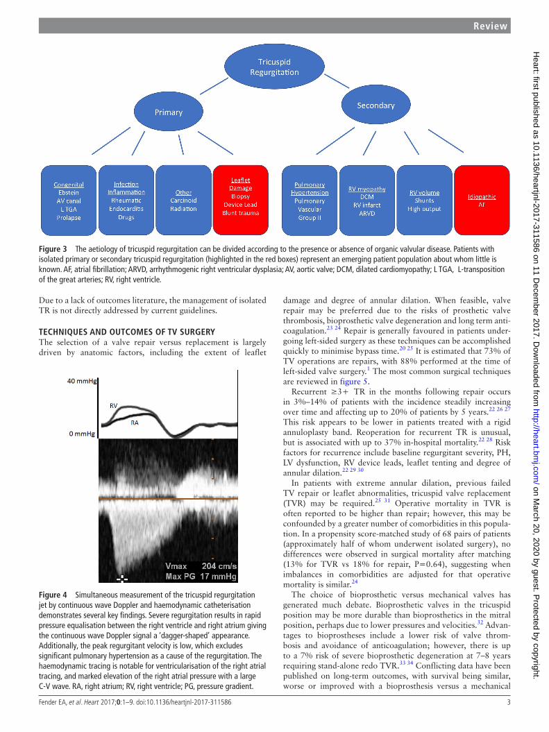
figure 3 The aetiology of tricuspid regurgitation can be divided according to the presence or absence of organic valvular disease. Patients with isolated primary or secondary tricuspid regurgitation (highlighted in the red boxes) represent an emerging patient population about whom little is known. AF, atrial fibrillation; ARVD, arrhythmogenic right ventricular dysplasia; AV, aortic valve; DCM, dilated cardiomyopathy; L TGA, L-transposition of the great arteries; RV, right ventricle.
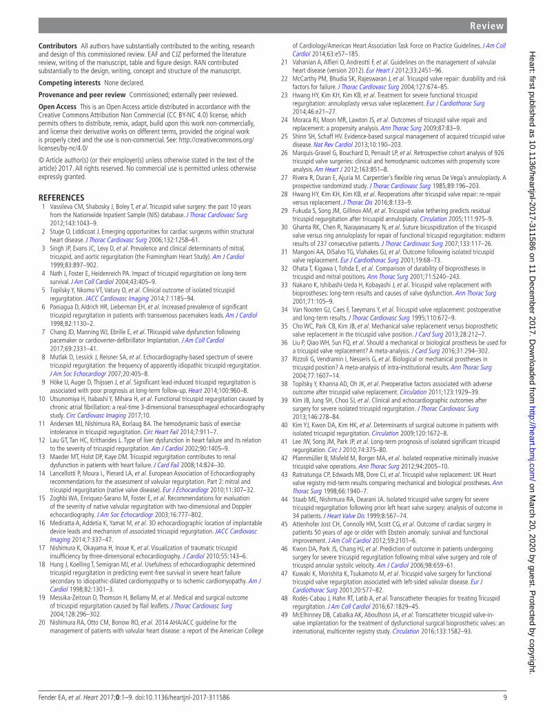
figure 4 Simultaneous measurement of the tricuspid regurgitation jet by continuous wave Doppler and haemodynamic catheterisation demonstrates several key findings. Severe regurgitation results in rapid pressure equalisation between the right ventricle and right atrium giving the continuous wave Doppler signal a ‘dagger-shaped’ appearance. Additionally, the peak regurgitant velocity is low, which excludes significant pulmonary hypertension as a cause of the regurgitation. The haemodynamic tracing is notable for ventricularisation of the right atrial tracing, and marked elevation of the right atrial pressure with a large C-V wave. RA, right atrium; RV, right ventricle; PG, pressure gradient.
on March 20, 2020 by guest. P
rotected by copyright.http://heart.bm
j.com/
Heart: first published as 10.1136/heartjnl-2017-311586 on 11 D
ecember 2017. D
ownloaded from

4 Fender EA, et al. Heart 2017;0:1–9. doi:10.1136/heartjnl-2017-311586
review
prosthesis.35–38 Overall, there is no definite survival benefit of a mechanical versus a bioprosthetic valve. The final choice should be a shared decision between the patient and the physi-cian. However, in older patients the lower rate of structural
deterioration and possible future need for a pacemaker favour a bioprosthetic valve.
While surgery is the only definitive treatment for isolated TR, it is rarely performed. Of the 4000–8000 TV operations performed annually in the USA, roughly 80% are at the time of left valve surgery.1 2 26 Stand-alone surgery makes up only 20% of operations, and many surgeries are performed for indica-tions such as endocarditis. The rarity of stand-alone surgery for isolated TR may result from concerns over in-hospital mortality (8%–10% in most series) and uncertainty surrounding long-term outcomes. Due to small sample size most studies span multiple decades and include patients undergoing left-sided heart surgery. This has resulted in widely variable estimations of in-hospital mortality with higher mortality associated with multivalvular surgery, advanced heart failure and redo sternotomy.1 19 22 38–44
Few studies have focused on the outcomes of surgery for isolated TR and these typically include heterogeneous patient populations (table 2). Additionally, there is a lack of comparative outcomes for medically and surgically treated patients. To date, only a single study has compared long-term survival in patients with isolated TR treated with medical therapy versus surgery. In a series of 45 pairs of propensity score-matched patients (66% secondary TR, 33% primary TR) treated with stand-alone surgery, there was a non-significant trend towards improved survival in the surgical patients (HR 0.29, 95% CI 0.08 to 1.10, P=0.07).41
While there are little data on long-term postoperative survival, studies have consistently demonstrated surgery can improve
table 1 Summary of existing society guidelines for tricuspid valve surgery for tricuspid regurgitation
2012 european society of cardiology recommendations212014 American heart Association/American college of cardiology recommendations20
class I
Severe primary or secondary TR at the time of left-sided valve surgery (level of evidence C)
Severe primary or secondary TR at the time of left-sided valve surgery (level of evidence C)
Symptomatic isolated severe primary TR without evidence of right ventricular dysfunction (level of evidence C)
class IIA
Surgery may be appropriate for moderate primary TR in patients at the time of left-sided valve surgery (level of evidence C)
Surgery may be appropriate for severe primary TR in patients unresponsive to medical therapy (level of evidence C)
Surgery may be appropriate for mild or moderate secondary TR in patients with annular dilation (≥40 mm or >21 mm/m2) at the time of left-sided valve surgery (level of evidence C)
Surgery may be appropriate for for mild or moderate secondary TR at the time of left-sided valve surgery if there is (A) dilation of the tricuspid annulus or (B) the patient has a history of right heart failure (level of evidence B)
Surgery may be appropriate for asymptomatic or mildly symptomatic patients with severe isolated primary TR and evidence of progressive RV dilation or decreased RV function (level of evidence C)
In patients with previous left-sided valve surgery; stand-alone tricuspid surgery may be appropriate for patients with severe secondary TR and either symptoms or evidence of right ventricular dilation or dysfunction, in the absence of left-sided valve dysfunction, severe RV or LV dysfunction and severe pulmonary hypertension (level of evidence C)
class IIb
Surgical tricuspid valve repair may be appropriate in patients with for mild or moderate secondary TR and pulmonary hypertension at the time of left-sided valve surgery (level of evidence C)
In patients with previous left-sided valve surgery; surgical repair or replacement may be appropriate in patients with symptomatic severe TR in the absence of severe RV dysfunction or severe pulmonary hypertension (level of evidence C)
Surgery may be appropriate for patients with asymptomatic or minimally symptomatic severe primary who have evidence of at least moderate right ventricular dilation or dysfunction (level of evidence C)
LV, left ventricle; RV, right ventricle; TR, tricuspid regurgitation.
figure 5 The most common tricuspid valve operations include the Kay bicuspidisation (A), DeVega suture annuloplasty (B), prosthetic annuloplasty band (C) and tricuspid valve replacement (D). AVN, atrioventricular node; CS, coronary sinus; A, anterior leaflet; P, posterior leaflet; S, septal leaflet.
on March 20, 2020 by guest. P
rotected by copyright.http://heart.bm
j.com/
Heart: first published as 10.1136/heartjnl-2017-311586 on 11 D
ecember 2017. D
ownloaded from

5Fender EA, et al. Heart 2017;0:1–9. doi:10.1136/heartjnl-2017-311586
review
tabl
e 2
Isol
ated
tric
uspi
d va
lve
surg
ery
is ra
re a
nd fe
w s
tudi
es h
ave
exam
ined
out
com
es in
this
pop
ulat
ion.
Stu
dies
are
ofte
n he
tero
gene
ous,
and
incl
ude
both
prim
ary
and
seco
ndar
y tr
icus
pid
valv
e di
seas
e
Aut
hor,
year
stud
y de
sign
stud
y po
pula
tion
out
com
esco
mm
ents
and
lim
itat
ions
Isol
ated
prim
ary
tric
uspi
dre
gurg
itatio
n
Man
goni
et a
l, 20
0131
Sing
le-c
entr
e,
retr
ospe
ctiv
e co
hort
st
udy
from
198
8 to
19
96
15 p
atie
nts
with
TV
repl
acem
ent f
or rh
eum
atic
dis
ease
(n=
12),
heal
ed e
ndoc
ardi
tis (n
=2)
and
sar
coid
osis
(n=
1)M
ean
age:
61
year
sBi
opro
sthe
tic v
alve
: 33%
Mec
hani
cal v
alve
: 67%
30-d
ay m
orta
lity:
20%
Med
ian
surv
ival
1.2
year
sSh
ort-
term
mor
talit
y hi
ghes
t in
thos
e w
ith a
nasa
rca
Long
-ter
m m
orta
lity
high
est i
n th
ose
with
ana
emia
, rhe
umat
ic
dise
ase,
pre
viou
s st
roke
or p
revi
ous
mitr
al v
alve
sur
gery
Incl
uded
pat
ient
s w
ith tr
icus
pid
sten
osis
(n
=1)
, reg
urgi
tatio
n (n
=8)
and
mix
ed
sten
osis
and
regu
rgita
tion
(n=
6)N
o co
mpa
rison
to m
edic
al th
erap
y
Atte
nhof
er e
t al,
2012
45Si
ngle
-cen
tre,
re
tros
pect
ive
coho
rt
stud
y fro
m 1
980
to
2010
81 a
dult
patie
nts
>50
year
s of
age
with
sev
ere
TR d
ue to
Ebs
tein
’s an
omal
yM
ean
age:
59
year
sTV
repa
ir: 2
5%Bi
opro
sthe
tic v
alve
: 52%
Mec
hani
cal v
alve
: 23%
Prev
ious
car
diac
sur
gery
: 16%
In-h
ospi
tal m
orta
lity:
4%
(all
prio
r to
1995
)At
84
mon
ths’
follo
w-u
p, d
eath
occ
urre
d in
17%
and
impr
oved
N
YHA
func
tiona
l cla
ss in
89%
.
Incl
uded
pat
ient
s w
ith E
bste
in’s
anom
aly
only
No
com
paris
on to
med
ical
ther
apy
Mes
sika
-Zei
toun
et
al, 2
00419
Sing
le-c
entr
e,
retr
ospe
ctiv
e co
hort
fro
m 1
980
to 2
000
60 p
atie
nts
with
TV
flail,
of w
hom
33
unde
rwen
t iso
late
d su
rger
yM
ean
age:
51
year
sTV
repa
ir: 8
2%Bi
opro
sthe
tic v
alve
: 12%
Mec
hani
cal v
alve
: 6%
In-h
ospi
tal m
orta
lity:
3%
(n=
1)Sy
mpt
omat
ic im
prov
emen
t in
88%
of s
urgi
cal p
atie
nts
Long
-ter
m s
urvi
val f
or s
urgi
cal v
ersu
s m
edic
al p
atie
nts
is n
ot re
port
ed.
Com
bine
d CA
BG o
r AVR
per
form
ed in
thre
e pa
tient
s
Isol
ated
seco
ndar
ytr
icus
pid
regu
rgita
tion
Kwon
et a
l, 20
0646
Sing
le-c
entr
e,
pros
pect
ive
coho
rt
stud
y fro
m 2
003
to
2005
18 p
atie
nts
with
sym
ptom
atic
sec
onda
ry T
R fo
llow
ing
prev
ious
mitr
al
valv
e su
rger
yM
ean
age:
58
year
sTV
repa
ir: 3
3%TV
repl
acem
ent:
67%
In-h
ospi
tal m
orta
lity:
11%
(n=
2)Im
prov
ed N
YHA
func
tiona
l cla
ss o
r 25%
incr
ease
in
resp
irato
ry v
aria
tion
of th
e in
ferio
r ven
a ca
va in
56%
of
surv
ivor
s (n
=9)
Adve
rse
outc
omes
pre
dict
ed b
y TV
ann
ular
sys
tolic
ve
loci
ty <
9.5
cm/s
Out
com
e w
as a
com
bine
d en
d-po
int
of im
prov
ed N
YHA
func
tiona
l cla
ss o
r in
crea
sed
resp
irato
ry v
aria
tion
in th
e in
ferio
r ven
a ca
va.
No
com
paris
on to
med
ical
ther
apy
Mix
edpo
pula
tion
prim
ary
and
seco
ndar
ytr
icus
pid
regu
rgita
tion
Topi
lsky
et a
l, 20
1138
Sing
le-c
entr
e,
retr
ospe
ctiv
e co
hort
st
udy
from
199
7 to
20
07
189
patie
nts
with
TV
repl
acem
ent f
or s
ympt
omat
ic s
ever
e TR
Mea
n ag
e: 6
7.5
year
sIs
olat
ed T
V su
rger
y: 3
6%Se
cond
ary
TR: 2
9%Co
mbi
ned
MV
or A
V su
rger
y: 4
6.5%
Com
bine
d CA
BG: 2
6.5%
Prev
ious
left-
side
d va
lve
surg
ery:
46.
5%Bi
opro
sthe
tic re
plac
emen
t: 81
.5%
Mec
hani
cal r
epla
cem
ent:
18.5
%
Ove
rall
in-h
ospi
tal m
orta
lity
for a
ll su
rger
ies:
10%
In-h
ospi
tal m
orta
lity
for i
sola
ted
surg
ery:
6%
At 2
9 m
onth
s’ fo
llow
-up,
dea
th o
ccur
red
in 3
7% a
nd h
eart
fa
ilure
adm
issi
on in
21.
7%.
All-c
ause
mor
talit
y w
as a
ssoc
iate
d w
ith in
crea
sed
Char
lson
co
mor
bidi
ty in
dex
scor
e, s
hort
er ri
ght i
ndex
of m
yoca
rdia
l pe
rform
ance
and
NYH
A cl
ass
IV s
ympt
oms.
46.5
% o
f the
stu
dy p
opul
atio
n ha
d co
mbi
ned
MV
or A
V su
rger
y.U
ncle
ar h
ow m
any
isol
ated
ope
ratio
ns
wer
e pe
rform
ed fo
r prim
ary
vers
us
seco
ndar
y TR
No
com
paris
on to
med
ical
ther
apy
Excl
uded
TV
repa
irs
Kim
et a
l, 20
0940
Sing
le-c
entr
e,
pros
pect
ive
coho
rt
stud
y fro
m 2
003
to
2008
61 p
atie
nts
with
sym
ptom
atic
TR
Mea
n ag
e: 5
7 ye
ars
Seco
ndar
y TR
: 84%
Prev
ious
left-
side
d va
lve
surg
ery:
93%
TV re
pair:
13%
Biop
rost
hetic
repl
acem
ent:
49%
Mec
hani
cal r
epla
cem
ent:
38%
In-h
ospi
tal m
orta
lity
9.8%
At 3
2 m
onth
s’ fo
llow
-up,
car
diac
dea
th o
ccur
red
in th
ree
patie
nts
and
six
had
card
iac
read
mis
sion
s.
Unc
lear
how
man
y ev
ents
occ
urre
d in
pa
tient
s w
ith p
rimar
y ve
rsus
sec
onda
ry T
RN
o co
mpa
rison
to m
edic
al th
erap
y
Vass
ileva
et a
l, 20
121
Mul
ticen
tre
clai
ms
data
from
the
Nat
ionw
ide
Inpa
tient
Sa
mpl
e fro
m 1
999
to
2008
5736
pat
ient
s w
ith is
olat
ed T
V su
rger
yTV
repl
acem
ent:
57%
In-h
ospi
tal m
orta
lity
for i
sola
ted
TV s
urge
ry: 9
%Ca
use
of tr
icus
pid
dise
ase
is n
ot re
port
ed,
uncl
ear i
f ste
nosi
s, re
gurg
itatio
n, p
rimar
y or
sec
onda
ry.
No
long
-ter
m o
utco
me
data
Cont
inue
d
on March 20, 2020 by guest. P
rotected by copyright.http://heart.bm
j.com/
Heart: first published as 10.1136/heartjnl-2017-311586 on 11 D
ecember 2017. D
ownloaded from

6 Fender EA, et al. Heart 2017;0:1–9. doi:10.1136/heartjnl-2017-311586
review
Aut
hor,
year
stud
y de
sign
stud
y po
pula
tion
out
com
esco
mm
ents
and
lim
itat
ions
Pfan
nmül
ler e
t al,
2012
42Si
ngle
-cen
tre,
re
tros
pect
ive
coho
rt
from
200
0 to
201
1
48 p
atie
nts
with
sev
ere
TR fo
llow
ing
prev
ious
left
card
iac
surg
ery
trea
ted
with
min
imal
ly in
vasi
ve T
V su
rger
yM
ean
age:
63.
8 ye
ars
Seco
ndar
y TR
: 90%
TV re
pair:
69%
Biop
rost
hetic
val
ve: 2
5%M
echa
nica
l: 6%
30-d
ay m
orta
lity:
4.2
%Ea
rly fa
ilure
of T
V re
pair:
12.
5%Ri
ng d
ehis
cenc
e: 9
% (l
imite
d to
sem
iflex
ible
ring
)5-
year
sur
viva
l 72%
am
ong
thos
e tr
eate
d w
ith e
lect
ive
surg
ery
No
com
paris
on to
med
ical
ther
apy
Lee
et a
l, 20
1041
Sing
le-c
entr
e,
retr
ospe
ctiv
e co
hort
st
udy
from
199
6 to
20
05
57 p
atie
nts
with
TV
surg
ery
Mea
n ag
e: 5
5 ye
ars
Seco
ndar
y TR
: 69%
TV re
pair:
75.
4%O
f the
57
patie
nts,
45 w
ere
prop
ensi
ty m
atch
ed a
ccor
ding
to s
urge
ry
or m
edic
al th
erap
y.
Ove
rall
hosp
ital m
orta
lity
for i
sola
ted
TV s
urge
ry: 8
.8%
10-y
ear s
urvi
val t
ende
d to
be
high
er in
the
surg
ical
pat
ient
s af
ter p
rope
nsity
mat
chin
g (H
R 0.
29, P
=0.
068)
.
This
is th
e on
ly s
tudy
to d
irect
ly c
ompa
re
outc
omes
of m
edic
al a
nd s
urgi
cal
man
agem
ent.
High
pre
vale
nce
of p
rimar
y TR
in s
urgi
cal
patie
nts
Kim
et a
l, 20
1339
Sing
le-c
entr
e,
retr
ospe
ctiv
e co
hort
st
udy
from
199
6 to
20
10
51 p
atie
nts
with
TV
surg
ery
Mea
n ag
e: 5
5.8
year
sSe
cond
ary
TR: 3
5%TV
repa
ir: 7
2.5%
Biop
rost
hetic
val
ve: 1
9.6%
Mec
hani
cal v
alve
: 7.8
%
In-h
ospi
tal m
orta
lity:
2.0
%5-
year
sur
viva
l: 83
.5%
5-ye
ar s
urvi
val f
ree
of C
HF, t
rans
plan
t or r
eope
ratio
n: 7
7.3%
Even
t-fre
e su
rviv
al p
redi
cted
by
RV e
nd-s
ysto
lic d
imen
sion
, pr
eope
rativ
e ha
emog
lobi
n, G
FR a
nd b
iliru
bin
No
com
paris
on to
med
ical
ther
apy
End-
syst
olic
dim
ensi
on re
port
ed
cont
inuo
usly
, it i
s un
clea
r if t
here
is a
th
resh
old
effe
ct.
Staa
b et
al,
1999
44Si
ngle
-cen
tre,
re
tros
pect
ive
coho
rt
stud
y of
pat
ient
s w
ith
TR fo
llow
ing
prev
ious
le
ft he
art v
alve
sur
gery
tr
eate
d fro
m 1
980
to
1997
34 p
atie
nts
with
sym
ptom
atic
TR
and
hist
ory
of p
rior l
eft-
side
d va
lve
surg
ery
Mea
n ag
e: 6
2.8
year
sAt
leas
t mod
erat
e org
anic
TV
dise
ase
seen
at s
urge
ry: 4
1.2%
TV re
pair:
21%
Biop
rost
hetic
val
ve: 3
8%M
echa
nica
l val
ve: 4
1%
In-h
ospi
tal m
orta
lity:
8.8
%M
ean
5-ye
ar s
urvi
val:
49%
Amon
g su
rviv
ors,
85%
exp
erie
nced
sym
ptom
atic
im
prov
emen
t.
Unc
lear
how
man
y ev
ents
occ
urre
d in
pa
tient
s w
ith p
rimar
y ve
rsus
sec
onda
ry T
RN
o co
mpa
rison
to m
edic
al th
erap
y
AV, a
ortic
val
ve; A
VR, a
ortic
val
ve re
plac
emen
t; CA
BG, c
oron
ary
arte
ry b
ypas
s gr
aftin
g; C
HF, c
onge
stiv
e he
art f
ailu
re; G
FR, g
lom
erul
ar fi
ltrat
ion
rate
; MV,
mitr
al v
alve
; NYH
A, N
ew Y
ork
Hear
t Ass
ocia
tion;
RV,
righ
t ven
tric
le; T
R, tr
icus
pid
regu
rgita
tion;
TV,
tric
uspi
d va
lve.
tabl
e 2
Cont
inue
d
on March 20, 2020 by guest. P
rotected by copyright.http://heart.bm
j.com/
Heart: first published as 10.1136/heartjnl-2017-311586 on 11 D
ecember 2017. D
ownloaded from

7Fender EA, et al. Heart 2017;0:1–9. doi:10.1136/heartjnl-2017-311586
review
symptoms.19 40 44–46 In a series of 33 patients with isolated TV flail, 88% of operative patients experienced symptomatic improvement.19 Similarly, a study of 34 patients with previous left-sided valve surgery undergoing stand-alone TV surgery demonstrated improved New York Heart Association functional class in 85% of operative patients, including 59% of whom had isolated secondary TR.44
predIctors of outcomes for tv surgeryRegardless of which series is examined, outcomes of TR surgery are worse than what is seen in mitral or aortic operations. Surgical mortality may be adversely impacted by the practice of delaying operative interventions, thereby allowing for the devel-opment of RV dysfunction and end-organ damage. For the aortic and mitral valves, the timing of surgical referral is based on an integration of symptoms, disease severity and early markers of LV dilation or dysfunction.20 There are little data to base timing of surgery for patients with severe TR, particularly those with isolated disease.
Several studies have examined the relationship between RA pressure and RV function with outcomes in patients undergoing TR surgery. An analysis of 260 patients treated surgically for secondary TR (the majority of whom underwent combined left valve surgery) found RA pressure was independently associated with hospital death (HR 5.6, 95% CI 1.7 to 78.0, P=0.01), with a mortality rate of 28% for patients with a preoperative RA pressure ≥15 mm Hg versus just 5% in patients with a pressure <15 mm Hg.47 RV dysfunction represents an advanced stage of chronic TR. In patients undergoing stand-alone TR surgery, RV
end-systolic dimension, RV end-systolic area and the RV index of myocardial performance have all been associated with survival free of death, heart failure, cardiac readmissions, heart transplant or TV reoperation.5 39 40 These findings suggest RA pressure and RV function are key determinates of postoperative outcomes. As with all surgeries for valve disease, severe symp-toms are associated with adverse short and long-term outcomes.
recommendAtIons for treAtment of IsolAted trTreatment of patients with severe isolated TR is based on several clinical observations:1. ‘Severe TR begets severe TR.’ Progressive RV dysfunction
drives further annular dilatation and results in more severe TR.
2. Diuretics may be effective in treating right heart failure in the early stages of isolated secondary TR and may potentially interrupt this cycle by lowering RA pressure.
3. Medical therapy will not reverse progressive RV dysfunction in severe isolated primary TR.
4. Isolated secondary TR associated with AF may improve with conversion to sinus rhythm.
5. Once severe right heart failure symptoms develop, if the TR remains untreated there is progressive clinical deterioration with development of end-stage liver and kidney failure.
6. Surgical intervention can improve the symptoms of right heart failure, but there are no data on enhanced survival in patients with isolated TR.
The current high mortality associated with surgery for isolated TR may be in part related to late surgical referral at which time
figure 6 Algorithm for the management of severe isolated tricuspid regurgitation. RA, right atrium; RV, right ventricle; TV, tricuspid valve.
on March 20, 2020 by guest. P
rotected by copyright.http://heart.bm
j.com/
Heart: first published as 10.1136/heartjnl-2017-311586 on 11 D
ecember 2017. D
ownloaded from

8 Fender EA, et al. Heart 2017;0:1–9. doi:10.1136/heartjnl-2017-311586
review
severe RV dysfunction and end-organ damage have already occurred.
Our recommendations for treatment of patient with severe isolated TR are shown in figure 6.
In patients without right heart failure symptoms surgical intervention is not recommended. However, it is reasonable to try diuretics if RA pressure elevation is seen on examination as diuretics may interrupt the cycle of ‘severe TR begets severe TR.’ Conversion to sinus rhythm may decrease the severity of TR in patients with persistent AF.
It is those patients with severe isolated TR and right heart failure who should be considered for surgical intervention. For patients with severe primary TR (frequently associated with intracardiac leads), surgical intervention may be indicated as this represents a structural problem which diuretics will not address. There have been reports of successful treatment of TR with lead extraction.7 However, in most cases the exact mechanism of the device-induced TR is unclear, and lead extraction could further damage the valve. Our current approach is surgical repair or TVR with exteriorisation of the lead outside the valve ring. In patients with isolated secondary TR an attempt to reduce annular size with diuretics or conversion to sinus rhythm for those with persistent AF may be effective. If symptoms remain surgical intervention is recommended prior to the onset of severe RV dysfunction, hepatic or renal dysfunction. Whether surgery may be effective at an earlier stage prior to the onset of symptomatic right heart failure remains to be determined.
percutAneous InterventIonsThe rarity of surgery has created a large population in whom percutaneous treatments could provide a viable alternative. No large experience exists for these devices but early studies are under way. Devices fall into two categories: transcatheter valves and coaptation devices48 (figure 7). Transcatheter valve replace-ment is usually performed within a previously placed biopros-thetic valve (‘valve-in-valve’), or as a stented valve implanted in the inferior vena cava and superior vena cava.48 49 Devices aimed at improving coaptation include off label use of the MitraClip (Abbot Vascular, Menlo Park, CA), a percutaneous annuloplasty system (TriAlign, Mitralign, Tewksbury, MA) and a spacer device (FORMA Edwards Lifesciences, Irvine, CA), which occupies the regurgitant orifice. The extent to which these devices result in a clinically meaningful improvement is not established and requires further research before these techniques can be adopted into clinical practice.
AreAs for further reseArchOur understanding of the management of isolated secondary TR is lacking in three key areas: identification of patients for surgical intervention, isolated TR surgical outcomes in the contemporary era and comparative survival analyses for medical and surgical patients.
Preoperative RV size and function are associated with postop-erative outcomes; however, threshold criteria for valve interven-tion are lacking.20 Similar to what is seen in the LV with mitral regurgitation, severe TR may mask early RV dysfunction. Overt RV dysfunction represents an end-stage condition and the prog-nosis cannot be altered surgically. Therefore, identification of echocardiographic markers of early RV dysfunction are needed to guide surgical referral.
Similarly, our understanding of surgical outcomes in patients with isolated TR is limited. Most series contain a heteroge-neous population treated over decades and therefore have
limited generalisability. Surgical referral for isolated TR is often delayed until the onset of frank right heart failure with end-organ damage which then further increases perioperative mortality. Efforts to define operative risk for truly isolated disease are critical to aid in decision-making, and to act as a benchmark against which studies of early operation can be compared.
A final, but major, limitation of existing surgical data is the paucity of comparative outcomes. Upfront operative mortality is high. Therefore, establishing long-term survival relative to medical therapy is critical to understanding the risks and benefits of surgery. A randomised trial of early surgery for isolated TR would fill an important gap in the literature.
conclusIonsIsolated severe TR results in progressive right heart failure and adversely impacts survival. Surgery remains the only definitive treatment but is rarely performed. There are little data to guide management of isolated TR; however, surgery is reasonable in patients with right heart failure who are at low operative risk prior to the development of RV dysfunction and end-organ damage. Further research is needed to identify markers of disease progression and to aid in the optimal timing for surgical referral.
figure 7 Multiple percutaneous devices are in development for the treatment of tricuspid regurgitation. Panel (A) is the FORMA device, a tricuspid spacer which occupies the regurgitant orifice and provides a surface against which coaptation can occur. Panel (B) demonstrates the TriAlign, which percutaneously reproduces a surgical Kay bicuspidisation. Panel (C) shows the MitraClip being used in the tricuspid position. Panel (D) demonstrates a stented caval valve implanted in the inferior vena cava.
on March 20, 2020 by guest. P
rotected by copyright.http://heart.bm
j.com/
Heart: first published as 10.1136/heartjnl-2017-311586 on 11 D
ecember 2017. D
ownloaded from
9Fender EA, et al. Heart 2017;0:1–9. doi:10.1136/heartjnl-2017-311586
review
contributors All authors have substantially contributed to the writing, research and design of this commissioned review. EAF and CJZ performed the literature review, writing of the manuscript, table and figure design. RAN contributed substantially to the design, writing, concept and structure of the manuscript.
competing interests None declared.
provenance and peer review Commissioned; externally peer reviewed.
open Access This is an Open Access article distributed in accordance with the Creative Commons Attribution Non Commercial (CC BY-NC 4.0) license, which permits others to distribute, remix, adapt, build upon this work non-commercially, and license their derivative works on different terms, provided the original work is properly cited and the use is non-commercial. See: http:// creativecommons. org/ licenses/ by- nc/ 4. 0/
© Article author(s) (or their employer(s) unless otherwise stated in the text of the article) 2017. All rights reserved. No commercial use is permitted unless otherwise expressly granted.
references 1 Vassileva CM, Shabosky J, Boley T, et al. Tricuspid valve surgery: the past 10 years
from the Nationwide Inpatient Sample (NIS) database. J Thorac Cardiovasc Surg 2012;143:1043–9.
2 Stuge O, Liddicoat J. Emerging opportunities for cardiac surgeons within structural heart disease. J Thorac Cardiovasc Surg 2006;132:1258–61.
3 Singh JP, Evans JC, Levy D, et al. Prevalence and clinical determinants of mitral, tricuspid, and aortic regurgitation (the Framingham Heart Study). Am J Cardiol 1999;83:897–902.
4 Nath J, Foster E, Heidenreich PA. Impact of tricuspid regurgitation on long-term survival. J Am Coll Cardiol 2004;43:405–9.
5 Topilsky Y, Nkomo VT, Vatury O, et al. Clinical outcome of isolated tricuspid regurgitation. JACC Cardiovasc Imaging 2014;7:1185–94.
6 Paniagua D, Aldrich HR, Lieberman EH, et al. Increased prevalence of significant tricuspid regurgitation in patients with transvenous pacemakers leads. Am J Cardiol 1998;82:1130–2.
7 Chang JD, Manning WJ, Ebrille E, et al. TRicuspid valve dysfunction following pacemaker or cardioverter-defibrillator Implantation. J Am Coll Cardiol 2017;69:2331–41.
8 Mutlak D, Lessick J, Reisner SA, et al. Echocardiography-based spectrum of severe tricuspid regurgitation: the frequency of apparently idiopathic tricuspid regurgitation. J Am Soc Echocardiogr 2007;20:405–8.
9 Höke U, Auger D, Thijssen J, et al. Significant lead-induced tricuspid regurgitation is associated with poor prognosis at long-term follow-up. Heart 2014;100:960–8.
10 Utsunomiya H, Itabashi Y, Mihara H, et al. Functional tricuspid regurgitation caused by chronic atrial fibrillation: a real-time 3-dimensional transesophageal echocardiography study. Circ Cardiovasc Imaging 2017;10.
11 Andersen MJ, Nishimura RA, Borlaug BA. The hemodynamic basis of exercise intolerance in tricuspid regurgitation. Circ Heart Fail 2014;7:911–7.
12 Lau GT, Tan HC, Kritharides L. Type of liver dysfunction in heart failure and its relation to the severity of tricuspid regurgitation. Am J Cardiol 2002;90:1405–9.
13 Maeder MT, Holst DP, Kaye DM. Tricuspid regurgitation contributes to renal dysfunction in patients with heart failure. J Card Fail 2008;14:824–30.
14 Lancellotti P, Moura L, Pierard LA, et al. European Association of Echocardiography recommendations for the assessment of valvular regurgitation. Part 2: mitral and tricuspid regurgitation (native valve disease). Eur J Echocardiogr 2010;11:307–32.
15 Zoghbi WA, Enriquez-Sarano M, Foster E, et al. Recommendations for evaluation of the severity of native valvular regurgitation with two-dimensional and Doppler echocardiography. J Am Soc Echocardiogr 2003;16:777–802.
16 Mediratta A, Addetia K, Yamat M, et al. 3D echocardiographic location of implantable device leads and mechanism of associated tricuspid regurgitation. JACC Cardiovasc Imaging 2014;7:337–47.
17 Nishimura K, Okayama H, Inoue K, et al. Visualization of traumatic tricuspid insufficiency by three-dimensional echocardiography. J Cardiol 2010;55:143–6.
18 Hung J, Koelling T, Semigran MJ, et al. Usefulness of echocardiographic determined tricuspid regurgitation in predicting event-free survival in severe heart failure secondary to idiopathic-dilated cardiomyopathy or to ischemic cardiomyopathy. Am J Cardiol 1998;82:1301–3.
19 Messika-Zeitoun D, Thomson H, Bellamy M, et al. Medical and surgical outcome of tricuspid regurgitation caused by flail leaflets. J Thorac Cardiovasc Surg 2004;128:296–302.
20 Nishimura RA, Otto CM, Bonow RO, et al. 2014 AHA/ACC guideline for the management of patients with valvular heart disease: a report of the American College
of Cardiology/American Heart Association Task Force on Practice Guidelines. J Am Coll Cardiol 2014;63:e57–185.
21 Vahanian A, Alfieri O, Andreotti F, et al. Guidelines on the management of valvular heart disease (version 2012). Eur Heart J 2012;33:2451–96.
22 McCarthy PM, Bhudia SK, Rajeswaran J, et al. Tricuspid valve repair: durability and risk factors for failure. J Thorac Cardiovasc Surg 2004;127:674–85.
23 Hwang HY, Kim KH, Kim KB, et al. Treatment for severe functional tricuspid regurgitation: annuloplasty versus valve replacement. Eur J Cardiothorac Surg 2014;46:e21–27.
24 Moraca RJ, Moon MR, Lawton JS, et al. Outcomes of tricuspid valve repair and replacement: a propensity analysis. Ann Thorac Surg 2009;87:83–9.
25 Shinn SH, Schaff HV. Evidence-based surgical management of acquired tricuspid valve disease. Nat Rev Cardiol 2013;10:190–203.
26 Marquis-Gravel G, Bouchard D, Perrault LP, et al. Retrospective cohort analysis of 926 tricuspid valve surgeries: clinical and hemodynamic outcomes with propensity score analysis. Am Heart J 2012;163:851–8.
27 Rivera R, Duran E, Ajuria M. Carpentier’s flexible ring versus De Vega’s annuloplasty. A prospective randomized study. J Thorac Cardiovasc Surg 1985;89:196–203.
28 Hwang HY, Kim KH, Kim KB, et al. Reoperations after tricuspid valve repair: re-repair versus replacement. J Thorac Dis 2016;8:133–9.
29 Fukuda S, Song JM, Gillinov AM, et al. Tricuspid valve tethering predicts residual tricuspid regurgitation after tricuspid annuloplasty. Circulation 2005;111:975–9.
30 Ghanta RK, Chen R, Narayanasamy N, et al. Suture bicuspidization of the tricuspid valve versus ring annuloplasty for repair of functional tricuspid regurgitation: midterm results of 237 consecutive patients. J Thorac Cardiovasc Surg 2007;133:117–26.
31 Mangoni AA, DiSalvo TG, Vlahakes GJ, et al. Outcome following isolated tricuspid valve replacement. Eur J Cardiothorac Surg 2001;19:68–73.
32 Ohata T, Kigawa I, Tohda E, et al. Comparison of durability of bioprostheses in tricuspid and mitral positions. Ann Thorac Surg 2001;71:S240–243.
33 Nakano K, Ishibashi-Ueda H, Kobayashi J, et al. Tricuspid valve replacement with bioprostheses: long-term results and causes of valve dysfunction. Ann Thorac Surg 2001;71:105–9.
34 Van Nooten GJ, Caes F, Taeymans Y, et al. Tricuspid valve replacement: postoperative and long-term results. J Thorac Cardiovasc Surg 1995;110:672–9.
35 Cho WC, Park CB, Kim JB, et al. Mechanical valve replacement versus bioprosthetic valve replacement in the tricuspid valve position. J Card Surg 2013;28:212–7.
36 Liu P, Qiao WH, Sun FQ, et al. Should a mechanical or biological prosthesis be used for a tricuspid valve replacement? A meta-analysis. J Card Surg 2016;31:294–302.
37 Rizzoli G, Vendramin I, Nesseris G, et al. Biological or mechanical prostheses in tricuspid position? A meta-analysis of intra-institutional results. Ann Thorac Surg 2004;77:1607–14.
38 Topilsky Y, Khanna AD, Oh JK, et al. Preoperative factors associated with adverse outcome after tricuspid valve replacement. Circulation 2011;123:1929–39.
39 Kim JB, Jung SH, Choo SJ, et al. Clinical and echocardiographic outcomes after surgery for severe isolated tricuspid regurgitation. J Thorac Cardiovasc Surg 2013;146:278–84.
40 Kim YJ, Kwon DA, Kim HK, et al. Determinants of surgical outcome in patients with isolated tricuspid regurgitation. Circulation 2009;120:1672–8.
41 Lee JW, Song JM, Park JP, et al. Long-term prognosis of isolated significant tricuspid regurgitation. Circ J 2010;74:375–80.
42 Pfannmüller B, Misfeld M, Borger MA, et al. Isolated reoperative minimally invasive tricuspid valve operations. Ann Thorac Surg 2012;94:2005–10.
43 Ratnatunga CP, Edwards MB, Dore CJ, et al. Tricuspid valve replacement: UK Heart valve registry mid-term results comparing mechanical and biological prostheses. Ann Thorac Surg 1998;66:1940–7.
44 Staab ME, Nishimura RA, Dearani JA. Isolated tricuspid valve surgery for severe tricuspid regurgitation following prior left heart valve surgery: analysis of outcome in 34 patients. J Heart Valve Dis 1999;8:567–74.
45 Attenhofer Jost CH, Connolly HM, Scott CG, et al. Outcome of cardiac surgery in patients 50 years of age or older with Ebstein anomaly: survival and functional improvement. J Am Coll Cardiol 2012;59:2101–6.
46 Kwon DA, Park JS, Chang HJ, et al. Prediction of outcome in patients undergoing surgery for severe tricuspid regurgitation following mitral valve surgery and role of tricuspid annular systolic velocity. Am J Cardiol 2006;98:659–61.
47 Kuwaki K, Morishita K, Tsukamoto M, et al. Tricuspid valve surgery for functional tricuspid valve regurgitation associated with left-sided valvular disease. Eur J Cardiothorac Surg 2001;20:577–82.
48 Rodés-Cabau J, Hahn RT, Latib A, et al. Transcatheter therapies for treating Tricuspid regurgitation. J Am Coll Cardiol 2016;67:1829–45.
49 McElhinney DB, Cabalka AK, Aboulhosn JA, et al. Transcatheter tricuspid valve-in-valve implantation for the treatment of dysfunctional surgical bioprosthetic valves: an international, multicenter registry study. Circulation 2016;133:1582–93.
on March 20, 2020 by guest. P
rotected by copyright.http://heart.bm
j.com/
Heart: first published as 10.1136/heartjnl-2017-311586 on 11 D
ecember 2017. D
ownloaded from


![Mitral regurgitation detected during the intraoperative period ......tricuspid regurgitation velocity have been identified as risk factors for MR [13]. As our patient had preopera-tive](https://img.pdfslide.net/doc/110x75/60f7a9c4d572506bd07f600b/mitral-regurgitation-detected-during-the-intraoperative-period-tricuspid.jpg)


