Embed Size (px)
Citation preview
INFECTION AND IMMUNITY, May 1989, p. 1399-14040019-9567/89/051399-06$02.00/0Copyright © 1989, American Society for Microbiology
Isolation and Immunological Characterization of a 55-KilodaltonSurface Protein from Salmonella typhimurium
KIRANI FOULAKI, W. GRUBER, AND S. SCHLECHT*
Max-Planck-Institut fur Immunbiologie, D-7800 Freiburg, Federal Republic of Germany
Received 12 August 1988/Accepted 15 January 1989
Surface proteins of different Salmonella R mutants were labeled selectively by treating live bacteria withcycloheptaamylose-dansylchloride. The labeled proteins were extracted from the cells with 6 M urea andanalyzed by sodium dodecyl sulfate-polyacrylamide gel electrophoresis. From the urea extract a 55-kilodaltonprotein common to numerous Salmonella strains could be isolated by ion-exchange chromatography and gelifitration free of lipopolysaccharide. Immunization of rabbits with isolated protein led to the formation ofspecific antibodies. Such antiprotein antisera could be employed in Western blots for the specific identificationof the 55-kilodalton protein in bacterial extracts containing mixtures of different SalmoneUa proteins. Theimportance of this antigen is emphasized by antisera against acetone-killed Salmonella bacteria, showing a
preferential interaction with the 55-kilodalton protein in Western blots. Active immunization of mice with the55-kilodalton protein afforded significant protection against experimental infection with S. typhimurium.
Among bacterial infections salmonellosis is still a world-wide problem. Numerous attempts have been made toestablish efficient vaccines and to get insight into mecha-nisms of antiinfectious immunity (1, 8, 17, 33). The immu-noprotective components of salmonellae, however, have notyet been identified. Generally, the 0 antigens are consideredto be responsible for the species-specific protection (8, 16).On the other hand, it is well known that Salmonella smoothstrains of different serovars and even rough mutants devoidof the 0 antigen also can act as effective vaccines (7, 11, 26).Many components of the Salmonella cell have been claimedto evoke antiinfectious immunity. However, in most cases
preparations used as vaccines were contaminated with othersubcellular fractions. In preceding papers (3, 28) it could bedemonstrated that the isolated and highly purified proteinfraction from a Salmonella typhimurium smooth strain as
well as those from various Salmonella rough mutants medi-ate protection against experimental Salmonella infection.Such preparations contained about 30 protein species (3).Species-overlapping protection became obvious. During nat-ural infection of humans (3, 6, 14, 32), antibodies againstSalmonella proteins are induced.The aim of this study was to identify proteins exposed on
the surface of Salmonella and to isolate relevant singleproteins with potential importance for vaccine development.Antibody formation against a 55-kilodalton (kDa) protein as
compared with that against whole bacteria, serological reac-tivity of this protein, and the protective capacity in compar-ison to a complex antigen extract are discussed.
MATERIALS AND METHODSBacteria and cultivation conditions. S. typhimurium his386
(Ra mutant), S. typhimurium SL1032 (Rd1 mutant), andSalmonella minnesota R345 (Rb2 mutant) derived from theculture collection of this Institute were used throughout thisstudy. The organisms were grown in a complex mediumconsisting of 7.5 g of tryptone (Difco Laboratories) 7.5 g oftryptose (Difco), 10 g of yeast extract (Difco), 10 g ofglucose, 3 g of NaCl, 8 g of Na2HPO4 12H20, 0.2 g ofMgSO4 7H20, and 0.3 ml of polyethylene glycol P 2000
* Corresponding author.
per liter of deionized water. Cultivation was performedunder aerobic conditions in a fermentor at 37°C and pH 7.2(27). Bacteria were harvested by centrifugation in the loga-rithmic phase or immediately after cells reached the station-ary phase, indicated by a sharp rise in oxygen partialpressure. The cells were used either for labeling or forpreparative extraction of proteins. For preparation of fla-gella, growth was performed in shaking cultures overnight inthe same medium with a reduced content of 0.3% glucose.
Cell labeling and analysis of labeled proteins. Labeling was
performed with the cycloheptaamylose-dansyl chloride com-plex (CDC) described by Kinoshita et al. (12) with themodification of Legrum et al. (13).About 1010 cells were washed three times with 3 ml of 5
mM phosphate buffer (pH 7.5) containing 140 mM NaCl and10 mM MgCl2. Bacteria were suspended in 2 ml of 50 mMphosphate buffer (pH 7.5). The 5 mg of CDC was added, andthe mixture was stirred for 1 h at 10°C. The suspension was
centrifuged for 10 min at 4,500 x g, and the supernatant was
removed.The cells were suspended in 6 M urea containing 10 mM
Tris hydrochloride (pH 7.5) and 5 mM EDTA and stirred for1 h (3). After centrifugation at 17,000 x g for 20 min, thesupernatant was concentrated by ultrafiltration (centricon10; Amicon Corp.) to a final volume of 70 ,ul. Samples were
analyzed by sodium dodecyl sulfate-polyacrylamide gel elec-trophoresis (SDS-PAGE) on 12% polyacrylamide gels. La-beled molecules were visualized by UV illumination andphotographed. Proteins were then stained with Coomassieblue.
Extraction and separation of proteins. Harvested bacteriawere washed once in Tricine {N-[Tris(hydroxymethyl)-meth-yl]glycine} buffer (pH 7.2) and extracted with buffered 6 Murea (see above). The extract was dialyzed for 3 days againstrunning tap water. The precipitated components were sepa-rated by centrifugation (34 800 x g, 60 min); both fractions,containing the soluble and insoluble material, were eitherkept at -20°C or lyophilized.
Isolation of 55-kDa protein. The 55-kDa protein was iso-lated from the soluble fraction of the extracts obtained fromS. typhimurium his386 and S. typhimurium SL1032. All stepswere carried out at 6°C with buffers without detergents.
1399
Vol. 57, No. 5
on June 18, 2018 by guesthttp://iai.asm
.org/D
ownloaded from
1400 FOULAKI ET AL.
In a first step the soluble fraction was separated byanion-exchange chromatography (2) with-DEAE-SepharoseCL 6B (Pharmacia Fine Chemicals). The extracted material,containing abouit 60 mg of protein, was dissolved in 100 ml ofstarting buffer (10 mM Tris hydrochloride [pH 8.0], 5 mMEDTA) and was loaded on a column of 2.4 by 11 cm. Thecolumn was washed with one bed volume of the startingbuffer, and bound material was eluted with a linear gradientof 0 to 500 mM NaCl dissolved in starting buffer at a flow rateof 20 ml/h. Fractions of S ml Were collected. Those contain-ing 55-kDa protein were combined and dialyzed againstbuffer containihg 20 mM Tris hydrochloride (pH 7.5)-5 mMEDTA-100 mM NaCl.
In a second purification step material was applied on aSepharose CL-6B (Pharmacia) column of 2.6 by 100 cm.Elution was performed with Tris buffer (pH 7.5) (see above)at a flow rate of 20 ml/h, and 5-ml fractions were collected.
Analytical methods. Protein content was determined by themethod of Bradford (5) with bovine serum albumin as astandard.
P-Hydroxymyristic acid, as an intrinsic component oflipopolysaccharide (LPS) was determined by gas-liquidchromatography (2, 9) with a Varian Aerograph 1400. Gen-erally 2 to 3 mg of substance was used; the amount,however, was increased to 5 mg in samples expected tocontain low amounts of LPS. The fatty acid methyl esterswere dissolved in S ,ul of chloroform, from which 1.5 ,ul wasinjected into the column. The sensitivity of the aerographwas normally adjusted to 10-10/2; with samples expected tohave low LPS content it was 10-11/2. The response factorwas taken as 0.9. The minimum amount of 3-hydroxy-myristic acid measured by this method was 0.001%. Sampleswith a smaller content were considered to be free from LPS.
Stimulation of antibodies in rabbits. Outbred Chinchillarabbits were used in all experiments. Samples (2 mg) of the55-kDa protein isolated from S. typhimurium his386 and S.typhimurium SL1032 were dissolved in 1.0 ml of 0.25%NaHCO3 solutioh, and 1.0 ml of Alu-Gel-S (Serva) wasadded. The mixture was kept at 4°C for 1 h; then 0.5 ml ofsorbitan trioleate (Span 85; Serva) and 2.5 ml of paraffin oilDAB 7 (Roth) were added, and the suspension was mixedthoroughly with an Ultra-Turrax (Janke and Kunkel, FederalRepublic of Germany) mixer. Two animals each were immu-nized three times subcutanously on days 1, 36, and 86 withthe suspension described above containing 1.0, 1.0, and 1.5mg of protein, respectively. Blood samples were taken ondays 93 and 100, i.e., 7 and 14 days after the last immuniza-tion.Another two animals were immunized intravenously with
acetone-killed S. typhimurium his386 (26). The suspensioncontained 2 x 10¹⁰ cells per ml of 0.9% sodium chloridesolution. Injections were performed on days 1, 8, 13, 18, and49 with 0.25, 0.5, 1.0, and 2.0 ml, respectively. Bloodsamples were taken on days 21, 25, 49, 56, and 61.Agar gel precipitation. Double diffusion tests by the
method of Ouchterlony (22) were performed in 1% agarosegel (Litex) with 0.05 M barbiturate buffer (pH 8.2). AnLPS-free 55-kDa protein isolated from S. typhimuriumhis386 was used as antigen with a concentration of 2 mg ofprotein per 100 ,ul of barbiturate buffer (pH 8.2). Antiserawere used undiluted. The gels were kept at 4°C and observedfor 5 days.
Preparation of flagella. Bacteria were suspended in 100mM Tris hydrochloride buffer (pH 7.2). Flagella weresheared off by treatment with an Omnimixer (Sorvall) for 10min at setting 2.8 in an ice-water bath. Bacteria and cell
debris were removed by two cycles of centrifugation at 4,000x g for 20 min and 8,000 x g for 15 min. From thesupernatant of the second centrifugation step the flagellawere spun down at 48,000 x g for 1 h, and the sediment wassuspended in 2 ml of sterile water and kept at -20°C untiluse.SDS-PAGE. SDS-PAGE was performed in a discontinuous
gel system by the method of Lugtenberg et al. (15). Proteinswere stained with Coomassie blue.Western blots (immunoblots). Proteins were transfered
electrophoretically from the SDS-polyacrylamide gel ontoImmobilon transfer membrane (Millipore) by the method ofTowbin et al. (31). The immune reaction was performed withantibodies obtained from immunized rabbits (see above).Bound rabbit immunoglobulin G was detected with alkalinephosphatase-conjugated goat antiserum as described byBlake et al. (4).
Vaccination and infection of mice. NMRI mice weighingabout 20 g were randomized in groups of eight animals.Soluble urea-extracted material obtained from S. typhimu-rium SL1032 and 55-kDa protein separated from this strainwere used as vaccines. Mice were immunized intraperitone-ally two times at intervals of 14 days with single doses of 100jig (3). Ten days after the booster vaccination the animalswere challenged intraperitoneally with graded, 10-fold-di-luted amounts of S. typhimurium C5. From the mortalityvalues obtained 19 days after challenge, the 50% lethal dosewas calculated by the method of Reed and Muench (23).Statistical significance was determined as described by Val-tonen (34).
RESULTSIdentification of surface proteins. Urea-extracted proteins
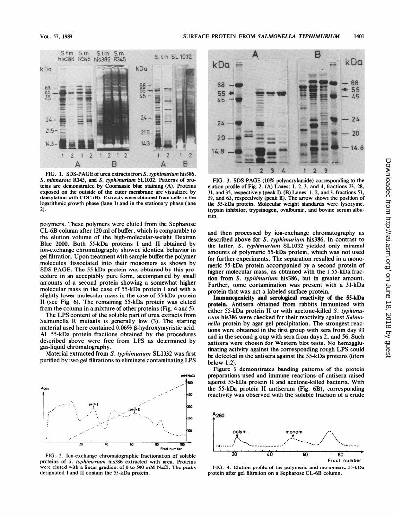
from S. typhimurium his386 and S. minnesota R345 showedvery similar patterns when analyzed by SDS-PAGE andstained with Coomassie blue (Fig. 1). A somewhat differentbanding could be observed with S. typhimurium SL1032; inaddition, the influence of the growth phase was apparenthere (Fig. 1). Fluoresceing bands in extracts from all of thesestrains, however, were closely related. Bands of high inten-sity were those with molecular masses of about 55 kDa andclose to 43 kDa. Some proteins with molecular masses ofabout 69 kDa and in the range of 24 kDa were also labeled.This indicates the presence of these proteins on the Salmo-nella surface and their possible importance as immunogens.One of them, with a molecular mass of 55 kDa, can beconsidered as a widespread component in Salmonella strains(3, 19) and as a most abundant component in the solublefraction of urea extracts. Therefore this protein was selectedfor further investigation. Isolation of flagellae and theiranalysis by SDS-PAGE revealed a molecular mass of 52kDa. This excluded the possibility that both proteins couldbe identical. Most probably the 55-kDa protein is an integralcomponent of the outer membrane.
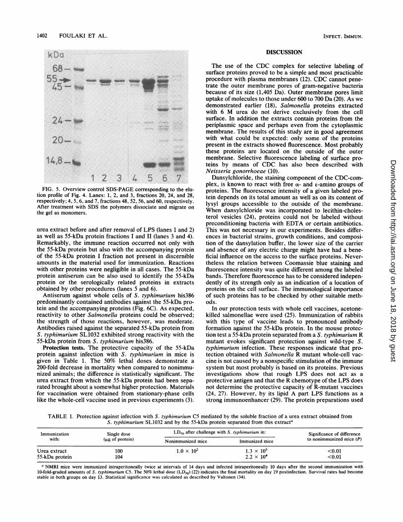
Separation of the 55-kDa protein. The soluble part of theurea extract obtained from S. typhimurium his386 wasfractionated by ion-exchange chromtography. The elutionprofile is given in Fig. 2. The 55-kDa protein was eluted intwo different ranges of salt concentrations, 110 through 165and 235 through 290 mM, still in a mixture with other proteinspecies (Fig. 3). According to elution ranges, the materialswere designated as 55-kDa protein I and 55-kDa protein II,respectively. In further processing both were treated sepa-rately.
After dialysis, the 55-kDa protein was further purified bygel filtration. Part of the 55-kDa protein apparently formed
INFECT. IMMUN.
on June 18, 2018 by guesthttp://iai.asm
.org/D
ownloaded from
SURFACE PROTEIN FROM SALMONELLA TYPHIMURIUM
Stm S mhis386 R345
__;~-I_u
S.tm S.mhis386 R345
kDa
68 - _ow _0a
55--_45 3# ..4_ : 1
24-s"21.5-
14.3 a
1 2 1 2 1 2 1 2
A B
S.tm SL1032
kDa
68 -
55 _a m `-45--"
24_-
21.5-
14.3- Aj I
1 2
A BkDa ;& kDa
68 -- -55 .,- m m555*;,;;?-- W"*-~~4545 w -
_-@-2
~--...
14.8~~~~~~~~^ -^ -2
I
1 2A B
FIG. 1. SDS-PAGE of urea extracts from S. typhimurium his386,S. minnesota R345, and S. typhimurium SL1032. Patterns of pro-teins are demonstrated by Coomassie blue staining (A). Proteinsexposed on the outside of the outer membrane are visualized bydansylation with CDC (B). Extracts were obtained from cells in thelogarithmic growth phase (lane 1) and in the stationary phase (lane2).
1 2 3 4 1 2 3
FIG. 3. SDS-PAGE (10% polyacrylamide) corresponding to theelution profile of Fig. 2. (A) Lanes: 1, 2, 3, and 4, fractions 23, 28,31, and 35, respectively (peak I). (B) Lanes: 1, 2, and 3, fractions 51,59, and 63, respectively (peak II). The arrow shows the position ofthe 55-kDa protein. Molecular weight standards were lysozyme,trypsin inhibitor, trypsinogen, ovalbumin, and bovine serum albu-min.
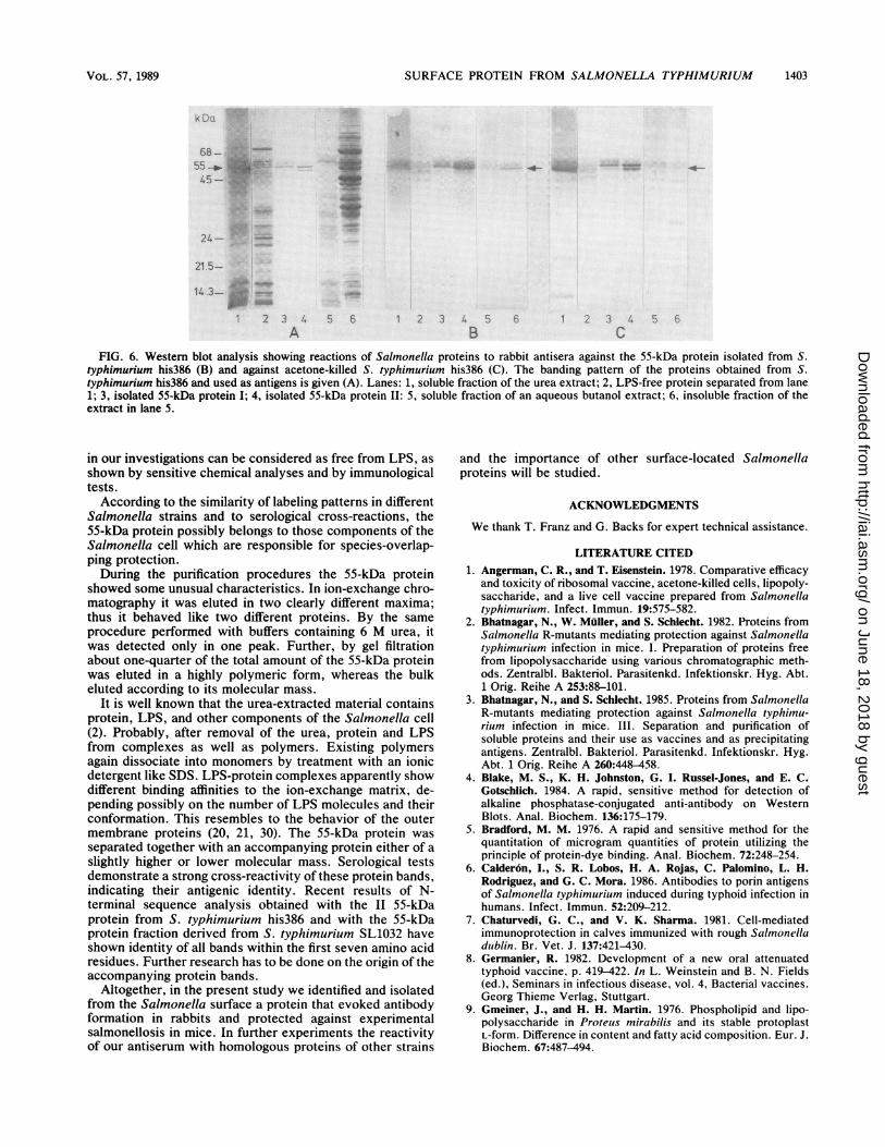
polymers. These polymers were eluted from the SepharoseCL-6B column after 120 ml of buffer, which is comparable tothe elution volume of the high-molecular-weight DextranBlue 2000. Both 55-kDa proteins I and II obtained byion-exchange chromatography showed identical behavior ingel filtration. Upon treatment with sample buffer the polymermolecules dissociated into their monomers as shown bySDS-PAGE. The 55-kDa protein was obtained by this pro-cedure in an acceptably pure form, accompanied by smallamounts of a second protein showing a somewhat highermolecular mass in the case of 55-kDa protein I and with aslightly lower molecular mass in the case of 55-kDa proteinII (see Fig. 6). The remaining 55-kDa protein was elutedfrom the column in a mixture of other proteins (Fig. 4 and 5).The LPS content of the soluble part of urea extracts from
Salmonella R mutants is generally low (3). The startingmaterial used here contained 0.06% ,B-hydroxymyristic acid.All 55-kDa protein fractions obtained by the proceduresdescribed above were free from LPS as determined bygas-liquid chromatography.
Material extracted from S. typhimurium SL1032 was firstpurified by two gel filtrations to eliminate contaminating LPS
mM NoCl1500
, . , peokI1
.II --
.' '', ",It
,peakI
~~~~~~~~~~II,
and then processed by ion-exchange chromatography asdescribed above for S. typhimurium his386. In contrast tothe latter, S. typhimurium SL1032 yielded only minimalamounts of polymeric 55-kDa protein, which was not usedfor further experiments. The separation resulted in a mono-meric 55-kDa protein accompanied by a second protein ofhigher molecular mass, as obtained with the I 55-kDa frac-tion from S. typhimurium his386, but in greater amount.Further, some contamination was present with a 31-kDaprotein that was not a labeled surface protein.Immunogenicity and serological reactivity of the 55-kDa
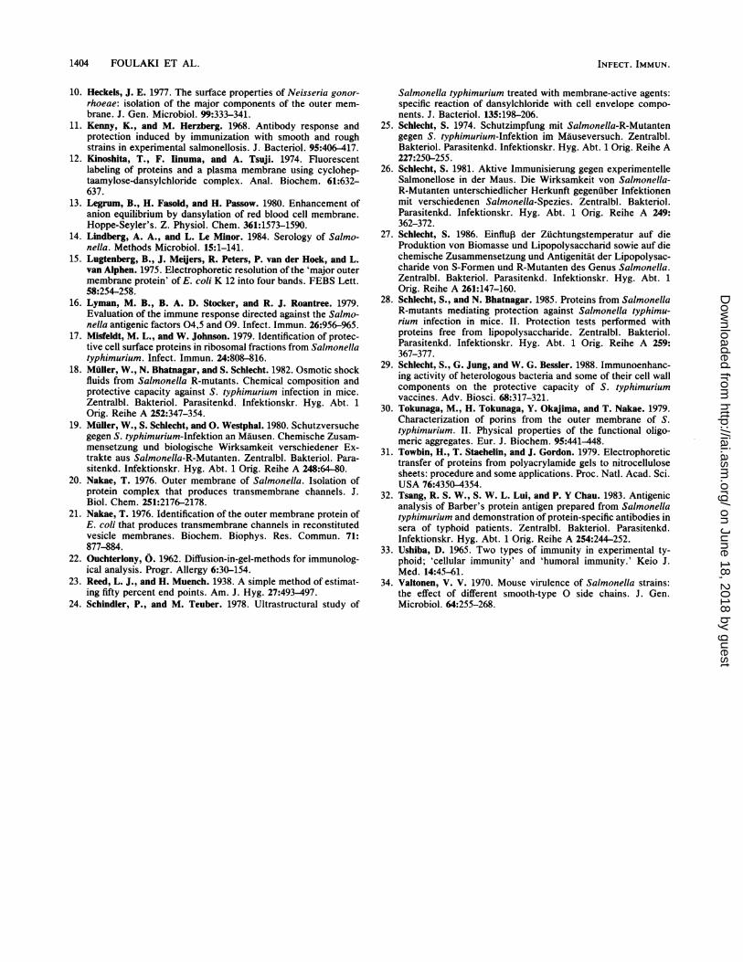
protein. Antisera obtained from rabbits immunized witheither 55-kDa protein II or with acetone-killed S. typhimu-rium his386 were checked for their reactivity against Salmo-nella protein by agar gel precipitation. The strongest reac-tions were obtained in the first group with sera from day 93and in the second group with sera from days 21 and 56. Suchantisera were chosen for Western blot tests. No hemagglu-tinating activity against the corresponding rough LPS couldbe detected in the antisera against the 55-kDa proteins (titersbelow 1:2).
Figure 6 demonstrates banding patterns of the proteinpreparations used and immune reactions of antisera raisedagainst 55-kDa protein II and acetone-killed bacteria. Withthe 55-kDa protein II antiserum (Fig. 6B), correspondingreactivity was observed with the soluble fraction of a crude
200
20 40 60 so0 'WFract.numbr
FIG. 2. Ion-exchange chromatographic fractionation of solubleproteins of S. typhimurium his386 extracted with urea. Proteinswere eluted with a linear gradient of 0 to 500 mM NaCl. The peaksdesignated I and II contain the 55-kDa protein.
Fract. numberFIG. 4. Elution profile of the polymeric and monomeric 55-kDa
protein after gel filtration on a Sepharose CL-6B column.
VOL. 57, 1989 1401
on June 18, 2018 by guesthttp://iai.asm
.org/D
ownloaded from
1402 FOULAKI ET AL.
DISCUSSION
55-
45-
24-_.
20 --
14,8- _O.
*361 23rn56
FIG. 5. Overview control SDS-PAGE corresponding to the elu-tion profile of Fig. 4. Lanes: 1, 2, and 3, fractions 20, 24, and 28,respectively; 4, 5, 6, and 7, fractions 48, 52, 56, and 60, respectively.After treatment with SDS the polymers dissociate and migrate onthe gel as monomers.
urea extract before and after removal of LPS (lanes 1 and 2)as well as 55-kDa protein fractions I and II (lanes 3 and 4).Remarkably, the immune reaction occurred not only withthe 55-kDa protein but also with the accompanying proteinof the 55-kDa protein I fraction not present in discernibleamounts in the material used for immunization. Reactionswith other proteins were negligible in all cases. The 55-kDaprotein antiserum can be also used to identify the 55-kDaprotein or the serologically related proteins in extractsobtained by other procedures (lanes 5 and 6).Antiserum against whole cells of S. typhimurium his386
predominantly contained antibodies against the 55-kDa pro-tein and the accompanying proteins (Fig. 6C). As expected,reactivity to other Salmonella proteins could be observed;the strength of those reactions, however, was moderate.Antibodies raised against the separated 55-kDa protein fromS. typhimurium SL1032 exhibited strong reactivity with the55-kDa protein from S. typhimurium his386.
Protection tests. The protective capacity of the 55-kDaprotein against infection with S. typhimurium in mice isgiven in Table 1. The 50% lethal doses demonstrate a200-fold decrease in mortality when compared to nonimmu-nized animals; the difference is statistically significant. Theurea extract from which the 55-kDa protein had been sepa-rated brought about a somewhat higher protection. Materialsfor vaccination were obtained from stationary-phase cellslike the whole-cell vaccine used in previous experiments (3).
The use of the CDC complex for selective labeling ofsurface proteins proved to be a simple and most practicableprocedure with plasma membranes (12). CDC cannot pene-trate the outer membrane pores of gram-negative bacteriabecause of its size (1,405 Da). Outer membrane pores limituptake of molecules to those under 600 to 700 Da (20). As wedemonstrated earlier (18), Salmonella proteins extractedwith 6 M urea do not derive exclusively from the cellsurface. In addition the extracts contain proteins from theperiplasmic space and perhaps even from the cytoplasmicmembrane. The results of this study are in good agreementwith what could be expected: only some of the proteinspresent in the extracts showed fluorescence. Most probablythese proteins are located on the outside of the outermembrane. Selective fluorescence labeling of surface pro-teins by means of CDC has also been described withNeisseria gonorrhoeae (10).
Dansylchloride, the staining component of the CDC-com-plex, is known to react with free a- and e-amino groups ofproteins. The fluorescence intensity of a given labeled pro-tein depends on its total amount as well as on its content oflysyl groups accessible to the outside of the membrane.When dansylchloride was incorporated to lecithin-choles-terol vesicles (24), proteins could not be labeled withoutpreconditioning bacteria with EDTA or certain antibiotics.This was not necessary in our experiments. Besides differ-ences in bacterial strains, growth conditions, and composi-tion of the dansylation buffer, the lower size of the carrierand absence of any electric charge might have had a bene-ficial influence on the access to the surface proteins. Never-theless the relation between Coomassie blue staining andfluorescence intensity was quite different among the labeledbands. Therefore fluorescence has to be considered indepen-dently of its strength only as an indication of a location ofproteins on the cell surface. The immunological importanceof such proteins has to be checked by other suitable meth-ods.
In our protection tests with whole cell vaccines, acetone-killed salmonellae were used (25). Immunization of rabbitswith this type of vaccine leads to pronounced antibodyformation against the 55-kDa protein. In the mouse protec-tion test a 55-kDa protein separated from a S. typhimurium Rmutant evokes significant protection against wild-type S.typhimurium infection. These responses indicate that pro-tection obtained with Salmonella R mutant whole-cell vac-cine is not caused by a nonspecific stimulation of the immunesystem but most probably is based on its proteins. Previousinvestigations show that rough LPS does not act as aprotective antigen and that the R chemotype of the LPS doesnot determine the protective capacity of R-mutant vaccines(24, 27). However, by its lipid A part LPS functions as astrong immunoenhancer (29). The protein preparations used
TABLE 1. Protection against infection with S. typhimurium CS mediated by the soluble fraction of a urea extract obtained fromS. typhimurium SL1032 and by the 55-kDa protein separated from this extracta
Immunization Single dose LD50 after challenge with S. typhimurium in: Significance of differencewith: (,ug of protein) Nonimmunized mice Immunized mice to nonimmunized mice (P)
Urea extract 100 1.0 x 10² 1.3 x 10⁵ <0.0155-kDa protein 104 2.2 x 10⁴ <0.01
a NMRI mice were immunized intraperitoneally twice at intervals of 14 days and infected intraperitoneally 10 days after the second immunization with10-fold-graded amounts of S. typhimurium C5. The 50% lethal dose (LD50) (22) indicates the final mortality on day 19 postinfection. Survival rates had becomestable in both groups on day 13. Statistical significance was calculated as described by Valtonen (34).
kDa
INFECT. IMMUN.
-Awl-ftw.'WANWAW 4100 mu-m "mm wow
I
on June 18, 2018 by guesthttp://iai.asm
.org/D
ownloaded from
SURFACE PROTEIN FROM SALMONELLA TYPHIMURIUM
68 - -55_--io45
;.
24- .
21 5-
14.3 w
2 3 4
A5 6 1 2 3 R5
BFIG. 6. Western blot analysis showing reactions of Salmonella proteins to rabbit antisera against the 55-kDa protein isolated from S.
typhimurium his386 (B) and against acetone-killed S. typhimurium his386 (C). The banding pattern of the proteins obtained from S.typhimurium his386 and used as antigens is given (A). Lanes: 1, soluble fraction of the urea extract; 2, LPS-free protein separated from lane1; 3, isolated 55-kDa protein I; 4, isolated 55-kDa protein II: 5, soluble fraction of an aqueous butanol extract; 6, insoluble fraction of theextract in lane 5.
in our investigations can be considered as free from LPS, as
shown by sensitive chemical analyses and by immunologicaltests.According to the similarity of labeling patterns in different
Salmonella strains and to serological cross-reactions, the55-kDa protein possibly belongs to those components of theSalmonella cell which are responsible for species-overlap-ping protection.During the purification procedures the 55-kDa protein
showed some unusual characteristics. In ion-exchange chro-matography it was eluted in two clearly different maxima;thus it behaved like two different proteins. By the sameprocedure performed with buffers containing 6 M urea, itwas detected only in one peak. Further, by gel filtrationabout one-quarter of the total amount of the 55-kDa proteinwas eluted in a highly polymeric form, whereas the bulkeluted according to its molecular mass.
It is well known that the urea-extracted material containsprotein, LPS, and other components of the Salmonella cell(2). Probably, after removal of the urea, protein and LPSfrom complexes as well as polymers. Existing polymersagain dissociate into monomers by treatment with an ionicdetergent like SDS. LPS-protein complexes apparently showdifferent binding affinities to the ion-exchange matrix, de-pending possibly on the number of LPS molecules and theirconformation. This resembles to the behavior of the outermembrane proteins (20, 21, 30). The 55-kDa protein was
separated together with an accompanying protein either of aslightly higher or lower molecular mass. Serological testsdemonstrate a strong cross-reactivity of these protein bands,indicating their antigenic identity. Recent results of N-terminal sequence analysis obtained with the II 55-kDaprotein from S. typhimurium his386 and with the 55-kDaprotein fraction derived from S. typhimurium SL1032 haveshown identity of all bands within the first seven amino acidresidues. Further research has to be done on the origin of theaccompanying protein bands.
Altogether, in the present study we identified and isolatedfrom the Salmonella surface a protein that evoked antibodyformation in rabbits and protected against experimentalsalmonellosis in mice. In further experiments the reactivityof our antiserum with homologous proteins of other strains
and the importance of other surface-located Salmonellaproteins will be studied.
ACKNOWLEDGMENTS
We thank T. Franz and G. Backs for expert technical assistance.
LITERATURE CITED1. Angerman, C. R., and T. Eisenstein. 1978. Comparative efficacy
and toxicity of ribosomal vaccine, acetone-killed cells, lipopoly-saccharide, and a live cell vaccine prepared from Salmonellatyphimurium. Infect. Immun. 19:575-582.
2. Bhatnagar, N., W. Muller, and S. Schlecht. 1982. Proteins fromSalmonella R-mutants mediating protection against Salmonellatyphimurium infection in mice. I. Preparation of proteins freefrom lipopolysaccharide using various chromatographic meth-ods. Zentralbl. Bakteriol. Parasitenkd. Infektionskr. Hyg. Abt.1 Orig. Reihe A 253:88-101.
3. Bhatnagar, N., and S. Schlecht. 1985. Proteins from SalmonellaR-mutants mediating protection against Salmonella typhimu-riuim infection in mice. III. Separation and purification ofsoluble proteins and their use as vaccines and as precipitatingantigens. Zentralbl. Bakteriol. Parasitenkd. Infektionskr. Hyg.Abt. 1 Orig. Reihe A 260:448-458.
4. Blake, M. S., K. H. Johnston, G. I. Russel-Jones, and E. C.Gotschlich. 1984. A rapid, sensitive method for detection ofalkaline phosphatase-conjugated anti-antibody on WesternBlots. Anal. Biochem. 136:175-179.
5. Bradford, M. M. 1976. A rapid and sensitive method for thequantitation of microgram quantities of protein utilizing theprinciple of protein-dye binding. Anal. Biochem. 72:248-254.
6. Calder6n, I., S. R. Lobos, H. A. Rojas, C. Palomino, L. H.Rodriguez, and G. C. Mora. 1986. Antibodies to porin antigensof Salmonella typhimurium induced during typhoid infection inhumans. Infect. Immun. 52:209-212.
7. Chaturvedi, G. C., and V. K. Sharma. 1981. Cell-mediatedimmunoprotection in calves immunized with rough Salmonelladuiblin. Br. Vet. J. 137:421-430.
8. Germanier, R. 1982. Development of a new oral attenuatedtyphoid vaccine, p. 419-422. In L. Weinstein and B. N. Fields(ed.), Seminars in infectious disease, vol. 4, Bacterial vaccines.Georg Thieme Verlag, Stuttgart.
9. Gmeiner, J., and H. H. Martin. 1976. Phospholipid and lipo-polysaccharide in Proteus mirabilis and its stable protoplastL-form. Difference in content and fatty acid composition. Eur. J.Biochem. 67:487-494.
1 22 3 4
C56e
VOL. 57, 1989 1403
on June 18, 2018 by guesthttp://iai.asm
.org/D
ownloaded from
1404 FOULAKI ET AL.
10. Heckels, J. E. 1977. The surface properties of Neisseria gonor-rhoeae: isolation of the major components of the outer mem-brane. J. Gen. Microbiol. 99:333-341.
11. Kenny, K., and M. Herzberg. 1968. Antibody response andprotection induced by immunization with smooth and roughstrains in experimental salmonellosis. J. Bacteriol. 95:406-417.
12. Kinoshita, T., F. Iinuma, and A. Tsuji. 1974. Fluorescentlabeling of proteins and a plasma membrane using cyclohep-taamylose-dansylchloride complex. Anal. Biochem. 61:632-637.
13. Legrum, B., H. Fasold, and H. Passow. 1980. Enhancement ofanion equilibrium by dansylation of red blood cell membrane.Hoppe-Seyler's. Z. Physiol. Chem. 361:1573-1590.
14. Lindberg, A. A., and L. Le Minor. 1984. Serology of Salmo-nella. Methods Microbiol. 15:1-141.
15. Lugtenberg, B., J. MeQers, R. Peters, P. van der Hoek, and L.van Alphen. 1975. Electrophoretic resolution of the 'major outermembrane protein' of E. coli K 12 into four bands. FEBS Lett.58:254-258.
16. Lyman, M. B., B. A. D. Stocker, and R. J. Roantree. 1979.Evaluation of the immune response directed against the Salmo-nella antigenic factors 04,5 and 09. Infect. Immun. 26:956-965.
17. Misfeldt, M. L., and W. Johnson. 1979. Identification of protec-tive cell surface proteins in ribosomal fractions from Salmonellatyphimurium. Infect. Immun. 24:808-816.
18. Muller, W., N. Bhatnagar, and S. Schlecht. 1982. Osmotic shockfluids from Salmonella R-mutants. Chemical composition andprotective capacity against S. typhimurium infection in mice.Zentralbl. Bakteriol. Parasitenkd. Infektionskr. Hyg. Abt. 1Orig. Reihe A 252:347-354.
19. Muller, W., S. Schlecht, and 0. Westphal. 1980. Schutzversuchegegen S. typhimurium-Infektion an Mausen. Chemische Zusam-mensetzung und biologische Wirksamkeit verschiedener Ex-trakte aus Salmonella-R-Mutanten. Zentralbl. Bakteriol. Para-sitenkd. Infektionskr. Hyg. Abt. 1 Orig. Reihe A 248:64-80.
20. Nakae, T. 1976. Outer membrane of Salmonella. Isolation ofprotein complex that produces transmembrane channels. J.Biol. Chem. 251:2176-2178.
21. Nakae, T. 1976. Identification of the outer membrane protein ofE. coli that produces transmembrane channels in reconstitutedvesicle membranes. Biochem. Biophys. Res. Commun. 71:877-884.
22. Ouchterlony, 0. 1962. Diffusion-in-gel-methods for immunolog-ical analysis. Progr. Allergy 6:30-154.
23. Reed, L. J., and H. Muench. 1938. A simple method of estimat-ing fifty percent end points. Am. J. Hyg. 27:493-497.
24. Schindler, P., and M. Teuber. 1978. Ultrastructural study of
Salmonella typhimurium treated with membrane-active agents:specific reaction of dansylchloride with cell envelope compo-nents. J. Bacteriol. 135:198-206.
25. Schlecht, S. 1974. Schutzimpfung mit Salmonella-R-Mutantengegen S. typhimurium-Infektion im Mauseversuch. Zentralbl.Bakteriol. Parasitenkd. Infektionskr. Hyg. Abt. 1 Orig. Reihe A227:250-255.
26. Schlecht, S. 1981. Aktive Immunisierung gegen experimentelleSalmonellose in der Maus. Die Wirksamkeit von Salmonella-R-Mutanten unterschiedlicher Herkunft gegenuber Infektionenmit verschiedenen Salmonella-Spezies. Zentralbl. Bakteriol.Parasitenkd. Infektionskr. Hyg. Abt. 1 Orig. Reihe A 249:362-372.
27. Schlecht, S. 1986. Einflup der Zuchtungstemperatur auf dieProduktion von Biomasse und Lipopolysaccharid sowie auf diechemische Zusammensetzung und Antigenitat der Lipopolysac-charide von S-Formen und R-Mutanten des Genus Salmonella.Zentralbl. Bakteriol. Parasitenkd. Infektionskr. Hyg. Abt. 1Orig. Reihe A 261:147-160.
28. Schlecht, S., and N. Bhatnagar. 1985. Proteins from SalmonellaR-mutants mediating protection against Salmonella typhimu-rium infection in mice. II. Protection tests performed withproteins free from lipopolysaccharide. Zentralbl. Bakteriol.Parasitenkd. Infektionskr. Hyg. Abt. 1 Orig. Reihe A 259:367-377.
29. Schlecht, S., G. Jung, and W. G. Bessler. 1988. Immunoenhanc-ing activity of heterologous bacteria and some of their cell wallcomponents on the protective capacity of S. typhimuriumvaccines. Adv. Biosci. 68:317-321.
30. Tokunaga, M., H. Tokunaga, Y. Okajima, and T. Nakae. 1979.Characterization of porins from the outer membrane of S.typhimurium. II. Physical properties of the functional oligo-meric aggregates. Eur. J. Biochem. 95:441-448.
31. Towbin, H., T. Staehelin, and J. Gordon. 1979. Electrophoretictransfer of proteins from polyacrylamide gels to nitrocellulosesheets: procedure and some applications. Proc. Natl. Acad. Sci.USA 76:4350-4354.
32. Tsang, R. S. W., S. W. L. Lui, and P. Y Chau. 1983. Antigenicanalysis of Barber's protein antigen prepared from Salmonellatyphimurium and demonstration of protein-specific antibodies insera of typhoid patients. Zentralbl. Bakteriol. Parasitenkd.Infektionskr. Hyg. Abt. 1 Orig. Reihe A 254:244-252.
33. Ushiba, D. 1965. Two types of immunity in experimental ty-phoid; 'cellular immunity' and 'humoral immunity.' Keio J.Med. 14:45-61.
34. Valtonen, V. V. 1970. Mouse virulence of Salmonella strains:the effect of different smooth-type 0 side chains. J. Gen.Microbiol. 64:255-268.
INFECT. IMMUN.
on June 18, 2018 by guesthttp://iai.asm
.org/D
ownloaded from