Embed Size (px)
Citation preview
Isolation and partial characterisation of a putative monoterpenesynthase from Melaleuca alternifolia
Dale Shelton a,*, Dimitrios Zabaras b, Shahid Chohan b, S. Grant Wyllie b, Peter Baverstock a,David Leach c, Robert Henry a
a Centre for Plant Conservation Genetics, Southern Cross University, Lismore, NSW 2480, Australiab Centre for Biomolecular and Biostructural Research, University of Western Sydney, Locked Bag 1767, Penrith South DC, NSW 1797, Australia
c Centre for Phytochemistry, Southern Cross University, Lismore, NSW 2480, Australia
Received 24 February 2004; accepted 25 October 2004
Available online 26 November 2004
Abstract
Melaleuca alternifolia (Cheel) is an Australia native tree harvested for its monoterpene-rich, essential oil. Monoterpene synthases (E.C.4.2.3.20) were partially purified from the flush growth of the commercially important, high terpinen-4-ol chemotype of M. alternifolia. Thepurified fractions produced an acyclic monoterpene, linalool that is not present in the essential oil. To further characterise the monoterpenesynthase, a cDNA library was constructed and 500 expressed sequence tags (ESTs) were sequenced to isolate putative terpene synthases. Asingle clone with similarity to the TspB gene sub-family of angiosperm monoterpene and isoprene synthases was isolated but was truncated atthe 5′ end. This single clone was used to design a probe for a cDNA library and was applied to isolate a full-length clone. This gene encodeda polypeptide 583 amino acids in length (67 kDa) including a putative transit peptide. Heterologous expression of the gene in Escherichia coliand subsequent assay of the recombinant enzyme did not result in the production of terpinen-4-ol, the major constituent of tea tree oil, or of itsprecursor sabinene hydrate. Significant quantities of linalool were observed in these assays, and in the assays of monoterpene synthase activityof a native enzyme in vitro, but the racemic nature of the linalool means that it may have a non-enzymatic origin.© 2004 Elsevier SAS. All rights reserved.
Keywords: Essential oil; Melaleuca alternifolia; Monoterpene synthase
1. Introduction
Melaleuca alternifolia (Cheel), commonly known as teatree, is an evergreen tree endemic to the mid-eastern coast ofAustralia. Tea tree was initially harvested from bush cut-tings, but is now grown in large plantations to supply com-mercial quantities of tea tree oil to the therapeutic and cos-
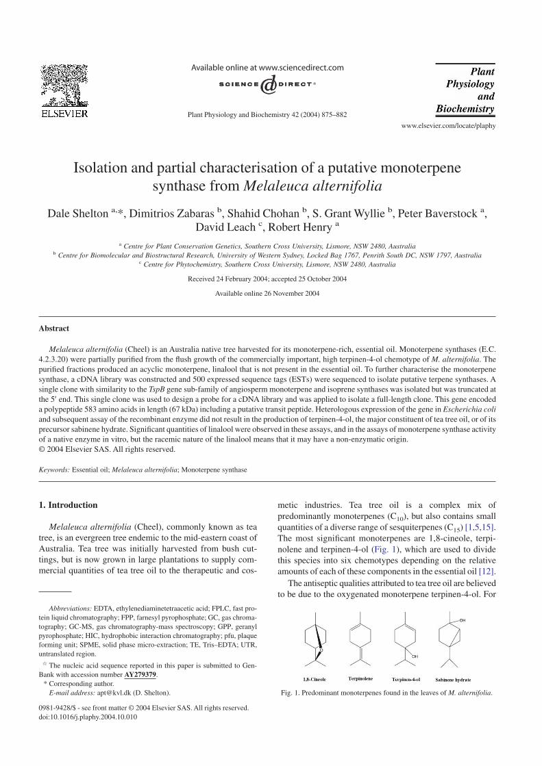
metic industries. Tea tree oil is a complex mix ofpredominantly monoterpenes (C10), but also contains smallquantities of a diverse range of sesquiterpenes (C15) [1,5,15].The most significant monoterpenes are 1,8-cineole, terpi-nolene and terpinen-4-ol (Fig. 1), which are used to dividethis species into six chemotypes depending on the relativeamounts of each of these components in the essential oil [12].
The antiseptic qualities attributed to tea tree oil are believedto be due to the oxygenated monoterpene terpinen-4-ol. For
Abbreviations: EDTA, ethylenediaminetetraacetic acid; FPLC, fast pro-tein liquid chromatography; FPP, farnesyl pyrophosphate; GC, gas chroma-tography; GC-MS, gas chromatography-mass spectroscopy; GPP, geranylpyrophosphate; HIC, hydrophobic interaction chromatography; pfu, plaqueforming unit; SPME, solid phase micro-extraction; TE, Tris–EDTA; UTR,untranslated region.
> The nucleic acid sequence reported in this paper is submitted to Gen-Bank with accession number AY279379.
* Corresponding author.E-mail address: [email protected] (D. Shelton). Fig. 1. Predominant monoterpenes found in the leaves of M. alternifolia.
Plant Physiology and Biochemistry 42 (2004) 875–882
www.elsevier.com/locate/plaphy
0981-9428/$ - see front matter © 2004 Elsevier SAS. All rights reserved.doi:10.1016/j.plaphy.2004.10.010
this reason international standards [13] dictate that tea treeoil has a minimum terpinen-4-ol content of 30%. Terpinen-4-ol in the essential oil of M. alternifolia is not an immediateproduct of a monoterpene synthase but is derived by rear-rangement from another oxygenated monoterpene, sabinenehydrate. In the leaves of M. alternifolia sabinene hydrate ispredominantly converted to terpinen-4-ol but also to smalleramounts c-terpinene and a-terpinene as the leaves age [21].The skeletal rearrangement of sabinene hydrates to terpinen-4-ol can be replicated in vitro by acidification and may beindicative of the mechanism of the conversion in M. alterni-folia [7]. Sabinene hydrates are not present in the distilled teatree oil as any sabinene hydrates in the tea tree leaf at thetime of harvest are converted to terpinen-4-ol by the steamdistillation process [7].
Numerous studies have been undertaken to examine thegenetic and environmental factors controlling essential oilcomposition in M. alternifolia. Analysis of the essential oilcomposition in the natural population of M. alternifolia showsa distinct clustering of chemotypes within certain geographicregions [12]. Isozyme [6] and microsatellite [17] analysis ofthe natural population indicate that there is gene flow amongmost of the population. These findings, in combination, sug-gest a strong environmental influence on the composition theessential oil. This is contrary to recent findings, in which con-trolled crosses among M. alternifolia chemotypes indicateda substantial level of genetic influence on oil composition inthis species [19]. This investigation aimed to isolate and char-acterise monoterpene synthases in M. alternifolia to partiallyunravel the complexity of genetic and environmental factorsinfluencing essential oil biosynthesis.
An in-depth understanding of the genetic and environmen-tal factors influencing the essential oil composition in M.alternifolia is desirable as this species is not only a commer-cially important crop, but also has the ability to serve as amodel species for essential oil production in other membersof the Myrtaceae family. The Myrtaceae family is predomi-nant in Australia and is emerging as a commercially impor-tant source of unique essential oils.
2. Results and discussion
2.1. Native enzyme purification and analysis
Isolation of the monoterpene synthases involved in essen-tial oil biosynthesis of M. alternifolia was initially attemptedusing a reverse genetics approach. Crude and partially puri-fied native enzyme extracts were prepared from the three pre-dominant M. alternifolia chemotypes, namely high 1,8-cineole, high terpinolene and high terpinen-4-ol, and assayedfor monoterpene synthase activity. After each round of puri-fication, the fractions collected were assayed to ascertain thosethat contained the monoterpene synthases and to assess whichmonoterpenes were produced. Native monoterpene syn-thases from the high 1,8-cineole and the high terpinolene
chemotypes yielded fractions that, when supplied with gera-nyl pyrophosphate (GPP), produced predominantly 1,8-cineole and terpinolene, respectively. Interestingly, when thestarting crude extract of the flush growth from the highterpinen-4-ol chemotype of M. alternifolia was supplied withGPP, none of the monoterpenes commonly observed in thisspecies could be detected. The predominant monoterpenedetected was linalool with minor quantities of limonene andE- and Z-ocimene also present (data not shown). The profileof the monoterpenes generated changed as the active frac-tions passed through the different purification steps. Furtherpurification of the active fractions failed to yield monoter-pene synthase activity, which produced monoterpenes com-mon in M. alternifolia. After the final step in the native pro-tein purification, a single fraction yielding only linalool wasobtained.
Although the protein purification from the high terpinen-4-ol chemotype did not display activity reflecting the essen-tial oil profile of the plant, the fact that volatiles were notdetected in all protein fractions (data not shown) and that sig-nificant linalool production was not observed in the proteinpurifications from the other chemotypes performed underidentical conditions, supports the view that the observed pro-duction of linalool was generated by enzymatic activity andwas not a solvolysis product or other chemical artefact.
The protein fraction showing monoterpene synthase activ-ity did not bind to a hydrophobic column. This indicates thatthe protein has little or no surface hydrophobicity. Themolecular weight of the putative monoterpene synthase inthe linalool producing fraction was determined to be about64 kDa by gel filtration chromatography. These characteris-tics are in agreement with those exhibited by most monoter-pene synthases [8].
2.2. Cloning, expression and analysis of a putativemonoterpene synthase
The application of degenerate PCR with primers designedto conserved amino acid motifs in other angiosperm terpenesynthases [23] met with little success. As an alternative, anexpressed sequence tag (EST) approach was employed.Sequencing of 500 ESTs derived from the flush growth of thecommercially important high terpinen-4-ol chemotype of M.alternifolia resulted in the isolation of a single, putativemonoterpene synthase clone. This clone was truncated to alength of approximately 600 bp, but was able to providesequence information that permitted the generation of a PCRproduct suitable for library screening. Screening of approxi-mately 20,000 plaque forming unit (pfu) isolated four puta-tive monoterpene synthases, which upon sequencing wererevealed to be the same gene.
Phylogenetically, the M. alternifolia synthase clusters mostclosely with the Tpsb terpene synthase sub-family of an-giosperm monoterpene synthases. The Tspb sub-family wasinitially defined as those terpene synthases sharing a mini-mum of 40% identity at the protein level [3]. The M. alterni-
876 D. Shelton et al. / Plant Physiology and Biochemistry 42 (2004) 875–882
folia synthase displayed the highest identity to the isoprenesynthase from poplar (Populus alba × Populus tremula) [16]and a myrcene synthase from Holm oak (Quercus ilex) [9]with 51% and 44% identity, respectively.
Along with sequence identity within the gene sub-families,the structural differences between the gene sub-families arealso indicative of the class of reaction catalysed. The mostobvious structural difference is the presence of the putativeplastid transit peptide at the amino terminus. The plastid tran-sit peptides in monoterpene synthases are characterised bybeing rich in serine and threonine residues prior to an abso-lutely conserved, RR(X8)W motif [4].
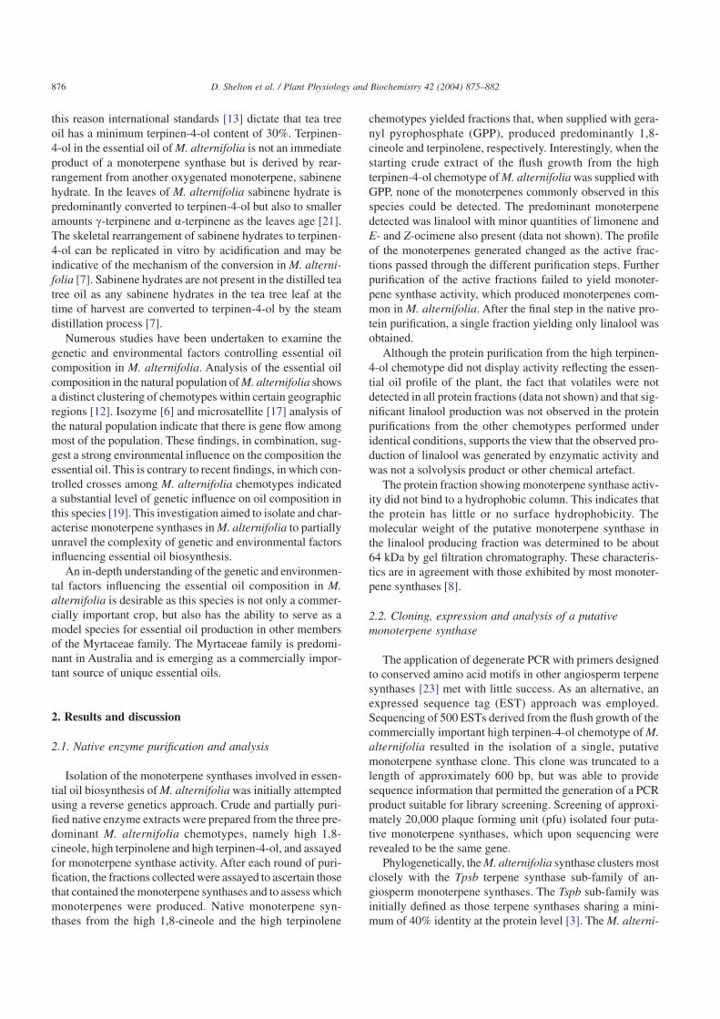
Examination of the putative M. alternifolia synthase aminoterminus revealed the presence four arginine doublets, Arg17
Arg18, Arg38 Arg39, Arg62 Arg63 and Arg65 Arg66, in theN-terminal region of the protein (Fig. 2). Sequence align-ment of the M. alternifolia synthase protein with the otherangiosperm and gymnosperm monoterpene synthases indi-cate that the Arg38 Arg39 doublet approximates the beginningof the mature peptide with the absolutely conserved tryp-tophan residue (Trp48) occurring only after this second dou-blet. In general the transit peptides of monoterpene synthasesare 50–70 amino acids in length [3], which is slightly longerthan that of the putative M. alternifolia synthase. The nucle-otide sequence immediately upstream of the putative startcodon of the M. alternifolia gene, assumed to be untrans-lated region (UTR), when translated could encode 19 aminoacids similar to the amino acid sequence of a transit peptide.It is feasible that the true start codon may be encoded furtherupstream of this putative UTR and that the methionine resi-due currently designated as the translation start site is coin-cidently contained within the true transit peptide sequence.Although this may be a likely scenario, it is believed to haveno consequence on the function of the enzyme in vitro as it isencoded upstream of the putative in vivo peptide cleavagesite.
Truncation of the protein immediately prior to Arg38 Arg39
results in a putative monoterpene synthase of 546 amino acidsin length with a molecular mass of 63 kDa and an isoelectricpoint at pH 5.9. This approximately corresponds with the esti-
mated molecular mass of the native monoterpene synthasesfrom M. alternifolia of 64 kDa. Despite the approximation ofthe beginning of the mature protein, two truncated constructswere made immediately prior to Arg17 Arg18 and Arg38 Arg39
in an attempt to produce a gene construct that closely mimicsthat found in planta and with optimal enzymatic activity.
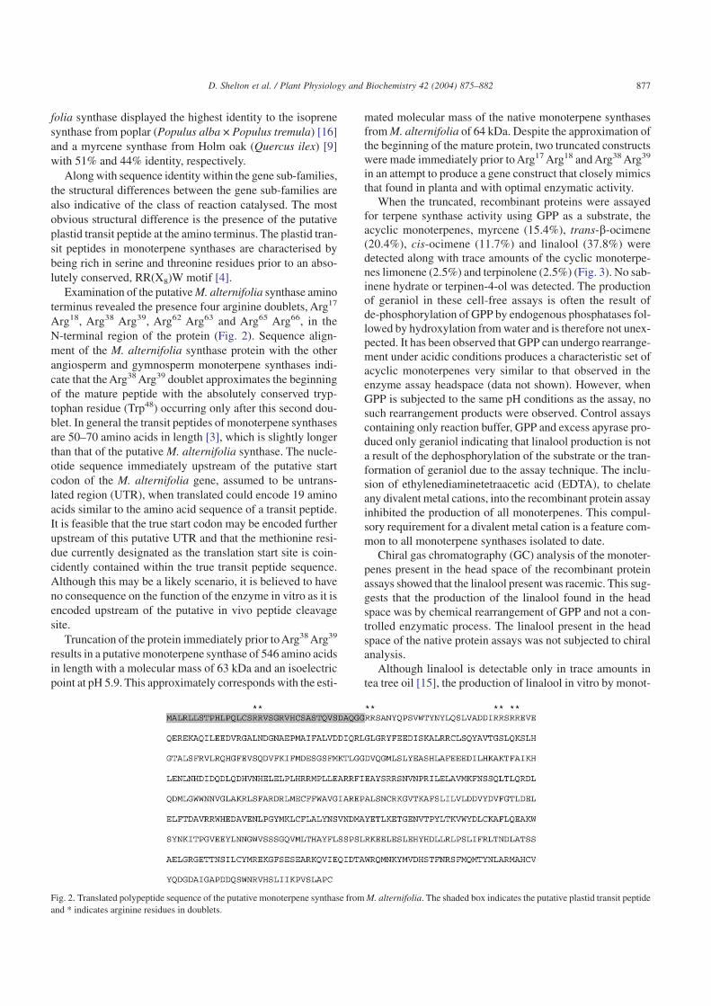
When the truncated, recombinant proteins were assayedfor terpene synthase activity using GPP as a substrate, theacyclic monoterpenes, myrcene (15.4%), trans-b-ocimene(20.4%), cis-ocimene (11.7%) and linalool (37.8%) weredetected along with trace amounts of the cyclic monoterpe-nes limonene (2.5%) and terpinolene (2.5%) (Fig. 3). No sab-inene hydrate or terpinen-4-ol was detected. The productionof geraniol in these cell-free assays is often the result ofde-phosphorylation of GPP by endogenous phosphatases fol-lowed by hydroxylation from water and is therefore not unex-pected. It has been observed that GPP can undergo rearrange-ment under acidic conditions produces a characteristic set ofacyclic monoterpenes very similar to that observed in theenzyme assay headspace (data not shown). However, whenGPP is subjected to the same pH conditions as the assay, nosuch rearrangement products were observed. Control assayscontaining only reaction buffer, GPP and excess apyrase pro-duced only geraniol indicating that linalool production is nota result of the dephosphorylation of the substrate or the tran-formation of geraniol due to the assay technique. The inclu-sion of ethylenediaminetetraacetic acid (EDTA), to chelateany divalent metal cations, into the recombinant protein assayinhibited the production of all monoterpenes. This compul-sory requirement for a divalent metal cation is a feature com-mon to all monoterpene synthases isolated to date.
Chiral gas chromatography (GC) analysis of the monoter-penes present in the head space of the recombinant proteinassays showed that the linalool present was racemic. This sug-gests that the production of the linalool found in the headspace was by chemical rearrangement of GPP and not a con-trolled enzymatic process. The linalool present in the headspace of the native protein assays was not subjected to chiralanalysis.
Although linalool is detectable only in trace amounts intea tree oil [15], the production of linalool in vitro by monot-
Fig. 2. Translated polypeptide sequence of the putative monoterpene synthase from M. alternifolia. The shaded box indicates the putative plastid transit peptideand * indicates arginine residues in doublets.
877D. Shelton et al. / Plant Physiology and Biochemistry 42 (2004) 875–882
erpene synthases isolated from plants that do not normallyproduce linalool has been observed previously. Two (3R)-linalool synthases have been isolated previously from Arte-misia annua, a herb that does not normally produce linalool[14]. The Km value of these enzymes was significantly higherthan that observed in any monoterpene synthases isolated priorand indicated a rate of linalool biosynthesis that is physiologi-cally unlikely.
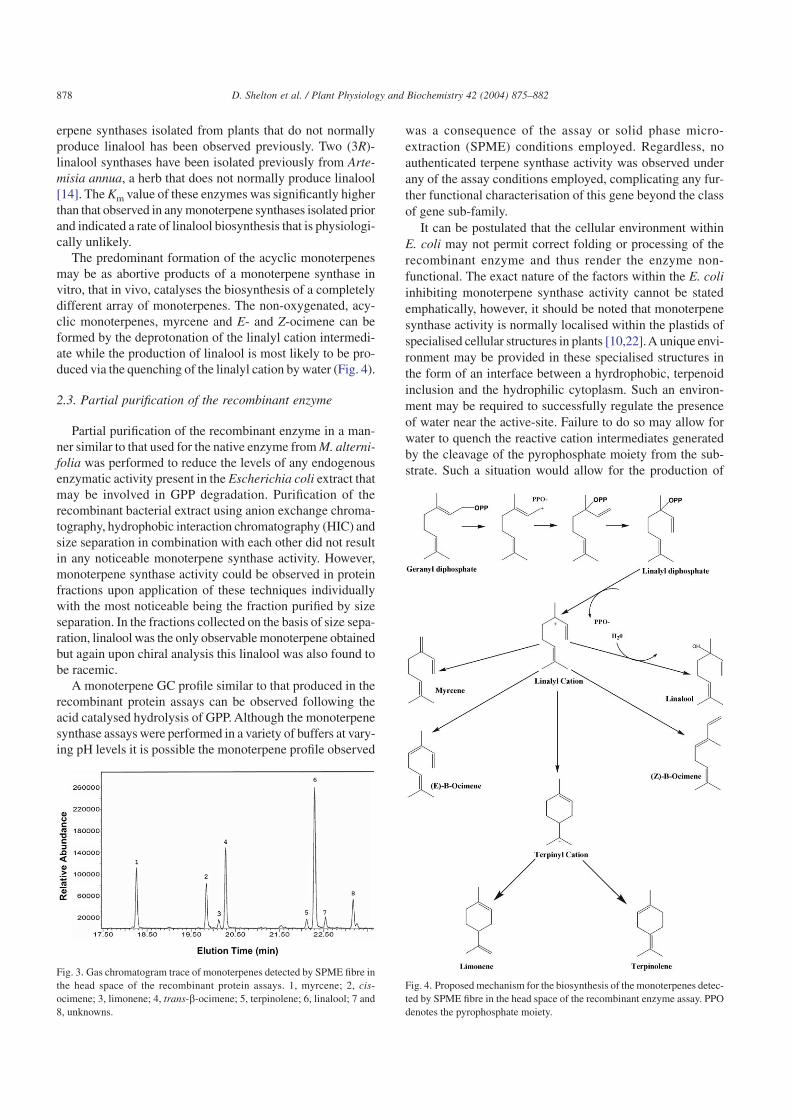
The predominant formation of the acyclic monoterpenesmay be as abortive products of a monoterpene synthase invitro, that in vivo, catalyses the biosynthesis of a completelydifferent array of monoterpenes. The non-oxygenated, acy-clic monoterpenes, myrcene and E- and Z-ocimene can beformed by the deprotonation of the linalyl cation intermedi-ate while the production of linalool is most likely to be pro-duced via the quenching of the linalyl cation by water (Fig. 4).
2.3. Partial purification of the recombinant enzyme
Partial purification of the recombinant enzyme in a man-ner similar to that used for the native enzyme from M. alterni-folia was performed to reduce the levels of any endogenousenzymatic activity present in the Escherichia coli extract thatmay be involved in GPP degradation. Purification of therecombinant bacterial extract using anion exchange chroma-tography, hydrophobic interaction chromatography (HIC) andsize separation in combination with each other did not resultin any noticeable monoterpene synthase activity. However,monoterpene synthase activity could be observed in proteinfractions upon application of these techniques individuallywith the most noticeable being the fraction purified by sizeseparation. In the fractions collected on the basis of size sepa-ration, linalool was the only observable monoterpene obtainedbut again upon chiral analysis this linalool was also found tobe racemic.
A monoterpene GC profile similar to that produced in therecombinant protein assays can be observed following theacid catalysed hydrolysis of GPP. Although the monoterpenesynthase assays were performed in a variety of buffers at vary-ing pH levels it is possible the monoterpene profile observed
was a consequence of the assay or solid phase micro-extraction (SPME) conditions employed. Regardless, noauthenticated terpene synthase activity was observed underany of the assay conditions employed, complicating any fur-ther functional characterisation of this gene beyond the classof gene sub-family.
It can be postulated that the cellular environment withinE. coli may not permit correct folding or processing of therecombinant enzyme and thus render the enzyme non-functional. The exact nature of the factors within the E. coliinhibiting monoterpene synthase activity cannot be statedemphatically, however, it should be noted that monoterpenesynthase activity is normally localised within the plastids ofspecialised cellular structures in plants [10,22].A unique envi-ronment may be provided in these specialised structures inthe form of an interface between a hyrdrophobic, terpenoidinclusion and the hydrophilic cytoplasm. Such an environ-ment may be required to successfully regulate the presenceof water near the active-site. Failure to do so may allow forwater to quench the reactive cation intermediates generatedby the cleavage of the pyrophosphate moiety from the sub-strate. Such a situation would allow for the production of
Fig. 3. Gas chromatogram trace of monoterpenes detected by SPME fibre inthe head space of the recombinant protein assays. 1, myrcene; 2, cis-ocimene; 3, limonene; 4, trans-b-ocimene; 5, terpinolene; 6, linalool; 7 and8, unknowns.
Fig. 4. Proposed mechanism for the biosynthesis of the monoterpenes detec-ted by SPME fibre in the head space of the recombinant enzyme assay. PPOdenotes the pyrophosphate moiety.
878 D. Shelton et al. / Plant Physiology and Biochemistry 42 (2004) 875–882
monoterpene alcohols such as linalool. Any future investiga-tions into the functional identity of this putative monoter-pene synthase should consider the possibility of an in plantaassay that may allow such a cellular environment to be repli-cated.
3. Conclusion
It can be ascertained that M. alternifolia putative terpenesynthase belongs to the Tspb gene sub-family of angiospermisoprene and monoterpene synthases. This conclusion is notonly supported by the level of identity at the amino acid levelbut also by the presence of a putative plastid transit peptideand the presence of various structural motifs, including theRR(X8)W motif. These latter two structural elements are abso-lutely conserved in isoprene and monoterpene synthases buthave not been observed in sesquiterpene synthases. Compari-son of the amino acid sequence of the putative M. alternifo-lia monoterpene synthase with that of other members of theTspb gene sub-family failed to reveal any conserved motifsor residues lacking from the M. alternifolia sequence thatmay render it non-functional.
The results of the assays of terpene synthase activity ofboth recombinant and native enzyme preparations of the highterpinen-4-ol chemotype of M. alternifolia are consistent. Nei-ther shows the expected sabinene hydrate or terpinen-4-olcompounds among the terpenes produced by these enzymes.While the terpenes that were produced had a composition andchirality expected from the chemical transformation of GPPno control experiments were able to demonstrate that this wasthe case. Our current interpretation of the results is that thesabinene hydrate synthase isolated by the molecular and bio-chemical approaches has been crippled in some way by theextraction or purification techniques used and no longer dis-plays its in vivo capacity to generate sabinene hydrate.
4. Methods
4.1. Plant material, substrates and reagents
M. alternifolia (Cheel) seedlings were grown in a green-house with regular watering and pruning to maintain a con-stant supply of flush growth. All chemical substrates andreagents were obtained from Sigma Chemical Co. unless oth-erwise stated.All DNA sequencing was performed by theAus-tralian Genomic Research Facility (AGRF).
4.2. RNA isolation and cDNA library construction
RNA isolation and cDNA library construction was per-formed as previously described [20] but briefly, approxi-mately 3 g of young shoot tips, approximately 2.5 cm inlength, from the high terpinen-4-ol chemotype of M. alterni-folia (Cheel) were ground in liquid nitrogen prior to RNA
extraction. Poly(A) RNA was isolated from the total RNAusing PolyATtract mRNA Isolation System (Promega) as perthe manufacturer’s guidelines. The synthesis of cDNA andthe ligation of adaptors were performed using the kZAPIIcDNA synthesis and Vector kit (Stratagene). The resultingcDNA was directionally ligated into the kZapII vector andpackaged using GigaPack Gold III (Stratagene) as per themanufacturer’s guidelines. The resulting primary library con-tained 1 × 10⁶ pfu.
4.3. Nucleotide sequencing and analysis
A mass excision of pBluescript SK+ from an amplifiedcDNA library was performed using VCS M13 helper phageand E. coli XL1 Blue MRF’ as recommended in the manu-facturer guidelines. Plasmids were extracted and ESTs wereobtained for approximately 500 clones. Each sequenceobtained was edited using AssemblyLIGN (Oxford Molecu-lar) to remove flanking vector sequence and assessed manu-ally to determine sequence quality. Comparison of the DNAsequences to non-redundant protein sequence databases at theNational Centre for Biotechnology (NCBI) and the Euro-pean Molecular Biology Laboratory (EMBL) was performedusing the Baylor College of Medicine Search Launcher(http://www.dot.imgen.bcm.tmc.edu:9331). A single clonewith similarity to a putative limonene synthase from Arabi-dopsis thaliana was isolated. This clone was severelytruncated to approximately 600 bp in length. Primers3B1F (5′-GGTTGCCGTCCTTGATTT-3′) and 3B1R (5′-ATCGCATCTCCGTCTTGGT-3′) were designed to thissequence to rapidly yield a 280 bp fragment that could bediagnostic in PCR-based library screening as well as used asa probe for conventional hybridisation-based library screen-ing.
4.4. cDNA library screening
To screen the library, a 280 bp probe was generated usingprimers 3B1R and 3B1F. The resulting fragment was gel puri-fied and labelled using [a-P32] dATP and the random primingbased Gigaprime DNA labelling kit (Bresatec) as per themanufacturer’s instructions. The labeled probe was used toscreen replica nylon membranes of 2.5 × 10⁴ pfu plated in E.coli XL1 Blue cells. The membranes were pre-hybridised in6× SSC, 5× Denhardt’s solution, 0.5% SDS, 15% formamideand 10 µg ml–1 sonicated denatured salmon sperm DNA solu-tion at 55 °C for 2 h prior to an 18 h hybridisation in the samesolution with the labelled probe added. The membranes werewashed three times in 2× SSC and 0.1% SDS solution at 55 °Cbefore being exposed to a phosphor screen overnight andscanned using a Storm Phosphoimager (Molecular Dynam-ics). A secondary round of PCR-based screening was per-formed using the primers 3B1R and 31BF to obtain singleclones putatively containing monoterpene synthases. The invivo excision of pBluescript SK+ from kZapII and subse-quent transfection into E. coli SOLR cells was performed as
879D. Shelton et al. / Plant Physiology and Biochemistry 42 (2004) 875–882
per the manufacture’s guidelines (Stratagene). Sequencing ofthe resultant plasmids from both the 3′ and 5′ ends usingM13 forward and reverse primers indicated that each of theseclones encoded the same gene truncated to varying lengths.A single, full-length clone was isolated and used for all fol-lowing steps.
4.5. cDNA modification and recombinant proteinexpression
To facilitate the efficient production of the recombinantenzyme in E. coli, all expression constructs were created usingthe expression vector pSBETa, which encodes the argU genefor tRNAArg4, to overcome translational difficulties associ-ated with codon usage between eukaryotes and prokaryotes[18]. To introduce suitable restriction sites for sub-cloninginto this vector and to create truncated gene constructs withthe presumptive plastid transit peptide removed a PCR-basedstrategy was adopted. Two truncated fragments were gener-ated, each containing a new start codon and NdeI restrictionsite, using the two forward primers Arg17F (5′ TGC TCTCAT ATG CGG AGG GTC AGT GGT CGA 3′), Arg35F (5′AGG ACA TAT GCG AAG ATC GGC CAA TTA TCA 3′)in combination with the reverse primer BamRev (5′ TTT TCCGGA TCC AAA GGG AGG ATA TGA TC 3′). The PCRreactions was performed using 1.5 units Pfx DNA poly-merase (GibcoBRL) in 1× Pfx amplification buffer (manu-facturer supplied) supplemented with 1 mM magnesium sul-phate, 400 mM of each dNTP, 0.4 µM of each primer and100 ng of template, in a final volume of 25 µl. The cyclingconditions for each PCR were as follows; 94 °C for 45 s fol-lowed by 35 cycles of 94 °C for 15 s, 55 °C for 15 s and 68 °Cfor 1 min 30 s, completion of the cycle was followed by afinal extension for 7 min at 68 °C. The resulting PCR prod-ucts were gel purified and digested with NdeI and Bam HI(New England Biolabs) as per the manufacturer’s guidelines.The expression vector pSBETa was also digested using NdeIand Bam HI followed by ethanol precipitation and resuspen-sion in Tris–EDTA (TE) buffer (pH 8.0). To inhibit selfre-ligation, cut pSBETa was treated with excess calf intesti-nal alkaline phosphatase (CIAP) (Promega). At the comple-tion of the reaction the CIAP was heat inactivated at 65 °Cfor 30 min. Each truncated fragment was ligated into the NdeIdigested plasmid and transformed into E. coli BL21 (DE3).The ligation resulted in the production of two expression con-structs designated pSMATS17 and pSMATS35 and re-sequenced to confirm sub-cloning fidelity.
4.6. Recombinant protein expression and productidentification
For heterologous expression, each construct was trans-formed in E. coli BL21 (DE3) and grown at 37 °C to OD600
0.5 in LB broth supplemented with 30 µg ml–1 kanamycin.Recombinant gene expression was induced at this point by theaddition of 1 mM isopropyl-1-thio-b-D-galactopyranoside and
grown for a further 12 h at 25 °C. For each construct, 10 mlof cells were harvested by centrifugation at 2000 × g for10 min and resuspended in 2 ml of assay buffer. Cells weredisrupted by sonication in an ice-water bath for 45 s at maxi-mum power using a Unisonics ultra sonic cleaner. The bac-terial lysates were cleared by centrifugation at 18,000 × g for10 min at 4 °C then transferred to 7 ml teflon-capped, headspace auto-sampler vials.
Three buffers similar to those used for recombinant monot-erpene synthase assays in Arabidopisis [2] and Salvia [23] atpH 6.5, 7.0 and 7.5 were used in the heterologous assays.These consisted of 50 mM MOPS (pH 6.5) or 50 mM Tris–HCl (pH 7.0 or 7.5) with 10% (v/v) glycerol, 10 mM MgCl2,1 mM MnCl2 and 5 mM dithiothreitol.A modified buffer usedto assay native sabinene hydrate synthase activity in Majo-rana hortensis [11] consisting of 10 mM sodium phosphate(pH 7.0), 10% (v/v) glycerol, 0.5 mM dithiothreitol, 1 mMascorbic acid and supplemented with 10 mM MgCl2 was alsoassayed. Terpene synthase activity of the bacterial extract wasinitiated by the addition of 10 µl of 2.74 mM GPP to eachextract. Each reaction was then sealed in the head space vialand incubated with shaking at 22 °C for 3–6 h. To confirmthe monoterpene products of the enzyme extract were not theproduct of contaminating phosphatase activity, aliquots ofeach buffer containing 10 µl of 2.74 mM GPP were treatedwith excess apyrase (Sigma). The products from the enzymeextract and buffer reactions were then compared. Reactionproduct sampling was performed using a polydimethylsilox-ane (PDMS)-coated SPME fibre (Supelco). Each sample washeated at 80 °C for 10 min prior to 30 min exposure of theSPME fiber to the sample head space. Reaction products ofthe recombinant synthase were identified using a Hewlett–Packard 6890 GC coupled to a Hewlett–Packard 5973 massselective detector and interfaced with a Hewlett–PackardChemstation. The GC was fitted with an SGE BPX5, fusedsilica, capillary column (50 m length × 0.22 mm inner diam-eter). Thermal desorption of the SPME fiber was performedin the front inlet at 260 °C with 24 psi injection pressure usinga helium carrier. Oven programming was as follows: initialtemperature 45 °C (2 min hold), 5 °C min–1 to 100 °C,4 °C min–1 to 260 °C (5 min hold) with splitless injection.Full spectra were recorded and peaks were identified by com-parison to Wiley275 and NBS75K libraries. Chirality of thereaction products was examined using a Hewlett–Packard6890 gas chromatograph-flame ionisation detector (GC-FID) (H2 carrier gas 1.6 ml min–1, splitless injection, inlettemperature 250 °C) fitted with a 50QC2/CYDEX column(50 m × 0.22 mm id × 0.25 µm film thickness) (SGE). Theoven programming was as follow: 50 °C (1 min hold) increas-ing 4 °C min–1 to 220 °C (1 min hold). Compounds wereidentified by the comparison of retention times with authen-tic standards.
4.7. Native protein isolation and product identification
4.7.1. Enzyme extractionLeaf material (~5 g) was ground with liquid N2 to a fine
powder. Extraction buffer (50 mM Tris, 1 mM MnCl2, 10 mM
880 D. Shelton et al. / Plant Physiology and Biochemistry 42 (2004) 875–882
MgCl2, 20% (v/v) glycerol, adjusted to pH 6.9 with HCl)(25 ml), polyvinylpolypyrrolidone (0.1 g) and amberliteXAD-4 (1 g) were added and the slurry filtered throughcheesecloth. The filtrate was centrifuged at 10,000 × g for10 min at 4 °C, the supernatant was collected and further cen-trifuged at 20,000 × g for 60 min at 4 °C. The supernatantwas then passed through a 0.45 µm syringe filter (Nalgate),placed in a centrifugal concentrator and centrifuged(6000 × g at 4 °C) to a volume of about 10 ml. An equalvolume of assay buffer (pH 7.2) was then added and the extractwas again concentrated (6000 × g at 4 °C) to about 10 ml.This process was repeated five times (5 × 10 ml) to ensure theextract was free of contaminating terpenoids. The resultingcrude enzyme extract was assayed for activity as above and,when required, was purified further by fast protein liquid chro-matography (FPLC). An ÄKTA™ FPLC equipped with aFrac-950 fraction collector was used. The system was con-trolled via UNICORN™ (ver. 3.11) software. All FPLC col-umns and accessories were from Amersham Pharmacia Bio-tech. A Hi-Trap Q HP (5 ml bed volume of Q-Sepharose)was used for ion-exchange chromatography. Before use thecolumn was pre-equilibrated (×5 column volumes) with assaybuffer. The crude enzyme extract was injected and the boundfraction was eluted with a gradient (0–100%) of a high saltbuffer (1 M KCl). The bound fraction was then desaltedthrough a Hi-Trap Desalting column (Sephadex G-25 Super-fine bed), assayed and passed through a MonoQ HR 5/5 col-umn (1 ml bed volume). The bound fraction was then elutedwith a gradient of 1 M KCl. A small portion (1 ml) of thebound fraction was desalted and assayed. The remainder ofthe bound fraction was concentrated by centrifugation (20,000× g at 4 °C) to about 2 ml and subjected to HIC. A ResourcePHE (1 ml bed volume, phenyl hydrophobic ligands phase)column was used for the HIC. The concentrated high salt frac-tion from above was passed through the pre-equilibrated (withhigh salt buffer) column using a reverse phase gradient thistime (i.e. concentration of salt from 100% to 0%).After assay-ing, the fraction of interest (unbound) was subjected to gelfiltration. The desalted unbound fraction (100 µl) from HICwas passed through a Superose 12HR 10/30 column andeluted isocratically (no salt buffer). The retention volume forthe peak showing activity was used to estimate molecular sizeagainst a calibration table constructed using four lyophilisedprotein standards. The proteins (Ribonuclease A 13.7 kDa,BSA 67 kDa, aldolase 158 kDa, catalase 232 kDa) (Amer-sham Pharmacia Biotech) were reconstituted with assay bufferto a 10 mg ml–1 concentration and mixed. The mixture (100 µl)was then passed through the Superose 12HR 10/30 column.
Acknowledgments
The authors would like to thank and acknowledge the con-tributions of Mr. Ashley Dowell from the Centre for Phy-tochemistry, Southern Cross University, Lismore, Australia,for training on the GC-MS and his assistance in establishing
GC methods for the analysis of monoterpenes in the headspace of the recombinant protein assay. The author wouldalso like to acknowledge that this work was supported by anAustralian Research Council SPIRT grant.
References
[1] G.R. Baker, R.F. Lowe, I.A. Southwell, Comparison of oil recoveredfrom tea tree leaf by ethanol extraction and steam distillation, J. Agric.Food Chem. 48 (2000) 4041–4043.
[2] J. Bohlmann, D. Martin, N.J. Oldham, J. Gershenzon, Terpenoidsecondary metabolism in Arabidopsis thaliana: cDNA cloning, char-acterization, and functional expression of a myrcene/(E)-beta-ocimene synthase, Arch. Biochem. Biophys. 375 (2000) 261–269.
[3] J. Bohlmann, G. Meyergauen, R. Croteau, Plant terpenoidsynthases—molecular biology and phylogenetic analysis, Proc. Natl.Acad. Sci. USA 95 (1998) 4126–4133.
[4] J. Bohlmann, C.L. Steele, R. Croteau, Monoterpene synthases fromgrand fir (Abies grandis)—cDNA isolation, characterization, andfunctional expression of myrcene synthase, (4s)-limonene synthase,and (1s,5s)-pinene synthase, J. Biol. Chem. 272 (1997) 21784–21792.
[5] J. Brophy, N. Davies, I. Southwell, I. Stiff, L. Williams, Gas chro-matographic quality control for oil of Melaleuca alternifolia terpinen-4-ol type (Australian Tea Tree), J. Agric. Food Chem. 37 (1989)1330–1335.
[6] P.A. Butcher, J.C. Bell, G.F. Moran, Patterns of genetic diversity andnature of breeding systems in Melaleuca alternifolia (Myrtaceae),Aust. J. Bot. 40 (1992) 365–375.
[7] C. Cornwell, D. Leach, S.G. Wyllie, Incorporation of oxygen-18 intoterpinen-4-ol from the H218O steam distillates of Melaleuca alterni-folia (Tea Tree), J. Essent. Oil Res. 7 (1995) 613–620.
[8] R. Croteau, E. Cane, Monoterpene and sesquiterpene cyclases, in:J.H. Law, H.C. Rilling (Eds.), Methods Enzymol, Academic PressInc., Orlando, 1985, pp. 383–405 vol. 110.
[9] R.J. Fischbach, W. Zimmer, J.P. Schnitzler, Isolation and functionalanalysis of a cDNA encoding a myrcene synthase from Holm oak(Quercus ilex L.), Eur. J. Biochem. 268 (2001) 5633–5638.
[10] J. Gershenzon, M. Maffei, R. Croteau, Biochemical and histochemi-cal localization of monoterpene biosynthesis in the glandular tri-chomes of spearmint (Mentha spicata), Plant Physiol. 89 (1989)1351–1357.
[11] T.W. Hallahan, R. Croteau, Monoterpene biosynthesis: demonstrationof a geranyl pyrophosphate: sabinene hydrate cyclase in solubleenzyme preparations from sweet marjoram (Majorana hortensis),Arch. Biochem. Biophys. 264 (1988) 618–631.
[12] L.E. Homer, D.N. Leach, D. Lea, L.S. Lee, R.J. Henry, P.R. Baver-stock, Natural variation in the essential oil content of Melaleucaalternifolia Cheel (Myrtaceae), Biochem. Syst. Ecol. 28 (2000) 367–382.
[13] International Standards Organisation, Oil of Melaleuca, Terpinen-4-ol Type, International Standards Organisation, Geneva, 1996.
[14] J.W. Jia, J. Crock, S. Lu, R. Croteau, X.Y. Chen, 3R-Linalool synthasefrom Artemisia annua L.: cDNA isolation, characterization, andwound induction, Arch. Biochem. Biophys. 372 (1999) 143–149.
[15] D.N. Leach, S.G. Wyllie, J.G. Hall, I. Kyratzis, Enantiomeric compo-sition of the principal components of the oil of Melaleuca alternifolia,J. Agric. Food Chem. 41 (1993) 1627–1632.
[16] B. Miller, C. Oschinski, W. Zimmer, First isolation of an isoprenesynthase gene from poplar and successful expression of the gene inEscherichia coli, Planta 231 (2001) 483–487.
[17] M. Rossetto, R.W. Slade, P.R. Baverstock, R.J. Henry, L.S. Lee,Microsatellite variation and assessment of genetic structure in tea tree(Melaleuca alternifolia Myrtaceae), Mol. Ecol. 8 (1999) 633–643.
881D. Shelton et al. / Plant Physiology and Biochemistry 42 (2004) 875–882

[18] P. Schenk, S. Baumann, R. Mattes, H. Steinbis, Improved high-levelexpression systems for eukaryotic genes in E. coli using T7 RNApolymerase and rare Arg tRNAs, Biotechniques 19 (1995) 196–198.
[19] D. Shelton, K. Aitken, L. Doimo, D. Leach, P. Baverstock, R. Henry,Genetic control of monoterpene composition in the essential oil ofMelaleuca alternifolia (Cheel), Theor. Appl. Genet. 105 (2002) 377–383.
[20] D. Shelton, D. Leach, P. Baverstock, R. Henry, Isolation of genesinvolved in secondary metabolism from Melaleuca alternifolia(Cheel) using expressed sequence tags (ESTs), Plant Sci. 162 (2002)9–15.
[21] I.A. Southwell, I. Stiff, Ontogenetical changes in monoterpenoids ofMelaleuca alternifolia leaf, Phytochemistry 28 (1989) 1047–1051.
[22] G. Turner, J. Gershenzon, E.E. Nielson, J.E. Froehlich, R. Croteau,Limonene synthase, the enzyme responsible for monoterpene biosyn-thesis in peppermint, is localized to leucoplasts of oil gland secretorycells, Plant Physiol. 120 (1999) 879–886.
[23] M.L. Wise, T.J. Savage, E. Katahira, R. Croteau, Monoterpene syn-thases from common sage (Salvia offıcinalis)—cDNA isolation, char-acterization, and functional expression of (+)-sabinene synthase, 1,8-cineole synthase, and (+)-bornyl diphosphate synthase, J. Biol. Chem.273 (1998) 14891–14899.
882 D. Shelton et al. / Plant Physiology and Biochemistry 42 (2004) 875–882