Embed Size (px)
Citation preview
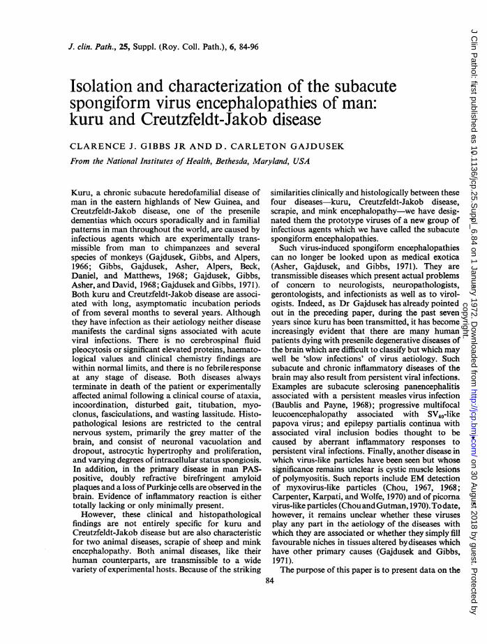
J. clin. Path., 25, Suppl. (Roy. Coll. Path.), 6, 84-96
Isolation and characterization of the subacutespongiform virus encephalopathies of man:kuru and Creutzfeldt-Jakob diseaseCLARENCE J. GIBBS JR AND D. CARLETON GAJDUSEK
From the National Institutes of Health, Bethesda, Maryland, USA
Kuru, a chronic subacute heredofamilial disease ofman in the eastern highlands of New Guinea, andCreutzfeldt-Jakob disease, one of the preseniledementias which occurs sporadically and in familialpatterns in man throughout the world, are caused byinfectious agents which are experimentally trans-missible from man to chimpanzees and severalspecies of monkeys (Gajdusek, Gibbs, and Alpers,1966; Gibbs, Gajdusek, Asher, Alpers, Beck,Daniel, and Matthews, 1968; Gajdusek, Gibbs,Asher, and David, 1968; Gajdusek and Gibbs, 1971).Both kuru and Creutzfeldt-Jakob disease are associ-ated with long, asymptomatic incubation periodsof from several months to several years. Althoughthey have infection as their aetiology neither diseasemanifests the cardinal signs associated with acuteviral infections. There is no cerebrospinal fluidpleocytosis or significant elevated proteins, haemato-logical values and clinical chemistry findings arewithin normal limits, and there is no febrileresponseat any stage of disease. Both diseases alwaysterminate in death of the patient or experimentallyaffected animal following a clinical course of ataxia,incoordination, disturbed gait, titubation, myo-clonus, fasciculations, and wasting lassitude. Histo-pathological lesions are restricted to the centralnervous system, primarily the grey matter of thebrain, and consist of neuronal vacuolation anddropout, astrocytic hypertrophy and proliferation,and varying degrees of intracellular status spongiosis.In addition, in the primary disease in man PAS-positive, doubly refractive birefringent amyloidplaques and a loss of Purkinje cells are observed in thebrain. Evidence of inflammatory reaction is eithertotally lacking or only minimally present.However, these clinical and histopathological
findings are not entirely specific for kuru andCreutzfeldt-Jakob disease but are also characteristicfor two animal diseases, scrapie of sheep and minkencephalopathy. Both animal diseases, like theirhuman counterparts, are transmissible to a widevariety of experimental hosts. Because of the striking
similarities clinically and histologically between thesefour diseases-kuru, Creutzfeldt-Jakob disease,scrapie, and mink encephalopathy-we have desig-nated them the prototype viruses of a new group ofinfectious agents which we have called the subacutespongiform encephalopathies.Such virus-induced spongiform encephalopathies
can no longer be looked upon as medical exotica(Asher, Gajdusek, and Gibbs, 1971). They aretransmissible diseases which present actual problemsof concern to neurologists, neuropathologists,gerontologists, and infectionists as well as to virol-ogists. Indeed, as Dr Gajdusek has already pointedout in the preceding paper, during the past sevenyears since kuru has been transmitted, it has becomeincreasingly evident that there are many humanpatients dying with presenile degenerative diseases ofthe brain which are difficult to classify but which maywell be 'slow infections' of virus aetiology. Suchsubacute and chronic inflammatory diseases of thebrain may also result from persistent viral infections.Examples are subacute sclerosing panencephalitisassociated with a persistent measles virus infection(Baublis and Payne, 1968); progressive multifocalleucoencephalopathy associated with SV40-likepapova virus; and epilepsy partialis continua withassociated viral inclusion bodies thought to becaused by aberrant inflammatory responses topersistent viral infections. Finally, another disease inwhich virus-like particles have been seen but whosesignificance remains unclear is cystic muscle lesionsof polymyositis. Such reports include EM detectionof myxovirus-like particles (Chou, 1967, 1968;Carpenter, Karpati, and Wolfe, 1970) and of picornavirus-like particles (ChouandGutman, 1970).To date,however, it remains unclear whether these virusesplay any part in the aetiology of the diseases withwhich they are associated or whether they simply fillfavourable niches in tissues altered bydiseases whichhave other primary causes (Gajdusek and Gibbs,1971).The purpose of this paper is to present data on the
84
copyright. on 30 A
ugust 2018 by guest. Protected by
http://jcp.bmj.com
/J C
lin Pathol: first published as 10.1136/jcp.25.S
uppl_6.84 on 1 January 1972. Dow
nloaded from
Isolation and characterization of the subacute spongiform virus encephalopathies ofman
C,,~ ~ ~N E E
.0 E Uc,,c
EE8
0 0
E E
6
._LD00
V; 4
E E
O LD
c U
.' m
w
aN E'oE<0< ,g" D . c m-
0~ ~ ~ ~
to NE E E
to*--
G)N 00
VoE Emcn
LOD-.
(NA..--.
LiLiic .0.2
VI'A
0 0
E E
(N
W; ;0 0
E ELD LD
(N.
E EN
85
C4
,a >
. -a
E °Em (n
r r-
4.. ..C C
Q0 -
_0
0 0E E
. . ,
,E
00E E
'C
*_E
I-4
00 0E E
,
.t_/
0
E ECZ0
.S:
.to
copyright. on 30 A
ugust 2018 by guest. Protected by
http://jcp.bmj.com
/J C
lin Pathol: first published as 10.1136/jcp.25.S
uppl_6.84 on 1 January 1972. Dow
nloaded from
86
isolation and characterization of the atypical virusesof kuru and Creutzfeldt-Jakob disease-two of theprototype subacute spongiform virus encephalo-pathies. The techniques employed in these studieshave been previously reported in detail (Gibbs andGajdusek, 1965) and have not undergone anysignificant change.
Kuru
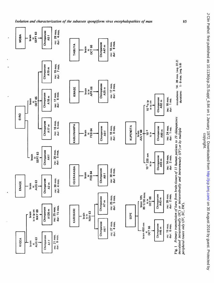
Kuru is an invariably fatal degenerative disease of thecentral nervous system, characterized by cerebellarataxia and trembling, progressive dysarthria and,finally, dysphagia, which has been observed only inthe Fore people and their neighbours who reside in arestricted part of the Eastern Highlands of NewGuinea (Gajdusek and Zigas, 1957; Gajdusek, 1963).The disease is caused by a filterable agent and is
Clarence J. Gibbs Jr and D. Carleton Gajdusek
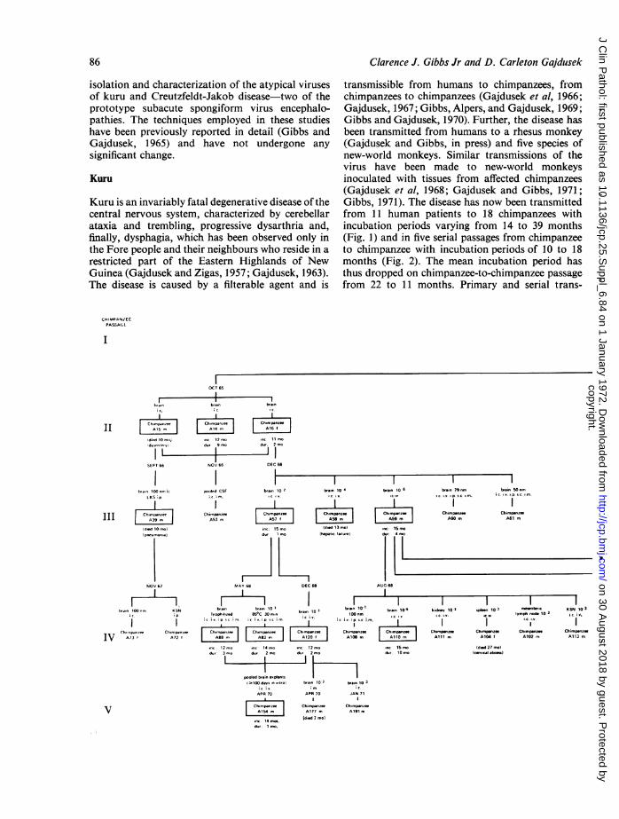
transmissible from humans to chimpanzees, fromchimpanzees to chimpanzees (Gajdusek et al, 1966;Gajdusek, 1967; Gibbs, Alpers, and Gajdusek, 1969;Gibbs and Gajdusek, 1970). Further, the disease hasbeen transmitted from humans to a rhesus monkey(Gajdusek and Gibbs, in press) and five species ofnew-world monkeys. Similar transmissions of thevirus have been made to new-world monkeysinoculated with tissues from affected chimpanzees(Gajdusek et al, 1968; Gajdusek and Gibbs, 1971;Gibbs, 1971). The disease has now been transmittedfrom 11 human patients to 18 chimpanzees withincubation periods varying from 14 to 39 months(Fig. 1) and in five serial passages from chimpanzeeto chimpanzee with incubation periods of 10 to 18months (Fig. 2). The mean incubation period hasthus dropped on chimpanzee-to-chimpanzee passagefrom 22 to 11 months. Primary and serial trans-
CHIMPANZCEPASSAGA
ic.
IIidird 10 mo}
SEPT 66
I.... 100nmiLKS. p.
III:Id.d 10 mo}
CNoiuo ChounoNOV 67
rVt"'n 100 n KSN
Ch -"J.*we Ch,mtpm.e.IV z3' A72
I~OCT 65I
;cv
|Ch,,mp..,,cI A14 m,
,.c: 12 mod., 9 I,,
NOV 66
i.c. m.
-1c,
biia 10 2.c,I".
b,.i 10 4
c v,
Chi.pa.zee Ch.opiToe ChimpaA53mA5f A5 I
inc: 1Smo. (died 13To)d., mo (hepattic tiure)
MAYb8DEC 68
[ I I I I-bVi, bb,.10 3bTi 101-
1voph,Ilted 85°C 30 m, rn 110itnm;cc; wip cimic iC ;. ip sc m c v. nm
Ch,mpono | ChFTmp.ne"'Ch.mp-ne| Chimpsiroe| A88 m A83 A 120 A1OS m
,c12 mo .c- 14 mo *c12 mod.,: 3 mo du,: 2 mo d., 2 mo
pooled biieiptamsIlO10 d.vsululCt b,.n 10 3 ts.n 10 3
APR 70 APR 70 JAN 71
Ch,mpn | Chimp-nee Ch..psnzttAl5 A177 m Al191 m
...1dietd 7 mo)
d.I
dduid1imo.
b,a.. 10 I
..C: 15 mo
d.r:4
AUG 68
ba6. 1065,.cF.
.nc 15S md.u: 10 m,o
bm. 79 nml.i. l.p.P. I0.
Chimp.uni"A60 m
b,sin 50 nmi'c. ..p. ' e. ..
ChimptnzecA61 m
I I1kidnev 101 .plen 10 3 ,Tuntiii KSN 10-3ffic. ... C lmyph nods 10-3 iiv.I ~~~Ii I
Chimpunze Chimpanzee Ch..puzee ChimopsteeAlil m A104 A102 m A113 m
Wind 27 mol(crvcitl abom
copyright. on 30 A
ugust 2018 by guest. Protected by
http://jcp.bmj.com
/J C
lin Pathol: first published as 10.1136/jcp.25.S
uppl_6.84 on 1 January 1972. Dow
nloaded from
Isolation and characterization of the subacute spongiform virus encephalopathies of man
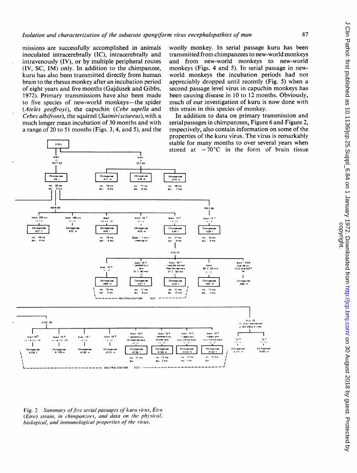
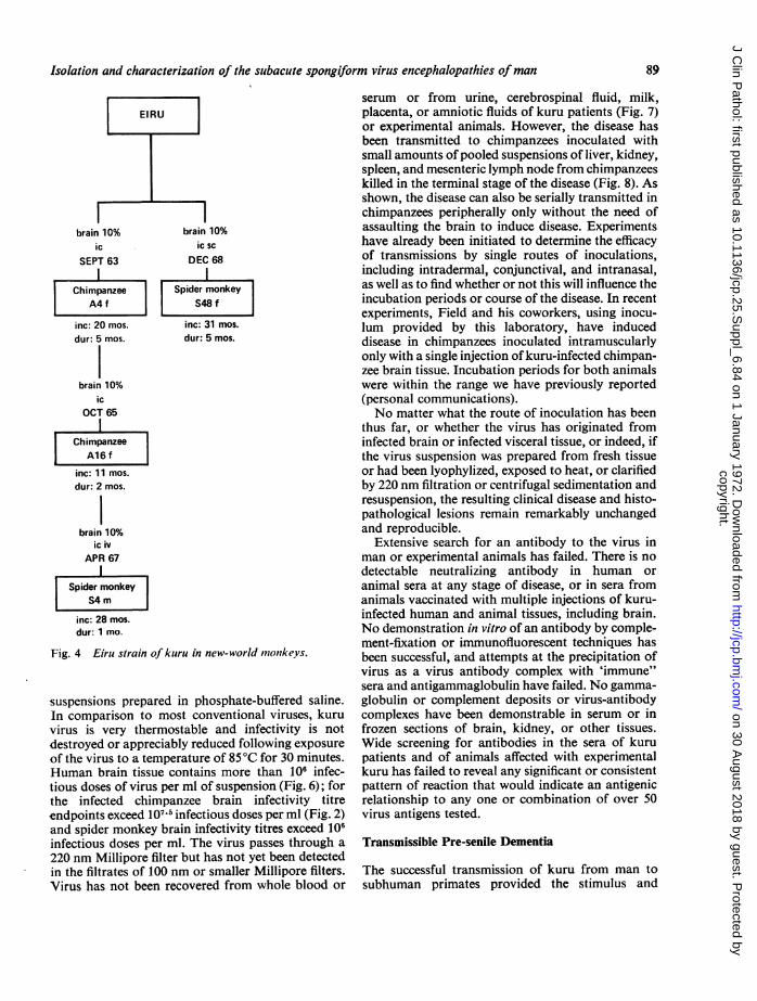
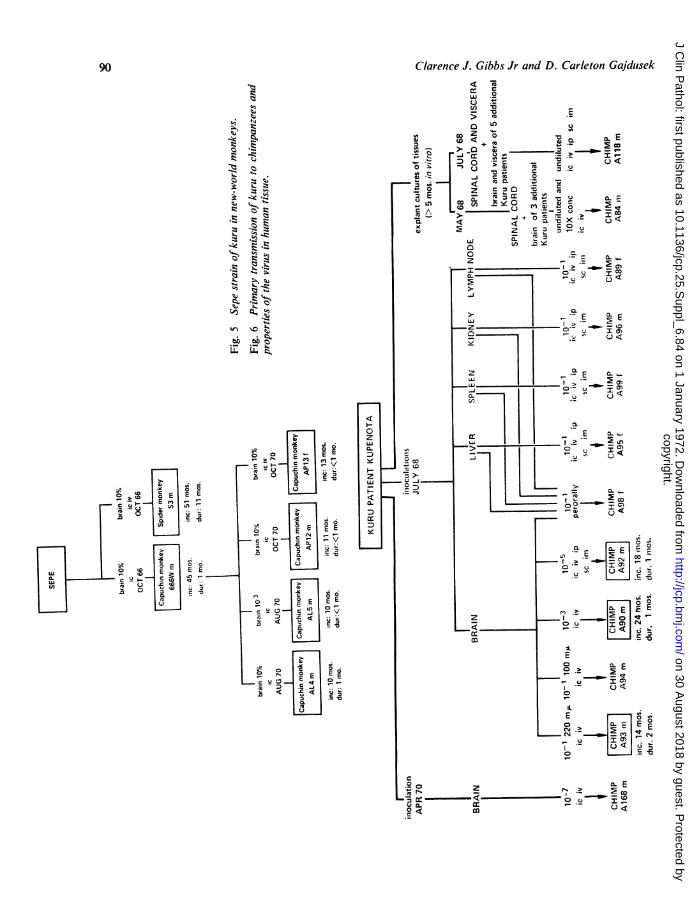
missions are successfully accomplished in animalsinoculated intracerebrally (IC), intracerebrally andintravenously (IV), or by multiple peripheral routes(IV, SC, IM) only. In addition to the chimpanzee,kuru has also been transmitted directly from humanbrain to the rhesus monkey after an incubation periodof eight years and five months (Gajdusek and Gibbs,1972). Primary transmissions have also been madeto five species of new-world monkeys-the spider(Ateles geoffroyi), the capuchin (Cebe sapella andCebes albifronis), the squirrel (Saimirisciureus), with amuch longer mean incubation of 30 months and witha range of 20 to 51 months (Figs. 3, 4, and 5), and the
IISEPT 63
.nc 20 modur- 5 mo
MAR 66
br&. 100 nm,.Icl.V
Ch.,6,ps.,A22 m,
b6,160 3
Ch-mp.nz-eA23 m,
(d.od 1 ..o..0.0 666)
woolly monkey. In serial passage kuru has beentransmitted from chimpanzees to new-world monkeysand from new-world monkeys to new-worldmonkeys (Figs. 4 and 5). In serial passage in new-world monkeys the incubation periods had notappreciably dropped until recently (Fig. 5) when asecond passage level virus in capuchin monkeys hasbeen causing disease in 10 to 12 months. Obviously,much of our investigation of kuru is now done withthis strain in this species of monkey.
In addition to data on primary transmission andserial passages in chimpanzees, Figure 6 and Figure 2,respectively, also contain information on some of theproperties of the kuru virus. The virus is remarkablystable for many months to over several years whenstored at - 70°C in the form of brain tissue
9dur: 7 mo
bra.. 10 5
nc 1 1 modur 5 mo
AUG 67
MAY b66
bran. 10 3
|Ch....z e |
LA29',nc 10 mo.d., 4 mo.
I ~ ~~~~~~IIbran 10 3 b,... 103
*pooled k.u,u Poco d n0orma.bran105 tFa ~~~~Ne G...* wea66c v 37 C 60 376C 60,666
|Chemganzz | |Ch.onpce | Ch.,o nzee|A68m | | ~~A71 | | A70 |
\ c15 mo .nc 1 1 m'o .c: 11 mIdur 3 mo dur 8 mo dur 15 m
-------NEUTRALIZATION TEST -------J-
br,a6 10 6 b6,6,6 10 6
*poled k.ur * .bb-t .nt,hum, n .r k.ru ch...p br..n
-I 1
bran, 10 6*r.bb.t ..t.
ltr h,.mo b,..n
Ch.,666p.66 Ch.,6mp..ze Ch.666p-tee Ch.-panzee Ch,,,p6azee Ch6, p.6e Ch, mP66,6 Ch66p.66-; A128 A 129 m, A130 m, A131 m, A134 I A1135 m, A13 I 3 I i
17 17 6o 6c 17 17 II du0 . 06du 2 m6 d06 16 d06 I
IL_____----__------------- NEUTRALIZATION TEST ---_ _ _ _ ____
AUG 7010'. b'al ,ed.-.nt,dd, 200 000 9 3 1.666
16'
66610. 10 6
Ch. p.nte- Ch.-p..,-A174 - A180 m,
Fig. 2 Summary offive serial passages of kuruii virus, Eiriu(Eiro) strain, in chimpanzees, and data on the physical,biological, and imnmunological properties of the virus.
87
br... 10-5iv. ip sc im.
b6... 10 6
.v p sc m.b6a6, 10
I66.66 10 8
IC jvb.,6. 10 6
* poolwed k.,uch.,mp-,-e ser
--I
----I.1
OCT 65
11
ch.",P-,*eI A18 ,, I
mc 17 ..o
d., 6,,,.
F--ch-pe-of
I A17 "I I..C. 16 .0d.,: 5 o
r-b,.,. 220 .,,,
Ch..pa.zwI A21 I
..C. 10 "'o
d.,. 4 ..o
b,&..A ". ",
I
Ch-pamseA20
..c 18 ..o
d.,: 5 o
b,a..85 C 30
,C '..
Ch,,.p..Z-I A67 M I
..c I "'od.,. 3 ,o
b,.,. - K SNt-1.1 36 ,,I
AUG ..d SEPTPO
Ch.,,,g.,,,-A64 ,,
JUNE 69
I
copyright. on 30 A
ugust 2018 by guest. Protected by
http://jcp.bmj.com
/J C
lin Pathol: first published as 10.1136/jcp.25.S
uppl_6.84 on 1 January 1972. Dow
nloaded from
Clarence J. Gibbs Jr and D. Carleton Gajdusek
0E0~~~~~~E
E
~~~~~~~~
0) -I
>0. E
WCO)0L
o o00 0
E E
1%4N
.8
UL:L
.& E
ml~~~~~~~~~
S~~~~~~~~~~~~~~~~mC
0, 0ELjCD>
C-
E E.
'4 .;
04
.0
D
0c 0
cOE E E
E~E m
-
0Er
LJ~~~~~~~~~~~~~~~~C
88
copyright. on 30 A
ugust 2018 by guest. Protected by
http://jcp.bmj.com
/J C
lin Pathol: first published as 10.1136/jcp.25.S
uppl_6.84 on 1 January 1972. Dow
nloaded from
Isolation and characterization of the subacute spongiform virus encephalopathies ofman
brain 10% brain 10%ic iC SC
SEPT 63 DEC 68l l
Chimpanzee Spider monkeyA4 f S48 f
inc: 20 mos. inc: 31 mos.
dur: 5 mos. dur: 5 mos.
brain 10%ic
OCT 65I
ChimpanzeelA16f
inc: 11 mos.
dur: 2 mos.
brain 10%ic iv
APR 67
1Spider monkey
inc: 28 mos.
dur: 1 mo.
Fig. 4 Eiru strain of kuru in new-world monkeys.
suspensions prepared in phosphate-buffered saline.In comparison to most conventional viruses, kuruvirus is very thermostable and infectivity is notdestroyed or appreciably reduced following exposureof the virus to a temperature of 85°C for 30 minutes.Human brain tissue contains more than 106 infec-tious doses of virus per ml of suspension (Fig. 6); forthe infected chimpanzee brain infectivity titreendpoints exceed 107X5 infectious doses per ml (Fig. 2)and spider monkey brain infectivity titres exceed 106infectious doses per ml. The virus passes through a
220 nm Millipore filter but has not yet been detectedin the filtrates of 100 nm or smaller Millipore filters.Virus has not been recovered from whole blood or
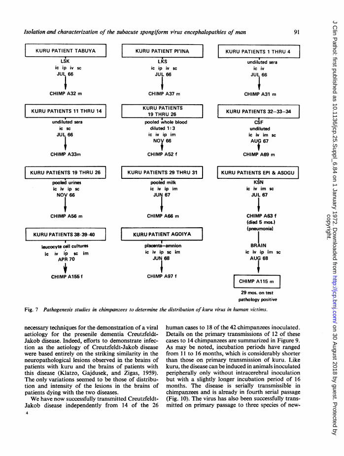
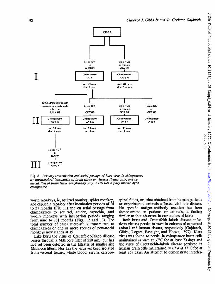
serum or from urine, cerebrospinal fluid, milk,placenta, or amniotic fluids of kuru patients (Fig. 7)or experimental animals. However, the disease hasbeen transmitted to chimpanzees inoculated withsmall amounts of pooled suspensions of liver, kidney,spleen, and mesenteric lymph node from chimpanzeeskilled in the terminal stage of the disease (Fig. 8). Asshown, the disease can also be serially transmitted inchimpanzees peripherally only without the need ofassaulting the brain to induce disease. Experimentshave already been initiated to determine the efficacyof transmissions by single routes of inoculations,including intradermal, conjunctival, and intranasal,as well as to find whether or not this will influence theincubation periods or course of the disease. In recentexperiments, Field and his coworkers, using inocu-lum provided by this laboratory, have induceddisease in chimpanzees inoculated intramuscularlyonly with a single injection of kuru-infected chimpan-zee brain tissue. Incubation periods for both animalswere within the range we have previously reported(personal communications).No matter what the route of inoculation has been
thus far, or whether the virus has originated frominfected brain or infected visceral tissue, or indeed, ifthe virus suspension was prepared from fresh tissueor had been lyophylized, exposed to heat, or clarifiedby 220 nm filtration or centrifugal sedimentation andresuspension, the resulting clinical disease and histo-pathological lesions remain remarkably unchangedand reproducible.
Extensive search for an antibody to the virus inman or experimental animals has failed. There is nodetectable neutralizing antibody in human oranimal sera at any stage of disease, or in sera fromanimals vaccinated with multiple injections of kuru-infected human and animal tissues, including brain.No demonstration in vitro of an antibody by comple-ment-fixation or immunofluorescent techniques hasbeen successful, and attempts at the precipitation ofvirus as a virus antibody complex with 'immune"sera and antigammaglobulin have failed. No gamma-globulin or complement deposits or virus-antibodycomplexes have been demonstrable in serum or infrozen sections of brain, kidney, or other tissues.Wide screening for antibodies in the sera of kurupatients and of animals affected with experimentalkuru has failed to reveal any significant or consistentpattern of reaction that would indicate an antigenicrelationship to any one or combination of over 50virus antigens tested.
Transmissible Pre-senile Dementia
The successful transmission of kuru from man tosubhuman primates provided the stimulus and
89
copyright. on 30 A
ugust 2018 by guest. Protected by
http://jcp.bmj.com
/J C
lin Pathol: first published as 10.1136/jcp.25.S
uppl_6.84 on 1 January 1972. Dow
nloaded from
Clarence J. Gibbs Jr and D. Carleton Gajdusek
E
20F-i700
0
0E E
LL1o
0 E 0
E E_
.CG-
0
Z0.DY
z
ccDze
a
E,
.04Iu
w o1
Ln
_cAXx
0c
U -J.
>
A
ECa
- > I'-
00
0<
<
-X -
__Q
0e,
-I .> - IQ<
0.
- 2coI_, 6 s---40-| Co °w (-_@L
V .
- Q g C -
0
0~~~~~
oo z
. -. .......
Nn
- 0~p
c
_.-.r. C- .> 2coOr- 075cc 0 u 30<
90
copyright. on 30 A
ugust 2018 by guest. Protected by
http://jcp.bmj.com
/J C
lin Pathol: first published as 10.1136/jcp.25.S
uppl_6.84 on 1 January 1972. Dow
nloaded from
Isolation and characterization of the subacute spongiform virus encephalopathies of man
KURU PATIENT TABUYA
LSKic ip iv ScJUL 66
CHIMP A32 m
KURU PATIENTS 11 THRU 14
undiluted seraic SC
JUL 66
CHIMP A33m
KURU PATIENT PI'INA
LKSL
ic ip iv scJUL 66
CHIMP A37 m
KURU PATIENTS19 THRU 26
pooled whole blooddiluted 1:3
ic iv ip imNOX 66
CHIMP A52 f.~~~~~~~~~~~~~~~~~~~~~~~J p KURU PATIENTS 1 THRU 4
undiluted sera
ic ivJUL 66
CHIMP A31 m
KURU PATIENTS 32-33-34
CSFundilutedic iv im scAUG 67
tCHIMP A69 m
KURU PATIENTS 19 THRU 26 KURU PATIENTS 29 THRU 315 I~~~~~~~~~~~~~
pooled urinesic iv ip scNOV 66
CHIMP A56 m
KURU PATIENTS 38-39-40
leucocyte cell culturesic iv ip Sc im
APR 70
CHIMP A155 f
pooled milkic iv ip imJUN 67
CHIMP A66 m
KURU PATIENT AGOIYA
placenta-amnionic iv ip Sc im
JUN 68
CHIMP A97 f
KURU PATIENTS EPI & ASOGU
KSNic iv im scJUL 67
CHIMP A63 f(died 5 mos.)(pneumonia)
BRAINic iv ip im ScAUG 68
CHIMP A115 m29 mos. on test
pathology positive
Fig. 7 Pathogenesis studies in chimpanzees to determine the distribution of kuru virus in human victims.
necessary techniques for the demonstration of a viralaetiology for the presenile dementia Creutzfeldt-Jakob disease. Indeed, efforts to demonstrate infec-tion as the aetiology of Creutzfeldt-Jakob diseasewere based entirely on the striking similarity in theneuropathological lesions observed in the brains ofpatients with kuru and the brains of patients withthis disease (Klatzo, Gajdusek, and Zigas, 1959).The only variations seemed to be those of distribu-tion and intensity of the lesions in the brains ofpatients dying with the two diseases.We have now successfully transmitted Creutzfeldt-
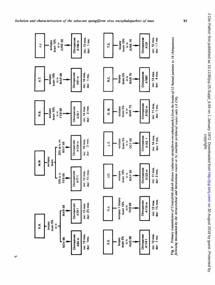
Jakob disease independently from 14 of the 264
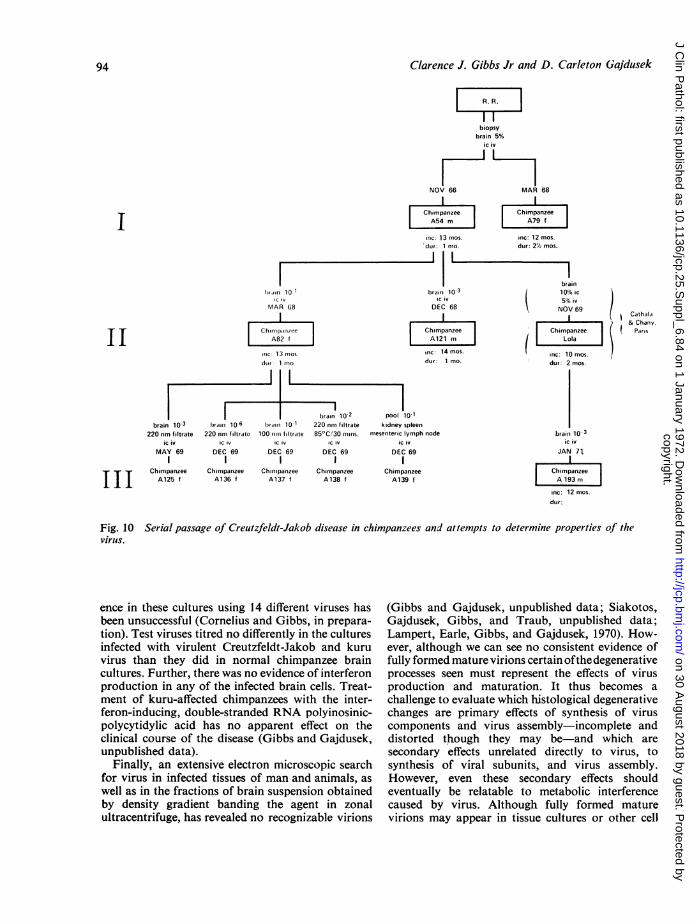
human cases to 18 of the 42 chimpanzees inoculated.Details on the primary transmissions of 12 of thesecases to 14 chimpanzees are summarized in Figure 9.As may be noted, incubation periods have rangedfrom 11 to 16 months, which is considerably shorterthan those on primary transmission of kuru. Likekuru. the disease can be induced in animals inoculatedperipherally only without intracerebral inoculationbut with a slightly longer incubation period of 16months. The disease is serially transmissible inchimpanzees and is already in fourth serial passage(Fig. 10). The virus has also been successfully trans-mitted on primary passage to three species of new-
91
L
I
copyright. on 30 A
ugust 2018 by guest. Protected by
http://jcp.bmj.com
/J C
lin Pathol: first published as 10.1136/jcp.25.S
uppl_6.84 on 1 January 1972. Dow
nloaded from
Clarence J. Gibbs Jr and D. Carleton Gajdusek
I
brain 10%ic
AUG 63
ChimpanzeeAl f
inc: 21 mos.
dur: 9 mos.
10% kidney liver spleenmesenteric lymph node brain 10%
ic iv ip sc icJULY 66 OCT 66
Chimpanzee Chimpanze flA34m A41 m
inc: 18 mos. inc: 11 mos.dur: 4 mos. dur: 1 mo.
spleen 10-2ic
JAN 71l
III
brain 10%ic iv ip imMAY 69
ChimpanzeeI A126 m l
inc: 26 mosdur: 1% mos
brain 10%ip iv SC imOCT 66
Chimpanzee| A44 f l
inc: 13 mos.dur: 6 mos.
brain 5%P0
OCT 66l
ChimpanzeeA48 f
ChimpanzeeA192 f
Fig. 8 Primary transmission and serial passage of kuru virus in chimpanzeesby intracerebral inoculation of brain tissue or visceral tissues only, and byinoculation of brain tissue peripherally only. A126 was a fully mature agedchimpanzee.
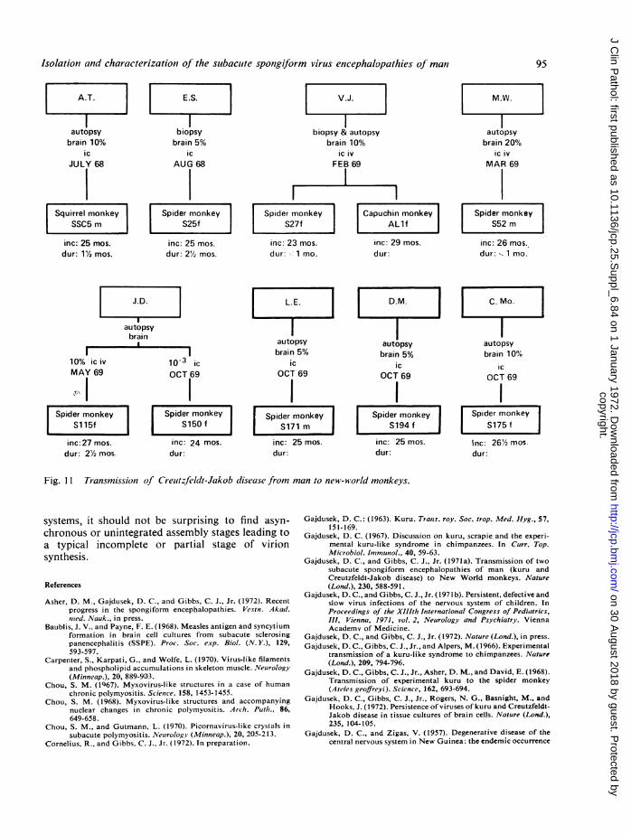
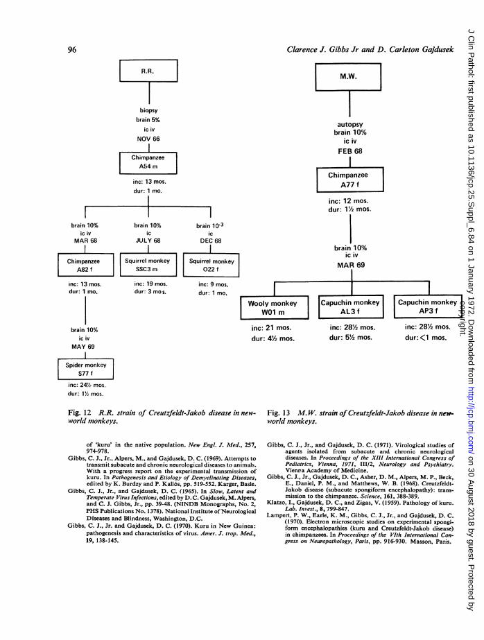
world monkeys, ie, squirrel monkey, spider monkey,and capuchin monkey, after incubation periods of 24to 27 months (Fig. 11) and on serial passage fromchimpanzees to squirrel, spider, capuchin, andwoolly monkeys with incubation periods rangingfrom nine to 281 months (Figs. 12 and 13). Thetotal number of cases successfully transmitted tochimpanzees or one or more species of new-worldmonkeys now stands at 19.
Like kuru the virus of Creutzfeldt-Jakob diseasepasses through a Millipore filter of 220 nm, but hasnot yet been detected in the filtrates of smaller sizeMillipore filters. Nor has the virus yet been isolatedfrom visceral tissues, whole blood, serum, cerebro-
spinal fluids, or urine obtained from human patientsor experimental animals affected with the disease.No specific antigen-antibody reaction has beendemonstrated in patients or animals, a findingsimilar to that observed in our studies of kuru.Both kuru and Creutzfeldt-Jakob disease infec-
tious viruses persist in vitro in cultures of explantedanimal and human tissues, respectively (Gajdusek,Gibbs, Rogers, Basnight, and Hooks, 1972). Kuruvirus was found to persist in chimpanzee brain cellsmaintained in vitro at 37°C for at least 70 days andthe virus of Creutzfeldt-Jakob disease persisted inhuman brain cells maintained in vitro at 37°C for atleast 255 days. An attempt to demonstrate interfer-
92
copyright. on 30 A
ugust 2018 by guest. Protected by
http://jcp.bmj.com
/J C
lin Pathol: first published as 10.1136/jcp.25.S
uppl_6.84 on 1 January 1972. Dow
nloaded from
Isolation and characterization of the subacute spongiform virus encephalopathies of man
c E E E
E , 'I
co [38
'0m0_ 0 v N
> E~~ E E
CLI
E C
E s
._N
o, 2
0 0
Eca E E_mm C N (
E < .'
N~~
ocoSECLir
~~ N
0~~~~~ ~20. Z
E
m~~~
r- LE E E
E
r =
~ ~ ~ ~
) 1C E E
L;>
v
.C
&mE
C4
Q4
AtIVs
N
o
Cq
*_
0-'
C.)
At
-o
s0 z
-0 o
o~-~
Iz
s
*0,
4*
93
copyright. on 30 A
ugust 2018 by guest. Protected by
http://jcp.bmj.com
/J C
lin Pathol: first published as 10.1136/jcp.25.S
uppl_6.84 on 1 January 1972. Dow
nloaded from
Clarence J. Gibbs Jr and D. Carleton Gajdusek
EI
biopsybrain 5%
ic iv
NOV 66 MAR 68
birain 10lIC IV
MAR 68
rc: 13 nmoscIlov 1 nmo
l ~~~~~~~ller
brain 10 2brain 10-3 biraii 10-6 brain 10 220 nne filtrate
220 nm filtrate 220 nns filtrate 100 11nm1 filtrate 850C/30 nuins.ic iv ic iv ic iv ic iv
MAY 69 DEC 69 DEC 69 DEC 691l
Chimpanzee Chinspanzee Chinmpanzee ChimpanzeeA125 f A136 f A137 f A138 f
1.ChimpanzeeA54 m
inc: 13 mos.dur: 1 mo.~~~~ Llbrain 10-3
IC iV
DEC 68
ChimpanzeeA121 n
Inc: 14 mos.
dur: 1 mo.
pool 10-1kidney spleen
mesenteric lymph nodeic iv
DEC 69I
ChimpanzeeA139 f
IChimpanzee
A79 f
inc: 12 mos.dur: 2/2 mos.
brain
10% ic
5% ivNOV 69
Cathala
I.I & ChanyChimpanzee ! Paris
Lola
Inc: 10 mos.
dur: 2 mos.
brain 10-3ic iv
JAN 71tI
ChimpanzeeA 193 m
inc: 12 mos.
dur:
Serial passage of Creutzfeldt-Jakob disease in chimpanzees and attempts to determine properties of the
ence in these cultures using 14 different viruses hasbeen unsuccessful (Cornelius and Gibbs, in prepara-tion). Test viruses titred no differently in the culturesinfected with virulent Creutzfeldt-Jakob and kuruvirus than they did in normal chimpanzee braincultures. Further, there was no evidence of interferonproduction in any of the infected brain cells. Treat-ment of kuru-affected chimpanzees with the inter-feron-inducing, double-stranded RNA polyinosinic-polycytidylic acid has no apparent effect on theclinical course of the disease (Gibbs and Gajdusek,unpublished data).
Finally, an extensive electron microscopic searchfor virus in infected tissues of man and animals, as
well as in the fractions of brain suspension obtainedby density gradient banding the agent in zonalultracentrifuge, has revealed no recognizable virions
(Gibbs and Gajdusek, unpublished data; Siakotos,Gajdusek, Gibbs, and Traub, unpublished data;Lampert, Earle, Gibbs, and Gajdusek, 1970). How-ever, although we can see no consistent evidence offully formed mature virions certainof the degenerativeprocesses seen must represent the effects of virusproduction and maturation. It thus becomes a
challenge to evaluate which histological degenerativechanges are primary effects of synthesis of viruscomponents and virus assembly-incomplete anddistorted though they may be-and which are
secondary effects unrelated directly to virus, tosynthesis of viral subunits, and virus assembly.However, even these secondary effects shouldeventually be relatable to metabolic interferencecaused by virus. Although fully formed maturevirions may appear in tissue cultures or other cell
94
I
II
III
Fig. 10virus.
copyright. on 30 A
ugust 2018 by guest. Protected by
http://jcp.bmj.com
/J C
lin Pathol: first published as 10.1136/jcp.25.S
uppl_6.84 on 1 January 1972. Dow
nloaded from
Isolation and characterization of the subacute spongiform virus encephalopathies of matn
A.T.ES V.J. M.W.
autopsy biopsy biopsy & autopsy autopsy
brain 10% brain 5% brain 10% brain 20%ic ic ic iv ic iv
JULY 68 AUG 68 FEB 69 MAR 69
[Squirrel monkey Spider monkey Spider monkey Capuchin monkey Spider monkey| SSC5 m l | S25f l S27f l l ALlf l | S52 m !
inc: 25 mos. inc: 25 mos. inc: 23 mos. inc: 29 mos. inc: 26 mos.
dur: 1t/2 mos. dur: 2/2 mos. dur: 1 mo. dur: dur: w- 1 mo.
| J.D. ll L.E. l l D.M. l l C. Mo. l
autopsybrain|autopsy autopsy autopsy
brain 5% brain 5% brain 10%10% ic iv 10-3 ic ic ic ic
MAY 69 OCT 69 OCT 69 OCT 69
PI I I
Spider monkey Spider monkey Spider monkey Spider monkey Spider monkey
inc:27 mos. inc: 24 mos. inc: 25 mos. inc: 25 mos. Inc: 26/2 mos.dur: 21/2 mos. dur: dur: dur: dur:
Fig. I I Transmission of Creutzfeldt-Jakob disease from man to new-world monkeys.
systems, it should not be surprising to find asyn-chronous or unintegrated assembly stages leading toa typical incomplete or partial stage of virionsynthesis.
References
Asher, D. M., Gajdusek, D. C., and Gibbs, C. J., Jr. (1972). Recentprogress in the spongiform encephalopathies. Vestn. Akad.nmed. Nauk., in press.
Baublis, J. V., and Payne, F. E. (1968). Measles antigen and syncytiumformation in brain cell cultures from subacute sclerosingpanencephalitis (SSPE). Proc. Soc. exp. Biol. (N. Y.), 129,593-597.
Carpenter, S., Karpati, G., and Wolfe, L. (1970). Virus-like filamentsand phospholipid accumulations in skeleton muscle. Neurology(Minneap.), 20, 889-903.
Chou, S. M. (1Q67). Myxovirus-like structures in a case of humanchronic polymyositis. Science, 158, 1453-1455.
Chou, S. M. (1968). Myxovirus-like structures and accompanyingnuclear changes in chronic polymyositis. Arch. Path., 86,649-658.
Chou, S. M., and Gutmann, L. (1970). Picornavirus-like crystals insubacute polymyositis. Neurology (Minneap.), 20, 205-213.
Cornelius, R., and Gibbs, C. J., Jr. (1972). In preparation.
Gajdusek, D. C.: (1963). Kuru. Trans. roy. Soc. trop. Med. Hyg., 57,151-169.
Gajdusek, D. C. (1967). Discussion on kuru, scrapie and the experi-mental kuru-like syndrome in chimpanzees. In Curr. Top.Microbiol. Immunol., 40, 59-63.
Gajdusek, D. C., and Gibbs, C. J., Jr. (1971a). Transmission of twosubacute spongiform encephalopathies of man (kuru andCreutzfeldt-Jakob disease) to New World monkeys. Nature(Lond.), 230, 588-591.
Gajdusek, D. C., and Gibbs, C. J., Jr. (1971 b). Persistent, defective andslow virus infections of the nervous system of children. InProceedings of the XlIlth International Congress of Pediatrics,III, Vienna, 1971, vol. 2, Neurology and Psychiatry. ViennaAcademv of Medicine.
Gajdusek, D. C., and Gibbs, C. J., Jr. (1972). Nature (Lond.), in press.Gajdusek, D. C., Gibbs, C. J., Jr., and Alpers, M. (1966). Experimental
transmission of a kuru-like syndrome to chimpanzees. Nature(Lond.), 209, 794-796.
Gajdusek, D. C., Gibbs, C. J., Jr., Asher, D. M., and David, E. (1968).Transmission of experimental kuru to the spider monkey(Ateles geoffreyi). Science, 162, 693-694.
Gajdusek, D. C., Gibbs, C. J., Jr., Rogers, N. G., Basnight, M., andHooks, J. (1972). Persistence of viruses of kuru and Creutzfeldt-Jakob disease in tissue cultures of brain cells. Nature (Lond.),235, 104-105.
Gajdusek, D. C., and Zigas, V. (1957). Degenerative disease of thecentral nervous system in New Guinea: the endemic occurrence
95
copyright. on 30 A
ugust 2018 by guest. Protected by
http://jcp.bmj.com
/J C
lin Pathol: first published as 10.1136/jcp.25.S
uppl_6.84 on 1 January 1972. Dow
nloaded from
Clarence J. Gibbs Jr and D. Carleton Gajdusek
biopsybrain 5%
ic ivNOV 66I
ChimpanzeeA54 m
inc: 13 mos.dur: 1 mo.
II I
brain 10% brain 10% brain 10-3ic iv ic ic
MAR 68 JULY 68 DEC 68
Chimpanzee Squirrel monkey Squirrel monkey|A82 f l SSC3 m 022 f
inc: 13 mos. inc: 19 mos. inc: 9 mos.
dur: 1 mo. dur: 3 mos. dur: 1 mo,
brain 10%ic iv
MAY 69
Spider monkeyS77 f
inc: 241/2 mos.
dur: 11/2 mos.
Fig. 12 R.R. strain of Creutzfeldt-Jakob disease in new-world monkeys.
of 'kuru' in the native population. New Engl. J. AMed., 257,974-978.
Gibbs, C. J., Jr., Alpers, M., and Gajdusek, D. C. (1969). Attempts totransmit subacute and chronic neurological diseases to animals.With a progress report on the experimental transmission ofkuru. In Pathogenesis and Etiology of Demyelinating Diseases,edited by K. Burdzy and P. Kall6s, pp. 519-552. Karger, Basle.
Gibbs, C. J., Jr., and Gajdusek, D. C. (1965). In Slow, Latent andTemperate Virus Infections, edited by D. C. Gajdusek, M. Alpers,and C. J. Gibbs, Jr., pp. 39-48. (NINDB Monographs, No. 2,PHS Publications No. 1378). National Institute ofNeurologicalDiseases and Blindness, Washington, D.C.
Gibbs, C. J., Jr. and Gajdusek, D. C. (1970). Kuru in New Guinea:pathogenesis and characteristics of virus. Amer. J. trop. Med.,19, 138-145.
autopsybrain 10%
ic iv
FEB 68
ChimpanzeeA77 f
inc: 12 mos.dur: 112 mos.
brain 10%ic iv
inc: 21 mos. inc: 281/2 mos. inc: 281/2 mos.dur: 41/2 mos. dur: 5/2 mos. dur: <1 mos.
Fig. 13 M. W. strain ofCreutzfeldt-Jakob disease in new-world monkeys.
Gibbs, C. J., Jr., and Gajdusek, D. C. (1971). Virological studies ofagents isolated from subacute and chronic neurologicaldiseases. In Proceedings of the XIII International Congress ofPediatrics, Vienna, 1971, 111/2, Neurology and Psychiatry.Vienpa Academy of Medicine.
Gibbs, C. J., Jr., Gajdusek, D. C., Asher, D. M., Alpers, M. P., Beck,E., Daniel, P. M., and Matthews, W. B. (1968). Creutzfeldt-Jakob disease (subacute spongiform encephalopathy): trans-mission to the chimpanzee. Science, 161, 388-389.
Klatzo, I., Gajdusek, D. C., and Zigas, V. (1959). Pathology of kuru.Lab. Invest., 8, 799-847.
Lampert, P. W., Earle, K. M., Gibbs, C. J., Jr., and Gajdusek, D. C.(1970). Electron microscopic studies on experimental spongi-form encephalopathies (kuru and Creutzfeldt-Jakob disease)in chimpanzees. In Proceedings of the VIth International Con-gress on Neuropathology, Paris, pp. 916-930. Masson, Paris.
96
copyright. on 30 A
ugust 2018 by guest. Protected by
http://jcp.bmj.com
/J C
lin Pathol: first published as 10.1136/jcp.25.S
uppl_6.84 on 1 January 1972. Dow
nloaded from