Embed Size (px)
Citation preview

IV. DIGESTIVE TRACT BACTERIA
1. Microbial Ecology of the Human Oral Cavity
The teeth and oral epithelial surfacesare colonised by a great variety of bacterial species. A partial list of indigenous (autochthonous) organisms found in the adult human mouth is shown in Table 11 (Gibbons and van Houte, 1975; Gibbons and van Houte, 1978; Rosebury, 1962; Socransky and Haffajee, 1988; van Houte, 1980). Characteristi
cally, such indigenous bacteria are well adjusted to their environment and can successfully colonise the mouth for appreciable periods of time or throughout life. Their relationship with the host is neither a purely mutually beneficial one (symbiosis) nor are oral bacteria obligately pathogenic (antibiosis) (Rosebury, 1962).
Table 11: Oral flora ——————————————————————————————————————— Gram+ cocci Gram- cocci Gram+ rods Gram- rods
——————————————————————————————————————— Staphylococcus sp. Veillonella sp. Actinomyces sp. Actinobacillus
A. viscosus actinomycetemcomitans
Streptococcus sp. Neisseria sp. A. naeslundii S. mutans group* A. odontolyticus Bacteroides sp.
S. mutans A. israelii B. forsythus S. sobrinus B. gingivalis
Arachnia B. intermedius S. milleri
Bacterionema Campylobacter S. mitior
Bifidobacterium Capnocytophaga sp. S. salivarius C. gingivalis
Clostridium C. ochracea S. sanguis
Corynebacterium Eikenella corrodens Enterococci
Eubacterium Fusobacterium Anaerobic strep.
Lactobacillus sp. Haemophilus L. casei L. plantarium Leptotrichia L. acidophilus L. fermenti Selenomonas sputigena L. brevis
Nocardia Spirochetes
Propionibacterium Wolinella recta
Rothia ——————————————————————————————————————— * Referred to as S. mutans in text.
41
Old Herborn University Seminar Monograph 1:Microbial ecology of the human digestive tract. Editors: Dirk van der Waaij, Peter J. Heidt, Volker Rusch and Jan-Olaf Gebbers. Institute for Microbiology and Biochemistry, Herborn-Dill, Germany: 41-83 (1987).
The term amphibiosis has been suggested for this situation by Rosebury (1962) to signify a spectrum of relationships between these two extremes, which may be more or less beneficial, or harmful depending on the conditions. Beneficial effects of the oral flora include its general contribution to host immunity by means of antibodies that cross-react with overt pathogens, the specific suppression of organisms such as Candida albicans, or the production of vitamins. The pathogenic potential of the oral flora is illustrated by many infectious processes such as dental caries, periodontal diseases, endodontic infections, sub-acute bacterial endocarditis, actinomycosis, bite wounds, etc., which require a variety of predisposing factors. For dental caries, the frequent consumption of dietary carbohydrates is required to permit expression of the cariogenic potential of dental plaque bacteria through their ability to produce high concentrations of organic acids which are responsible for the demineralisation of the underlying tooth surface (Gibbons and van Houte, 1978; van Houte, 1980). Sub-acute endocarditis reflects a combination of the dissemination of oral organisms such as S. mutans and S. sanguis and previously damaged heart tissue (van Houte, Jordan and Bellack, 1971).
Acquisition of the Oral Flora Exposure of the oral tissues to bacte
ria during and directly after birth leads rapidly to the development of an oral flora (Gibbons and van Houte, 1975; Gibbons and van Houte, 1978; Socransky and Haffajee, 1988). The selection of bacteria from the outer environment is based on their ability to adhere to oral surfaces and to grow under the prevailing conditions; the "cleansing" activity of saliva plays an important role in bacterial disposal. The evidence indicates that the mouth harbours a unique flora,
which meets very specific requirements for adherence and growth. This indicates that the new-born acquires its characteristic flora from other human beings. Indeed, bacteriocin-typing studies with S. mutans, demonstrating the transmission of this organism from parents to their children, directly support this contention (Berkowitz, Turner and Creen, 1981). Furthermore, transmission of S. mutans appears to require generally direct contact (e.g. contaminated food, kissing) and is influenced by the cell numbers transferred; its emergence in children of parents with low cell numbers may be greatly delayed (Berkowitz, Turner and Green, 1981).
Initially, many different organisms can be found in the infant's edentulous mouth reflecting contact with a variety of sources (Gibbons and van Houte, 1978; Socransky and Haffajee, 1988). However, the presence of many of these organisms is transient. Indigenous organisms capable of colonising the epithelial surfaces e.g. S. salivarius can be isolated consistently already within a few weeks or even days and the spectrum of bacterial species and their number gradually increases with time. In this early phase of bacterial colonisation, streptococci, actinomyces, neisseriae, and veillonellae can be regularly isolated. Most of these organisms are facultative with respect to oxygen and many overtly anaerobic species are still absent; bacterial oxygen utilisation may permit the colonisation of some anaerobic organisms such as veillonellae (Ritz, 1967).
The eruption of teeth permits a major addition to the already complex flora and is indispensable for the colonisation of organisms such as S. mutans and S . sanguis and Lactobacillus species; in this regard, teeth may provide a specific non-shedding surface which enables their attachment, or specific sites e.g.
42
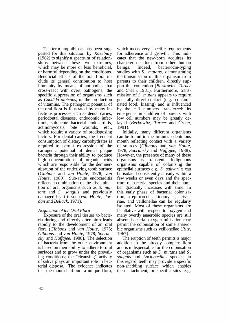
deep fissures which are relatively sheltered from the oral cleansing forces and which may further facilitate their colonisation (Gibbons and van Houte, 1975; Gibbons and van Houte, 1978; Socransky and Haffajee, 1988). Tooth eruption also leads to the development of the gingival sulcus around the teeth and the emergence of a major anaerobic flora, particularly Gram-negative rods (Table 11) that are associated with different types of periodontal disease (Socransky and Haffajee, 1988). The low oxidation-reduction potential prevailing within the sulcus plays an important role in this regard; specific nutrients may also promote bacterial growth e.g. hostderived haemin or hormones and vitamin K analogues, produced by other bacteria, for growth of B. gingivalis (Loesche, 1968). The preferential habitat of some oral organisms, based on adherence and growth factors, is shown in Table 12. Such organisms can also be found, albeit less frequently and in lower concentrations, in other sites of the dentulous adult mouth. Furthermore, the indicated sites are probably in all cases indispensable for their persistent colonisation.
The most densely-populated surfaces are generally the teeth (Gibbons and van Houte, 1975; Gibbons and van Houte, 1978). Here tightly adherent masses of
bacteria, termed dental plaque, can reach concentrations of 2 x 10¹¹ cells (viable and non-viable) per gram wet weight. The concentrations on the tongue dorsum may average 100 bacteria per cell whereas those on the buccal mucosa average about 10-20 bacteria per cell. The bacterial concentrations in saliva are generally between 107 and 108 per ml. The "flora" of saliva originates from bacterial cells dislodged from the teeth and epithelial surfaces. Bacterial growth on the oral surfaces, particularly when the cell density is high, is relatively low and it has been estimated that the biomass doubles only a few times per day. The high cell density in dental plaque limits the inward diffusion of nutrients. Nutrient limitation is responsible for a bacterial growth rate far below its optimal level, particularly in the deeper parts of plaque (Critchley, 1969).
Dental Plaque Formation and Ecology Dental plaques form on the enamel
covering the crowns of teeth, on enamel near or in the healthy gingival sulcus (about 1 mm deep), on cementum or underlying dentin of roots of teeth exposed to the oral environment by periodontal disease, or in the pathologically deepened gingival sulcus ("pocket") as sociated with loss of tooth-supporting
Table 12: Preferential colonisation sites of some oral bacteria ———————————————————————————————————————
Coronal Gingival Tongue Organism plaque sulcus dorsum
——————————————————————————————————————— S. mutans + S. sanguis + S. salivarius + A. viscosus + A. naeslundii + Lactobacillus sp. +*
Anaerobes (e.g. Bacteroides, + Fusobacteria, Spirochetes)
——————————————————————————————————————— * Particularly caries lesions
43

bone (periodontal disease) (Gibbons and van Houte, 1975; Gibbons and van Houte, 1978; Socransky and Haffajee, 1988; van Houte, 1980). In view of the powerful oral cleansing forces, pre-selection sites for plaque are the relatively protected tooth fissures, areas between teeth, and areas near the gingival sulcus. Exposed enamel, or cementum or dentin on the roots of teeth, are covered by an acquired pellicle on which plaque can form. Plaque formation under natural conditions may involve bacterial colonisation of this pellicle when all plaque has been removed (e.g. oral hygiene) as well as continuous replacement of lost cell mass not involving an exposed pellicle.
Plaque formation on the pellicle entails initial attachment of bacteria that are present in saliva or on contacting surfaces. This adhesion is bacterium-specific and is based on the interaction with different pellicle components such as various salivary glycoproteins or enzymes (Gibbons and van Houte, 1975; Gibbons and van Houte, 1978; van Houte, 1982). Some of these interactions may involve lectin-like (carbohydrate-protein) binding between specific bacterial adhesins and pellicle receptors. Initial bacterial adhesion is probably sub-optimal because components in saliva to which cells available for attachment are exposed, will bind to and block the specific bacterial adhesins which mediate bacterial attachment to the pellicle and will also cause bacterial aggregation (Gibbons and van Houte, 1975).
Further increase in the size of plaque involves in situ proliferation of bacteria and additional adherence. Cell accumulation involves adhesion between bacteria and the interbacterial matrix, which contains salivary glycoproteins, and extracellular polymers synthesised by bacteria as well as direct cell-to-cell binding. These interactions, possibly
together with mechanical entrapment of bacteria in matrix components, are responsible for the structural integrity of plaque. The adhesive interactions between matrix components and different bacteria as well as cell-to-cell binding are also highly specific and may also involve lectin-like binding mechanisms (Gibbons and van Houte, 1975; Gibbons and van Houte, 1978; van Houte, 1982).
The relative significance of bacterial adhesion and growth during plaque formation may be summarised as follows: 1. The initial composition of dental
plaque is governed by bacteriumspecific interactions with the pellicle. In fact, these interactions and bacterial adhesive interactions with oral epithelial surfaces constitute an important ecological determinant (Gibbons and van Houte, 1975; Gibbons and van Houte, 1978; van Houte, 1982) and form the basis for the preferential colonisation of different oral bacteria in different oral sites (Table 12),
2. depending on the conditions, factors influencing bacterial growth may cause shifts in bacterial composition during plaque development (see below), and
3. the total biomass of dental plaque, allowed to develop for a significant period of time, is mainly determined by in situ bacterial proliferation rather than bacterial adhesion. With respect to the composition of the flora on epithelial surfaces, bacterial adhesion appears as the most significant determinant since maintenance of the flora requires continuous bacterial attachment due to the constant shedding of epithelial cells (Gibbons and van Houte, 1975; Gibbons and van Houte, 1978; van Houte, 1982).
The growth of oral indigenous bacteria is influenced by a vast array of factors.
44

They include oxygen tension, dietary carbohydrates, bacterial acids (pH), bacterial toxins (e.g. bacteriocins) and a variety of enzymes (Gibbons and van Houte, 1975; Gibbons and van Houte, 1978; Loesche, 1968; Ritz, 1967; Socransky and Haffajee, 1988). Many of these factors have been studied particularly in their relation to dental plaque. The presence of oxygen may play a role in the gingival sulcus (see earlier) but also in the deep layers of plaque (Ritz, 1967). In view of their significance for dental caries development, much is known about the effect of dietary carbohydrates. Many oral bacteria are fastidious and require carbohydrates for growth and absence of carbohydrate consumption causes major shifts in the oral flora. Especially organisms such as S. mutans and lactobacilli are very adversely affected; other organisms may be less dependent upon dietary carbohydrate by utilising host sources of carbohydrate or by the metabolism of other substrate types (van Houte, 1980).
Bacterial metabolism of dietary carbohydrates, comprised mainly of sucrose and starches with lesser amounts of lactose, fructose, glucose and some other sugars, may lead to a variety of products with varying ecological effects (van Houte, 1979). These include: 1. A variety of organic acid end prod
ucts (e.g. lactate, propionate, acetate) from all carbohydrates including starches (prior hydrolysis by host- or bacterium-derived amylase),
2. synthesis and degradation of a variety of extracellular polysaccharides from sucrose, specifically. These include glucans (dextran, rich in 1.6 linkages and mutan rich in 1.3 linkages) synthesised by S. mutans or S . sanguis, fructose (levan) synthesised by S. salivarius and A. viscosus, and heteropolysaccharides synthe
sised from other sugars as well. Examples of the latter are a polymer consisting of N-acetyl-glucosamine, glucose and galactose synthesised by A. viscosus and a glucose-rhamnose "capsule" synthesized by L. casei,
3. synthesis of intracellular glycogentype polysaccharides by a wide variety of bacteria from all carbohydrates.
Bacterial acidogenesis in dental plaque constitutes an important ecological force in view of the widely-varying pH tolerance of plaque bacteria (Gibbons and van Houte, 1978; van Houte, 1980). Extracellular glucans, particularly insoluble mutan, are important matrix components of plaque exposed regularly to sucrose. They contribute to voluminous plaque formation by promoting the structural integrity of plaque through intercellular binding or, possibly, cell entrapment; glucans are not readily degraded by enzymatic action. Levan as well as glycogen and the glucose-rhamnose capsule of L. casei are rapidly degraded to acids when the environmental carbohydrate supply is depleted (Gibbons and van Houte, 1975; Gibbons and van Houte, 1978; van Houte, 1982). Glycogen metabolism has been implicated in caries aetiology by causing prolonged acid production in plaque during its degradation and an enhanced acid production during its synthesis; it may also promote bacterial survival (van Houte, 1979).
In view of the above, the earlier mentioned effect of dietary carbohydrate on the oral populations of S. mutans and lactobacilli appear to be due to: 1. their particular dependence on dietary
carbohydrates for growth, 2. their high acid tolerance providing
them with a selective advantage over other organisms in acidic environments; this correlates with the uniquely high proportions of lactobacilli in caries lesions, exhibiting
45

often a low pH, and the proportions of S. mutans in caries-associated and frequently acidic plaques, and
3. extracellular glucan synthesis in the case of S. mutans which promotes cellular adhesion and thereby an increase of its plaque proportions.
Ecological Aspects of Dental Caries Dental plaque is a prerequisite for the
development of dental caries, periodontal diseases, and dental calculus (Gibbons and van Houte, 1978; Socransky and Haffajee, 1988; van Houte, 1980). Another prerequisite factor for dental caries is dietary carbohydrate. The pathogenesis reflects essentially a disturbance of the equilibrium between the tooth surface and protective saliva on the one hand and dietary carbohydrate and dental plaque on the other. Acid production by plaque bacteria from carbohydrate leads to an increased H+-concentration in the plaque milieu (lower pH). This may lead to undersaturation of calcium and phosphate ions with respect to tooth mineral (hydroxyapatite, Ca10(PO4)6(OH)2) and loss of Ca and P from the tooth surface (demineralisation); this process is reversible with increasing plaque pH (remineralisation).
In view of the pivotal role of plaque pH in dental caries development, a primary aetiological role is restricted to acidogenic organisms; this is true not only for enamel caries but probably also for root surface caries (Jordan 1986). Dental caries may be defined as a dietary-modified bacterial infectious disease. This concept implies a dynamic interaction between dietary carbohydrate and the plaque flora composition i.e. a higher carbohydrate intake induces an ecological shift towards higher proportions of acid-tolerant, acidogenic bacteria which is accompanied by a higher pH-lowering and cariogenic potential of plaque (van Houte, 1980). This concept
is supported, among others, by the following observations: 1. plaques associated with caries activity
on enamel, when exposed to carbohydrate, exhibit a pH profile which is in a lower pH range than that of plaques associated with caries inactivity,
2. the flora of the former is characterised by increased proportions of S . mutans and, less frequently and mostly in fissures, of lactobacilli, whereas the proportions of other predominant acidogenic organisms, mainly streptococci and actinomyces, remain unchanged or are decreased, and
3. S. mutans and lactobacilli are among the most acid-tolerant plaque organisms. This trait is of special significance because the lower the pH reached in plaque during carbohydrate utilisation, the greater is the probability and extent of tooth surface demineralisation.
Differences among plaque bacteria with respect to acidogenicity and acid tolerance are a matter of degree. Nevertheless, there is considerable evidence that quantitative differences with respect to these traits are important. Consequently, the aetiology of dental caries in enamel appears to entail considerably bacterial specificity. S. mutans is presently considered as a prime aetiologic agent; lactobacilli probably play a much lesser role whereas the significance of other acidogenic organisms requires further clarification. The spectrum of aetiologically significant plaque organisms is probably larger in the case of root surface caries which requires less stringent acidic plaque conditions (Jordan, 1986).
Johannes van Houte, Forsyth Dental Center, Boston, Massachussetts 02115, U.S.A.
46

2. Stomach Microbial Ecology
Traditionally, the normal stomach with its acidic gastric juice has been considered a sterile organ protecting the upper gut so that only transient oral bacteria occur following ingestion of food. With a reduction in gastric acid, different bacteria can colonise the stomach. Typically, this is seen in patients with pernicious anaemia. With pH 4 to 5 only acid resistant lactobacilli plus streptococci can survive but when the pH becomes greater than 5, many oral and faecal organisms can be isolated (Hill, 1983).
In 1983 and 1984, Marshal, and Marshal and Warren, respectively reported the isolation of a micro-aerophilic spiral organism from the mucosa of the majority of patients with chronic gastritis. This association has been confirmed world-wide. The organism is now called Helicobacter pylori; it is motile by multiple sheathed flagellae and adheres to gastric epithelium beneath the mucus gel. The organism is poorly seen on Haematoxilin-Eosin-stained sections but can be easily seen with a number of stains such as silver or a modified Giemsa (Rathbone, Wyatt and Heatly, 1986a). Colonisation has never been seen in an entirely normal stomach (antrum and corpus) and colonisation of the intestinal epithelium has also not yet been observed. The only time the organism is seen in the duodenum was in association with gastric metaplasia. Ultrastructurally, the organism adheres mostly in the intercellular gutters where they are associated with microvillous depletion and epithelial mucus depletion. Biochemically, a marked feature of the organism is its strong urease activity.
Helicobacter pylori is associated with both a systemic as well as a local immune response (Rathbone et al., 1986b). Studies looking at antibody
coating of the bacteria demonstrate IgG, IgA and IgM coating on the bacteria. What was noted in these investigations was that in all subjects studied the bacteria deep in the gastric pits were uncoated (Wyatt, Rathbone and Heatly, 1986). Mucosal culture studies demonstrate that the gastric plasma cells are producing Helicobacter pylori-specific antibodies. Gastric T cell studies show increased stimulated T helper cells in the patients with Helicobacter pylori-associated gastritis.
There is evidence for acute infection from reported episodes of 'epidemic' gastritis with deliberate human ingestion of Helicobacter pylori (Rathbone et al. 1986c). Treatment studies have demonstrated that clearance of the organisms is associated with an improvement in the histological picture (McNulty et al., 1986). Recolonisation occurs and has been demonstrated to be with the same strain (Langenberg et al., 1986). In patients with normal acid secretion, Helicobacter pylori is the only organism colonising the human stomach. This colonisation appears stable and long term. The organism is well adapted to living in its niche, protected from gastric acid by the mucus bicarbonate barrier The histological entity we call chronic B-gastritis, would appear at least in part to be the gastric immune reaction to Helicobacter pylori. Interestingly, the inflammation and colonisation is long term with the immune reaction failing to eliminate the organism. One presumes this highly specialised organism living in its unique niche derives benefit from the chronic inflammatory reaction.
Barry J. Rathbone, Department of Medicine, St. James University Hospital, Leeds, U.K. LS9 7TF.
47
3. Sequential Development of the Human Intestinal Microbial Flora
Sources of Neonatal Bacterial Colonisation Before Birth:
As long as the amniotic membrane remains intact, the normal foetus is sterile until shortly before birth. During Birth:
As a result of passing through the vagina the gastrointestinal tract of the neonate is seeded with a wide variety of microorganisms originating from both the maternal microbial flora and the environment. For example, studies examining Escherichia coli from maternal and infant stool have shown that many babies acquire strains of E. coli identical to strains isolated from their mothers (Gothefors et al., 1976). Organisms best suited for survival in the intestinal environment become established by a
process of natural selection. Many of the microorganisms are not able to colonise habitats in the neonatal gastrointestinal tract and disappear from it soon after birth. After Birth:
Factors other than the maternal microbial flora are also undoubtedly important in the development of the neonate intestinal flora since infants are not always colonised with strains of bacteria from their mothers. The external environment (e.g., air and hospital personnel) is major source of colonising bacteria. In addition, external forces, such as food source and antimicrobial agents, can have dramatic influences on bacterial colonisation of the infant intestinal tract.
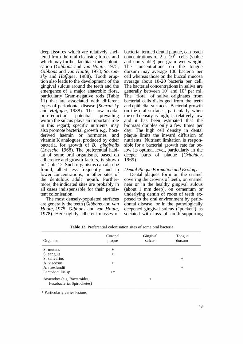
Figure 22: Sequential development of the normal intestinal flora in breast-fed infants. n: Enterobacteriaceae; §: Bifidobacterium; s: Streptococcus; Ë: Clostridium; ↕: other anaerobes
48

Succession of Microbial Colonisation of the Infant Large Intestine
Despite the wide variety of bacteria constantly infiltrating the intestinal tract of infants, the pattern of colonisation is predictable and dependent upon complex regulatory mechanisms. For purposes of discussion the sequential microbial colonisation of the infant intestinal tract can be divided into four major phases: Phase I (0-2 Weeks):
Phase I is the initial period of bacterial colonisation which occurs during the first two weeks of life. The intestinal flora is very unstable at this time and numerous changes in bacterial populations are occurring. Phase II (2 Weeks-Preweaning):
Phase II is the remaining period during which breast milk and/or formula milk is the exclusive form of nutrition.
The intestinal flora stabilises during this period but the actual composition of the flora is dependent on the food source. Phase III (Weaning):
Phase III is initiated, in breast-fed infants, at the time that dietary supplements such as formula or cereal are introduced into the infants diet. The introduction of alternate food sources to the breast-fed infant causes major perturbations in the intestinal ecosystem. Phase IV (Post-Weaning):
Phase IV is the period after weaning is completed. The intestinal bacterial flora of breast-fed and formula-fed infants approaches that of adults during this phase.
Sequential Development of the Normal Intestinal Flora in Breast-Fed Infants
Regardless of the animal species, Enterobacteriaceae and enteric strepto
Figure 23: Sequential development of the normal intestinal flora in formula-fed infants. n: Enterobacteriaceae; §: Bifidobacterium; s: Streptococcus; Ë: Clostridium; ↕: other anaerobes
49
cocci are almost universally the first mi- The introduction of solid food to croorganisms to appear in the colon of the new-born (Figure 22). Bifidobacterium usually appear in the intestinal tract shortly after Enterobacteriaceae and achieve levels of approximately 109
CFU/gram of faeces by the end of the second week of life (Phase I). In many animal species, transient colonisation of the colon with a variety of bacteria, including clostridia and other anaerobes, may occur during the first few days of life.
From two weeks of age until solid foods are given (Phase II), breast-fed infants have a very simple flora consisting primarily of high numbers of Bifidobacterium (109 CFU/g faeces) and lower numbers of Enterobacteriaceae and Streptococcus. Colonisation with Bacteroides and Clostridium, which may occur during the first week of life, decrease to very low numbers during the period of exclusive breast-feeding.
breast-fed infants causes major disturbances in the microbial ecology of the colon (Phase III). The numbers of Enterobacteriaceae and streptococci increase while colonisation by Bacteroides, Clostridium and other anaerobes takes place.
By the second year of life, the major bacterial populations of the intestinal flora resemble those of adults (Phase IV). During this time the numbers of Bifidobacterium decrease while the numbers of Enterobacteriaceae, Streptococcus, Clostridium and other anaerobes continue to increase. The typical adult microbial intestinal flora consists of over 300 different species of aerobic, facultative and anaerobic bacteria.
Sequential Development of the Normal Intestinal Flora in Formula-Fed Infants
The intestinal flora of infants fed a diet based on cow's milk differs markedly from the intestinal flora of breast-
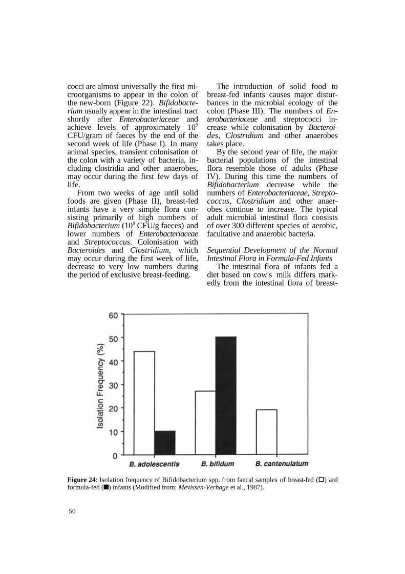
Figure 24: Isolation frequency of Bifidobacterium spp. from faecal samples of breast-fed (§) and formula-fed (n) infants (Modified from: Mevissen-Verhage et al., 1987).
50
fed infants (Figure 23). In in fants, unlike adults, relatively small changes in diet will have major effects on the intestinal flora.
In phase I, the sequence of bacterial colonisation in infants fed exclusively formula milk is quite similar to that of breast-fed neonates. Both groups are rapidly colonised with Enterobacteriaceae and Streptococcus.
During phase II, the intestinal flora of formula-fed infants is much more complex than that of breast-fed infants. The number of aerobic bacteria, such as Enterobacteriaceae and Streptococcus is typically higher in the faeces of formulafed neonates than in the faeces of breastfed neonates. Bifidobacterium species are the predominant faecal bacteria in both groups.
However, the total counts of other anaerobic bacteria, such as Bacteroides, Eubacterium, Peptostreptococcus, Veillonella and Clostridium are signifi
cantly higher in the formula-fed group than in the breast-fed group. During phase II, relatively small amounts of formula milk supplementing otherwise exclusive breast-feeding usually results in shifts of the intestinal flora toward formula-fed patterns. The intestinal flora may not return to its original composition for a period of up to four weeks following the resumption of exclusive breast-feeding.
It is during phase III that the bacterial populations of the large intestine of breast-fed and formula-fed infants are beginning to resemble each other in both number and composition of bacteria. This is primarily a result of alterations in the anaerobic intestinal flora of breastfed neonates since the introduction of solid foods has little effect on the composition of the intestinal flora of formula-fed neonates.
Finally, during phase IV, the aerobic and anaerobic intestinal flora of for-
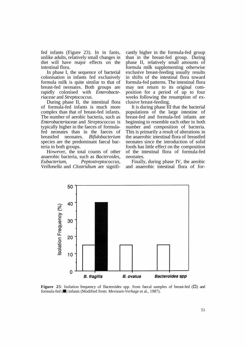
Figure 25 : Isolation frequency of Bacteroides spp. from faecal samples of breast-fed (§) and formula-fed (n) infants (Modified from: Mevissen-Verhage et al., 1987).
51
mula-fed and breast-fed infants are essentially identical and resemble the intestinal flora of the adult.
Other Differences in the Development of the Intestinal Flora of Formula-Fed and Breast-Fed Infants
When comparing the intestinal flora of breast-fed and formula-fed neonates most studies have identified bacteria just to the genus level.
However, differences can also be demonstrated between species within a genus in the two groups of infants. As described above, Bifidobacterium species are the predominant bacteria in both formula-fed and breast-fed infants.
However, the actual species of Bifidobacterium in infants fed exclusively breast milk have been shown to be quite different from the Bifidobacterium species found in the intestinal tracts of formula-fed infants. Mevissen-Verhage and co-workers (1987) have shown that the Bifidobacterium species most frequently isolated from infants are B . adolescentis, B. bifidum and B . catenulatum (Figure 24). Breast-fed infants have all three species in their intestine with B. adolescentis being the most common. On the other hand, bottle-fed infants have only B. adolescentis and B. bifidum, with the latter being isolated most frequently. These investigators found similar results with species of Bacteroides (Figure 25). Formula-fed infants have only B. fragilis in their faeces while breast-fed infants have B . fragilis and B. ovatus, as well as other species of Bacteroides in their intestine.
In addition to the actual species of bacteria differing between breast-fed and formula-fed neonates, the physicochemical characteristics of a particular species may also differ between the two groups. For example, fewer E. coli serogroups are found in the faeces of breast-fed infants than in the faeces of formula-fed infants (Braun, 1981). This
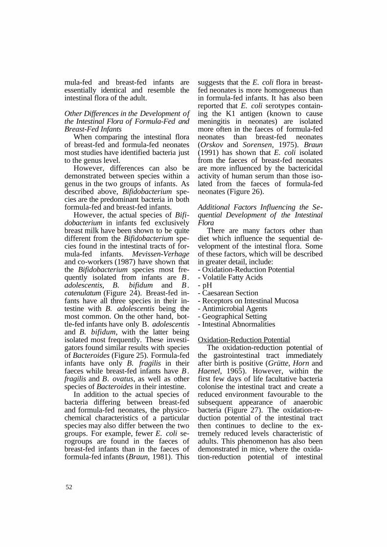
suggests that the E. coli flora in breastfed neonates is more homogeneous than in formula-fed infants. It has also been reported that E. coli serotypes containing the K1 antigen (known to cause meningitis in neonates) are isolated more often in the faeces of formula-fed neonates than breast-fed neonates (Orskov and Sorensen, 1975). Braun (1991) has shown that E. coli isolated from the faeces of breast-fed neonates are more influenced by the bactericidal activity of human serum than those isolated from the faeces of formula-fed neonates (Figure 26).
Additional Factors Influencing the Sequential Development of the Intestinal Flora
There are many factors other than diet which influence the sequential development of the intestinal flora. Some of these factors, which will be described in greater detail, include: - Oxidation-Reduction Potential - Volatile Fatty Acids - pH - Caesarean Section - Receptors on Intestinal Mucosa - Antimicrobial Agents - Geographical Setting - Intestinal Abnormalities
Oxidation-Reduction Potential The oxidation-reduction potential of
the gastrointestinal tract immediately after birth is positive (Grütte, Horn and Haenel, 1965). However, within the first few days of life facultative bacteria colonise the intestinal tract and create a reduced environment favourable to the subsequent appearance of anaerobic bacteria (Figure 27). The oxidation-reduction potential of the intestinal tract then continues to decline to the extremely reduced levels characteristic of adults. This phenomenon has also been demonstrated in mice, where the oxidation-reduction potential of intestinal
52
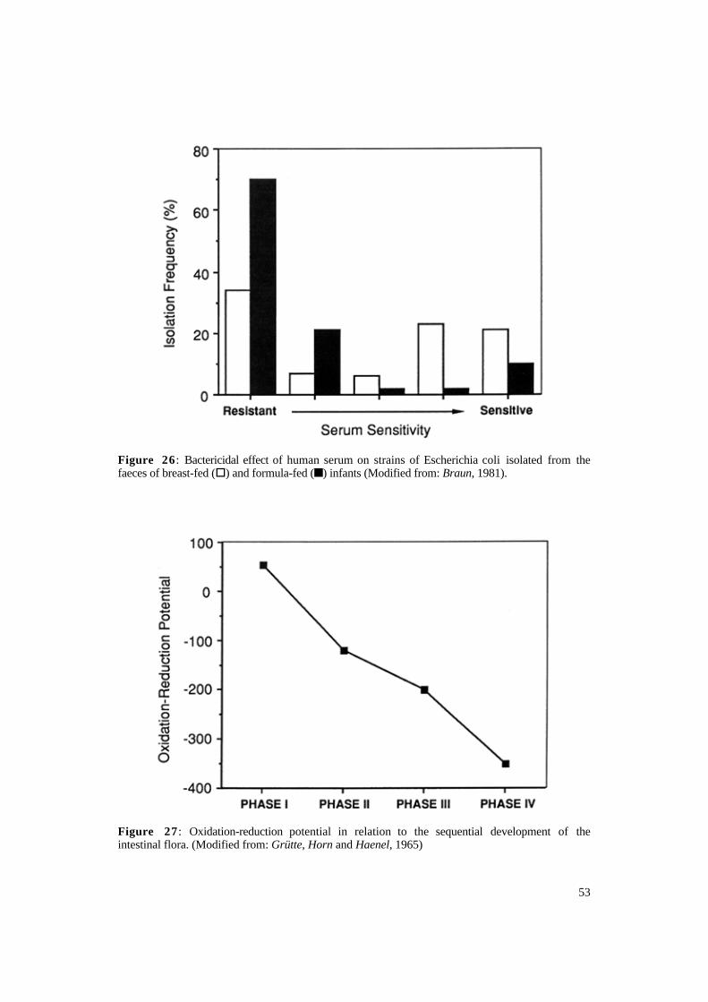
Figure 26 : Bactericidal effect of human serum on strains of Escherichia coli isolated from the faeces of breast-fed (§) and formula-fed (n) infants (Modified from: Braun, 1981).
Figure 27 : Oxidation-reduction potential in relation to the sequential development of the intestinal flora. (Modified from: Grütte, Horn and Haenel, 1965)
53

contents is considerably higher in germfree mice than in gnotobiotic mice colonised by facultative bacteria (Celesk, Asano and Wagner, 1976).
Volatile Fatty Acids Volatile fatty acids also play an im
portant role in the sequential development of the neonate intestinal flora. Acetic acid is the major, and often the only, fatty acid in stools of breast-fed neonates during the first few days of life (Bullen, Tearle and Stewart, 1977) (Figure 28). The presence of acetic acid is presumably due to the predominance of Bifidobacterium, a major acetic acid producer. Later in life, some breast-fed infants may also have low levels of propionic and butyric acids in their intestine. On the other hand, a variety of volatile fatty acids are usually present in the intestine of formula-fed infants, in
cluding acetic, butyric and propionic acids. Volatile fatty acids have been shown by numerous investigators to be inhibitory to a wide variety of bacteria (Hentges, 1983). Interestingly, it is during the period in which the concentrations of volatile fatty acids are increasing that there is a marked decline in the numbers of E. coli and streptococci.
pH The pH of stool obtained from
breast-fed neonates stabilises at a mean of about 5.0 to 5.5 after the first week of life and remains at this level as long as the infant receives a diet of only breast milk (Bullen, Tearle and Stewart, 1977) (Figure 29). In contrast, the mean pH of stool obtained from formula-fed neonates rises over time and may reach values as high as 8.5, suggesting that the intraluminal metabolic
Figure 28: Volatile fatty acids in the faeces of breast-fed and formula-fed infants. n: Breast-fed, acetic acid; s: Breast-fed, propionic acid; u: Breast-fed, butyric acid; §: Formula-fed, acetic acid; Ë: Formula-fed, propionic acid; ↕: Formula-fed, butyric acid (Modified from: Bullen, Tearle and Stewart, 1977).
54
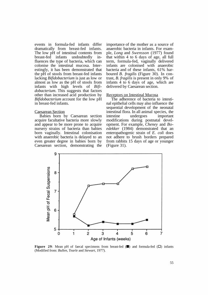
events in formula-fed infants differ dramatically from breast-fed infants. The low pH of intestinal contents from breast-fed infants undoubtedly influences the type of bacteria, which can colonise the intestinal mucosa. Interestingly, it has been demonstrated that the pH of stools from breast-fed infants lacking Bifidobacterium is just as low or almost as low as the pH of stools from infants with high levels of Bifidobacterium. This suggests that factors other than increased acid production by Bifidobacterium account for the low pH in breast-fed infants.
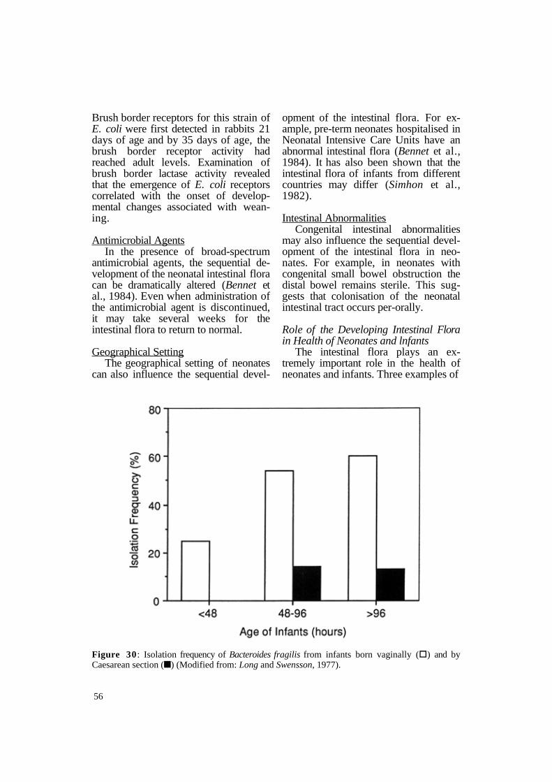
Caesarean Section Babies born by Caesarean section
acquire facultative bacteria more slowly and appear to be more prone to acquire nursery strains of bacteria than babies born vaginally. Intestinal colonisation with anaerobic bacteria is delayed to an even greater degree in babies born by Caesarean section, demonstrating the
importance of the mother as a source of anaerobic bacteria in infants. For example, Long and Swensson (1977) found that within 4 to 6 days of age, all full term, formula-fed, vaginally delivered infants are colonised with anaerobic bacteria and of these infants, 61% harboured B. fragilis (Figure 30). In contrast, B. fragilis is present in only 9% of infants 4 to 6 days of age, which are delivered by Caesarean section.
Receptors on Intestinal Mucosa The adherence of bacteria to intesti
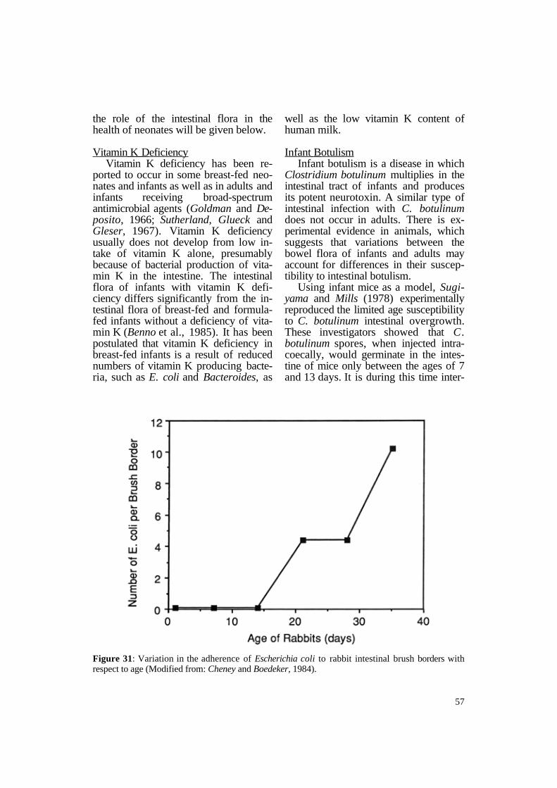
nal epithelial cells may also influence the sequential development of the neonatal intestinal flora. In all animal species, the intestine undergoes important modifications during postnatal development. For example, Cheney and Boedekker (1984) demonstrated that an enteropathogenic strain of E. coli does not adhere to brush borders prepared from rabbits 15 days of age or younger (Figure 31).
Figure 29 : Mean pH of faecal specimens from breast-fed (n) and formula-fed (§) infants (Modified from: Bullen, Tearle and Stewart, 1977).
55
Brush border receptors for this strain of E. coli were first detected in rabbits 21 days of age and by 35 days of age, the brush border receptor activity had reached adult levels. Examination of brush border lactase activity revealed that the emergence of E. coli receptors correlated with the onset of developmental changes associated with weaning.
Antimicrobial Agents In the presence of broad-spectrum
antimicrobial agents, the sequential development of the neonatal intestinal flora can be dramatically altered (Bennet et al., 1984). Even when administration of the antimicrobial agent is discontinued, it may take several weeks for the intestinal flora to return to normal.
Geographical Setting The geographical setting of neonates
can also influence the sequential devel
opment of the intestinal flora. For example, pre-term neonates hospitalised in Neonatal Intensive Care Units have an abnormal intestinal flora (Bennet et al., 1984). It has also been shown that the intestinal flora of infants from different countries may differ (Simhon et al., 1982).
Intestinal Abnormalities Congenital intestinal abnormalities
may also influence the sequential development of the intestinal flora in neonates. For example, in neonates with congenital small bowel obstruction the distal bowel remains sterile. This suggests that colonisation of the neonatal intestinal tract occurs per-orally.
Role of the Developing Intestinal Flora in Health of Neonates and lnfants
The intestinal flora plays an extremely important role in the health of neonates and infants. Three examples of
Figure 30 : Isolation frequency of Bacteroides fragilis from infants born vaginally (§) and by Caesarean section (n) (Modified from: Long and Swensson, 1977).
56
the role of the intestinal flora in the well as the low vitamin K content of health of neonates will be given below. human milk.
Vitamin K Deficiency Vitamin K deficiency has been re
ported to occur in some breast-fed neonates and infants as well as in adults and infants receiving broad-spectrum antimicrobial agents (Goldman and Deposito, 1966; Sutherland, Glueck and Gleser, 1967). Vitamin K deficiency usually does not develop from low intake of vitamin K alone, presumably because of bacterial production of vitamin K in the intestine. The intestinal flora of infants with vitamin K deficiency differs significantly from the intestinal flora of breast-fed and formulafed infants without a deficiency of vitamin K (Benno et al., 1985). It has been postulated that vitamin K deficiency in breast-fed infants is a result of reduced numbers of vitamin K producing bacteria, such as E. coli and Bacteroides, as
Infant Botulism Infant botulism is a disease in which
Clostridium botulinum multiplies in the intestinal tract of infants and produces its potent neurotoxin. A similar type of intestinal infection with C. botulinum does not occur in adults. There is experimental evidence in animals, which suggests that variations between the bowel flora of infants and adults may account for differences in their susceptibility to intestinal botulism.
Using infant mice as a model, Sugiyama and Mills (1978) experimentally reproduced the limited age susceptibility to C. botulinum intestinal overgrowth. These investigators showed that C. botulinum spores, when injected intracoecally, would germinate in the intestine of mice only between the ages of 7 and 13 days. It is during this time inter-
Figure 31: Variation in the adherence of Escherichia coli to rabbit intestinal brush borders with respect to age (Modified from: Cheney and Boedeker, 1984).
57
val that the intestinal flora of mice undergo dramatic quantitative and qualitative changes.
There is additional evidence, which demonstrates the importance of the intestinal flora in controlling C. botulinum intestinal overgrowth. Moberg and Sugiyama (1978) showed that the intestines of adult germfree mice are colonised with C. botulinum when as few as 10 spores of this microorganism are given orally. On the other hand, adult mice with a conventional microbial flora are resistant to C. botulinum intestinal colonisation even when 105 spores are inoculated orally. When the adult germfree mice are housed with conventional animals, they become resistant to challenge with 105 spores. Burr and Sugiyama (1982) showed that adult mice with a normal microbial flora but treated with large oral doses of two broad spectrum antimicrobial agents are at
least 50-fold more susceptible to C. botulinum intestinal colonisation than untreated control mice. The increased susceptibility of antimicrobial treated mice to C. botulinum intestinal colonisation is abolished by stopping the antibiotics and housing the mice with untreated control mice.
Clostridium difficile colonisation of infants
Clostridium difficile is an important aetiologic agent of antimicrobial agentassociated diarrhoeal disease in adults. Asymptomatic adults seldom have toxigenic C. difficile in their intestinal tract. On the other hand, up to 90% of infants less than one year of age are asymptomatically colonised with toxigenic C. difficile. This suggests that a developmental change in resistance to C. difficile intestinal colonisation occurs between infancy and adulthood. How-
Figure 32: Concentration of volatile fatty acids in the caeca of infant hamsters. n: Acetic acid; §: Butyric acid; Ë: Propionic acid; s: Valeric acid; u: Isovaleric acid (Modified from: Rolfe, 1984).
58
ever, the mechanisms accounting for the resistance to C. difficile colonisation in healthy, untreated adults are unknown.
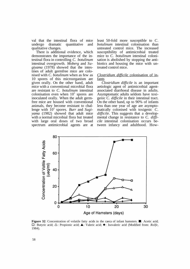
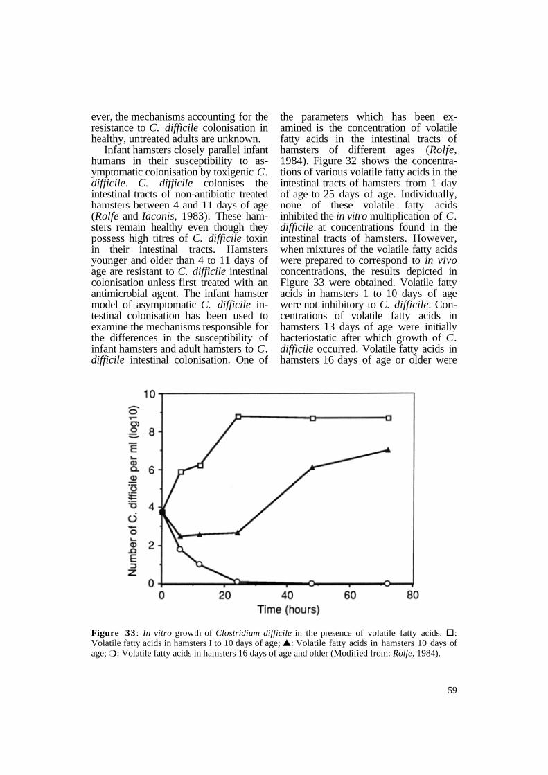
Infant hamsters closely parallel infant humans in their susceptibility to asymptomatic colonisation by toxigenic C. difficile. C. difficile colonises the intestinal tracts of non-antibiotic treated hamsters between 4 and 11 days of age (Rolfe and Iaconis, 1983). These hamsters remain healthy even though they possess high titres of C. difficile toxin in their intestinal tracts. Hamsters younger and older than 4 to 11 days of age are resistant to C. difficile intestinal colonisation unless first treated with an antimicrobial agent. The infant hamster model of asymptomatic C. difficile intestinal colonisation has been used to examine the mechanisms responsible for the differences in the susceptibility of infant hamsters and adult hamsters to C. difficile intestinal colonisation. One of
the parameters which has been examined is the concentration of volatile fatty acids in the intestinal tracts of hamsters of different ages (Rolfe, 1984). Figure 32 shows the concentrations of various volatile fatty acids in the intestinal tracts of hamsters from 1 day of age to 25 days of age. Individually, none of these volatile fatty acids inhibited the in vitro multiplication of C. difficile at concentrations found in the intestinal tracts of hamsters. However, when mixtures of the volatile fatty acids were prepared to correspond to in vivo concentrations, the results depicted in Figure 33 were obtained. Volatile fatty acids in hamsters 1 to 10 days of age were not inhibitory to C. difficile. Concentrations of volatile fatty acids in hamsters 13 days of age were initially bacteriostatic after which growth of C. difficile occurred. Volatile fatty acids in hamsters 16 days of age or older were
Figure 33 : In vitro growth of Clostridium difficile in the presence of volatile fatty acids. §: Volatile fatty acids in hamsters I to 10 days of age; s: Volatile fatty acids in hamsters 10 days of age; m: Volatile fatty acids in hamsters 16 days of age and older (Modified from: Rolfe, 1984).
59
bactericidal to C. difficile. These results suggest that volatile fatty acids may be one mechanism regulating the growth of C. difficile in vivo.
Conclusion A completely satisfactory under
standing of the succession of the normal flora in neonates and infants may not soon be achieved because of the com
plexity of the ecosystem. However, this is basic information, which must be understood if we ever hope to control intestinal diseases of infancy.
Rial D. Rolfe, Department of Microbiology, Texas Tech University Health Sciences Center, Lubbock, Texas 79430, U.S.A.
4. Mechanisms that Predispose to Ecological Stability in the Gut
One of the most remarkable features of the adult indigenous flora of humans and animals is its stability. The populations of microorganisms comprising the flora continually exert strong forces to maintain the community status quo. The practical effect of these activities is the exclusion of invading populations of non-indigenous microorganisms, including pathogens that attempt to colonise the intestinal tract from time to time. Only the most extreme stress situations, such as antibiotic administration, have major effect on the stability of the initial flora.
Factors responsible for the exclusion of non-indigenous organisms from the intestine and therefore maintenance of flora stability have not been identified, although several inhibitory mechanisms have been proposed. They include: 1. competition between the flora and
non-indigenous organisms for nutrients present in limited quantities,
2. elaboration of substances by the flora that inhibit multiplication of non-indigenous organisms,
3. competition between the flora and non-indigenous organisms for attachment sites on the intestinal mucosal cells, and
4. establishment of environmental conditions by the flora that adversely affect non-indigenous organisms.
Any one or all may be operative.
Considerable work has been done to identify compounds elaborated by the microflora that inhibit non-indigenous organisms. Although antibiotic substances produced by the flora, such as colicines, interfere with multiplication of non-indigenous organisms in vitro, there is no evidence that they function in the intestinal tract. However, there is evidence that volatile fatty acids, elaborated by components of the microflora as metabolic products, play a role in excluding non-indigenous organisms from the intestinal tract. Some time ago, Meynell (1963) and Bohnhoff and coinvestigators (1964a, 1964b) demonstrated that the multiplication of Salmonella enteritidis, a non-indigenous pathogen, was inhibited by suspensions of intestinal contents obtained from conventional mice. The contents contained volatile fatty acids in concentrations that prevented multiplication of S . enteritidis at low pH and oxidation-reduction potential measured in the intestine.
Treatment of the animals with streptomycin eliminated components of intestinal bacterial flora; this was associated with a decrease in total volatile fatty acids and an accompanying increase in oxidation-reduction potential and pHproducing conditions that favoured the multiplication of S. enteritidis. Maier and co-investigators (1972) obtained
60
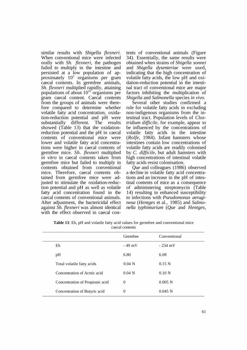
similar results with Shigella flexneri. When conventional mice were infected orally with Sh. flexneri, the pathogen failed to multiply in the intestine and persisted at a low population of approximately 103 organisms per gram caecal contents. In germfree animals, Sh. flexneri multiplied rapidly, attaining populations of about 1010 organisms per gram caecal content. Caecal contents from the groups of animals were therefore compared to determine whether volatile fatty acid concentration, oxidation-reduction potential and pH were substantially different. The results showed (Table 13) that the oxidationreduction potential and the pH in caecal contents of conventional mice were lower and volatile fatty acid concentrations were higher in caecal contents of germfree mice. Sh. flexneri multiplied in vitro in caecal contents taken from germfree mice but failed to multiply in contents obtained from conventional mice. Therefore, caecal contents obtained from germfree mice were adjusted to stimulate the oxidation-reduction potential and pH as well as volatile fatty acid concentration found in the caecal contents of conventional animals. After adjustment, the bactericidal effect against Sh. flexneri was almost identical with the effect observed in caecal con
tents of conventional animals (Figure 34). Essentially, the same results were obtained when strains of Shigella sonnei and Shigella dysenteriae were used, indicating that the high concentration of volatile fatty acids, the low pH and oxidation-reduction potential in the intestinal tract of conventional mice are major factors inhibiting the multiplication of Shigella and Salmonella species in vivo.
Several other studies confirmed a role for volatile fatty acids in excluding non-indigenous organisms from the intestinal tract. Population levels of Clostridium difficile, for example, appear to be influenced by the concentrations of volatile fatty acids in the intestine (Rolfe, 1984). Infant hamsters whose intestines contain low concentrations of volatile fatty acids are readily colonised by C. difficile, but adult hamsters with high concentrations of intestinal volatile fatty acids resist colonisation.
Que and colleagues (1986) observed a decline in volatile fatty acid concentrations and an increase in the pH of intestinal contents of mice as a consequence of administering streptomycin (Table 14) resulting in enhanced susceptibility to infections with Pseudomonas aeruginosa (Hentges et al., 1985) and Salmonella typhimurium (Que and Hentges,
Table 13: Eh, pH and volatile fatty acid values for germfree and conventional mice caecal contents
——————————————————————————————————————— Germfree Conventional
——————————————————————————————————————— Eh - 49 mV - 234 mV
pH 6.80 6.08
Total volatile fatty acids 0.04 N 0.15 N
Concentration of Acetic acid 0.04 N 0.10 N
Concentration of Propionic acid 0 0.005 N
Concentration of Butyric acid 0 0.045 N ———————————————————————————————————————
61
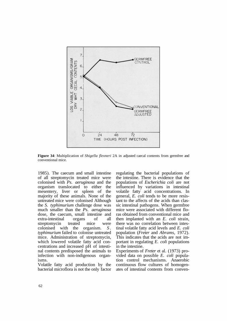
Figure 34: Multiplication of Shigella flexneri 2A in adjusted caecal contents from germfree and conventional mice.
1985). The caecum and small intestine of all streptomycin treated mice were colonised with Ps. aeruginosa and the organism translocated to either the mesentery, liver or spleen of the majority of these animals. None of the untreated mice were colonised Although the S. typhimurium challenge dose was much smaller than the Ps. aeruginosa dose, the caecum, small intestine and extra-intestinal organs of all streptomycin treated mice were colonised with the organism. S . typhimurium failed to colonise untreated mice. Administration of streptomycin, which lowered volatile fatty acid concentrations and increased pH of intestinal contents predisposed the animals to infection with non-indigenous organisms. Volatile fatty acid production by the bacterial microflora is not the only factor
regulating the bacterial populations of the intestine. There is evidence that the populations of Escherichia coli are not influenced by variations in intestinal volatile fatty acid concentrations. In general, E. coli tends to be more resistant to the affects of the acids than classic intestinal pathogens. When germfree mice were associated with different floras obtained from conventional mice and then implanted with an E. coli strain, there was no correlation between intestinal volatile fatty acid levels and E. coli population (Freter and Abrams, 1972). This indicates that the acids are not important in regulating E. coli populations in the intestine. Experiments of Freter et al. (1973) provided data on possible E. coli population control mechanisms. Anaerobic continuous flow cultures of homogenates of intestinal contents from conven
62

Table 14: Effects of streptomycin administration on various environmental conditions of caecal contents
——————————————————————————————————————— Mean + SD of results fromA:
Condition ————————————————————— Untreated mice Treated mice
——————————————————————————————————————— Eh (mV) -128.90 ± 7.61 -118.57 ± 21.80 ProteinB 6.17 ± 1.53 5.95 ± 1.79 CarbohydrateB 4.54 ± 3.22 6.90 ± 4.81 Dry wt/wet wt 0.24 ± 0.03 0.20 ± 0.06C
pH 6.42 ± 0.13 6.73 ± 0.28C
AceticacidD 74.8 ± 9.0 53.1 ± 7.9C
Propionic acidD 19.6 ± 4.0 13.0 ± 4.4C
Butyric acidD 60.8 ± 9.0 20.7 ± 4.9C
Valeric acidD 2.5 ± 0.9 NDC,E
——————————————————————————————————————— A 10 Experiments. B Results given in milligrams per gram (wet weight) of caecal contents. C Significantly different compared with untreated mice (p<0.05) by the two-tailed Student t-test. D Results given in micro-equivalents per gram (wet weight) of caecal contents. E ND: None detected.
tional mice mixed with E. coli in veal infusion broth, suppressed E. coli populations when compared with pure continuous flow cultures of E. coli. The diminished E. coli population levels were of magnitude observed in vivo in conventional mice suggesting that the control mechanisms in the flow culture and in the animal intestines were similar. Effluents from the continuous flow cultures inhibited E. coli multiplication, but the inhibition was reversed by addition of glucose. The results suggest that competition for nutrients, which are replaceable by glucose, is the activity overriding importance in the regulation of E. coli population levels in continuous flow cultures and possibly in the intestinal contents.
Bile acids, which are modified by indigenous flora components, may also function to exclude non-indigenous organisms from the intestinal tract. The primary bile acids, cholic acid and chenodeoxycholic acid, are synthesised by the liver and are conjugated to either taurine or glycine. Human bile also contains conjugates of a secondary bile
acid, dehydrocholic acid, which is formed by the dehydroxylation of cholic acid.
In the intestine the conjugates are hydrolysed to release free bile acids by a variety of bacteria, particularly anaerobes. Only free acids are present in the faeces. The bacteria also convert the primary bile acids to secondary bile acids by oxidoreduction of hydroxyl groups and dehydroxylation reactions. Floch et al. (1972) demonstrated that a variety of both Gram-positive and Gram-negative bacteria are inhibited by free bile acids but are not affected by either human whole bile or by conjugated bile acids. Free bile acid may very well be responsible for the exclusion of non-indigenous organisms from the intestinal ecosystem.
Competition for colonisation sites on intestinal mucosa surfaces is another mechanism that has been proposed to explain the role of the indigenous bacterial microflora in the rejection of nonindigenous organisms from the intestine. This was suggested by discovery of the colonisation by normal flora bac
63

teria of the mucus layer on the intestinal mucosal cells. The resulting mat of microflora presumably prevents contact and colonisation by non-indigenous bacteria, which need to adhere to the mucosa in order to survive in this open system. Specificity of attachment appears to be greater between indigenous microflora and host cells than between non-indigenous organisms and host cells. The non-indigenous organisms either fail to attach or are displaced by indigenous microflora once attached and are eliminated from the intestinal ecosystem.
The body of evidence suggests that multiple mechanisms are employed by the flora to exclude non-indigenous organisms from the intestinal tract. Recent data reported by Hentges et al. (1989) tend to confirm this hypothesis. Using Swiss white mice, they examined the influence of several oral antibiotics, administered at therapeutic levels, on resistance against colonisation by enterotoxigenic E. coli, S. typhimurium and Sh. flexneri.
In every case where an antibiotic produced an effect, resistance to colonisation with S. typhimurium and Sh. flexneri was enhanced. However, the situation with E. coli was the reverse. When an effect was observed the colonisation resistance for this pathogen was decreased. Therefore, identical factors appear to regulate colonisation of the intestine by Sh. flexneri and S . typhimurium. These factors differ from those that regulate the colonisation of the intestine by E. coli. This provides strong evidence that a universal mechanism governing colonisation against non-indigenous organisms in the intestinal tract does not exist but that several mechanisms are involved to actively exclude non-indigenous organisms from the ecosystem. In this way the indigenous intestinal flora maintains the ecological stability of the gut.
David J. Hentges, Texas Tech. University, Health Science Center, Lubbock, Texas 79430, U.S.A.
5. Individual Variation in the Microbial Population of the Human Gut
Microbial colonisation of new-born infants begins immediately after birth; infants are colonised by flora from the body of the mother and other (human) contacts as has been reviewed earlier in this seminar. Initial colonisation is fortuitous, depending on the first suitable organism to arrive at a particular site as well as on factors such as route of delivery, the type of nourishment received (breast milk or formula) and the degree of exposure to 'hospital environment'. In most cases, after only a few weeks, the representation of microbial species within the neonatal flora is remarkably similar to the adult pattern of colonisation (Table 15). This Table shows the conventional summary of the composi
tion of the digestive tract microflora. Even more detailed descriptions which go into genera and subgenera surpass perhaps the reality as such detailed list of flora components does not take in consideration that genera and even species and subspecies can be further subdivided in different serotypes. Particularly the latter may be of 'practical importance', since an important way in which the host organism 'decides' about the composition of its flora is not only by offering it a species - and possibly even strain - specific source of nutrients but by looking at it immunologically. Both selective mechanisms have been mentioned in this seminar several times.
64

Table 15: Microorganisms which may inhabit various sites of the digestive tract ——————————————————————————————————————— Oropharynx Nasopharynx Upper intestines Lower intestines
——————————————————————————————————————— Viridans streptococci Staphylococci Streptococci Staphylococci Staphylococci Corynebacteria Lactobacilli Streptococci Str. pyogenes Haemophilus spp. (Candida spp.) (including Branhamella cattharhalis enterococci) Neisseria spp. Lactobacilli Lactobacilli Corynebacteria Corynebacteria Neisseria spp. Haemophilus spp. Obligate Gram-pos. Obligate anaerobes and Gram-neg. (Candida, protozoa) anaerobes
Aerobic Gram-negative enterobacilli
C. albicans (Protozoa)
———————————————————————————————————————
The fact that the composition of the gastrointestinal flora of the new-born is completely fortuitous as it depends on the flora of the mother and many other sources encountered early in life and the fact that since a flora has settled in the digestive tract it remains stable in composition over long periods, may indicate that indeed in the first weeks or months after birth, the immune system may determine which bacteria, capable of digesting their nutrients (mucus, cells) can be 'tolerated' and which should be 'rejected'. This then may explain why several investigators have reported that different humans often are colonised by a different intestinal flora (Holdeman, Good and Moore, 1976; Mitsuoka and Ohno, 1977; Moore, Cato and Holde-man, 1978).
Van de Merwe and co-authors (1983) have investigated to which extent family members (parents and their children) match when the composition of their faecal flora is carefully studied. They reported that in man not only the aerobic (for most part transient) but also the anaerobic part of the faecal flora differs between individuals. In ten young
(several months to several years old) human twins (five monozygotic and five dizygotic twin pairs) van de Merwe, Stegeman and Hazenberg (1983) being faced with the difficult task to analyse the faecal flora of these children in great detail sought for a method by which they could handle a great number of samples with the limited technical assistance available. They found a rather elegant solution which enabled them to answer their question to which extent the human flora differs from one to another within one family and whether there is a genetic component involved in the selection made of the fortuitously encountered flora after birth. Fresh faeces were plated on selective media under carefully maintained strict anaerobic circumstances. After incubation, Gramstains were made of the various different (looking) colonies. In this way the Gram-positive bacteria could be classified into six different morphology groups: group 1, cocci growing in clusters or in chains; group 2, ovoid larger cocci and short rods;
65
group 3, typical Bifidobacterium; group 4, typical Eubacterium; group 5, very short rods; group 6, all other (unclassable) types of rods.
Gram-negative bacteria (rods) were put in a seventh group. Per faecal sample the seven morphological groups were expressed in percentages of the total number of bacteria in that particular sample. Individual faecal floras were represented by a single point in an Euclidian space with seven dimensions, one for each group of bacteria. Dissimilarity of faecal flora of twin siblings and between children of different siblings was expressed as a distance in the Euclidian space between the point representing each individual. These distances were calculated by taking the square root of the sum of square raised differ
ences of corresponding parameters. From the results of their study van
de Merwe and co-workers concluded that the composition of the faecal flora is under influence of genetic determinants of the host. In their study the floras of monozygotic twin siblings, being individually constant, were less distant (different) than siblings of the dizygotic twins. As one may expect on the bases of observations reported by other groups, the distance in the Euclidian space between unrelated subjects appeared much greater than between siblings of dizygotic origin.
Maarten P. Hazenberg, Department of Immunology, Erasmus University, P.O. Box 1738, NL-3000 Rotterdam, The Netherlands.
6. Microbial Ecology of the Human Bile Duct
If in patients the bile duct gets occluded, either from outside by for example a pancreatic carcinoma, or from within by foreign material such as a Ttube used to drain the common bile duct for some time after surgery, stone formation may occur. A study by Speer and co-workers (unpublished data) suggests that bacteria are involved in this stone formation. This is not confined to the bile duct only but may also be the case in the ureters as will also be discussed in this paper.
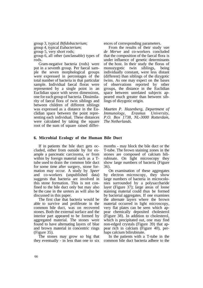
The first clue that bacteria would be able to survive and proliferate in the common bile duct, was on recovered stones. Both the external surface and the interior part appeared to be formed by aggregated material. The stones were found to have alternating layers of blue and brown material in concentric rings (Figure 35).
The stones may grow so big that they eventually - in less than one to six
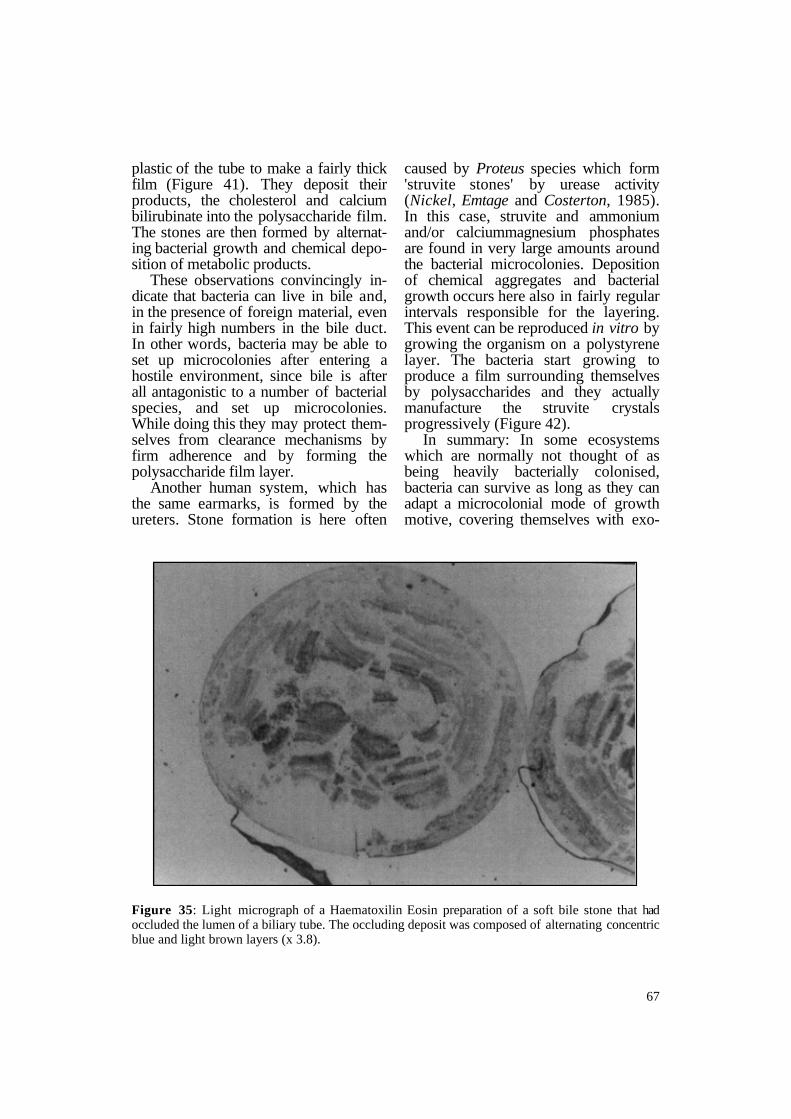
months - may block the bile duct or the T-tube. The brown staining zones in the stones are composed of calcium bilirubinate. On light microscopy they show large numbers of bacteria (Figure 36).
On examination of these aggregates by electron microscopy, they show large numbers of bacteria in microcolonies surrounded by a polysaccharide layer (Figure 37); large areas of loose staining material could thus be formed by bacterial aggregates. If one examines the alternate layers where the brown material occurred in light microscopy, very flat plates can be seen which appear chemically deposited cholesterol (Figure 38). In addition to cholesterol, which is precipitated out, one may find non-edged crystals (Figure 39) that appear rich in calcium (Figure 40), perhaps calcium bilirubinate.
In the patients with a T-tube in the common bile duct bacteria adhere to the
66
plastic of the tube to make a fairly thick film (Figure 41). They deposit their products, the cholesterol and calcium bilirubinate into the polysaccharide film. The stones are then formed by alternating bacterial growth and chemical deposition of metabolic products.
These observations convincingly indicate that bacteria can live in bile and, in the presence of foreign material, even in fairly high numbers in the bile duct. In other words, bacteria may be able to set up microcolonies after entering a hostile environment, since bile is after all antagonistic to a number of bacterial species, and set up microcolonies. While doing this they may protect themselves from clearance mechanisms by firm adherence and by forming the polysaccharide film layer.
Another human system, which has the same earmarks, is formed by the ureters. Stone formation is here often
caused by Proteus species which form 'struvite stones' by urease activity (Nickel, Emtage and Costerton, 1985). In this case, struvite and ammonium and/or calciummagnesium phosphates are found in very large amounts around the bacterial microcolonies. Deposition of chemical aggregates and bacterial growth occurs here also in fairly regular intervals responsible for the layering. This event can be reproduced in vitro by growing the organism on a polystyrene layer. The bacteria start growing to produce a film surrounding themselves by polysaccharides and they actually manufacture the struvite crystals progressively (Figure 42).
In summary: In some ecosystems which are normally not thought of as being heavily bacterially colonised, bacteria can survive as long as they can adapt a microcolonial mode of growth motive, covering themselves with exo-
Figure 35: Light micrograph of a Haematoxilin Eosin preparation of a soft bile stone that had occluded the lumen of a biliary tube. The occluding deposit was composed of alternating concentric blue and light brown layers (x 3.8).
67
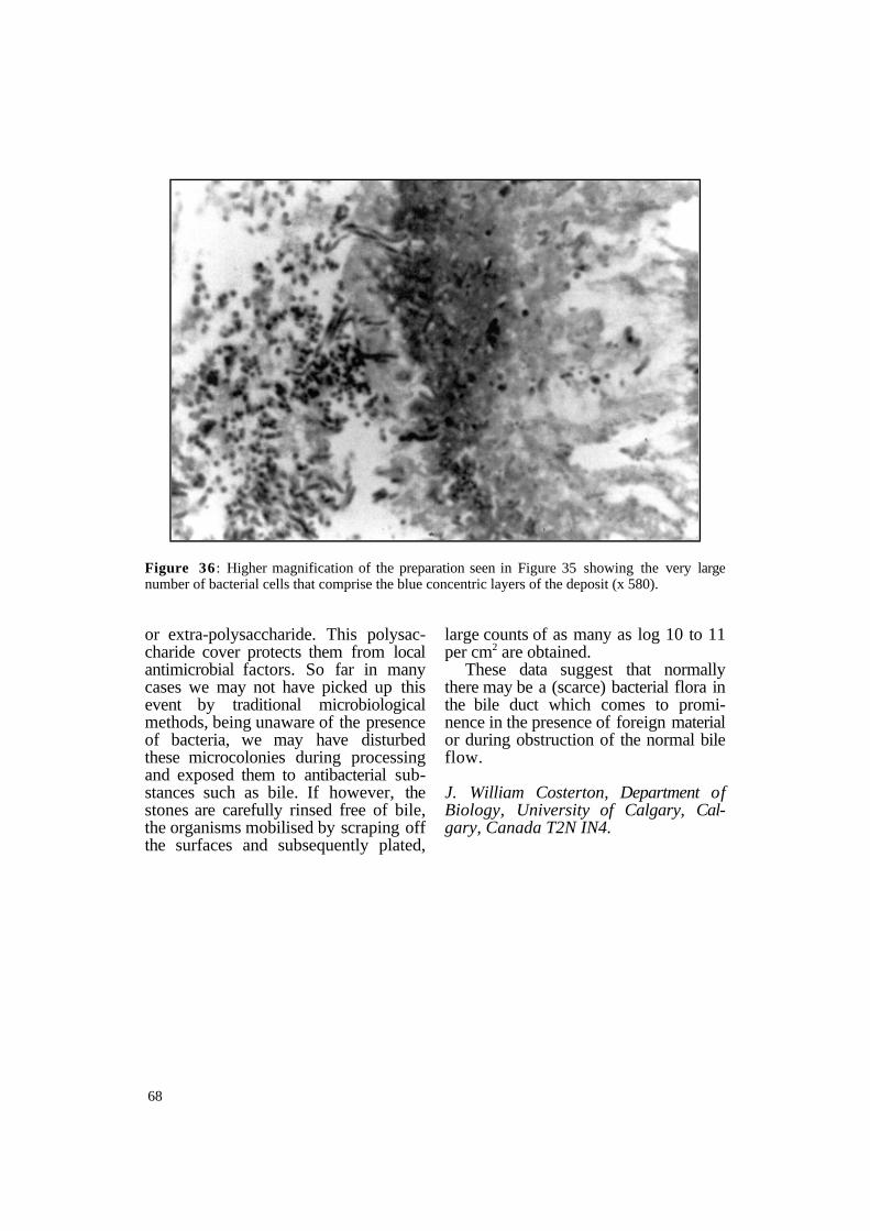
Figure 36 : Higher magnification of the preparation seen in Figure 35 showing the very large number of bacterial cells that comprise the blue concentric layers of the deposit (x 580).
or extra-polysaccharide. This polysaccharide cover protects them from local antimicrobial factors. So far in many cases we may not have picked up this event by traditional microbiological methods, being unaware of the presence of bacteria, we may have disturbed these microcolonies during processing and exposed them to antibacterial substances such as bile. If however, the stones are carefully rinsed free of bile, the organisms mobilised by scraping off the surfaces and subsequently plated,
large counts of as many as log 10 to 11 per cm2 are obtained.
These data suggest that normally there may be a (scarce) bacterial flora in the bile duct which comes to prominence in the presence of foreign material or during obstruction of the normal bile flow.
J. William Costerton, Department of Biology, University of Calgary, Calgary, Canada T2N IN4.
68
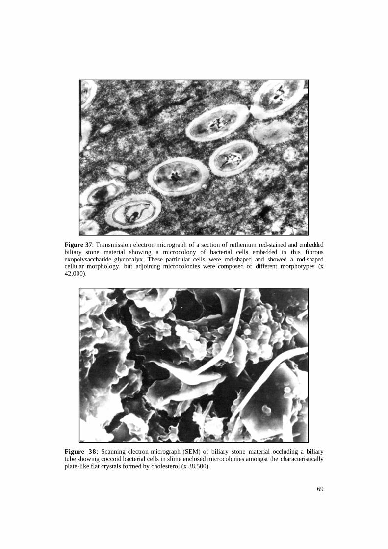
Figure 37: Transmission electron micrograph of a section of ruthenium red-stained and embedded biliary stone material showing a microcolony of bacterial cells embedded in this fibrous exopolysaccharide glycocalyx. These particular cells were rod-shaped and showed a rod-shaped cellular morphology, but adjoining microcolonies were composed of different morphotypes (x 42,000).
Figure 38 : Scanning electron micrograph (SEM) of biliary stone material occluding a biliary tube showing coccoid bacterial cells in slime enclosed microcolonies amongst the characteristically plate-like flat crystals formed by cholesterol (x 38,500).
69
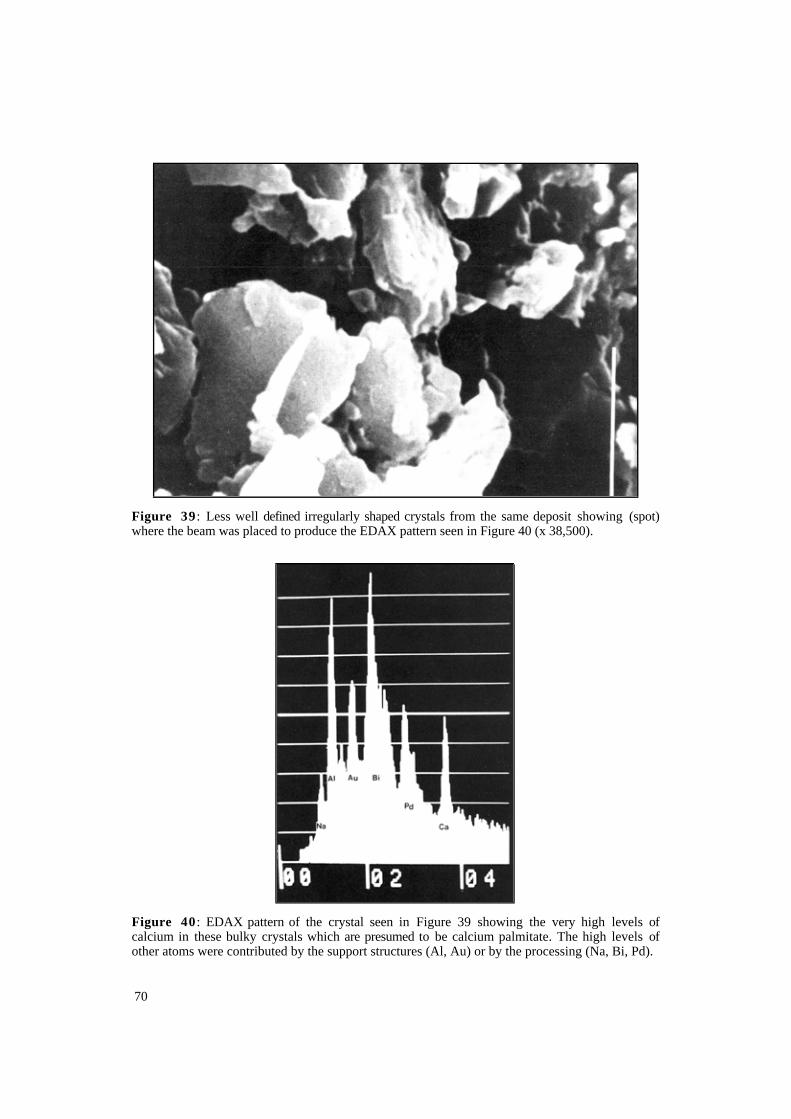
Figure 39 : Less well defined irregularly shaped crystals from the same deposit showing (spot) where the beam was placed to produce the EDAX pattern seen in Figure 40 (x 38,500).
Figure 40 : EDAX pattern of the crystal seen in Figure 39 showing the very high levels of calcium in these bulky crystals which are presumed to be calcium palmitate. The high levels of other atoms were contributed by the support structures (Al, Au) or by the processing (Na, Bi, Pd).
70
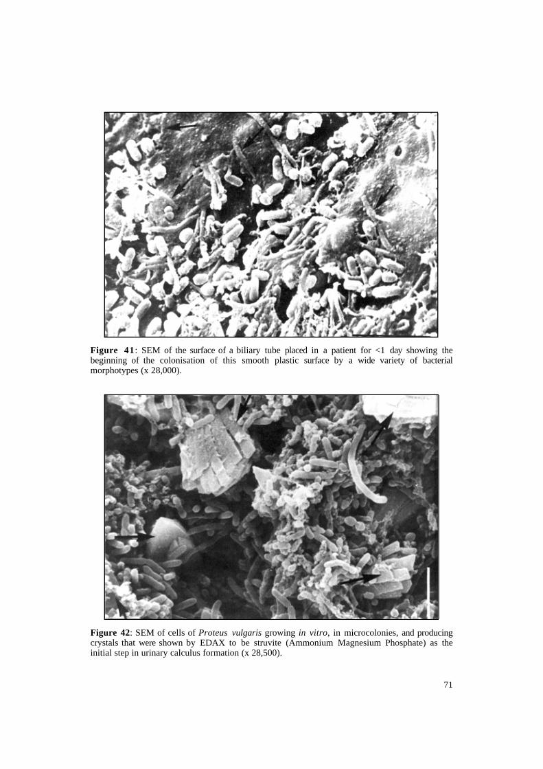
Figure 41 : SEM of the surface of a biliary tube placed in a patient for <1 day showing the beginning of the colonisation of this smooth plastic surface by a wide variety of bacterial morphotypes (x 28,000).
Figure 42: SEM of cells of Proteus vulgaris growing in vitro, in microcolonies, and producing crystals that were shown by EDAX to be struvite (Ammonium Magnesium Phosphate) as the initial step in urinary calculus formation (x 28,500).
71

7 . Effects of Dietary, Genetic and Stress Factors on the Microbial Ecology of the Gut
A review of the literature reveals that the colonic microbial flora can be characterised in several different ways (Lee, 1985; Holdeman, Good and Moore, 1976; Moore et al., 1981); i.e. by: a. the particular microbial species, b. the metabolic characteristics of the
microflora, c. the quantity or mass of bacteria in a
particular ecological niche and d. genetic characteristics of the micro
organisms which define particular metabolic or enzymatic capabilities.
In the fasting individual, only limited material enters the colon in the form of mucus, desquamated and obligatory products of cell membrane turnover. Among bacterial species which characterise the colonic microflora no qualitative changes have been observed during fasting, starvation or during ingestion of a chemically defined diet which is fully absorbed in the small intestine (Hentges, 1980; McNabb and Tomasi, 1981; Simon and Gorbach, 1987; Tannock and Savage, 1974; Tomkins et al., 1981). Expansion of the indigenous colonic flora occurs with introduction of growth substrates for this microflora. These are usually complex polysaccha
rides such as starch cellulose, pectins and other dietary constituents. Cellulose is only partly degraded in the human colon (Wolin, 1981). Such an expansion of the human colonic flora may be associated with changes in the population density of certain bacterial species in the caecum which favour lactic acid production and reduced acetic, butyric and propionic acid generation. A change in content of complex polysaccharides degraded in the caecum in some animal species, may alter the distribution of other bacterial species and their capability for survival in this ecological niche (Mathiesen et al., 1987) (Table 16). Changes in faecal microbial populations associated with chronic dietary intake of specific foodstuffs (Tomkins et al., 1981) have not been characterised consistently (Moore et al., 1981). However, review of the literature suggests that changes of bacterial species may occur in human groups on high fat high meat protein intake associated with increased activity of ß-glucuronidase, azoreductase and bile acid excretion (Hentges, 1980; Gorbach et al., 1967; Goldin et al., 1980). It is now well recognised, that many bacterial metabolic
Table 16: Seasonal change in caecal microflora (reindeer) ———————————————————————————————————————
Fall in bacterial populations in winter (17%) ———————————————————————————————————————
Species: Summer: Winter ———————— ————— ————— Butyrivibrio 23 % 18 % Streptococcus bovis 17 % 5 % Bacteroides 10 % 26 %
Metabolic activity: Summer: Winter ———————— ————— ————— Fibre 36 % 48 % Cellulolysis 10 % 6 % Xylanolysis 33 % 48 % Starch 77 % 71%
———————————————————————————————————————
72
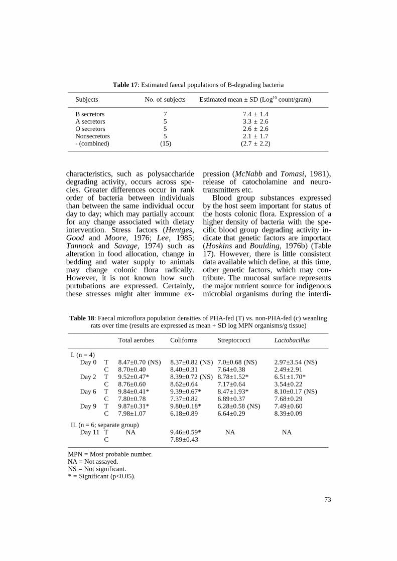
Table 17: Estimated faecal populations of B-degrading bacteria ———————————————————————————————————————
Subjects No. of subjects Estimated mean ± SD (Log10 count/gram) ———————————————————————————————————————
B secretors 7 7.4 ± 1.4 A secretors 5 3.3 ± 2.6 O secretors 5 2.6 ± 2.6 Nonsecretors 5 2.1 ± 1.7 - (combined) (15) (2.7 ± 2.2)
———————————————————————————————————————
characteristics, such as polysaccharide degrading activity, occurs across species. Greater differences occur in rank order of bacteria between individuals than between the same individual occur day to day; which may partially account for any change associated with dietary intervention. Stress factors (Hentges, Good and Moore, 1976; Lee, 1985; Tannock and Savage, 1974) such as alteration in food allocation, change in bedding and water supply to animals may change colonic flora radically. However, it is not known how such purtubations are expressed. Certainly, these stresses might alter immune ex
pression (McNabb and Tomasi, 1981), release of catocholamine and neurotransmitters etc.
Blood group substances expressed by the host seem important for status of the hosts colonic flora. Expression of a higher density of bacteria with the specific blood group degrading activity indicate that genetic factors are important (Hoskins and Boulding, 1976b) (Table 17). However, there is little consistent data available which define, at this time, other genetic factors, which may contribute. The mucosal surface represents the major nutrient source for indigenous microbial organisms during the interdi-
Table 18: Faecal microflora population densities of PHA-fed (T) vs. non-PHA-fed (c) weanling rats over time (results are expressed as mean + SD log MPN organisms/g tissue)
——————————————————————————————————————— Total aerobes Coliforms Streptococci Lactobacillus
——————————————————————————————————————— I. (n = 4)
Day 0 T 8.47±0.70 (NS) 8.37±0.82 (NS) 7.0±0.68 (NS) 2.97±3.54 (NS) C 8.70±0.40 8.40±0.31 7.64±0.38 2.49±2.91
Day 2 T 9.52±0.47* 8.39±0.72 (NS) 8.78±1.52* 6.51±1.70* C 8.76±0.60 8.62±0.64 7.17±0.64 3.54±0.22
Day 6 T 9.84±0.41* 9.39±0.67* 8.47±1.93* 8.10±0.17 (NS) C 7.80±0.78 7.37±0.82 6.89±0.37 7.68±0.29
Day 9 T 9.87±0.31* 9.80±0.18* 6.28±0.58 (NS) 7.49±0.60 C 7.98±1.07 6.18±0.89 6.64±0.29 8.39±0.09
II. (n = 6; separate group) Day 11 T NA 9.46±0.59* NA NA
C 7.89±0.43 ——————————————————————————————————————— MPN = Most probable number.
NA = Not assayed. NS = Not significant. * = Significant (p<0.05).
73
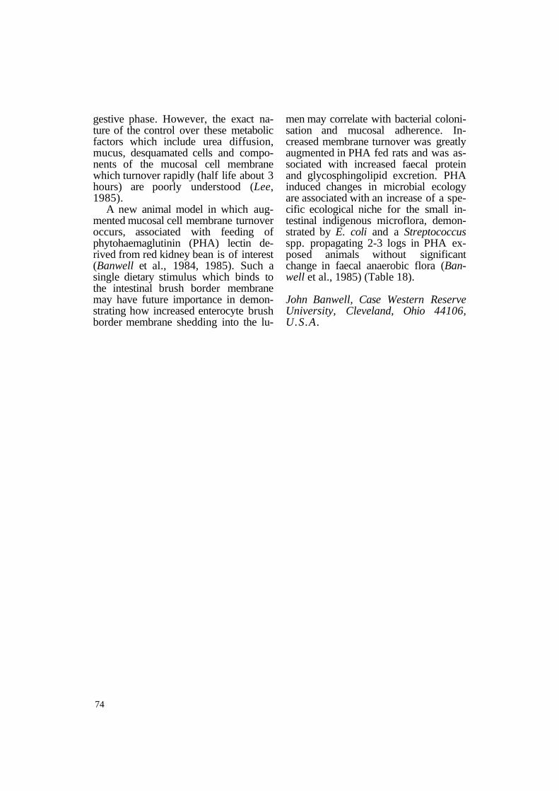
gestive phase. However, the exact nature of the control over these metabolic factors which include urea diffusion, mucus, desquamated cells and components of the mucosal cell membrane which turnover rapidly (half life about 3 hours) are poorly understood (Lee, 1985).
A new animal model in which augmented mucosal cell membrane turnover occurs, associated with feeding of phytohaemaglutinin (PHA) lectin derived from red kidney bean is of interest (Banwell et al., 1984, 1985). Such a single dietary stimulus which binds to the intestinal brush border membrane may have future importance in demonstrating how increased enterocyte brush border membrane shedding into the lu
men may correlate with bacterial colonisation and mucosal adherence. Increased membrane turnover was greatly augmented in PHA fed rats and was associated with increased faecal protein and glycosphingolipid excretion. PHA induced changes in microbial ecology are associated with an increase of a specific ecological niche for the small intestinal indigenous microflora, demonstrated by E. coli and a Streptococcus spp. propagating 2-3 logs in PHA exposed animals without significant change in faecal anaerobic flora (Banwell et al., 1985) (Table 18).
John Banwell, Case Western Reserve University, Cleveland, Ohio 44106, U.S.A.
74
V. LITERATURE
Abrams, G.D., Bauer, A., and Sprinz, H.: In- 6765 (1987). fluence of the normal flora on mucosal morphology and cellular renewal in the ileum. Lab. Invest. 12, 355-364 (1963).
Allen, A. and Snary, D.: The structure and function of gastric mucus. Gut 13, 666-672 (1972).
Allen, A. and Carroll, N.J.H.: Adherent and soluble mucus in the stomach and duodenum. Dig. Dis. Sci. 30, 55S-62S (1985).
Allen, A. and Hoskins, L.C.: Colonic mucus in health and disease. In: Diseases of the Colon, Rectum and Anal Canal (Eds.: Kirsner, J.B., and Shorter, R.G.). Williams and Wilkins, Baltimore, 65-94 (1988).
Aminoff, D.: 1,2-α-L-Fucosidase from Clostridium perfringens. Meth. Enzymol. 28, 763-769 (1972).
Andre, C., Lambert, R., and Descons, F.: Stimulation of gastric mucous secretions in man by secretin. Digestion 7, 284-293 (1972).
Banwell, J.G., Abramowsky, C.R., Weber, F., and Howard, R.: Phytohaemagglutinin-induced diarrheal dehydration. Dig. Dis. Sci. 29, 921-929 (1984).
Banwell, J.G., Howard, R., Cooper, D., and Costerton, J.W.: Intestinal microbial flora after feeding phytohemagglutinin lectins (Phasueolus vulgaris) to rats. Appl. Environ. Microbiol. 50, 60-80 (1985).
Bayliss, C.E. and Houston, A.P.: Characterization of plant polysaccharide- and mucinfermenting anaerobic bacteria from human feces. Appl. Environ. Microbiol. 48, 626632 (1986).
Bennet, R., Erickssen, M., Nord, C.E., and Zetterstrom, R.: Impact of various antibiotics on the fecal flora of newborn infants. Microecol. Ther. 14, 251 (1984).
Benno, Y., Sawada, K., and Mitsuoka, T.: The intestinal microflora of infants: Fecal flora of infants with vitamin K deficiency. Microbiol. Immunol. 29, 243-250 (1985).
Berkowitz, R.J., Turner, J., and Green, P.: Maternal salivary levels of Streptococcus mutans and primary oral infection of infants. Arch. oral Biol. 26, 147-149 (1981).
Bjork, S., Breimer, M., Hansson, G.C., Karlsson, K-A., and Leffler, H.: Structures of blood group glycosphingolipids of human small intestine. J. Biol. Chem. 262, 6758
Black, J.W., Bradbury, J.E., and Wyllie, J.H.: Stimulation of colonic mucus output in rat. Br. J. Pharmacol. 66, 456-463 (1979).
Bock, K., Breimer, M.E., Brignole, A., Hansson, G.C., Karlsson, KA., Larson, G., Leffler, H., Samuelsson, B.E., Stromberg, N., Svanborg-Eden, K., and Thurin, J.: Specificity of binding of a strain of uropathogenic Escherichia coli to Gal-α(1-4)Galcontaining glycosphingolipids. J. Biol. Chem. 260, 8545-8551 (1985).
Bohnhoff, M., Miller, P.C., and Martin, W.R.: Resistance of the mouse's intestinal tract to experimental Salmonella infection. I. Factors which interfere with initiation of infection by oral inoculation. J. Exp. Med. 120, 805-816 (1964a).
Bohnhoff, M., Miller, P.C., and Martin, W.R.: Resistance of the mouse's intestinal tract to experimental Salmonella infection. II. Factors responsible for its loss following streptomycin treatment. J. Exp. Med. 120, 817-828 (1964b).
Bolton, J.P., Palmer, D., and Cohen, M.M.: Stimulation of mucus and nonparietal cell secretion by the E2 prostaglandin. Am. J. Dig. Dis. 23, 539-364 (1978).
Braun, O.H.: Effect of consumption of human milk and other formulas on intestinal bacterial flora in infants. In: Textbook of Gastroenterology and Nutrition in Infancy (Ed.: Lebenthal, E.). Raven Press, New York (1981).
Brook, I., Walker, R.I., and MacVittie, T.J.: Effect of antimicrobial therapy on the bowel flora and bacterial infection in irradiated mice. Int. J. Radiat. Biol. 53, 709-716 (1988).
Bryant, M.P.: Nutritional features and ecology of predominant anaerobic bacteria of the intestinal tract. Am. J. Clin. Nutr. 27, 13131323 (1974).
Bullen, C.L., Tearle, P.V., and Stewart, M.G.: The effect of "humanized" milks and supplemented breast feeding on the faecal flora of infants. J. Med. Microbiol. 10, 403-413 (1977).
Burr, D.H. and Sugiyama, H.: Susceptibility to enteric botulinum colonization of antibiotic-treated adult mice. Infect. Immun. 36, 103-106 (1982).
75

Burr, D.H., Kerner, D.T., Blanco, C.S., Bourgeois, A.L., and Wistar, Jr.: Gastric lavage: A simple method to obtain IgA-rich intestinal secretions from the rabbit. J. Immunol. Methods 99, 277-281 (1987).
Burr, D.H., Caldwell, M.B., Bourgeois, A.L., Morgan, H.R., Wistar, Jr., and Walker, R.I.: Mucosal and systemic immunity to Campylobacter jejuni in rabbits following gastric inoculation. Infect. Immun. 56 (1), 99-105 (1988).
Caldwell, M.B., Walker, R.I., Stewart, S.D., and Rogers, J.E.: Simple adult rabbit model for Campylobacter jejuni enteritis. Infect. Immun. 42, 1176-1182 (1983).
Caldwell, M.B. and Walker, R.I.: Animal model of human disease. Adult rabbit model for Campylobacter jejuni enteritis. Am. J. Pathol. 122, 573-576 (1986).
Carlstedt-Duke, B., Midtvedt, T., Norin, C.E., and Gustafsson, B.E.: Isolation and characterization of a mucin-degrading strain of Peptostreptococcus from rat intestinal tract. Acta Path. Microbiol. Immunol. Scand. Sect. B94, 293-300 (1986).
Celesk, R.A., Asano, T., and Wagner, M.: The size, pH and redox potential of the cecum in mice associated with various microbial floras. Proc. Soc. Exp. Biol. Med. 151, 260266 (1976).
Cheney, C.P., and Boedeker, E.C.: Rabbit mucosal receptors for an enteropathogenic Escherichia coli strain: Appearance of bacterial receptor activity at weaning. Gastroenterology 87, 821-826 (1984).
Cheng, K.J., Irvin, R.T., and Costerton, J.W.: Autochthonous and pathogenic colonization of animal tissues by bacteria. Can. J. Microbiol. 27, 461-490 (1981).
Cooper, R., Fraser, S.M., Sturrock, R.D., and Gemmell, C.G.: Raised titers of anti-Klebsiella IgA in ankylosing spondylitis, rheumatoid arthritis and inflammatory bowel disease. Brit. Med. J. 296, 1432-1434 (1988).
Costerton, J.W., Rozee, K.R., and Cheng, K.J.: Colonization of particulates, mucous and intestinal tissue. Prog. Fd. Nutr. Sci. 7, 191-195 (1983).
Critchley, P.: The breakdown of the carbohydrate and protein matrix of dental plaque. Caries Res. 3, 249265 (1969).
Croft, D.N. and Cotton, P.B.: Gastrointestinal cell loss in man. Digestion 8, 144-160
(1973). Cromwell, C.L., and Hoskins, L.C.: Antigen
degradation in human colon ecosystems. Host's ABO blood type influences enteric bacterial degradation of a cell surface anti-gen on Escherichia coli 086. Gastroenterology 73 (1), 37-41 (1977).
Curatolo, W.: Glycolipid function. Biochem. Biophys. Acta 906, 137-160 (1987).
DeVay, J.E. and Adler, H.E.: Antigens common to hosts and parasites. Ann. Rev. Microbiol. 30, 147-168 (1976).
Dinoso, V.P., Ming, S., and Meniff, J.: Ultrastructural changes of the canine gastric mucosa after topical application of graded concentrations of ethanol. Am. J. Dig. Dis. 21, 626-632 (1976).
Dische, Z., Kahn, N., Rothschild, C., Danilchenko, A., Liebling, J., and Wang, S.C.: Glycoproteins of submaxillary saliva of the cat: differences in composition produced by sympathetic and parasympathetic nerve stimulation. J. Neurochem. 17, 649658 (1970).
Domschke, W., Domschke, S., Classen, S., and Demling, L.: Prostaglandin-stimulated gastric mucus secretion in man. Acta Hepatogastroenterologica 25, 292-294 (1972).
Dykhuizen, D.E. and Hart, D.L.: Selection in chemostats. Microbiol. Rev. 47, 150-168 (1982).
Ebringer, A., Cooke, D., Cadwell, D.R., Cowling, P., and Ebringer, A.: Ankylosing spondylitis Klebsiella and HLA-B27. Rheumatol. Rehabil. 16, 190-196 (1977).
Ebringer, A. (ed): Proceedings of the international symposium on the pathogenesis of HLA-B27 associated diseases. Br. J. Rheumatol. 22, Suppl. 2. (1983).
Englyst, H.N. and Cummings, J.H.: Digestion of the carbohydrates of banana (Musa paradisiaca sapientum) in the human small intestine. Am. J. Clin. Nutr. 44, 42-50 (1986).
Ferguson, A. and Parrott, D.M.V.: Histopathology and time course of rejection of allografts of mouse small intestine. Transplantation 15, 546554 (1973).
Filipe, M.I.: Mucins in the human gastrointestinal epithelium: A review. Invest. Cell. Pathol. 2, 195-216 (1979).
Fiocchi, C. and Farmer, R.G.: Autoimmunity in inflammatory bowel disease. Clin. Aspects of Autoimmun. 1, 12-19 (1987).
76

Floch, M.H., Binder, J.J., Filburn, B., and Gershengoren, W.: The effect of bile acids on intestinal microflora. Am. J. Clin. Nutr. 25, 1418-1426 (1972).
Formal, S.B., Hale, T.L., and Sansonetti, P.J. Invasive enteric pathogens. Rev. Infect. Dis. 5 (suppl. 4), 8702-8707 (1983).
Forstner, J.F.: Intestinal mucins in health and disease. Digestion 17, 234-263 (1978).
Forstner, J.F., Roomi, N.W., Fahim, R.E.F., and Forstner, G.G.: Cholera toxin stimulates secretion of immunoreactive intestinal mucin. Am. J. Physiol. 240 (Gastrointest. Liver Physiol.), G10-G16 (1981).
Forstner, J.F., Maxwell, B., and Romui, N.: Intestinal secretion of mucin in chronically reserpine-treated rats. Am. J. Physiol. 241 (Gastrointest. Liver Physiol.), G443-G450 (1981).
Freter, R. and Abrams, G.D.: Function of various intestinal bacteria in converting germfree mice to the normal state. Infect. Immun. 6, 119-126 (1972).
Freter, R., Abrams, G.D., and Aranki, A.: Patterns of interaction in gnotobiotic mice among bacteria of a synthetic "normal" intestinal flora. In: Germfree Research: Biological effect of gnotobiotic environments (Ed.: Heneghan, J.B.). Academic Press, New York, London, 429-434 (1973)
Geczy, A.F., Alexander, K., Bashir, H.V., Edmonds, J.P., Upfold, L.I., and Sullivan, J.: HLA-B27, Klebsiella and ankylosing spondilytis: Biological and chemical studies. Immunol. Rev. 70, 23-50 (1983).
Geczy, A.F., Prendergast, J.K., Sullivan, J.S., Upfold, L.I., McGuigan, L.E., Bashir, H.V., Prendergast, M., and Edmonds, J.P.: HLA-B27, molecular mimicry and ankylosing spondylitis: popular misconceptions. Ann. Rheum. Dis. 46, 171-172 (1987).
George, M.H. and Leitch, G.J.: Separation of rabbit ileum mucus secretion from electrolyte and water secretion by cholera enterotoxin, verapamil and A23187. Life Sciences 32, 839-846 (1983).
Gibbons, R.J. and van Houte, J.: Bacterial adherence in oral microbial ecology. Ann. Rev. Microbiol. 29, 19-44 (1975).
Gibbons, R.J. and van Houte, J.: Bacteriology of Dental Caries. In: Textbook of Oral Biology (Eds.: Shaw, J.H, Sweeney, E.A., Cappuccino, C.C., and Meller, S.M.). W.B. Saunders Co., Philadelphia, 975-991
(1978). Gibbons, R.J. and van Houte, J.: Oral Bacterial
Ecology. In: Textbook of Oral Biology (Eds.: Shaw, J.H, Sweeney, E.A., Cappuccino, C.C., and Meller, S.M.). W.B. Saunders Co., Philadelphia, 684-705 (1978).
Goldin, R., Swenson, L., Dwyer, J., Sexton, M., and Gorbach, S.L.: Effect of diet and Lactobacillus acidophilus supplements on human fecal bacterial enzymes. J. Natl. Cancer Inst. 64, 255-261 (1980).
Goldman, H.I. and Deposito, F.: Hypoprothrombinemic bleeding in young infants. Am. J. Dis. Child. 111, 430-432 (1966).
Gorbach, S.L., Plaut, A.G., Nahas, L., and Weinstein, L.: Studies of intestinal microflora. II. Microorganisms of the small intestine and their relations to oral and fecal flora. Gastroenterology 53, 856-864 (1967).
Gothefors, L., Carlsson, B., Ahlstedt, S., Hanson, L.A., and Winberg, J.: Influence of maternal gut flora and colostral and cord serum antibodies on presence of Escherichia coli in faeces of the newborn infant. Acta Paediatr. Scand. 65, 225-232 (1976).
Grütte, F.K., Horn, R., and Haenel, H.: Ernährung und biochemischmikrookologische Vorgänge im Enddarm von Säuglingen. Z. Kinderheilkd. 93, 28-39 (1965).
Gustafsson, B.E., and Carlstedt-Duke, B.: Intestinal water-soluble mucins in germfree, exgermfree, and conventional animals. Acta Path. Microbiol. Immunol. Scand. Sect. B 92, 247-252 (1984).
Gustafsson, B.E., Karlsson, K.-A., Larson, G., Midtvedt, T., Stromberg, N., Teneberg, S., and Thurin, J.: Glycosphingolipid patterns of the gastrointestinal tract and feces of germ-free and conventional rats. J. Biol. Chem. 261, 15294-15300 (1986).
Hansson, G., Karlsson, K-A., Larson, G., Lindberg, G., and Stromberg, T.J.: Lactosylceramide as the probable adhesion site for major indigenous bacteria of the gastrointestinal tract. In: Proc. 7th Intern. Symp. on Glycoconjugates (Eds.: Chester, M.A., Heinegard, D., Lundblad, A., and Svensson, S.). Rahms, Lund, 631-632 (1983).
Harouny, V. and Hoskins, L.C.: A role for mucin-degrading bacteria in nutritional support of human enteric bacteria. Gastroenterology 84, 1405 (Abstr) (1983).
Hentges, L.V., Good, I.J., and Moore, W.E.C.:
77

Human faecal flora: Variation in bacterial composition within individuals and a possible effect of emotional stress. Appl. Environ. Microbiol. 31, 359-375 (1976).
Hentges, D.J.: Does diet influence human fecal microflora composition? Nutrition Reviews 38, 329-336 (1980).
Hentges, D.J.: Role of the intestinal microflora in host defense against infection. In: Human Intestinal Microflora in Health and Disease (Ed.: Hentges, D.J.). Academic Press, New York, 311-331 (1983).
Hentges, D.J., Stein, A.J., Casey, S.W., and Que, J.U.: Protective role of intestinal flora against infection with Pseudomonas aerigunosa in mice: Influence of antibiotics on colonization resistance. Infect. Immun. 47, 118-122 (1985).
Hentges, D.J., Marsh, W.W., Dougherty, S.H., Thal, W.R., and Adams, M.K.: Effects of antibiotics on resistance to colonization with enteric pathogens in mice. In: Proceedings of the III. International Symposium on the influence of Antibiotics on the Host-Parasitic Relationship (Eds.: Peters, P., and Pulverer, G.). Springer Verlag, Berlin, 204-208 (1989).
Heyma, P., Harrison, L.C., and Robins-Browne. R.: Thyrotrophin (TSH) binding sites on Yersinia enterocolitica recognized by immunoglobulins from humans with Graves' disease. Clin. Exp. Immunol. 64, 249-254 (1986).
Hill, M.: Bacterial factors. In: Gut defences in clinical practice (Eds.: Losawsky, M.S., and Heatly, R.V.). Churchill Livingstone, Edinburgh, 147-154 (1983).
Holdeman, L.V., Good, I.J., and Moore, W.E.C.: Human fecal flora: variation in bacterial composition within individuals and a possible effect of emotional stress. Appl. Environ. Microbiol. 31, 359-375 (1976).
Holgersson, J., Karlsson, K-A., Karlsson, P., Norrby, E., Orvell, C., and Stromberg, N.: Approaches to the study of receptors. Wistar Symp. Ser. 3, 273-301 (1985).
Horowitz, M.I. and Hollander, F.: Evidence regarding the chemical complexicity of acetylcholine stimulated gastric mucus. Gastroenterology 40, 1185-793 (1961).
Hoskins, L.C. and Zamcheck, N.: Bacterial degradation of gastrointestinal mucins. I. Comparison of mucus constituents in the
stools of germfree and conventional rats. Gastroenterology 54 (2), 210-217 (1968).
Hoskins, L.C. and Boulding, E.T.: Degradation of blood group antigens in human colon ecosystems. I. In vitro production of ABH blood group-degrading enzymes by enteric bacteria. J. Clin. Invest. 57, 63-73 (1976a).
Hoskins, L.C. and Boulding, E.T.: Degradation of blood group antigens in human colon ecosystems. J. Clin. Invest. 57, 74-82 (1976b).
Hoskins, L.C. and Boulding, E.T.: Mucin degradation in human colon ecosystems. Evidence for the existence and role of bacterial subpopulations producing glycosidases as extracellular enzymes. J. Clin. Invest. 67, 163-172 (1981).
Hoskins, L.C., Agustines, M., McKee, W.B., Boulding, E.T., Kriaris, M., and Niedermeyer, G.: Mucin degradation m human colon ecosystems. Isolation and properties of fecal strains that degrade ABH blood group antigens and oligosaccharides from mucin glycoproteins. J. Clin. Invest. 75, 944-953 (1985).
Hughes, R., Olander, H.J., and Williams, C.B.: Swine dysentery: pathogenicity of Treponema hyodysenteriae. Am. J. Vet. Res. 36, 971-977 (1975).
Inman, R.D.: Arthritis and enteritis - an interface of protein manifestations. J. Rheumatol. 14, 406-410 (1987).
Ivy, A.C. and Oyama, Y.: Studies on the secretion of pars pylorica gastri. Am. J. Physiol. 57, 51-60 (1921).
Jordan, H.V.: Microbial etiology of root surface caries. Gerodontology 5, 1320 (1986).
Kelly, D.G., Code, C.F., Lechago, J., Bugajski, J., and Schelgel, J.F.: Physiological and morphological characteristics of progressive disruption of the canine gastric mucosal barrier. Am. J. Dig. Dis. 24, 424441 (1979).
Kerss, S., Allen, A., and Garner, A.: A simple method for measuring thickness of the mucus gel layer adherent to rat, frog and human gastric mucosa: Influence of feeding, prostaglandin, N-acetylcysteine and other agents. Clinical Science 63, 187-195 (1982).
Khavkin, T.N., Kudryavtseva, M.V., Dragunskaya, E.M., Polotsky, Y.E., and Kudryavtsev, B.N.: Fluorescent PAS-reaction study of the epithelium of normal rabbit il
78
eum and after challenge with enterotoxigenic Escherichia coli. Gastroenterology 78, 782-790 (1980).
Kowalewski, K., Pachkowski, T., and Kolodej, A.: Effect of secretin on mucinous secretion by the isolated canine stomach perfused extracorporally. Pharmacology 16, 78-82 (1979).
Langenberg, W., Rauws, E.A.J., Widjojokusumo, A., Tytgat, G.N.J., and Zanen, H.C.: Identification of Campylobacter pyloridis isolates by restriction of endonuclease DNA-analysis. J. Clin. Microbiol. 24, 414417 (1983).
Larson, G., Walsfeldt, P., Falk, P., Leffler, H., and Kaprowski, H.: Fecal excretion of intestinal glycosphingolipids by newborns and young children. FEBS Lett. 214, 41-44 (1987).
Larson, G., Falk, P., and Hoskins, L.C.: Degradation of human intestinal glycosphingolipids by extracellular glycosidases from mucin-degrading bacteria of the human fecal flora. J. Biol. Chem. 263, 10790-10798 (1988).
Lee, A.: Neglected niches. The microbial ecology of the gastrointestinal tract. In: Microbial Ecology Vol. 8 (Ed. Marshall, K.C.). Plenum Press, New York, 115-160 (1985).
Lee, A., O'Rourke, J.L., Barrington, P.J., and Trust, T.J.: Mucus colonization as determinant of pathogenicity in intestinal infection by Campylobacter jejuni: a mouse cecal model. Infect. Immun. 51, 536-546 (1986).
Lindstedt, G., Lindstedt, S., and Gustafsson, B.E.: Mucus in intestinal contents of germfree rats. J. Exp. Med. 121, 201-213 (1965).
Loesche, W.J.: Importance of nutrition in gingival crevice microbial ecology. Periodontics 6, 245-249 (1968).
Long, S.S. and Swenson, R.M.: Development of anaerobic fecal flora in healthy newborn infants. J. Pediat. 91, 298-301 (1977).
MacDermott, R.P., Donaldson, R.M., and Trier, J.S.: Glycoprotein synthesis and secretion by mucosal biopsies of rabbit colon and human rectum. J. Clin. Invest. 54, 545-554 (1974).
MacDonald, T.T. and Ferguson, A.: Hypersensitivity reactions in the small intestine. 3. The effects of allograft rejection and graftversus-host disease on epithelial cell kinetics. Cell Tissue Kinetics 10, 301-312
(1977). MacDonald, T.T. and Ferguson, A.: Small
intestinal epithelial cell kinetics and protozoal infection in mice. Gastroenterology 74, 496-500 (1978).
Maier, B.R., Onderdonk, A.B., Baskett, R.C., and Hentges, D.J.: Shigella. Indigenous flora interactions in mice. Am. J. Clin. Nutr. 25, 1433-1440 (1972).
Mantle, M., Thakore, E., Hardin, J., and Gall, D.G.: Effect of Yersinia enterocolitica on intestinal mucin secretion. Am. J. Physiol. 256, G319-G327 (1989).
Marshall, B.: Unidentified curved bacilli on gastric epithelium in active chronic gastritis. Lancet, i, 12731275 (1983).
Marshall, B.J., and Warren, J.R.: Unidentified curved bacilli in the stomach of patients with gastritis and peptic ulceration. Lancet, ii, 13111315 (1984).
Mathiesen, S.D., Orpin, C.G., Greenwood, Y., and Blix, A.S.: Seasonal changes in the cecal microflora of the high-arctic Svalbard reindeer (Rangifer tarandus platyrhynchus). Appl. Environ. Microbiol. 53, 114-118 (1987).
McGuire, E.J., Chipowsky, S., and Roseman, S.: ß-N-Acetylglucosaminidase, α-N-Acetylgalactosaminidase, and ß-Galactosidase from Clostridium perfringens. Meth. Enzymol. 28, 755-763 (1972).
McNabb, P.C. and Tomasi, T.B.: Host defense mechanisms at mucosal surfaces. Ann. Rev. Microbiol. 35, 477-496 (1981).
McNulty, C.A.M., Gearty, J.C., Crump, B., Davis, M., Donovan, I.A., Melikian, V., Lister, D.M., and Wise, R.: Campylobacter pyloridis and associated gastritis. Investigator blind, placebo controlled trial of bismuth salicylate and erythromycin ethylsuccinate. Brit. Med. J. 293, 645-649 (1986).
McSweegan, E., and Walker, R.I.: Identification and characterization of two Campylobacter jejuni adhesins for cellular and mucous substrates. Infect. Immun. 53, 141148 (1986).
McSweegan, E., Burr, D.H., and Walker, R.I.: Intestinal mucus gel and secretory antibody are barriers to Campylobacter jejuni adherence to INT 407 cells. Infect. Immun. 55, 1431-1435 (1987).
Mevissen-Verhage, E.A.E., Marcelis, J.H., de Vos, M.N., Harmsen-van Amerongen, W.C.M., and Verhoef, J.: Bifidobacterium,
79
Bacteroides, and Clostridium spp. in fecal samples from breast-fed and bottle-fed infants with and without iron supplement. J. Clin. Microbiol. 25, 285-289 (1987).
Meynell, G.G.: Antibacterial mechanisms of the mouse gut. II. The role of Eh and volatile fatty acids in the normal gut. Brit. J. Exp. Pathol. 44, 209-219 (1963).
Mielants, H., Veys, E.M., Cuvelier, C., De-Vos, M., and Botelberghe, L.: HLA-B27 related arthritis and bowel inflammation. Part 2: Ileicolonoscopy and bowel histology in patients with HLA-B27 related arthritis. J. Rheumatol. 12, 294-298 (1985).
Miller, R.S. and Hoskins, L.C.: Mucin degradation in human colon ecosystems. Fecal population densities of mucin-degrading bacteria estimated by a "most probable number" method. Gastroenterology 81, 759-765 (1981).
Mitsuoka, T. und Ohno, K.: Die Faekalflora bei Menschen. V. Mitteilung: Die Schwankungen in der Zusammensetzung der Faekalflora gesunder Erwachsener. Zbl. Bakt. Hyg.; I.Abt.A 238, 228-236 (1977).
Moberg, L.J. and Sugiyama, H.: Microbial ecological basis of infant botulism as studied with germfree mice. Infect. Immun. 25, 653-657, 1978.
Moon, H.W., Whipp, S.C., and Baetz, A.L.: Comparative effects of enterotoxins from Escherichia coli and Vibrio cholerae on rabbit and swine small intestine. Lab. Invest. 25, 133-140 (1971).
Moore, W.E.C. and Holdeman, L.V.: Human fecal flora: the normal flora of 20 Japanese-Hawaiians. Appl. Microbiol. 27, 961-979 (1974).
Moore, W.E.C., Cato, E.P., and Holdeman, L.V.: Some current concepts in intestinal bacteriology. Am. J. Clin. Nutr. 31, 533542 (1978).
Moore, W.E.C., Cato, E.P., Good, I.J., and Holdeman, L.V.: The effect of diet on the human fecal flora. Banbury Report 7, 11-21 (1981).
Mowat, A. McI., and Ferguson, A.: Intraepithelial lymphocyte count and crypt hyperplasia measure the mucosal component of graft-versus-host reaction in mouse small intestine. Gastroenterology 83, 417-423 (1981).
Mowat, A. McI., Boreland, A., and Parrott, D.M.V.: Hypersensitivity reactions in the
small intestine. VII. The intestinal phase of immune graft-versus-host reaction is induced by Lyt-2- T cells activated by 1-A alloantigens. Transplantation 41,192-198 (1986)
Mowat, A. McI. and Felstein, M.V.: Experimental studies of immunologically mediated enteropathy. II. Role of natural killer cells in the intestinal phase of murine graftversus-host reaction. Immunology 61, 179183 (1987).
Mowat, A. McI., Felstein, M.V., and Baca, M.E.: Experimental studies of immunologically mediated enteropathy. III. Severe and progressive enteropathy during graft-versus-host reaction in athymic mice. Immunology 61, 185-188 (1987).
Mowat, A. McI., Felstein, M.W., Borland, A., and Parrott, D.M.V.: Experimental studies in immunologically mediated enteropathy. I. Development of cell mediated immunity and intestinal pathology during a graft-versus-host reaction in irradiated mice. Gut, 29, 949-956 (1988).
Neutra, M.R., O'Malley, L.J., and Specian, R.D.: Regulation of intestinal goblet cell secretion. II. A survey of potential secretagogues. Am. J. Physiol. 242 (Gastrointest. Liver Physiol.), G380-G387 (1982).
Nickel, J.C., Emtage, J., and Costerton, J.W.: Ultrastructural microbial ecology of infection-induced urinary stones. J. Urol. 133, 622-627 (1985).
Njoku, O.O. and Leitch, G.J.: Separation of cholera toxin-induced mucus secretion from electrolyte secretion in rabbit ileum by acetazolamide, colchicine, cytochalasin B and indomethacin. Digestion 27, 174-184 (1983).
Norin, K.E., Gustafsson, B.E,, Lindblad, B.S., and Midtvedt, T.: The establishment of some microflora associated biochemical characteristics in feces from children during the first years of life. Acta Pediatr. Scand. 74, 207-212 (1985).
O'Loughlin, E.V., Humphreys, G., Dunn, I., Kelly, J., Lian, C.J., Pai, C., and Gall, D.G.: Clinical, morphological and biochemical alterations in acute intestinal yersiniosis. Pediatr. Res. 20, 602-608 (1986).
O'Loughlin, E.V., Pai, C.H., and Gall, D.G. Effect of acute Yersinia enterocolitica infection on in vivo and in vitro small intestinal
80
solute and fluid absorption in the rabbit. Gastroenterology 94, 664-672 (1988).
Orskov, F. and Sorensen, K.B.: Escherichia coli serogroups in breastfed and bottle-fed infants. Acta Pathol. Microbiol. Scand., Sect. B. 83B, 25-30 (1975).
Owen, R.L.: Macrophage function in Peyer's patch epithelium. Adr. Exp. Med. Biol. 149, 507-513 (1982)
Owen, R.L., Pierce, N.F., Apple, R.T., and Cray, Jr, W.C.: M cell transport of Vibrio cholerae from the intestinal lumen into Peyer's patches: a mechanism for antigen sampling and for microbial transepithelial migration. J. Inf. Dis. 153, 1108-1118 (1986).
Parrott, D.M.V.: The structure and organization of lymphoid tissue in the gut. In: Food Allergy and Tolerance (Eds. Bosroff, J., and Challacombe, S.J.), 3-26 (1987).
Pearson, J.P. and Allen, A.: A protein, 70,000 molecular weight, is joined by disulphide bridges to pig gastric mucus glycoprotein. Trans. Biochem. Soc. 8, 388-389 (1980).
Podolsky, D.K.: Oligosaccharide structures of human colonic mucin. J. Biol. Chem. 260, 8262-8271 (1985).
Prizont, R. and Koningsberg, N.: Identification of bacterial glycosidases in rat caecal contents. Dig. Dis. Sci. 26, 773-777 (1981).
Que, J.U. and Hentges, D.J.: Effect of streptomycin administration on colonization resistance to Salmonella typhimurium in mice. Infect. Immun. 48, 169-174 (1985).
Que, J.U., Casey, S.W., and Hentges, D.J.: Factors responsible for increased susceptibility of mice to intestinal colonization after treatment with streptomycin. Infect. Immun. 53, 116-123 (1986).
Rathbone, B.J., Wyatt, J.I., and Heatly, B.W.: Campylobacter pyloridis; a new factor in peptic ulcer disease? Gut 27, 635-641 (1986a).
Rathbone, B.J., Wyatt, J.I., Worsely, B.W., Shires, S.E., Trejdosiewicz, L.K., Heatly, R.V., and Losowsky, M.S.: Systemic and local response to gastric Campylobacter pyloridis in non-ulcer dyspepsia. Gut 27, 642647 (1986b).
Rathbone, B.J., Wyatt, J.I., Tampkins, D., Heatly, R.V., and Losansky, M.S.: In vitro production of Campylobacter pyloridis specific antibodies by gastric mucosal biopsies. Gut 27, A 607 (1986c).
Ritz, H.L.: Microbial population shifts in developing human dental plaque. Arch. Oral Biol. 12, 1561-1568 (1967).
Rolfe, R.D. and Iaconis, J.P.: Intestinal colonization of infant hamsters with Clostridium difficile. Infect. Immun. 42, 480-486 (1983).
Rolfe, R.D.: Role of volatile fatty acids in colonization resistance to Clostridium difficile. Infect. Immun. 45, 185-191 (1984).
Roomi, N.W., Laburthe, M., Fleming, N., Crowther, R.S., and Forstner, J.F.: Cholera-induced mucin secretion from rat intestine: Lack of effect of cAMP, cycloheximide, VIP and colchicine. Am. J. Physiol. 247 (Gastrointest. Liver Physiol.), G140G148 (1984).
Rosebury, T.: Microorganisms indigenous to man. McGraw Hill Book Co., Inc., New York (1962).
Rozee, K.R., Cooper, D., Lam, K., and Costerton, J.W.: Microbial flora of the mouse ileum mucous layer and epithelial surface. Appl. Environ. Microbiol. 43, 1451-1463 (1982).
Rout, W.R., Formal, S.B., Dammin, G.J., and Giannella, R.A.: Pathophysiology of Salmonella diarrhea in the rhesus monkey: Intestinal transport, morphological and bacteriological studies. Gastroenterology 67, 5970 (1974).
Rout, W.R., Formal, S.B., Giannella, R.A., and Dammin, G.J.: Pathophysiology of Shigella diarrhea in the rhesus monkey: intestinal transport, morphological and bacteriological studies. Gastroenterology 68, 270-278 (1975).
Russell, J.B.: Fermentation of peptides by Bacteroides ruminocola B,4. Appl. Environ. Microbiol. 45, 1566-1574 (1983).
Sakata, T. and Engelhardt, W.V.: Luminal mucin in the large intestine of mice, rats and guinea pigs. Cell Tissue Res. 219, 629-635 (1981).
Salyers, A.A., West, S.E.H., Vercellotti, J.R., and Wilkins, T.D.: Fermentation of mucins and plant polysaccharides by anaerobic bacteria from the human colon. Appl. Environ. Microbiol. 34, 529-533 (1977a).
Salyers, A.A., Vercellotti, J.R., West, S.E.H., and Wilkins, T.D.: Fermentation of mucin and plant polysaccharides by strains of Bacteroides from the human colon. Appl. Environ. Microbiol. 33, 319-323 (1977b).
81

Schattner, A.: The origin of autoantibodies. Immunol. Letters 14, 143-153 (1986).
Schwab, J.H.: Biological properties of the chronic nodular lesion of connective tissues. J. Bacteriol. 90, 1405-1411 (1965).
Sherman, P., Forstner, J.F., Roomi, N.W., Khatri, I., and Forstner, G.G.: Mucin depletion in the intestine of malnourished rats. Am. J. Physiol. 248 (Gastrointest. Liver Physiol.), G418-G423 (1985).
Sherman, P., Fleming, N., Forstner, J.F., Roomi, N., and Forstner, G.G.: Bacteria and the mucus blanket in experimental small bowel bacterial overgrowth. Am. J. Pathol. 126, 527-534 (1987).
Simhon, A., Douglas, J.R., Drasar, B.S., and Soothill, J.F.: Effect of feeding on infants' faecal flora. Arch. Dis. Child. 57, 54-58 (1982).
Simon, G.L. and Gorbach, S.L.: Intestinal flora and gastrointestinal function. In: Physiology of the Gastrointestinal Tract, second edition (Ed. Johnson, L.R.). Raven Press, New York, 1729-1747 (1987).
Slomiany, B.L., Varahabhotla, L., Murta, N., and Slomiany, A.: Isolation and chararacterization of oligosaccharides from rat colonic mucus glycoprotein. J. Biol. Chem. 255, 9719-9723 (1980).
Smith, C.J. and Bryant, M.P.: Introduction to metabolic activities of intestinal bacteria. Am. J. Clin. Nutr. 32, 149-157 (1979).
Snary, D., Allen, A., and Pain, R.H.: Structural studies on gastric mucoproteins. Lowering of molecular weight after reduction with 2-mercaptoethanol. Biochem. Biophys. Res. Commun. 40, 844-851 (1970).
Sneller, M.C. and Strober, W.: M cells and host defense. J. Inf. Dis. 154, 737-741 (1986).
Socransky, S.S. and Haffajee, A.D.: Microbiology (plaque). In: Periodontics (Eds.: Grant, D.A., Stern, I.B., and Listgarten, M.A.). The C.V. Mosby Co., St. Louis, 147-197 (1988).
Specian, R.D. and Neutra, M.R.: Regulation of intestinal goblet cell secretion. I. Role of parasympathetic stimulation. Am. J. Physiol. 242 (Gastrointest. Liver Physiol.), G370-G379 (1982).
Spira, W.M., Sack, R.B., and Froehlich, J.L.: Simple adult rabbit model for Vibrio cholerae and enterotoxogenic Escherichia coli diarrhea. Infect. Immun. 32, 739-747
(1981). Springer, G.F.: Blood-group and Forssman
antigenic determinants shared between microbes and mammalian cells. Progr. Allergy 15, 9-77 (1971).
Stanley, R.A., Ram, S.P., Wilkinson, R.K., and Roberton, A.M.: Degradation of pig gastric and colonic mucins by bacteria isolated from the pig colon. Appl. Environ. Microbiol. 51, 1104-1109 (1986).
Steinberg, S.E., Banwell, J.G., Yardley, J.H., Keusch, G.T., and Hendrix, T.R.: Comparison of secretory and histological effects of Shigella and cholera enterotoxins in rabbit jejunum. Gastroenterology 68, 309317 (1975).
Stokes, R., Miller, B.G., and Bourne, F.J.: Animal models of food sensitivity. In: Food Allergy and Tolerance (Eds. Bosrof, J., and Challacombe, S.J.), 286-300 (1987).
Sugiyama, H. and Mills, D.C.: Intraintestinal toxin in infant mice challenged intragastrically with Clostridium botulinum spores. Infect. Immun. 21, 59-63 (1978).
Sutherland, J.M., Glucck, H.I., and Gleser, G.: Hemorrhagic disease of the newborn. Breast feeding as a necessary factor in the pathogenesis. Am. J. Dis. Child. 113, 524-533 (1967).
Tannock, G.W. and Savage, D.C.: Influence of dietary and environmental stress on microbial populations in the murine gastrointestinal tract. Infect. Immun. 9, 591-598 (1974).
Tomkins, A.M., Bradley, A.K., Oswald, S., and Drasar, B.S.: Diet and feacal flora of infants, children and adults in rural Nigeria and urban U.K. J. Hyg. Camb. 86, 285-293 (1981).
Vagne, M. and Perret, G.: Effect of duodenal acidification on gastric mucus and acid secretion in conscious cats. Digestion 14, 332-341 (1976).
van Bohemen, Ch.G., Weterings, E., Nabbe, A.J.J.M., Mulder, C.J.J., Goei The, H.S., and Zanen, H.C.: Raised serum IgA to common cell envelope antigens supports enterobacterial inductive contribution to pathogenesis of secundary ankylosing spondylitis. Immunol. Letters 14, 303-306 (1986a).
van Bohemen, Ch.G., Nabbe, A.J.J.M., Landheer, J.E., Grumet, F.C., Mazurkiewicz,
82

E.S., Dinant, H.J., Lionarons, R.J., van Bodegom, P.C., and Zanen, H.C.: HLAB27 M1M2 and high immune responsiveness to Shigella flexneri in post-dysenteric arthritis. Immunol. Letters 13, 71-74 (1986b).
van de Merwe, J.P., Stegeman, J.H., and Hazenberg, M.P.: The resident faecal flora is determined by genetic characteristics of the host. Implications for Crohn's disease. Antonie van Leeuwenhoek 49, 119-124 (1983).
van Houte, J., Jordan, H.V., and Bellack, S.: Proportions of Streptococcus sanguis, an organism associated with s.b.e., in human feces and dental plaque. Infect. Immun. 4, 658-695 (1971).
van Houte, J.: Carbohydrates, sugar substitutes and oral bacterial colonization. ERGOB conference on Sugar Substitutes, Zurich. In: Health and Sugar Substitutes. Proceedings of the ERGOB Conference on Sugar Substitutes (Ed.: Guggenheim, B.) Karger, Basel, 199-204 (1979).
van Houte, J.: Bacterial specificity in the etiology of dental caries. Intern. Dent. J. 30, 305-326 (1980).
van Houte, J.: Bacterial adherence and plaque formation. Infection 4, 252-260 (1982).
Variyam, E.P. and Hoskins, L.C.: Mucin degradation in human colon ecosystems. Degradation of hog gastric mucin by fecal extracts and fecal cultures. Gastroenterology 81, 751-758 (1981).
Variyam, E.P. and Hoskins, L.C.: In vitro degradation of gastric mucin. Carbohydrate side chains protect polypeptide core from pancreatic proteases. Gastroenterology 84;
533-537 (1983). Vercellotti, J.R., Salyers, A.A., Bullard, W.S.,
and Wilkins, T.D.: Breakdown of mucin and plant polysaccharides in the human colon. Can. J. Biochem. 55, 1190-1196 (1977).
Walker, R.I. and Porvaznik, M.: Association of bacteria and endotoxin with posttrauma events. In: Traumatic injury: Infection and other immunologic sequellae. University Park Press, Baltimore, 1-15 (1983).
Walker, R.I., Brook,, I., Costerton, W., MacVittie, T., and Myhal, M.L.: Possible association of mucus blanket integrity with postirradiation colonization resistance. Radiat. Res. 104, 346-357 (1985).
Walker, R.I., Caldwell, M.B., Lee, E.C., Guerry, P., Trust, T.J., and Ruiz-Palacios, G.M.: Pathophysiology of Campylobacter enteritis. Microbiol. Rev. 50, 81-94 (1986).
Wolin, M.J.: Fermentation in the rumen and human large intestine. Science 213, 14631468 (1981).
Wong, K.H., Skelton, S.K., and Feeley, J.C.: Interaction of Campylobacter jejuni and Campylobacter coli with lectins and blood group antibodies. J. Clin. Microbiol. 22, 134-135 (1985).
Wyatt, J.I., Rathbone, B.J., and Heatly, R.V.: Local immune response to gastric Campylobacter pyloridis in non-ulcer dyspepsia. J. Clin. Pathol. 39, 863-870 (1986).
Zalewsky, C.A. and Moody, F.G.: Mechanisms of mucus release in exposed canine gastric mucosa. Gastroenterology 77, 719728 (1979).
83