Embed Size (px)
Citation preview

JACO Journal of the Academy of Chiropractic Orthopedists
2017
Volume 14
Issue 4
December 2017

JACO Journal of the Academy of
Chiropractic Orthopedists
The Open Access, Peer-Reviewed and Indexed Publication of
the Academy of Chiropractic Orthopedists
December 2017 – Volume 14, Issue 4
Editorial Board Editor-In-Chief
Shawn M. Neff, DC, MAS, FACO
Managing Editor
Tracey A. Littrell, DC, DACBR, DACO, CCSP®
Associate Editors
James Demetrious, DC, FACO
David Swensen, DC, FACO
Alicia M. Yochum, RN, DC, DACBR, RMSK
Current Events Editor
James R. Brandt, DC, MS, FACO
Editorial Advisory Board
James R. Brandt, DC, MS, FACO
Ronald C Evans, DC, FACO
James Demetrious, DC, FACO
Michael Henrie, DO
Robert Morrow, MD
Bruce Gundersen, DC, FACO
Editorial Review Board Scott D. Banks, DC MS Ward Beecher, D.C., FACO
Thomas F. Bergmann, DC Gary Carver, DC, FACO
Jeffrey R. Cates, DC, FACO Rick Corbett, DC, DACBR, FCCO(C)
Donald S. Corenman, MD, DC, FACO Clinton Daniels, DC, MS, DAAPM
Anthony Vincent D'Antoni, MS, DC, PhD James Demetrious, DC, FACO
Daniel P. Dock, DC, FACO Neil L. Erickson, DC, DABCO, CCSP®
Simon John Forster, DC, DABCO Jaroslaw P. Grod, DC, FCCS(C)
Evan M. Gwilliam, DC, MBA Tony Hamm, DC, FACO
Nathan Hinkeldey, DC, DACRB Dale Huntington, DC, FACO
Charmaine Korporaal, M.Tech: Chiropractic Ralph Kruse, DC, FACO
Thomas Mack, DC, FACO Joyce Miller, DC, FACO
Loren C. Miller DC, FACO William E. Morgan, DC, DAAPM
Raymond S Nanko, DC, MD, DAAPM, FACO Deanna O'Dwyer, DC, FACO
Casey Okamoto, DC Joni Owen, DC, FACO
Gregory C. Priest, DC, FACO J Christopher Roecker, DC, MS, DACO, DACSP
Chris Romney, DC, FACO Roger Russell, DC, MS, FACO
Stephen M. Savoie, DC, FACO Alec Schielke, DC
Brandon Steele, DC John Stites, DC, DACBR, DACO
David Swensen, DC, FACO Cliff Tao, DC, DACBR
John M. Ventura, DC, FACO Michelle A Wessely BSc, DC, DACBR
Michael R. Wiles, DC, MEd, MS James A. Wyllie, DC DABCO
Steve Yeomans, DC, FACO Alicia M. Yochum, RN, DC, DACBR, RMSK
Articles, abstracts, opinions and comments appearing in this journal are the work of submitting authors, have been reviewed by
members of the editorial board and do not reflect the positions, opinions, endorsements or consensus of the Academy.
Journal of the Academy of Chiropractic Orthopedists December 2017- Volume 14, Issue 4
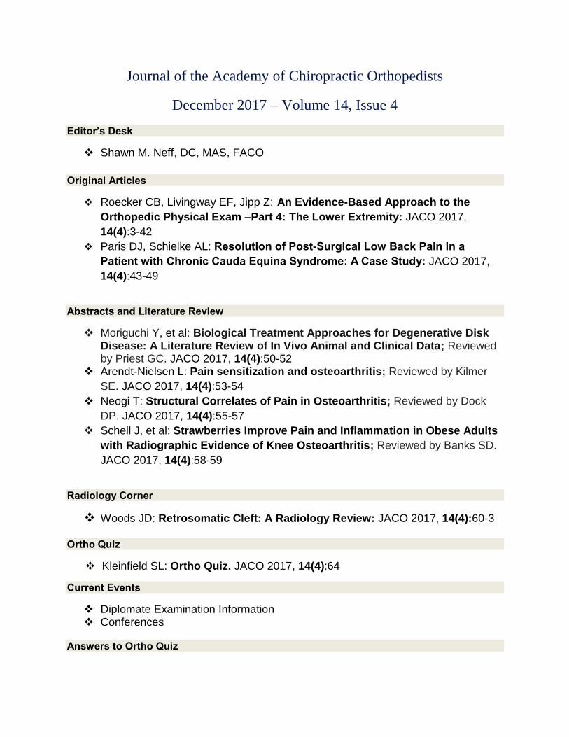
Journal of the Academy of Chiropractic Orthopedists
December 2017 – Volume 14, Issue 4
Editor’s Desk
❖ Shawn M. Neff, DC, MAS, FACO
Original Articles
❖ Roecker CB, Livingway EF, Jipp Z: An Evidence-Based Approach to the
Orthopedic Physical Exam –Part 4: The Lower Extremity: JACO 2017,
14(4):3-42
❖ Paris DJ, Schielke AL: Resolution of Post-Surgical Low Back Pain in a
Patient with Chronic Cauda Equina Syndrome: A Case Study: JACO 2017,
14(4):43-49
Abstracts and Literature Review
❖ Moriguchi Y, et al: Biological Treatment Approaches for Degenerative Disk Disease: A Literature Review of In Vivo Animal and Clinical Data; Reviewed by Priest GC. JACO 2017, 14(4):50-52
❖ Arendt-Nielsen L: Pain sensitization and osteoarthritis; Reviewed by Kilmer
SE. JACO 2017, 14(4):53-54
❖ Neogi T: Structural Correlates of Pain in Osteoarthritis; Reviewed by Dock
DP. JACO 2017, 14(4):55-57
❖ Schell J, et al: Strawberries Improve Pain and Inflammation in Obese Adults
with Radiographic Evidence of Knee Osteoarthritis; Reviewed by Banks SD.
JACO 2017, 14(4):58-59
Radiology Corner
❖ Woods JD: Retrosomatic Cleft: A Radiology Review: JACO 2017, 14(4):60-3
Ortho Quiz
❖ Kleinfield SL: Ortho Quiz. JACO 2017, 14(4):64
Current Events
❖ Diplomate Examination Information ❖ Conferences
Answers to Ortho Quiz

Journal of the Academy of Chiropractic Orthopedists
Volume 14, Issue 4
2
❖ Check your knowledge on page 66
The Editor’s Desk
Shawn M. Neff, DC, MAS, FACO
Editor-in-Chief
Welcome to the December 2017 issue of the Journal of the Academy of Chiropractic
Orthopedists. We at the Journal wish to express our appreciation to our readers and contributors.
We are grateful to have the opportunity to help the chiropractic orthopedic community share and
stay abreast of the newest evidence in the specialty.
We are aware of the issues we are having
with the archives of the Journal. We are
working to correct this issue. There were
files corrupted and lost in the transition of
webhosting. If you have copies of any of
the archived journals which are currently
corrupted, please contact me at
The new year will bring changes as it
always does. Our commitment to the
doctors and patients of our profession and
specialty will not change. We look
forward to another year of advancing
knowledge in conservative orthopedics
with all of you.
I hope you all enjoy this issue.
Sincerely,
-Shawn
3
Original Article
An Evidence-Based Approach to the Orthopedic Physical Exam –
Part 4: The Lower Extremity
Christopher B. Roecker, DC, MS, DACO1, Emma Forlow Livingway, ATC2, Zachary Jipp, DC3
1Assistant Professor, Palmer College of Chiropractic Life Science & Foundations Department 2 Student, Doctor of Chiropractic Program, Palmer College of Chiropractic, Davenport, IA
3Doctor of Chiropractic, Colorado Occupational Medical Partners, Denver, CO
Published: December 2017 Journal of the Academy of Chiropractic Orthopedists
December 2017, Volume 14, Issue 4
This is an Open Access article which permits unrestricted use, distribution, and reproduction in any medium, provided the original work is properly cited. The article copyright belongs to the author and the Academy of Chiropractic Orthopedists and is available at:
http://www.dcorthoacademy.com. © 2017 Roecker/Livingway/Jipp and the Academy of Chiropractic Orthopedists.
ABSTRACT
This narrative review article aims to examine the current evidence surrounding the orthopedic
physical exam procedures related to various lower extremity conditions and provide an overview
of these examinations. A narrative review was performed using online databases, authoritative
textbooks, and a mobile orthopedic exam application. When multiple studies existed for a single
orthopedic test, we reported results from the highest-quality studies and used studies with the
highest Quality Assessment of Diagnostic Accuracy Studies (QUADAS) scores. Additionally,
we attempted to highlight when orthopedic physical exams were described, but have yet to be
evaluated in order to establish diagnostic accuracy. The purpose of this article is to provide an
overview of the evidence-based orthopedic physical exams for many lower extremity conditions.
KEY WORDS (MeSH terms)
Evidence Based Practice; Injury, Leg; Injuries, Hip; Injury, Knee; Injury, Ankle; Injuries, Foot
Journal of the Academy of Chiropractic Orthopedists
Volume 14, Issue 4
4
INTRODUCTION
Clinical decision-making involves clinicians engaging in the process of probabilistic thinking,
which evaluates the probability that a patient’s clinical presentation is due to a given
pathology.1,2 Clinicians tend to represent this decision-making process in the context of
establishing a list of differential diagnoses, but at its core the process of winnowing down a list
of differential diagnoses is the same as probabilistic thinking.2
Framing clinical decision-making in the context of probabilistic thinking is not new. Pauker and
Kassirer first described this approach in 1980.1 We feel this work of Pauker and Kassirer clearly
outlines the process of clinical decision-making. They describe the concepts of a testing
threshold and a diagnostic threshold in the context of establishing a differential diagnosis. Every
diagnostic challenge incorporates multitudes of information, such as the patient’s chief
complaint, findings from the physical examination, and even the clinician’s instincts (likely from
his/her education or clinical experience);3 each of these pieces of information adds to or subtracts
from the probability that any given pathology is responsible for the patient’s condition (i.e.
disease state).
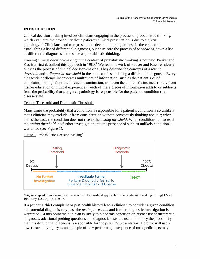
Testing Threshold and Diagnostic Threshold
Many times the probability that a condition is responsible for a patient’s condition is so unlikely
that a clinician may exclude it from consideration without consciously thinking about it; when
this is the case, the condition does not rise to the testing threshold. When conditions fail to reach
the testing threshold, no further investigation into the presence of such an unlikely condition is
warranted (see Figure 1).
Figure 1: Probabilistic Decision-Making*
*Figure adapted from Pauker SG, Kassirer JP. The threshold approach to clinical decision making. N Engl J Med.
1980 May 15;302(20):1109-17.
If a patient’s chief complaint or past health history lead a clinician to consider a given condition,
this potential diagnosis may pass the testing threshold and further diagnostic investigation is
warranted. At this point the clinician is likely to place this condition on his/her list of differential
diagnoses; additional probing questions and diagnostic tests are used to modify the probability
that this differential diagnosis is responsible for the patient’s presentation. Here we will use a
lower extremity injury as an example of how performing a sequence of orthopedic tests may
5
influence the probability of a condition being present, to the point of crossing a diagnostic
threshold.
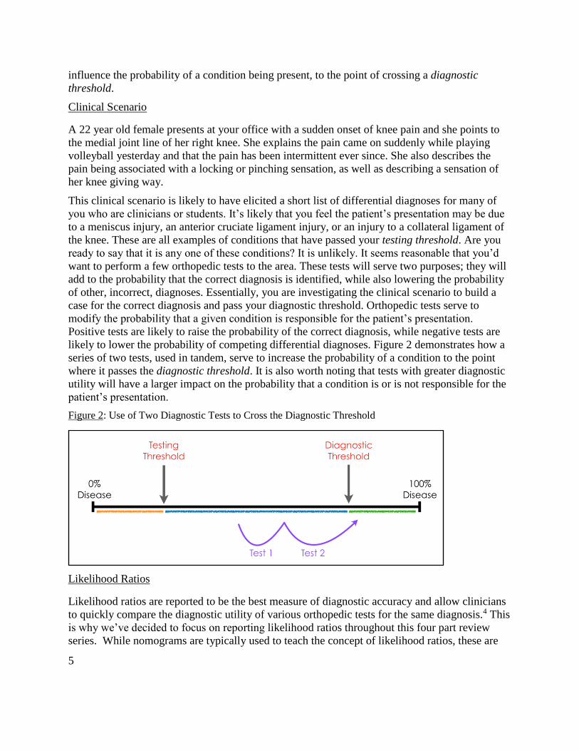
Clinical Scenario
A 22 year old female presents at your office with a sudden onset of knee pain and she points to
the medial joint line of her right knee. She explains the pain came on suddenly while playing
volleyball yesterday and that the pain has been intermittent ever since. She also describes the
pain being associated with a locking or pinching sensation, as well as describing a sensation of
her knee giving way.
This clinical scenario is likely to have elicited a short list of differential diagnoses for many of
you who are clinicians or students. It’s likely that you feel the patient’s presentation may be due
to a meniscus injury, an anterior cruciate ligament injury, or an injury to a collateral ligament of
the knee. These are all examples of conditions that have passed your testing threshold. Are you
ready to say that it is any one of these conditions? It is unlikely. It seems reasonable that you’d
want to perform a few orthopedic tests to the area. These tests will serve two purposes; they will
add to the probability that the correct diagnosis is identified, while also lowering the probability
of other, incorrect, diagnoses. Essentially, you are investigating the clinical scenario to build a
case for the correct diagnosis and pass your diagnostic threshold. Orthopedic tests serve to
modify the probability that a given condition is responsible for the patient’s presentation.
Positive tests are likely to raise the probability of the correct diagnosis, while negative tests are
likely to lower the probability of competing differential diagnoses. Figure 2 demonstrates how a
series of two tests, used in tandem, serve to increase the probability of a condition to the point
where it passes the diagnostic threshold. It is also worth noting that tests with greater diagnostic
utility will have a larger impact on the probability that a condition is or is not responsible for the
patient’s presentation.
Figure 2: Use of Two Diagnostic Tests to Cross the Diagnostic Threshold
Likelihood Ratios
Likelihood ratios are reported to be the best measure of diagnostic accuracy and allow clinicians
to quickly compare the diagnostic utility of various orthopedic tests for the same diagnosis.4 This
is why we’ve decided to focus on reporting likelihood ratios throughout this four part review
series. While nomograms are typically used to teach the concept of likelihood ratios, these are
Journal of the Academy of Chiropractic Orthopedists
Volume 14, Issue 4
6
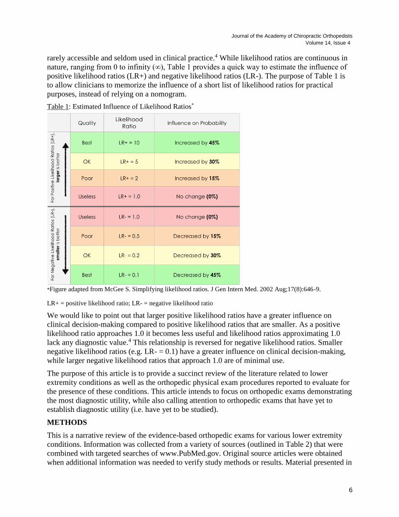
rarely accessible and seldom used in clinical practice.4 While likelihood ratios are continuous in
nature, ranging from 0 to infinity (∞), Table 1 provides a quick way to estimate the influence of
positive likelihood ratios (LR+) and negative likelihood ratios (LR-). The purpose of Table 1 is
to allow clinicians to memorize the influence of a short list of likelihood ratios for practical
purposes, instead of relying on a nomogram.
Table 1: Estimated Influence of Likelihood Ratios*
*Figure adapted from McGee S. Simplifying likelihood ratios. J Gen Intern Med. 2002 Aug;17(8):646-9.
LR+ = positive likelihood ratio; LR- = negative likelihood ratio
We would like to point out that larger positive likelihood ratios have a greater influence on
clinical decision-making compared to positive likelihood ratios that are smaller. As a positive
likelihood ratio approaches 1.0 it becomes less useful and likelihood ratios approximating 1.0
lack any diagnostic value.4 This relationship is reversed for negative likelihood ratios. Smaller
negative likelihood ratios (e.g. LR- = 0.1) have a greater influence on clinical decision-making,
while larger negative likelihood ratios that approach 1.0 are of minimal use.
The purpose of this article is to provide a succinct review of the literature related to lower
extremity conditions as well as the orthopedic physical exam procedures reported to evaluate for
the presence of these conditions. This article intends to focus on orthopedic exams demonstrating
the most diagnostic utility, while also calling attention to orthopedic exams that have yet to
establish diagnostic utility (i.e. have yet to be studied).
METHODS
This is a narrative review of the evidence-based orthopedic exams for various lower extremity
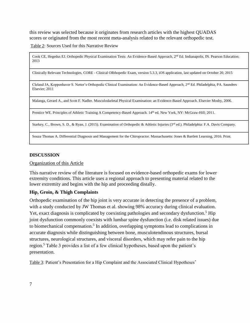
conditions. Information was collected from a variety of sources (outlined in Table 2) that were
combined with targeted searches of www.PubMed.gov. Original source articles were obtained
when additional information was needed to verify study methods or results. Material presented in
7
this review was selected because it originates from research articles with the highest QUADAS
scores or originated from the most recent meta-analysis related to the relevant orthopedic test.
Table 2: Sources Used for this Narrative Review
Cook CE, Hegedus EJ. Orthopedic Physical Examination Tests: An Evidence-Based Approach, 2nd Ed. Indianapolis, IN. Pearson Education;
2013
Clinically Relevant Technologies, CORE - Clinical ORthopedic Exam, version 5.3.3, iOS application, last updated on October 20, 2015
Cleland JA, Koppenhaver S. Netter’s Orthopedic Clinical Examination: An Evidence-Based Approach, 2nd Ed. Philadelphia, PA. Saunders
Elsevier; 2011
Malanga, Gerard A., and Scott F. Nadler. Musculoskeletal Physical Examination: an Evidence-Based Approach. Elsevier Mosby, 2006.
Prentice WE. Principles of Athletic Training A Competency-Based Approach. 14th ed. New York, NY: McGraw-Hill; 2011.
Starkey, C., Brown, S. D., & Ryan, J. (2015). Examination of Orthopedic & Athletic Injuries (3rd ed.). Philadelphia: F.A. Davis Company.
Souza Thomas A. Differential Diagnosis and Management for the Chiropractor. Massachusetts: Jones & Bartlett Learning, 2016. Print.
DISCUSSION
Organization of this Article
This narrative review of the literature is focused on evidence-based orthopedic exams for lower
extremity conditions. This article uses a regional approach to presenting material related to the
lower extremity and begins with the hip and proceeding distally.
Hip, Groin, & Thigh Complaints
Orthopedic examination of the hip joint is very accurate in detecting the presence of a problem,
with a study conducted by JW Thomas et al. showing 98% accuracy during clinical evaluation.
Yet, exact diagnosis is complicated by coexisting pathologies and secondary dysfunction.5 Hip
joint dysfunction commonly coexists with lumbar spine dysfunction (i.e. disk related issues) due
to biomechanical compensation.5 In addition, overlapping symptoms lead to complications in
accurate diagnosis while distinguishing between bone, musculotendinous structures, bursal
structures, neurological structures, and visceral disorders, which may refer pain to the hip
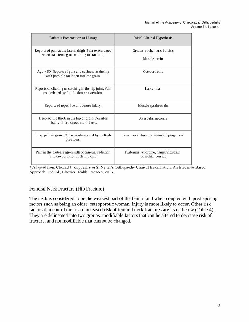
region.5 Table 3 provides a list of a few clinical hypotheses, based upon the patient’s
presentation.
Table 3: Patient’s Presentation for a Hip Complaint and the Associated Clinical Hypotheses*
Journal of the Academy of Chiropractic Orthopedists
Volume 14, Issue 4
8
Patient’s Presentation or History Initial Clinical Hypothesis
Reports of pain at the lateral thigh. Pain exacerbated
when transferring from sitting to standing.
Greater trochanteric bursitis
Muscle strain
Age > 60. Reports of pain and stiffness in the hip
with possible radiation into the groin.
Osteoarthritis
Reports of clicking or catching in the hip joint. Pain
exacerbated by full flexion or extension.
Labral tear
Reports of repetitive or overuse injury. Muscle sprain/strain
Deep aching throb in the hip or groin. Possible
history of prolonged steroid use.
Avascular necrosis
Sharp pain in groin. Often misdiagnosed by multiple
providers.
Femoroacetabular (anterior) impingement
Pain in the gluteal region with occasional radiation
into the posterior thigh and calf.
Piriformis syndrome, hamstring strain,
or ischial bursitis
* Adapted from Cleland J, Koppenhaver S. Netter’s Orthopaedic Clinical Examination: An Evidence-Based
Approach. 2nd Ed., Elsevier Health Sciences; 2015.
Femoral Neck Fracture (Hip Fracture)
The neck is considered to be the weakest part of the femur, and when coupled with predisposing
factors such as being an older, osteoporotic woman, injury is more likely to occur. Other risk
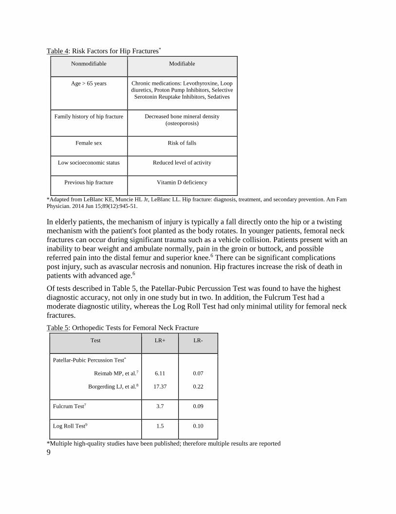
factors that contribute to an increased risk of femoral neck fractures are listed below (Table 4).
They are delineated into two groups, modifiable factors that can be altered to decrease risk of
fracture, and nonmodifiable that cannot be changed.
9
Table 4: Risk Factors for Hip Fractures*
Nonmodifiable Modifiable
Age > 65 years Chronic medications: Levothyroxine, Loop
diuretics, Proton Pump Inhibitors, Selective
Serotonin Reuptake Inhibitors, Sedatives
Family history of hip fracture Decreased bone mineral density
(osteoporosis)
Female sex Risk of falls
Low socioeconomic status Reduced level of activity
Previous hip fracture Vitamin D deficiency
*Adapted from LeBlanc KE, Muncie HL Jr, LeBlanc LL. Hip fracture: diagnosis, treatment, and secondary prevention. Am Fam
Physician. 2014 Jun 15;89(12):945-51.
In elderly patients, the mechanism of injury is typically a fall directly onto the hip or a twisting
mechanism with the patient's foot planted as the body rotates. In younger patients, femoral neck
fractures can occur during significant trauma such as a vehicle collision. Patients present with an
inability to bear weight and ambulate normally, pain in the groin or buttock, and possible
referred pain into the distal femur and superior knee.6 There can be significant complications
post injury, such as avascular necrosis and nonunion. Hip fractures increase the risk of death in
patients with advanced age.6
Of tests described in Table 5, the Patellar-Pubic Percussion Test was found to have the highest
diagnostic accuracy, not only in one study but in two. In addition, the Fulcrum Test had a
moderate diagnostic utility, whereas the Log Roll Test had only minimal utility for femoral neck
fractures.
Table 5: Orthopedic Tests for Femoral Neck Fracture
Test LR+ LR-
Patellar-Pubic Percussion Test*
Reimab MP, et al.7
Borgerding LJ, et al.8
6.11
17.37
0.07
0.22
Fulcrum Test7 3.7 0.09
Log Roll Test9 1.5 0.10
*Multiple high-quality studies have been published; therefore multiple results are reported
Journal of the Academy of Chiropractic Orthopedists
Volume 14, Issue 4
10
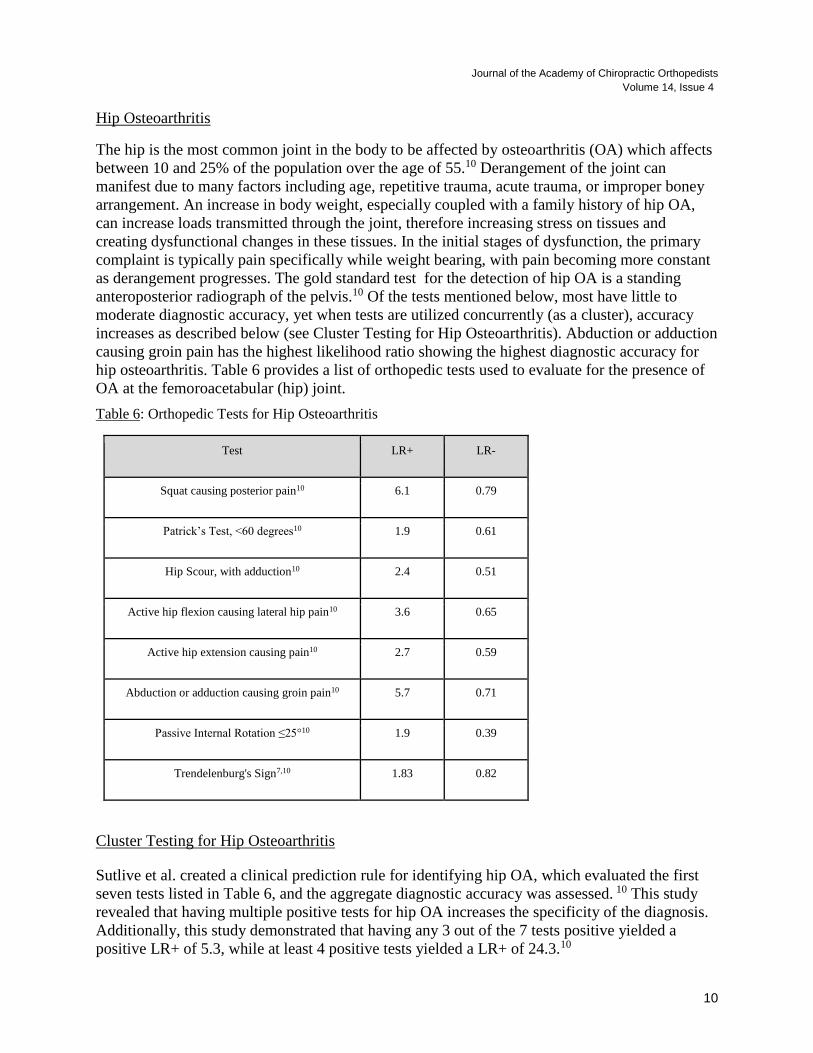
Hip Osteoarthritis
The hip is the most common joint in the body to be affected by osteoarthritis (OA) which affects
between 10 and 25% of the population over the age of 55.10 Derangement of the joint can
manifest due to many factors including age, repetitive trauma, acute trauma, or improper boney
arrangement. An increase in body weight, especially coupled with a family history of hip OA,
can increase loads transmitted through the joint, therefore increasing stress on tissues and
creating dysfunctional changes in these tissues. In the initial stages of dysfunction, the primary
complaint is typically pain specifically while weight bearing, with pain becoming more constant
as derangement progresses. The gold standard test for the detection of hip OA is a standing
anteroposterior radiograph of the pelvis.10 Of the tests mentioned below, most have little to
moderate diagnostic accuracy, yet when tests are utilized concurrently (as a cluster), accuracy
increases as described below (see Cluster Testing for Hip Osteoarthritis). Abduction or adduction
causing groin pain has the highest likelihood ratio showing the highest diagnostic accuracy for
hip osteoarthritis. Table 6 provides a list of orthopedic tests used to evaluate for the presence of
OA at the femoroacetabular (hip) joint.
Table 6: Orthopedic Tests for Hip Osteoarthritis
Test LR+ LR-
Squat causing posterior pain10 6.1 0.79
Patrick’s Test, <60 degrees10 1.9 0.61
Hip Scour, with adduction10 2.4 0.51
Active hip flexion causing lateral hip pain10 3.6 0.65
Active hip extension causing pain10 2.7 0.59
Abduction or adduction causing groin pain10 5.7 0.71
Passive Internal Rotation ≤25°10 1.9 0.39
Trendelenburg's Sign7,10 1.83 0.82
Cluster Testing for Hip Osteoarthritis
Sutlive et al. created a clinical prediction rule for identifying hip OA, which evaluated the first
seven tests listed in Table 6, and the aggregate diagnostic accuracy was assessed. 10 This study
revealed that having multiple positive tests for hip OA increases the specificity of the diagnosis.
Additionally, this study demonstrated that having any 3 out of the 7 tests positive yielded a
positive LR+ of 5.3, while at least 4 positive tests yielded a LR+ of 24.3.10
11
Femoroacetabular Impingement
Femoroacetabular impingement (FAI) is associated with the abnormal anatomical relationship
between the acetabulum and femur. The classifications that cause dysfunction can be described
as either a cam lesion, pincer lesion, or combination lesion where both are present. The cam
deformity is characterized by an additional bony prominence in the femoral head or neck region.
The pincer lesion features an additional bony protrusion on the acetabulum. The existence of FAI
is a recent discovery for clinicians, yet FAI has proven important to recognize due to its
contribution to a multitude of hip related morbidities such as labral injuries and early OA in the
active population.11 Imaging techniques such as radiograph and MRI can be utilized for
diagnosis. In addition, relief post anesthetic injection can be utilized as a confirmatory test.11
As established in the table below, there is only minimal evidence to support the diagnostic
accuracy of test that are typically utilized to identify FAI pathology. Patient history and physical
examination are crucial to early detection and treatment. Table 7 provides a list of orthopedic
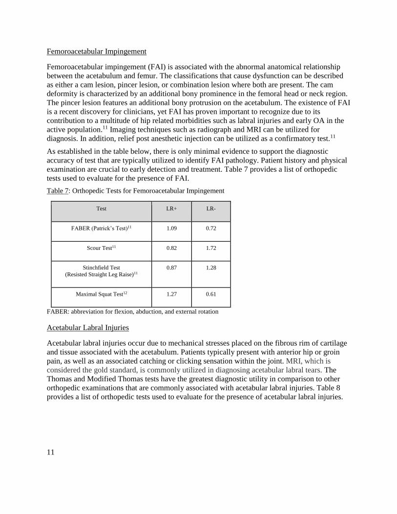
tests used to evaluate for the presence of FAI.
Table 7: Orthopedic Tests for Femoroacetabular Impingement
Test LR+ LR-
FABER (Patrick’s Test)11 1.09 0.72
Scour Test11 0.82 1.72
Stinchfield Test
(Resisted Straight Leg Raise)11
0.87 1.28
Maximal Squat Test12 1.27 0.61
FABER: abbreviation for flexion, abduction, and external rotation
Acetabular Labral Injuries
Acetabular labral injuries occur due to mechanical stresses placed on the fibrous rim of cartilage
and tissue associated with the acetabulum. Patients typically present with anterior hip or groin
pain, as well as an associated catching or clicking sensation within the joint. MRI, which is
considered the gold standard, is commonly utilized in diagnosing acetabular labral tears. The
Thomas and Modified Thomas tests have the greatest diagnostic utility in comparison to other
orthopedic examinations that are commonly associated with acetabular labral injuries. Table 8
provides a list of orthopedic tests used to evaluate for the presence of acetabular labral injuries.
Journal of the Academy of Chiropractic Orthopedists
Volume 14, Issue 4
12
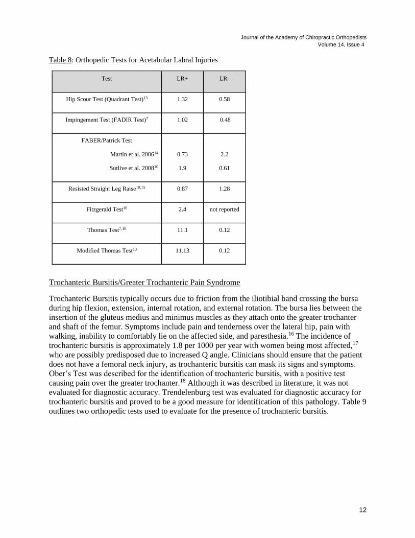
Table 8: Orthopedic Tests for Acetabular Labral Injuries
Test LR+ LR-
Hip Scour Test (Quadrant Test)13 1.32 0.58
Impingement Test (FADIR Test)7 1.02 0.48
FABER/Patrick Test
Martin et al. 200614
Sutlive et al. 200810
0.73
1.9
2.2
0.61
Resisted Straight Leg Raise10,15 0.87 1.28
Fitzgerald Test10 2.4 not reported
Thomas Test7,10 11.1 0.12
Modified Thomas Test13 11.13 0.12
Trochanteric Bursitis/Greater Trochanteric Pain Syndrome
Trochanteric Bursitis typically occurs due to friction from the iliotibial band crossing the bursa
during hip flexion, extension, internal rotation, and external rotation. The bursa lies between the
insertion of the gluteus medius and minimus muscles as they attach onto the greater trochanter
and shaft of the femur. Symptoms include pain and tenderness over the lateral hip, pain with
walking, inability to comfortably lie on the affected side, and paresthesia.16 The incidence of
trochanteric bursitis is approximately 1.8 per 1000 per year with women being most affected,17
who are possibly predisposed due to increased Q angle. Clinicians should ensure that the patient
does not have a femoral neck injury, as trochanteric bursitis can mask its signs and symptoms.
Ober’s Test was described for the identification of trochanteric bursitis, with a positive test
causing pain over the greater trochanter.18 Although it was described in literature, it was not
evaluated for diagnostic accuracy. Trendelenburg test was evaluated for diagnostic accuracy for
trochanteric bursitis and proved to be a good measure for identification of this pathology. Table 9
outlines two orthopedic tests used to evaluate for the presence of trochanteric bursitis.
13
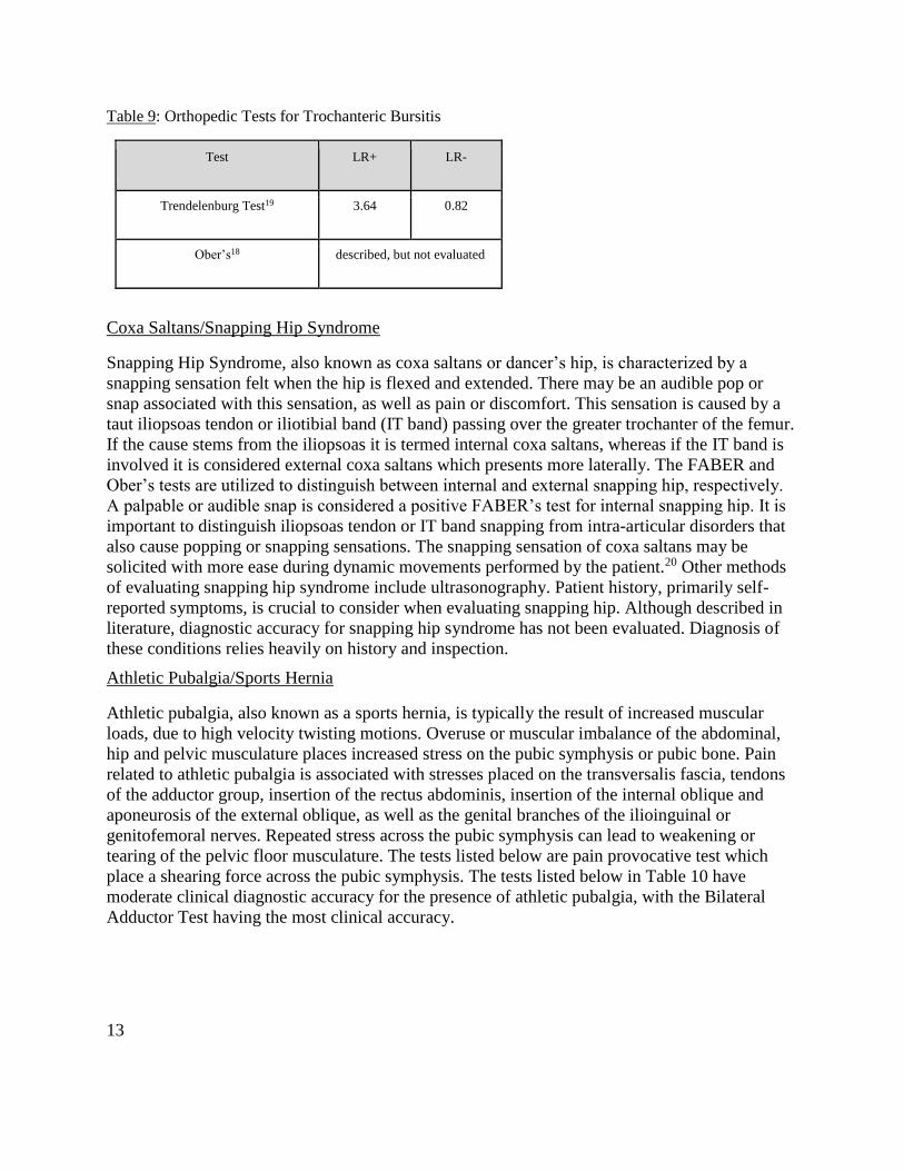
Table 9: Orthopedic Tests for Trochanteric Bursitis
Test LR+ LR-
Trendelenburg Test19 3.64 0.82
Ober’s18 described, but not evaluated
Coxa Saltans/Snapping Hip Syndrome
Snapping Hip Syndrome, also known as coxa saltans or dancer’s hip, is characterized by a
snapping sensation felt when the hip is flexed and extended. There may be an audible pop or
snap associated with this sensation, as well as pain or discomfort. This sensation is caused by a
taut iliopsoas tendon or iliotibial band (IT band) passing over the greater trochanter of the femur.
If the cause stems from the iliopsoas it is termed internal coxa saltans, whereas if the IT band is
involved it is considered external coxa saltans which presents more laterally. The FABER and
Ober’s tests are utilized to distinguish between internal and external snapping hip, respectively.
A palpable or audible snap is considered a positive FABER’s test for internal snapping hip. It is
important to distinguish iliopsoas tendon or IT band snapping from intra-articular disorders that
also cause popping or snapping sensations. The snapping sensation of coxa saltans may be
solicited with more ease during dynamic movements performed by the patient.20 Other methods
of evaluating snapping hip syndrome include ultrasonography. Patient history, primarily self-
reported symptoms, is crucial to consider when evaluating snapping hip. Although described in
literature, diagnostic accuracy for snapping hip syndrome has not been evaluated. Diagnosis of
these conditions relies heavily on history and inspection.
Athletic Pubalgia/Sports Hernia
Athletic pubalgia, also known as a sports hernia, is typically the result of increased muscular
loads, due to high velocity twisting motions. Overuse or muscular imbalance of the abdominal,
hip and pelvic musculature places increased stress on the pubic symphysis or pubic bone. Pain
related to athletic pubalgia is associated with stresses placed on the transversalis fascia, tendons
of the adductor group, insertion of the rectus abdominis, insertion of the internal oblique and
aponeurosis of the external oblique, as well as the genital branches of the ilioinguinal or
genitofemoral nerves. Repeated stress across the pubic symphysis can lead to weakening or
tearing of the pelvic floor musculature. The tests listed below are pain provocative test which
place a shearing force across the pubic symphysis. The tests listed below in Table 10 have
moderate clinical diagnostic accuracy for the presence of athletic pubalgia, with the Bilateral
Adductor Test having the most clinical accuracy.
Journal of the Academy of Chiropractic Orthopedists
Volume 14, Issue 4
14
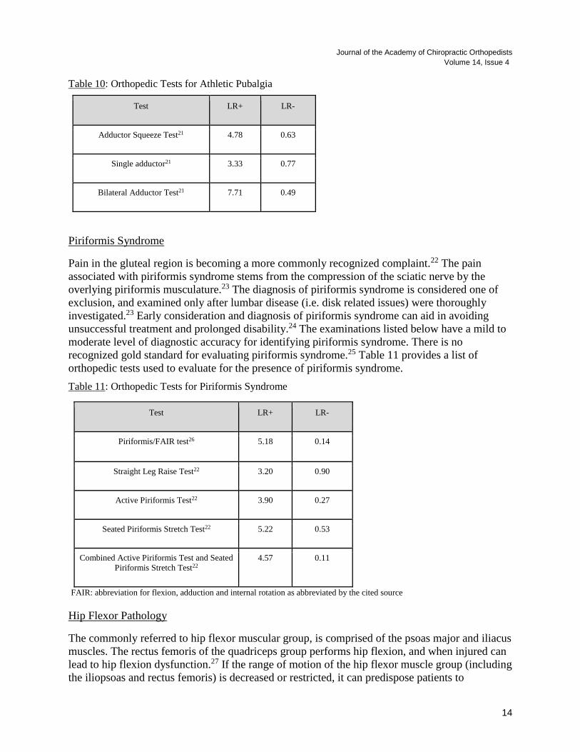
Table 10: Orthopedic Tests for Athletic Pubalgia
Test LR+ LR-
Adductor Squeeze Test21 4.78 0.63
Single adductor21 3.33 0.77
Bilateral Adductor Test21 7.71 0.49
Piriformis Syndrome
Pain in the gluteal region is becoming a more commonly recognized complaint.22 The pain
associated with piriformis syndrome stems from the compression of the sciatic nerve by the
overlying piriformis musculature.23 The diagnosis of piriformis syndrome is considered one of
exclusion, and examined only after lumbar disease (i.e. disk related issues) were thoroughly
investigated.23 Early consideration and diagnosis of piriformis syndrome can aid in avoiding
unsuccessful treatment and prolonged disability.24 The examinations listed below have a mild to
moderate level of diagnostic accuracy for identifying piriformis syndrome. There is no
recognized gold standard for evaluating piriformis syndrome.25 Table 11 provides a list of
orthopedic tests used to evaluate for the presence of piriformis syndrome.
Table 11: Orthopedic Tests for Piriformis Syndrome
Test LR+ LR-
Piriformis/FAIR test26 5.18 0.14
Straight Leg Raise Test22 3.20 0.90
Active Piriformis Test22 3.90 0.27
Seated Piriformis Stretch Test22 5.22 0.53
Combined Active Piriformis Test and Seated
Piriformis Stretch Test22
4.57 0.11
FAIR: abbreviation for flexion, adduction and internal rotation as abbreviated by the cited source
Hip Flexor Pathology
The commonly referred to hip flexor muscular group, is comprised of the psoas major and iliacus
muscles. The rectus femoris of the quadriceps group performs hip flexion, and when injured can
lead to hip flexion dysfunction.27 If the range of motion of the hip flexor muscle group (including
the iliopsoas and rectus femoris) is decreased or restricted, it can predispose patients to
15
musculoskeletal injuries of the lower extremity. In fact, it has been theorized that pathology of
the hip flexors can act as a reciprocal inhibitor to the gluteus maximus.27,28 The inhibition of the
gluteus maximus then causes other hip extensors to be overworked, causing greater tissue stress
and dysfunction.27,28 According to a study conducted by Eckard et al. assessing the epidemiology
of hip flexor injury in sport, men’s soccer and men’s hockey have the highest number of hip
flexor strains than any other sport; Furthermore, men’s soccer athletes injured their hip flexors
more often than women’s soccer athletes.27 Of the orthopedic examinations listed below, Ely’s
test has the highest diagnostic usefulness, whereas the Modified Thomas test has very limited
usefulness during an assessment. The Thomas Test, which is commonly described in assessing
hip flexor pathologies, has not been evaluated for diagnostic accuracy. Table 12 provides a list of
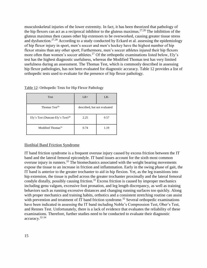
orthopedic tests used to evaluate for the presence of hip flexor pathology.
Table 12: Orthopedic Tests for Hip Flexor Pathology
Test LR+ LR-
Thomas Test29 described, but not evaluated
Ely’s Test (Duncan-Ely’s Test)30 2.25 0.57
Modified Thomas31 0.74 1.19
Iliotibial Band Friction Syndrome
IT band friction syndrome is a frequent overuse injury caused by excess friction between the IT
band and the lateral femoral epicondyle. IT band issues account for the sixth most common
overuse injury in runners.32 The biomechanics associated with the weight bearing movements
expose the tissue to an increase in friction and inflammation. Early in the swing phase of gait, the
IT band is anterior to the greater trochanter to aid in hip flexion. Yet, as the leg transitions into
hip extension, the tissue is pulled across the greater trochanter proximally and the lateral femoral
condyle distally, possibly causing friction.32 Excess friction is caused by improper mechanics
including genu valgum, excessive foot pronation, and leg length discrepancy, as well as training
behaviors such as running excessive distances and changing running surfaces too quickly. Along
with proper mechanics and training habits, orthotics and a consistent stretching routine can assist
with prevention and treatment of IT band friction syndrome.32 Several orthopedic examinations
have been indicated in assessing the IT band including Noble’s Compression Test, Ober’s Test,
and Rennes Test. Unfortunately, there is a lack of evidence that evaluates the reliability of these
examinations. Therefore, further studies need to be conducted to evaluate their diagnostic
accuracy.32-34
Journal of the Academy of Chiropractic Orthopedists
Volume 14, Issue 4
16
Knee Complaints
Knee pain is a common reason for patients to seek care from health care providers, and knee
injuries are among the most common sports-related injuries.35 A wide variety of pathologic
conditions may cause knee pain, which include acute trauma to the knee, overuse injuries, or
inflammatory arthritides. Soft tissue injuries to the knee commonly present with a sudden onset
of pain and are characteristically associated with specific mechanisms of onset. Clinicians are
presented with a diagnostic challenge each time a patient presents with knee pain and are
challenged with formulating a working list of diagnoses during the history and physical exam
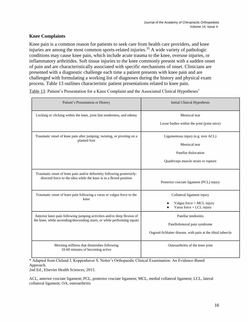
process. Table 13 outlines characteristic patient presentations related to knee pain.
Table 13: Patient’s Presentation for a Knee Complaint and the Associated Clinical Hypotheses*
Patient’s Presentation or History Initial Clinical Hypothesis
Locking or clicking within the knee, joint line tenderness, and edema Meniscal tear
Loose bodies within the joint (joint mice)
Traumatic onset of knee pain after jumping, twisting, or pivoting on a
planted foot
Ligamentous injury (e.g. torn ACL)
Meniscal tear
Patellar dislocation
Quadriceps muscle strain or rupture
Traumatic onset of knee pain and/or deformity following posteriorly-
directed force to the tibia while the knee is in a flexed position
Posterior cruciate ligament (PCL) injury
Traumatic onset of knee pain following a varus or valgus force to the
knee
Collateral ligament injury
● Valgus force = MCL injury
● Varus force = LCL injury
Anterior knee pain following jumping activities and/or deep flexion of
the knee, while ascending/descending stairs, or while performing squats
Patellar tendonitis
Patellofemoral pain syndrome
Osgood-Schlatter disease, with pain at the tibial tubercle
Morning stiffness that diminishes following
10-60 minutes of becoming active
Osteoarthritis of the knee joint
* Adapted from Cleland J, Koppenhaver S. Netter’s Orthopaedic Clinical Examination: An Evidence-Based
Approach.
2nd Ed., Elsevier Health Sciences; 2015.
ACL, anterior cruciate ligament; PCL, posterior cruciate ligament; MCL, medial collateral ligament; LCL, lateral
collateral ligament; OA, osteoarthritis
17
Evaluating for Fracture around the Knee
Acute injury to the knee may require advanced imaging. While ultrasound or MRI are frequently
used to assist in the evaluation of soft tissue injuries of the knee, plain film radiography may be
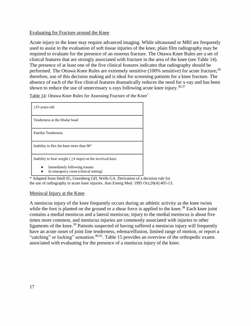
required to evaluate for the presence of an osseous fracture. The Ottawa Knee Rules are a set of
clinical features that are strongly associated with fracture in the area of the knee (see Table 14).
The presence of at least one of the five clinical features indicates that radiography should be
performed. The Ottawa Knee Rules are extremely sensitive (100% sensitive) for acute fracture;36
therefore, use of this decision making aid is ideal for screening patients for a knee fracture. The
absence of each of the five clinical features dramatically reduces the need for x-ray and has been
shown to reduce the use of unnecessary x-rays following acute knee injury.36,37
Table 14: Ottawa Knee Rules for Assessing Fracture of the Knee*
≥55-years-old
Tenderness at the fibular head
Patellar Tenderness
Inability to flex the knee more than 90°
Inability to bear weight ( ≥4 steps) on the involved knee
● Immediately following trauma
● In emergency room (clinical setting)
* Adapted from Stiell IG, Greenberg GH, Wells GA. Derivation of a decision rule for
the use of radiography in acute knee injuries. Ann Emerg Med. 1995 Oct;26(4):405-13.
Meniscal Injury at the Knee
A meniscus injury of the knee frequently occurs during an athletic activity as the knee twists
while the foot is planted on the ground or a shear force is applied to the knee.38 Each knee joint
contains a medial meniscus and a lateral meniscus; injury to the medial meniscus is about five
times more common, and meniscus injuries are commonly associated with injuries to other
ligaments of the knee.39 Patients suspected of having suffered a meniscus injury will frequently
have an acute onset of joint line tenderness, edema/effusion, limited range of motion, or report a
“catching” or locking” sensation.40,41. Table 15 provides an overview of the orthopedic exams
associated with evaluating for the presence of a meniscus injury of the knee.
Journal of the Academy of Chiropractic Orthopedists
Volume 14, Issue 4
18
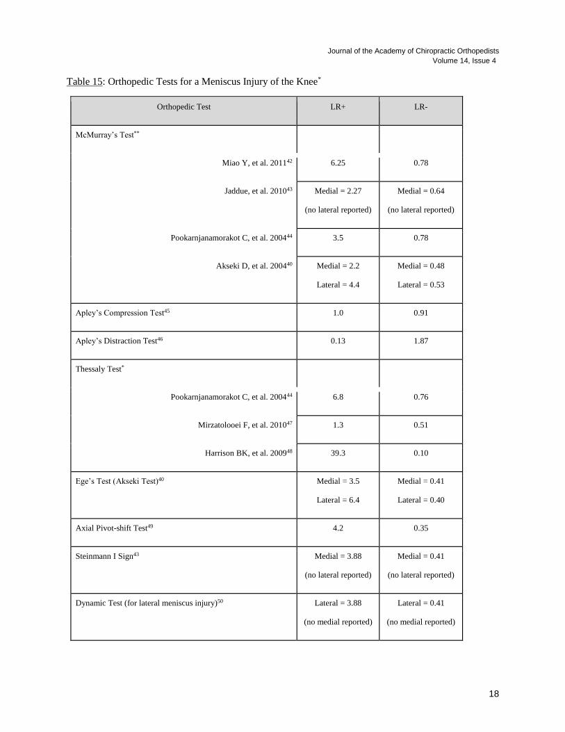
Table 15: Orthopedic Tests for a Meniscus Injury of the Knee*
Orthopedic Test LR+ LR-
McMurray’s Test**
Miao Y, et al. 201142 6.25 0.78
Jaddue, et al. 201043 Medial = 2.27
(no lateral reported)
Medial = 0.64
(no lateral reported)
Pookarnjanamorakot C, et al. 200444 3.5 0.78
Akseki D, et al. 200440 Medial = 2.2
Lateral = 4.4
Medial = 0.48
Lateral = 0.53
Apley’s Compression Test45 1.0 0.91
Apley’s Distraction Test46 0.13 1.87
Thessaly Test*
Pookarnjanamorakot C, et al. 200444 6.8 0.76
Mirzatolooei F, et al. 201047 1.3 0.51
Harrison BK, et al. 200948 39.3 0.10
Ege’s Test (Akseki Test)40 Medial = 3.5
Lateral = 6.4
Medial = 0.41
Lateral = 0.40
Axial Pivot-shift Test49 4.2 0.35
Steinmann I Sign43 Medial = 3.88
(no lateral reported)
Medial = 0.41
(no lateral reported)
Dynamic Test (for lateral meniscus injury)50 Lateral = 3.88
(no medial reported)
Lateral = 0.41
(no medial reported)
19
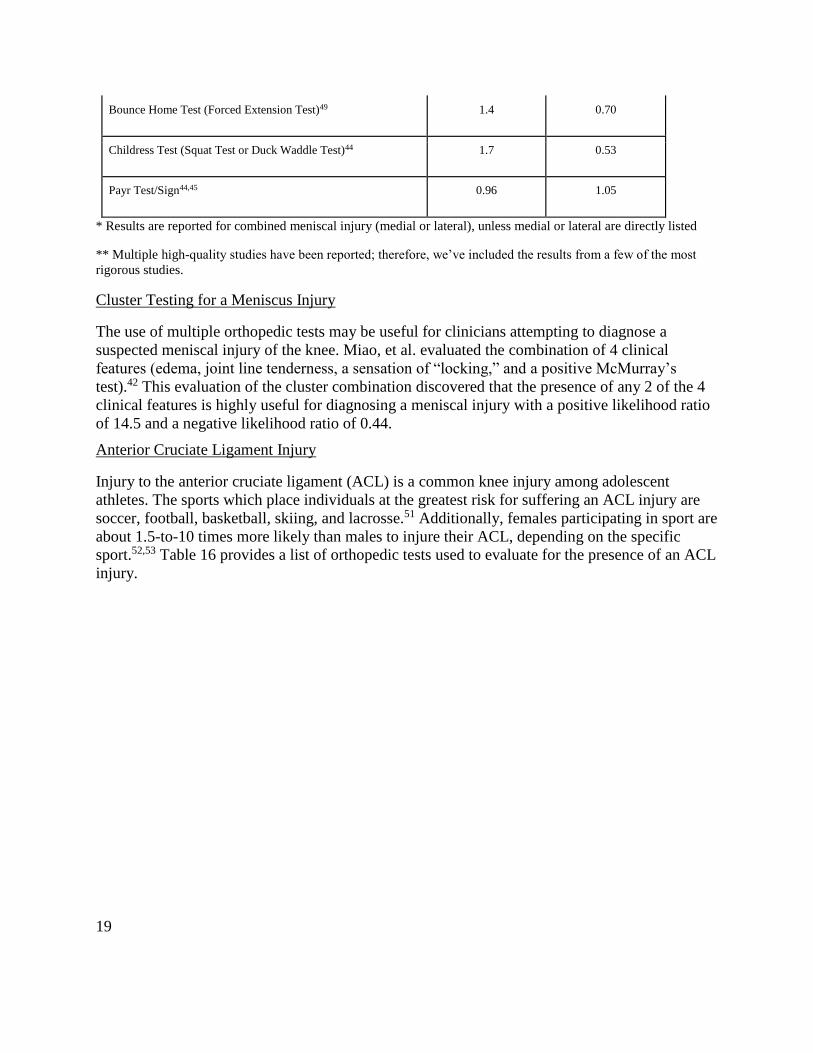
Bounce Home Test (Forced Extension Test)49 1.4 0.70
Childress Test (Squat Test or Duck Waddle Test)44 1.7 0.53
Payr Test/Sign44,45 0.96 1.05
* Results are reported for combined meniscal injury (medial or lateral), unless medial or lateral are directly listed
** Multiple high-quality studies have been reported; therefore, we’ve included the results from a few of the most
rigorous studies.
Cluster Testing for a Meniscus Injury
The use of multiple orthopedic tests may be useful for clinicians attempting to diagnose a
suspected meniscal injury of the knee. Miao, et al. evaluated the combination of 4 clinical
features (edema, joint line tenderness, a sensation of “locking,” and a positive McMurray’s
test).42 This evaluation of the cluster combination discovered that the presence of any 2 of the 4
clinical features is highly useful for diagnosing a meniscal injury with a positive likelihood ratio
of 14.5 and a negative likelihood ratio of 0.44.
Anterior Cruciate Ligament Injury
Injury to the anterior cruciate ligament (ACL) is a common knee injury among adolescent
athletes. The sports which place individuals at the greatest risk for suffering an ACL injury are
soccer, football, basketball, skiing, and lacrosse.51 Additionally, females participating in sport are
about 1.5-to-10 times more likely than males to injure their ACL, depending on the specific
sport.52,53 Table 16 provides a list of orthopedic tests used to evaluate for the presence of an ACL
injury.
Journal of the Academy of Chiropractic Orthopedists
Volume 14, Issue 4
20
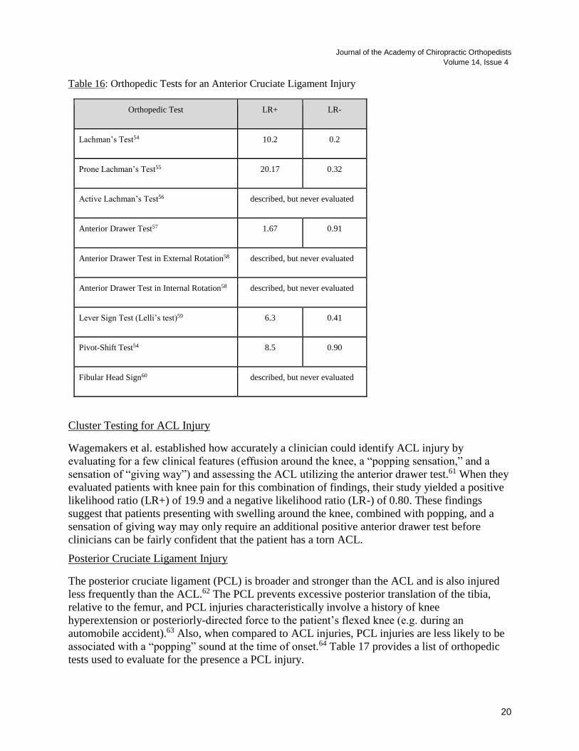
Table 16: Orthopedic Tests for an Anterior Cruciate Ligament Injury
Orthopedic Test LR+ LR-
Lachman’s Test54 10.2 0.2
Prone Lachman’s Test55 20.17 0.32
Active Lachman’s Test56 described, but never evaluated
Anterior Drawer Test57 1.67 0.91
Anterior Drawer Test in External Rotation58 described, but never evaluated
Anterior Drawer Test in Internal Rotation58 described, but never evaluated
Lever Sign Test (Lelli’s test)59 6.3 0.41
Pivot-Shift Test54 8.5 0.90
Fibular Head Sign60 described, but never evaluated
Cluster Testing for ACL Injury
Wagemakers et al. established how accurately a clinician could identify ACL injury by
evaluating for a few clinical features (effusion around the knee, a “popping sensation,” and a
sensation of “giving way”) and assessing the ACL utilizing the anterior drawer test.61 When they
evaluated patients with knee pain for this combination of findings, their study yielded a positive
likelihood ratio (LR+) of 19.9 and a negative likelihood ratio (LR-) of 0.80. These findings
suggest that patients presenting with swelling around the knee, combined with popping, and a
sensation of giving way may only require an additional positive anterior drawer test before
clinicians can be fairly confident that the patient has a torn ACL.
Posterior Cruciate Ligament Injury
The posterior cruciate ligament (PCL) is broader and stronger than the ACL and is also injured
less frequently than the ACL.62 The PCL prevents excessive posterior translation of the tibia,
relative to the femur, and PCL injuries characteristically involve a history of knee
hyperextension or posteriorly-directed force to the patient’s flexed knee (e.g. during an
automobile accident).63 Also, when compared to ACL injuries, PCL injuries are less likely to be
associated with a “popping” sound at the time of onset.64 Table 17 provides a list of orthopedic
tests used to evaluate for the presence a PCL injury.
21
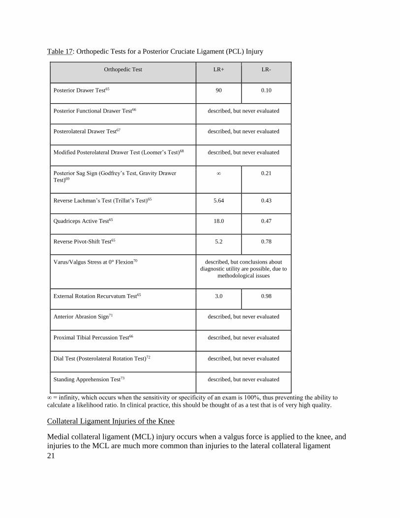
Table 17: Orthopedic Tests for a Posterior Cruciate Ligament (PCL) Injury
Orthopedic Test LR+ LR-
Posterior Drawer Test65 90 0.10
Posterior Functional Drawer Test66 described, but never evaluated
Posterolateral Drawer Test67 described, but never evaluated
Modified Posterolateral Drawer Test (Loomer’s Test)68 described, but never evaluated
Posterior Sag Sign (Godfrey’s Test, Gravity Drawer
Test)69
∞ 0.21
Reverse Lachman’s Test (Trillat’s Test)65 5.64 0.43
Quadriceps Active Test65 18.0 0.47
Reverse Pivot-Shift Test65 5.2 0.78
Varus/Valgus Stress at 0° Flexion70 described, but conclusions about
diagnostic utility are possible, due to
methodological issues
External Rotation Recurvatum Test65 3.0 0.98
Anterior Abrasion Sign71 described, but never evaluated
Proximal Tibial Percussion Test66 described, but never evaluated
Dial Test (Posterolateral Rotation Test)72 described, but never evaluated
Standing Apprehension Test73 described, but never evaluated
∞ = infinity, which occurs when the sensitivity or specificity of an exam is 100%, thus preventing the ability to
calculate a likelihood ratio. In clinical practice, this should be thought of as a test that is of very high quality.
Collateral Ligament Injuries of the Knee
Medial collateral ligament (MCL) injury occurs when a valgus force is applied to the knee, and
injuries to the MCL are much more common than injuries to the lateral collateral ligament
Journal of the Academy of Chiropractic Orthopedists
Volume 14, Issue 4
22
(LCL).74 Many MCL injuries occur when a traumatic force is applied to a partially flexed knee
and patients may be able to ambulate following the injury.
The Valgus Stress Test is performed at 0° and 30° of flexion and is the hallmark orthopedic test
for evaluating for an MCL injury. Pain on the Valgus Stress Test equals a LR+ of 2.3 and the
absence of pain equals a LR- of 0.30, while laxity on the Valgus Stress Tests equals a LR+ of 1.8
and the absence of laxity equals a LR- of 0.20.75
A test cluster for evaluating an MCL injury has also been developed.75 Kastelein et al.
discovered that patients who have experienced either trauma by external force to the leg or
rotational trauma to the leg along with laxity and pain on the Valgus Stress Test have a LR+ of
6.4 and a LR- of 0.5.75 This shows that combining information from the history and orthopedic
exam may provide the most useful information when evaluating potential MCL injuries.
The LCL is one component of a complex of ligaments known as the posterolateral corner of the
knee. Injury to the LCL occurs when a varus force is applied to the knee, which results in
localized pain, edema, and a sensation of instability in more severe cases.
The Varus Stress Test is also performed at 0° and 30° of flexion and is the hallmark orthopedic
test for evaluating for an LCL injury. Unfortunately, the studies that have attempted to evaluate
the clinical utility of the Varus Stress Test have methodological issues, which limits the ability to
draw conclusions from these studies.7677
Anterior Knee Pain
Anterior knee pain, sometimes called patellofemoral pain is a symptom that may originate from
various pathologies.78 Anterior knee pain is most likely to affect adolescents and young adults
and is more commonly reported among females.79 While the features of anterior knee pain vary
by individual diagnosis, common symptoms include pain with activity, such as those that
involving walking down stairs, squatting, prolonged sitting, or when wearing high-heels.7880 We
have provided a brief overview of the common causes of anterior knee pain in Table 18, along
with the relevant orthopedic tests for each diagnosis.
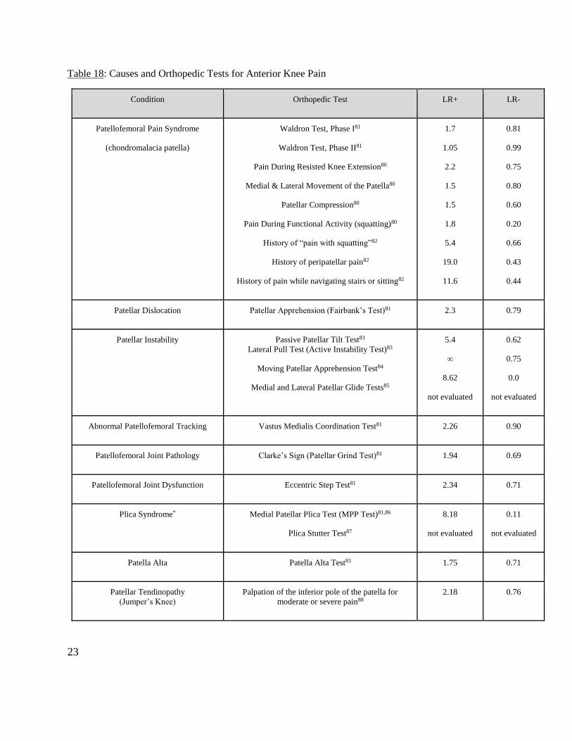
23
Table 18: Causes and Orthopedic Tests for Anterior Knee Pain
Condition Orthopedic Test LR+ LR-
Patellofemoral Pain Syndrome
(chondromalacia patella)
Waldron Test, Phase I81
Waldron Test, Phase II81
Pain During Resisted Knee Extension80
Medial & Lateral Movement of the Patella80
Patellar Compression80
Pain During Functional Activity (squatting)80
History of “pain with squatting”82
History of peripatellar pain82
History of pain while navigating stairs or sitting82
1.7
1.05
2.2
1.5
1.5
1.8
5.4
19.0
11.6
0.81
0.99
0.75
0.80
0.60
0.20
0.66
0.43
0.44
Patellar Dislocation Patellar Apprehension (Fairbank’s Test)81 2.3 0.79
Patellar Instability Passive Patellar Tilt Test83
Lateral Pull Test (Active Instability Test)83
Moving Patellar Apprehension Test84
Medial and Lateral Patellar Glide Tests85
5.4
∞
8.62
not evaluated
0.62
0.75
0.0
not evaluated
Abnormal Patellofemoral Tracking Vastus Medialis Coordination Test81 2.26 0.90
Patellofemoral Joint Pathology Clarke’s Sign (Patellar Grind Test)81 1.94 0.69
Patellofemoral Joint Dysfunction Eccentric Step Test81 2.34 0.71
Plica Syndrome* Medial Patellar Plica Test (MPP Test)81,86
Plica Stutter Test87
8.18
not evaluated
0.11
not evaluated
Patella Alta Patella Alta Test83 1.75 0.71
Patellar Tendinopathy
(Jumper’s Knee)
Palpation of the inferior pole of the patella for
moderate or severe pain88
2.18 0.76
Journal of the Academy of Chiropractic Orthopedists
Volume 14, Issue 4
24
Chondral Fracture
(osteochondritis dissecans)
Wilson’s Test89 described, but not evaluated
Osgood-Schlatter’s
Physical examination and radiography90 described, but not evaluated
Patellofemoral Bursitis
(Housemaid’s Knee)
Physical examination combined with a history of
frequent kneeling91
described, but not evaluated
Infrapatellar Fat Pad Injury
(Hoffa’s disease)
Physical examination and MRI92,93 described, but not evaluated
∞ = infinity, which occurs when the sensitivity or specificity of an exam is 100%, thus preventing the ability to
calculate a likelihood ratio
* Many tests have been described with the intention to diagnoses plica syndrome, but nearly all have not been
evaluated for diagnostic accuracy
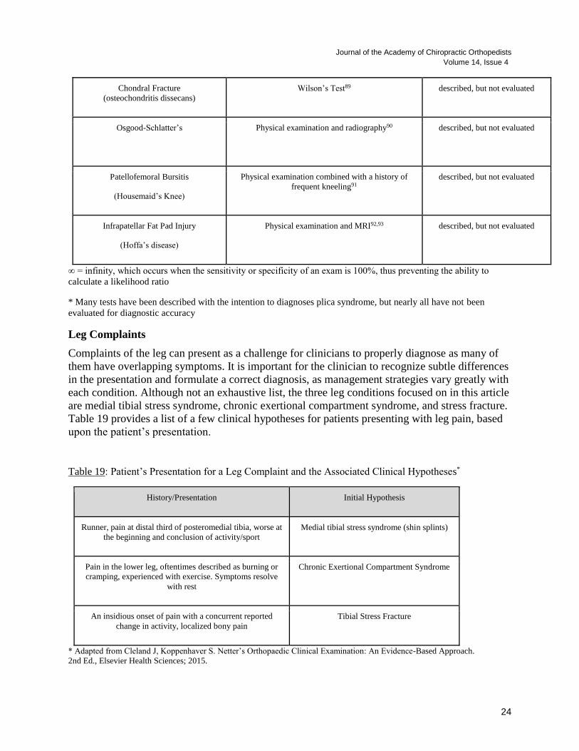
Leg Complaints
Complaints of the leg can present as a challenge for clinicians to properly diagnose as many of
them have overlapping symptoms. It is important for the clinician to recognize subtle differences
in the presentation and formulate a correct diagnosis, as management strategies vary greatly with
each condition. Although not an exhaustive list, the three leg conditions focused on in this article
are medial tibial stress syndrome, chronic exertional compartment syndrome, and stress fracture.
Table 19 provides a list of a few clinical hypotheses for patients presenting with leg pain, based
upon the patient’s presentation.
Table 19: Patient’s Presentation for a Leg Complaint and the Associated Clinical Hypotheses*
History/Presentation Initial Hypothesis
Runner, pain at distal third of posteromedial tibia, worse at
the beginning and conclusion of activity/sport
Medial tibial stress syndrome (shin splints)
Pain in the lower leg, oftentimes described as burning or
cramping, experienced with exercise. Symptoms resolve
with rest
Chronic Exertional Compartment Syndrome
An insidious onset of pain with a concurrent reported
change in activity, localized bony pain
Tibial Stress Fracture
* Adapted from Cleland J, Koppenhaver S. Netter’s Orthopaedic Clinical Examination: An Evidence-Based Approach.
2nd Ed., Elsevier Health Sciences; 2015.
25
Medial Tibial Stress Syndrome
Medial tibial stress syndrome (MTSS), commonly referred to as shin splints, affects many
runners and persons involved in other activities that involve running and jumping on hard
surfaces. Although the exact etiology of MTSS is unknown, it is believed that bony overload and
periosteal inflammation or traction are involved.94,95 There are no orthopedic tests designed to
evaluate for MTSS. Research has shown that it can be reliably clinically diagnosed using history
and physical examination findings.96 Common features of MTSS include;
● Pain provoked upon palpation of the posteromedial tibia over an area of at least 5 cm
● Pain located within the distal third of the tibia
● Pain improves with relative rest
● Pain exacerbated with physical activity, especially at the beginning and end
Chronic Exertional Compartment Syndrome
Chronic exertional compartment syndrome (CECS) is a rare condition that typically affects
young adult distance runners and other running athletes. There is an increase in pressure within
the confinement of a closed fascial compartment during exercise. There are currently no
orthopedic tests used to evaluate CECS. A good history is paramount, as the physical exam is
often unrevealing.97 It is important to note for the chiropractor that CECS can mimic other
conditions, such as MTSS, and that there is an average 2 year delay in diagnosis, making it
important to rule out other causes.98 The gold standard in diagnosis is intracompartmental
pressure testing, which is outside the scope of a chiropractor. Common history findings for
CECS include;
● Bilateral symptoms 70-80% of the time
● Pain, swelling, sensation of burning, cramping, tightness develop during exercise
● Development of pain in a certain area of the leg develops at the same time, distance, or
intensity of the exercise
● Pain is relieved with rest
Stress Fracture
Stress fractures of the leg are associated with repetitive activities of impact, such as running and
marching. They most commonly occur in the tibia, although they can occur in the fibula.
Common physical exam findings associated with a stress fracture include;99,100
● Recent increase in physical activity
● Gradual onset
● Pain with weight bearing
● Localized bony pain
● Begins as pain with stress, eventually progressing to pain at rest and at night
Journal of the Academy of Chiropractic Orthopedists
Volume 14, Issue 4
26
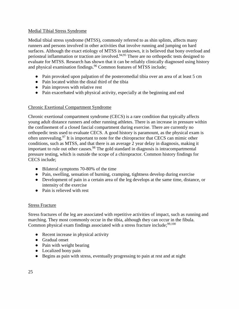
Although radiographs are commonly the first image ordered, they have a poor sensitivity in the
diagnosis of stress fractures, as they are not visible on radiographs for 2-6 weeks post injury.
Scintigraphy and MRI are considered the gold standard in diagnosing stress fractures. Although
there are no traditional orthopedic tests used in helping diagnose stress fractures, the use of a
tuning fork and therapeutic ultrasound have been studied; reproduction of pain following the
application of the tuning fork or ultrasound to the bone is a positive finding for a stress fracture.
Table 20 provides an overview of the exams associated with evaluating for the presence of a
stress fracture in the lower leg.
Table 20: Orthopedic Tests for a Stress Fracture
Exam LR+ LR-
Tuning Fork (128-Hz)101 2.3 0.3
Ultrasound102 2.1 0.3
Ankle Complaints
Ankle sprains are the most common musculoskeletal injuries seen by primary care providers.103
Although sprains are the most common injury to the ankle, other conditions (ex. tendinopathy,
fracture, nerve compression, arthritis, etc.) can cause pain and dysfunction in the ankle. Table 21
outlines characteristic patient presentations associated with ankle pain.
Table 21: Patient’s Presentation for an Ankle Complaint and the Associated Clinical Hypotheses*
Patient’s Presentation or History Initial Clinical Hypothesis
Patient reports a traumatic incident in either
forced inversion or eversion
Possible ankle sprain
Possible fracture
Possible peroneal nerve involvement (with inversion)
Patient reports trauma to ankle that included
tibial rotation on a planted foot
Possible syndesmotic sprain
Patient reports traumatic event resulting in
inability to plantarflex the ankle
Possible Achilles tendon rupture
Patient reports pain with stretch of calf muscles
and during gait (toe push off)
Possible Achilles tendonitis
Possible Sever’s disease
* Adapted from Cleland J, Koppenhaver S. Netter’s Orthopaedic Clinical Examination: An Evidence-Based
Approach.
2nd Ed., Elsevier Health Sciences; 2015.
27
Ankle Sprain
An ankle sprain is the most frequent injury to the ankle, with inversion sprain being the most
prevalent. An inversion ankle sprain damages the anterior talofibular ligament most commonly,
but can also affect the calcaneofibular and posterior talofibular ligaments.104 Eversion ankle
sprains can damage the deltoid ligament, and are commonly associated with fractures of the
medial malleolus and syndesmotic injuries.105 Common exam findings include tenderness,
swelling, and bruising around the ankle with an inability or difficulty bearing weight on the
affected side. Acute injury to the ankle may necessitate advanced imaging to screen for fracture.
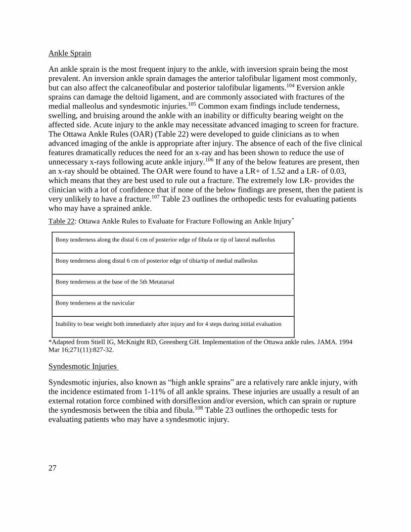
The Ottawa Ankle Rules (OAR) (Table 22) were developed to guide clinicians as to when
advanced imaging of the ankle is appropriate after injury. The absence of each of the five clinical
features dramatically reduces the need for an x-ray and has been shown to reduce the use of
unnecessary x-rays following acute ankle injury.106 If any of the below features are present, then
an x-ray should be obtained. The OAR were found to have a LR+ of 1.52 and a LR- of 0.03,
which means that they are best used to rule out a fracture. The extremely low LR- provides the
clinician with a lot of confidence that if none of the below findings are present, then the patient is
very unlikely to have a fracture.107 Table 23 outlines the orthopedic tests for evaluating patients
who may have a sprained ankle.
Table 22: Ottawa Ankle Rules to Evaluate for Fracture Following an Ankle Injury*
Bony tenderness along the distal 6 cm of posterior edge of fibula or tip of lateral malleolus
Bony tenderness along distal 6 cm of posterior edge of tibia/tip of medial malleolus
Bony tenderness at the base of the 5th Metatarsal
Bony tenderness at the navicular
Inability to bear weight both immediately after injury and for 4 steps during initial evaluation
*Adapted from Stiell IG, McKnight RD, Greenberg GH. Implementation of the Ottawa ankle rules. JAMA. 1994
Mar 16;271(11):827-32.
Syndesmotic Injuries
Syndesmotic injuries, also known as “high ankle sprains” are a relatively rare ankle injury, with
the incidence estimated from 1-11% of all ankle sprains. These injuries are usually a result of an
external rotation force combined with dorsiflexion and/or eversion, which can sprain or rupture
the syndesmosis between the tibia and fibula.108 Table 23 outlines the orthopedic tests for
evaluating patients who may have a syndesmotic injury.
Journal of the Academy of Chiropractic Orthopedists
Volume 14, Issue 4
28
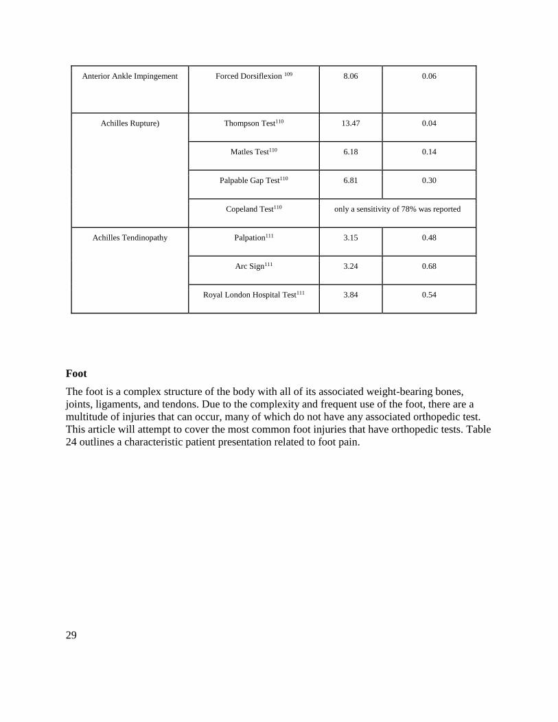
Ankle Impingement
Ankle impingement is divided into anterior and posterior ankle impingement. Anterior
impingement is a condition in which pain is experienced at the front of the ankle due to
compression of bony or soft tissue structures in the ankle mortise joint during maximal
dorsiflexion. Posterior impingement refers to pain felt at the back of the ankle due to
compression of structures in the ankle mortise during maximal plantar flexion. There are no
orthopedic tests in the literature that have been studied for posterior ankle impingement. There
has been one orthopedic test studied for anterior impingement, the forced dorsiflexion test. It
should be noted that this study had a high risk of bias.109 Table 23 outlines the orthopedic test for
evaluating patients who are suspected of ankle impingement.
Achilles Rupture and Achilles Tendinopathy
The Achilles tendon is the most commonly ruptured tendon. The main causes are a forceful
contraction of the calf muscles, overstretching of the tendon, and a fall from a height. As
opposed to a complete rupture, some patients’ tendon will still be intact and will have Achilles
tendinopathy, which is usually associated with overuse. Table 23 outlines the orthopedic tests for
evaluating patients who may have Achilles tendon rupture or tendinopathy.
Table 23: Causes and Orthopedic Tests for Ankle Pain
Condition Orthopedic Test LR+ LR-
Inversion Ankle Sprain
Anterior Drawer Test
Croy T, et al.104
Schwieterman B, et al.110
1.4
∞
0.41
0.42
Inversion Talar Tilt Test110 4.00 0.57
Posterior Drawer Test described, but never evaluated
Eversion Ankle Sprain Eversion Talar Tilt Test described, but never evaluated
Syndesmotic Injury
External Rotation Stress Test
(Kleiger’s Test)110
only a specificity of 99% was reported
Squeeze Test110 4.60 0.75
Fibular Translation110 6.30 0.28
29
Anterior Ankle Impingement
Forced Dorsiflexion 109 8.06 0.06
Achilles Rupture)
Thompson Test110 13.47 0.04
Matles Test110 6.18 0.14
Palpable Gap Test110 6.81 0.30
Copeland Test110 only a sensitivity of 78% was reported
Achilles Tendinopathy Palpation111 3.15 0.48
Arc Sign111 3.24 0.68
Royal London Hospital Test111 3.84 0.54
Foot
The foot is a complex structure of the body with all of its associated weight-bearing bones,
joints, ligaments, and tendons. Due to the complexity and frequent use of the foot, there are a
multitude of injuries that can occur, many of which do not have any associated orthopedic test.
This article will attempt to cover the most common foot injuries that have orthopedic tests. Table
24 outlines a characteristic patient presentation related to foot pain.
Journal of the Academy of Chiropractic Orthopedists
Volume 14, Issue 4
30
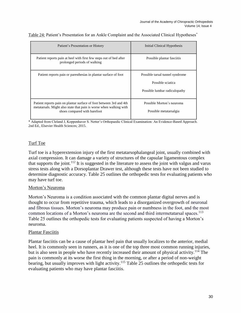
Table 24: Patient’s Presentation for an Ankle Complaint and the Associated Clinical Hypotheses*
Patient’s Presentation or History Initial Clinical Hypothesis
Patient reports pain at heel with first few steps out of bed after
prolonged periods of walking
Possible plantar fasciitis
Patient reports pain or paresthesias in plantar surface of foot Possible tarsal tunnel syndrome
Possible sciatica
Possible lumbar radiculopathy
Patient reports pain on plantar surface of foot between 3rd and 4th
metatarsals. Might also state that pain is worse when walking with
shoes compared with barefoot
Possible Morton’s neuroma
Possible metatarsalgia
* Adapted from Cleland J, Koppenhaver S. Netter’s Orthopaedic Clinical Examination: An Evidence-Based Approach.
2nd Ed., Elsevier Health Sciences; 2015.
Turf Toe
Turf toe is a hyperextension injury of the first metatarsophalangeal joint, usually combined with
axial compression. It can damage a variety of structures of the capsular ligamentous complex
that supports the joint.112 It is suggested in the literature to assess the joint with valgus and varus
stress tests along with a Dorsoplantar Drawer test, although these tests have not been studied to
determine diagnostic accuracy. Table 25 outlines the orthopedic tests for evaluating patients who
may have turf toe.
Morton’s Neuroma
Morton’s Neuroma is a condition associated with the common plantar digital nerves and is
thought to occur from repetitive trauma, which leads to a disorganized overgrowth of neuronal
and fibrous tissues. Morton’s neuroma may produce pain or numbness in the foot, and the most
common locations of a Morton’s neuroma are the second and third intermetatarsal spaces.113
Table 25 outlines the orthopedic tests for evaluating patients suspected of having a Morton’s
neuroma.
Plantar Fasciitis
Plantar fasciitis can be a cause of plantar heel pain that usually localizes to the anterior, medial
heel. It is commonly seen in runners, as it is one of the top three most common running injuries,
but is also seen in people who have recently increased their amount of physical activity.114 The
pain is commonly at its worse the first thing in the morning, or after a period of non-weight
bearing, but usually improves with light activity.115 Table 25 outlines the orthopedic tests for
evaluating patients who may have plantar fasciitis.
31
Sever’s Disease
Sever’s disease, also called calcaneal apophysitis, is a traction apophysitis that occurs where the
Achilles tendon attaches to the calcaneus. It causes inferior heel pain in children and adolescents.
The pain is usually absent in the mornings and is aggravated by physical activity, particularly
running and jumping.116 Table 25 outlines the orthopedic tests that have been reported for
evaluating Sever’s disease.
Tarsal Tunnel Syndrome
Tarsal tunnel syndrome is a compressive neuropathy of the posterior tibial nerve as it passes
under the flexor retinaculum of the tarsal tunnel. Common symptoms include paresthesia and
pain on the plantar surface of the foot and the medial ankle.117 Table 25 outlines the orthopedic
tests for evaluating patients suspected of having tarsal tunnel syndrome.
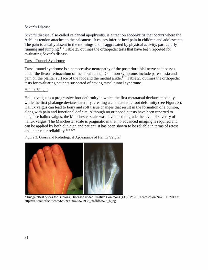
Hallux Valgus
Hallux valgus is a progressive foot deformity in which the first metatarsal deviates medially
while the first phalange deviates laterally, creating a characteristic foot deformity (see Figure 3).
Hallux valgus can lead to bony and soft tissue changes that result in the formation of a bunion,
along with pain and functional deficits. Although no orthopedic tests have been reported to
diagnose hallux valgus, the Manchester scale was developed to grade the level of severity of
hallux valgus. The Manchester scale is pragmatic in that no advanced imaging is required and
can be applied by both clinician and patient. It has been shown to be reliable in terms of retest
and inter-rater reliability.118-120
Figure 3: Gross and Radiological Appearance of Hallux Valgus*
* Image “Best Shoes for Bunions," licensed under Creative Commons (CC) BY 2.0, accesses on Nov. 11, 2017 at:
https://c2.staticflickr.com/6/5599/30475577936_94dbfba526_b.jpg
Journal of the Academy of Chiropractic Orthopedists
Volume 14, Issue 4
32
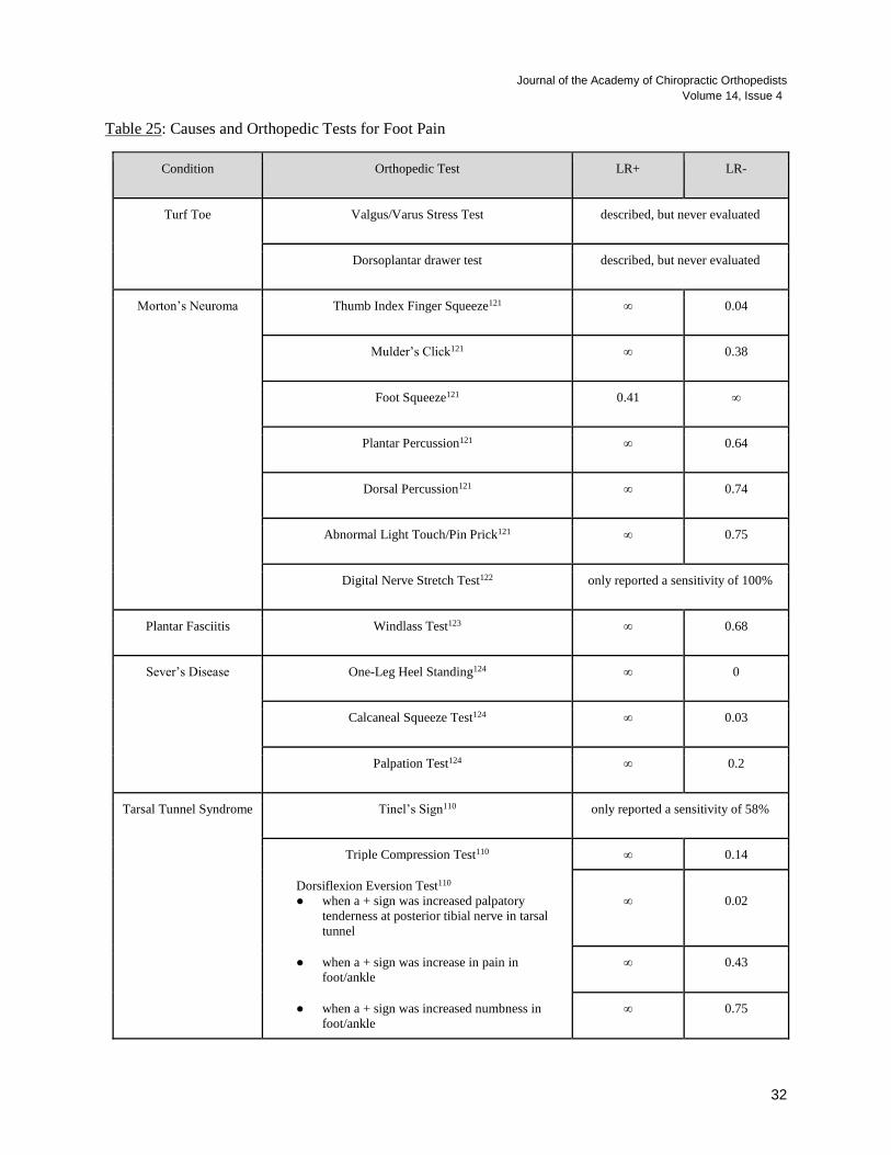
Table 25: Causes and Orthopedic Tests for Foot Pain
Condition Orthopedic Test LR+ LR-
Turf Toe Valgus/Varus Stress Test described, but never evaluated
Dorsoplantar drawer test described, but never evaluated
Morton’s Neuroma Thumb Index Finger Squeeze121 ∞ 0.04
Mulder’s Click121 ∞ 0.38
Foot Squeeze121 0.41 ∞
Plantar Percussion121 ∞ 0.64
Dorsal Percussion121 ∞ 0.74
Abnormal Light Touch/Pin Prick121 ∞ 0.75
Digital Nerve Stretch Test122 only reported a sensitivity of 100%
Plantar Fasciitis Windlass Test123 ∞ 0.68
Sever’s Disease One-Leg Heel Standing124 ∞ 0
Calcaneal Squeeze Test124 ∞ 0.03
Palpation Test124 ∞ 0.2
Tarsal Tunnel Syndrome
Tinel’s Sign110 only reported a sensitivity of 58%
Triple Compression Test110 ∞ 0.14
Dorsiflexion Eversion Test110
● when a + sign was increased palpatory
tenderness at posterior tibial nerve in tarsal
tunnel
∞
0.02
● when a + sign was increase in pain in
foot/ankle
∞ 0.43
● when a + sign was increased numbness in
foot/ankle
∞ 0.75
33
LIMITATIONS
Due to the nature of this being a narrative review, selection bias may have influenced our
selection of relevant reference articles or source materials. Also, the lack of search criteria,
which would have been involved with a systematic review, make it so our results are less
reproducible. While we attempted to select the highest quality reference materials, using the
QUADAS grading scale, we did not systematically grade every source article that was used in
this report.
CONCLUSION
The purpose of this article is to provide clinicians with an evidence-based overview of the
available orthopedic tests for a variety of lower extremity conditions. Additionally, we have
attempted to emphasize when orthopedic tests do not exist or when they have only been
described, but have never been evaluated for accuracy. Many orthopedic tests for lower
extremity conditions have limited utility. When no test exists or when the available tests are not
of a high quality, we would like to emphasize that this is when clinical decision-making should
rely more heavily on the other aspects of an evaluation such as the history, patient’s presentation,
doctor’s experience, and even the patient’s preferences.
Throughout this 4-part orthopedic review series the authors have attempted to encourage
clinicians to adopt an evidence-based approach to clinical decision-making. We have suggested
that clinicians adapt the use of likelihood ratios as one of the best ways to quickly judge the
usefulness of orthopedic exams. Our hope is that this review series will assist clinicians in their
ability to provide a concise orthopedic exam. Using the fewest number of exams that are of the
highest utility is intended to yield the most accurate results. Additionally, the use of high-quality
exams is intended to reduce the rate of false positive and false negative exam findings, which
may mislead a clinician in achieving an accurate diagnosis. Orthopedic tests are tools to assist
clinicians, and the usefulness of these tests is highly variable. While clinical decision-making
must be made in the face of uncertainty, we hope this 4-part series will help to provide a
framework for clinicians to successfully navigate this process.
List of Abbreviations
ACL = anterior cruciate ligament of the knee
CECS = Chronic Exertional Compartment Syndrome
FABER = abbreviation for flexion, abduction, and external rotation
FAI = Femoroacetabular impingement
IT band = Iliotibial band
LCL = lateral collateral ligament of the knee
Journal of the Academy of Chiropractic Orthopedists
Volume 14, Issue 4
34
LR+ = Positive Likelihood Ratio
LR- = Negative Likelihood Ratio
MCL = medial collateral ligament of the knee
MRI = Magnetic Resonance Imaging
MTSS = Medial Tibial Stress Syndrome
OAR= Ottawa Ankle Rules
OA = Osteoarthritis
PCL = posterior cruciate ligament of the knee
∞ = infinity
Competing Interests
Each of the three authors declare that they have no competing interests related to this work.
Author’s Contributions
CBR conceived this project and CBR, EFL, and ZJ each contributed to the literature review and
participated in the drafting and revisions of this work. Each of the three authors of this article
have met the criteria for authorship.
Acknowledgements
The lead author (CBR) of this 4-part orthopedic review series would like to thank each of the co-
authors that have agreed to take part in the writing of this series of articles. Each of the co-
authors (listed below) who were gracious enough to share their time, talents, and unique
perspectives in order to make this review series as useful as possible. This series of articles
would not have been possible without help from each of you.
List of co-authors involved with this this 4-part series:
● Dr. Rebecca Warnecke: Palmer College of Chiropractic DC student at the time of writing
● Dr. Milad Asefi: practicing doctor of chiropractic
● Dr. Casey Okamoto: practicing doctor of chiropractic
● Mrs. Emma Forlow Livingway: Palmer College of Chiropractic DC student at the time of
writing
● Dr. Zachary Jipp: practicing doctor of chiropractic

35
REFERENCES
1. Pauker SG, Kassirer JP. The threshold approach to clinical decision making. N Engl J Med. 1980
May 15;302(20):1109–17.
2. Cahan A, Gilon D, Manor O, Paltiel O. Probabilistic reasoning and clinical decision-making: do
doctors overestimate diagnostic probabilities? QJM. 2003 Oct;96(10):763–9.
3. Mukherjee S. The Laws of Medicine: Field Notes from an Uncertain Science. Simon and Schuster;
2015.
4. McGee S. Simplifying likelihood ratios. J Gen Intern Med. 2002 Aug;17(8):646–9.
5. Byrd JWT, Jones KS. Diagnostic accuracy of clinical assessment, magnetic resonance imaging,
magnetic resonance arthrography, and intra-articular injection in hip arthroscopy patients. Am J
Sports Med. 2004 Oct;32(7):1668–74.
6. LeBlanc KE, Muncie HL Jr, LeBlanc LL. Hip fracture: diagnosis, treatment, and secondary
prevention. Am Fam Physician. 2014 Jun 15;89(12):945–51.
7. Reiman MP, Mather RC 3rd, Cook CE. Physical examination tests for hip dysfunction and injury. Br
J Sports Med. 2015 Mar;49(6):357–61.
8. Borgerding LJ, Kikillus PJ, Boissonnault WG. Use of the patellar-pubic percussion test in the
diagnosis and management of a patient with a non-displaced hip fracture. J Man Manip Ther.
2007;15(4):E78–84.
9. Shin AY, Morin WD, Gorman JD, Jones SB, Lapinsky AS. The superiority of magnetic resonance
imaging in differentiating the cause of hip pain in endurance athletes. Am J Sports Med. 1996
Mar;24(2):168–76.
10. Sutlive TG, Lopez HP, Schnitker DE, Yawn SE, Halle RJ, Mansfield LT, et al. Development of a
clinical prediction rule for diagnosing hip osteoarthritis in individuals with unilateral hip pain. J
Orthop Sports Phys Ther. 2008 Sep;38(9):542–50.
11. Pacheco-Carrillo A, Medina-Porqueres I. Physical examination tests for the diagnosis of
femoroacetabular impingement. A systematic review. Phys Ther Sport. 2016 Sep;21:87–93.
12. Ayeni O, Chu R, Hetaimish B, Nur L, Simunovic N, Farrokhyar F, et al. A painful squat test provides
limited diagnostic utility in CAM-type femoroacetabular impingement. Knee Surg Sports Traumatol
Arthrosc. 2014 Apr;22(4):806–11.
13. Burgess RM, Rushton A, Wright C, Daborn C. The validity and accuracy of clinical diagnostic tests
used to detect labral pathology of the hip: a systematic review. Man Ther. 2011 Aug;16(4):318–26.
14. Martin RL, Enseki KR, Draovitch P, Trapuzzano T, Philippon MJ. Acetabular labral tears of the hip:
examination and diagnostic challenges. J Orthop Sports Phys Ther. 2006 Jul;36(7):503–15.
15. Maslowski E, Sullivan W, Forster Harwood J, Gonzalez P, Kaufman M, Vidal A, et al. The
diagnostic validity of hip provocation maneuvers to detect intra-articular hip pathology. PM R. 2010
Mar;2(3):174–81.
Journal of the Academy of Chiropractic Orthopedists
Volume 14, Issue 4
36
16. Lustenberger DP, Ng VY, Best TM, Ellis TJ. Efficacy of treatment of trochanteric bursitis: a
systematic review. Clin J Sport Med. 2011 Sep;21(5):447–53.
17. Williams BS, Cohen SP. Greater trochanteric pain syndrome: a review of anatomy, diagnosis and
treatment. Anesth Analg. 2009 May;108(5):1662–70.
18. Herrington L, Rivett N, Munro S. The relationship between patella position and length of the
iliotibial band as assessed using Ober’s test. Man Ther. 2006 Aug;11(3):182–6.
19. Woodley SJ, Nicholson HD, Livingstone V, Doyle TC, Meikle GR, Macintosh JE, et al. Lateral hip
pain: findings from magnetic resonance imaging and clinical examination. J Orthop Sports Phys
Ther. 2008 Jun;38(6):313–28.
20. Byrd JWT. Evaluation of the hip: history and physical examination. N Am J Sports Phys Ther. 2007
Nov;2(4):231–40.
21. Verrall GM, Slavotinek JP, Barnes PG, Fon GT. Description of pain provocation tests used for the
diagnosis of sports-related chronic groin pain: relationship of tests to defined clinical (pain and
tenderness) and MRI (pubic bone marrow oedema) criteria. Scand J Med Sci Sports. 2005
Feb;15(1):36–42.
22. Martin HD, Kivlan BR, Palmer IJ, Martin RL. Diagnostic accuracy of clinical tests for sciatic nerve
entrapment in the gluteal region. Knee Surg Sports Traumatol Arthrosc. 2014 Apr;22(4):882–8.
23. Rodrigue T, Hardy RW. Diagnosis and treatment of piriformis syndrome. Neurosurg Clin N Am.
2001 Apr;12(2):311–9.
24. Foster MR. Piriformis syndrome. Orthopedics. 2002 Aug;25(8):821–5.
25. Filler AG, Haynes J, Jordan SE, Prager J, Villablanca JP, Farahani K, et al. Sciatica of nondisc origin
and piriformis syndrome: diagnosis by magnetic resonance neurography and interventional magnetic
resonance imaging with outcome study of resulting treatment. J Neurosurg Spine. 2005 Feb;2(2):99–
115.
26. Fishman LM, Dombi GW, Michaelsen C, Ringel S, Rozbruch J, Rosner B, et al. Piriformis
syndrome: diagnosis, treatment, and outcome--a 10-year study. Arch Phys Med Rehabil. 2002
Mar;83(3):295–301.
27. Eckard TG, Padua DA, Dompier TP, Dalton SL, Thorborg K, Kerr ZY. Epidemiology of Hip Flexor
and Hip Adductor Strains in National Collegiate Athletic Association Athletes, 2009/2010-
2014/2015. Am J Sports Med. 2017 Oct;45(12):2713–22.
28. Mills M, Frank B, Goto S, Blackburn T, Cates S, Clark M, et al. Effect of restricted hip flexor muscle
length on hip extensor muscle activity and lower extremity biomechanics in college-aged female
soccer players. Int J Sports Phys Ther. 2015 Dec;10(7):946–54.
29. Cook C, Hegedus E. Orthopedic Physical Examination Tests: Pearson New International Edition: An
Evidence-Based Approach. Pearson Higher Ed; 2013. 552 p.
30. Marks MC, Alexander J, Sutherland DH, Chambers HG. Clinical utility of the Duncan-Ely test for
rectus femoris dysfunction during the swing phase of gait. Dev Med Child Neurol. 2003
Nov;45(11):763–8.
37
31. Vigotsky AD, Lehman GJ, Beardsley C, Contreras B, Chung B, Feser EH. The modified Thomas test
is not a valid measure of hip extension unless pelvic tilt is controlled. PeerJ. 2016 Aug 11;4:e2325.
32. Lucas CA. Iliotibial band friction syndrome as exhibited in athletes. J Athl Train. 1992;27(3):250–2.
33. Rosenthal MD. Clinical testing for extra-articular lateral knee pain. A modification and combination
of traditional tests. N Am J Sports Phys Ther. 2008 May;3(2):107–9.
34. Khaund R, Flynn SH. Iliotibial band syndrome: a common source of knee pain. Am Fam Physician.
2005 Apr 15;71(8):1545–50.
35. Hyde TE, Gengenbach MS. Conservative Management of Sports Injuries. Jones & Bartlett Learning;
2007. 1173 p.
36. Jackson JL, O’Malley PG, Kroenke K. Evaluation of acute knee pain in primary care. Ann Intern
Med. 2003 Oct 7;139(7):575–88.
37. Bachmann LM, Haberzeth S, Steurer J, ter Riet G. The accuracy of the Ottawa knee rule to rule out
knee fractures: a systematic review. Ann Intern Med. 2004 Jan 20;140(2):121–4.
38. Stanitski CL, Harvell JC, Fu F. Observations on acute knee hemarthrosis in children and adolescents.
J Pediatr Orthop. 1993 Jul;13(4):506–10.
39. Hagino T, Ochiai S, Senga S, Yamashita T, Wako M, Ando T, et al. Meniscal tears associated with
anterior cruciate ligament injury. Arch Orthop Trauma Surg. 2015 Dec;135(12):1701–6.
40. Akseki D, Ozcan O, Boya H, Pinar H. A new weight-bearing meniscal test and a comparison with
McMurray’s test and joint line tenderness. Arthroscopy. 2004 Nov;20(9):951–8.
41. Lowery DJ, Farley TD, Wing DW, Sterett WI, Steadman JR. A clinical composite score accurately
detects meniscal pathology. Arthroscopy. 2006 Nov;22(11):1174–9.
42. Miao Y, Yu J-K, Ao Y-F, Zheng Z-Z, Gong X, Leung KKM. Diagnostic values of 3 methods for
evaluating meniscal healing status after meniscal repair: comparison among second-look arthroscopy,
clinical assessment, and magnetic resonance imaging. Am J Sports Med. 2011 Apr;39(4):735–42.
43. Jaddue DAK, Tawfiq FH, Sayed-Noor AS. The utility of clinical examination in the diagnosis of
medial meniscus injury in comparison with arthroscopic findings. Eur J Orthop Surg Traumatol.
2010;20(5):389–92.
44. Pookarnjanamorakot C, Korsantirat T, Woratanarat P. Meniscal lesions in the anterior cruciate
insufficient knee: the accuracy of clinical evaluation. J Med Assoc Thai. 2004 Jun;87(6):618–23.
45. Jerosch J, Riemer S. [How good are clinical investigative procedures for diagnosing meniscus
lesions?]. Sportverletz Sportschaden. 2004 Jun;18(2):59–67.
46. Fowler PJ, Lubliner JA. The predictive value of five clinical signs in the evaluation of meniscal
pathology. Arthroscopy. 1989;5(3):184–6.
47. Mirzatolooei F, Yekta Z, Bayazidchi M, Ershadi S, Afshar A. Validation of the Thessaly test for
detecting meniscal tears in anterior cruciate deficient knees. Knee. 2010 Jun;17(3):221–3.
Journal of the Academy of Chiropractic Orthopedists
Volume 14, Issue 4
38
48. Harrison BK, Abell BE, Gibson TW. The Thessaly test for detection of meniscal tears: validation of
a new physical examination technique for primary care medicine. Clin J Sport Med. 2009
Jan;19(1):9–12.
49. Kurosaka M, Yagi M, Yoshiya S, Muratsu H, Mizuno K. Efficacy of the axially loaded pivot shift
test for the diagnosis of a meniscal tear. Int Orthop. 1999;23(5):271–4.
50. Mariani PP, Adriani E, Maresca G, Mazzola CG. A prospective evaluation of a test for lateral
meniscus tears. Knee Surg Sports Traumatol Arthrosc. 1996;4(1):22–6.
51. Gornitzky AL, Lott A, Yellin JL, Fabricant PD, Lawrence JT, Ganley TJ. Sport-Specific Yearly Risk
and Incidence of Anterior Cruciate Ligament Tears in High School Athletes: A Systematic Review
and Meta-analysis. Am J Sports Med. 2016 Oct;44(10):2716–23.
52. Agel J, Arendt EA, Bershadsky B. Anterior cruciate ligament injury in national collegiate athletic
association basketball and soccer: a 13-year review. Am J Sports Med. 2005 Apr;33(4):524–30.
53. Arendt EA, Agel J, Dick R. Anterior cruciate ligament injury patterns among collegiate men and
women. J Athl Train. 1999 Apr;34(2):86–92.
54. Benjaminse A, Gokeler A, van der Schans CP. Clinical diagnosis of an anterior cruciate ligament
rupture: a meta-analysis. J Orthop Sports Phys Ther. 2006 May;36(5):267–88.
55. Mulligan EP, Harwell JL, Robertson WJ. Reliability and diagnostic accuracy of the Lachman test
performed in a prone position. J Orthop Sports Phys Ther. 2011 Oct;41(10):749–57.
56. Cross MJ, Schmidt DR, Mackie IG. A no-touch test for the anterior cruciate ligament. J Bone Joint
Surg Br. 1987 Mar;69(2):300.
57. Ostrowski JA. Accuracy of 3 diagnostic tests for anterior cruciate ligament tears. J Athl Train. 2006
Jan;41(1):120–1.
58. Larson RL. Physical examination in the diagnosis of rotatory instability. Clin Orthop Relat Res. 1983
Jan;(172):38–44.
59. Jarbo KA, Hartigan DE, Scott KL, Patel KA, Chhabra A. Accuracy of the Lever Sign Test in the
Diagnosis of Anterior Cruciate Ligament Injuries. Orthop J Sports Med. 2017
Oct;5(10):2325967117729809.
60. al-Duri Z. Relation of the fibular head sign to other signs of anterior cruciate ligament insufficiency.
A follow-up letter to the editor. Clin Orthop Relat Res. 1992 Feb;(275):220–5.
61. Wagemakers HP, Luijsterburg PA, Boks SS, Heintjes EM, Berger MY, Verhaar JA, et al. Diagnostic
accuracy of history taking and physical examination for assessing anterior cruciate ligament lesions
of the knee in primary care. Arch Phys Med Rehabil. 2010 Sep;91(9):1452–9.
62. Amis AA, Gupte CM, Bull AMJ, Edwards A. Anatomy of the posterior cruciate ligament and the
meniscofemoral ligaments. Knee Surg Sports Traumatol Arthrosc. 2006 Mar;14(3):257–63.
63. Wang C-J. Injuries to the posterior cruciate ligament and posterolateral instabilities of the knee.
Chang Gung Med J. 2002 May;25(5):288–97.
64. Roberts DM, Stallard TC. Emergency department evaluation and treatment of knee and leg injuries.
39
Emerg Med Clin North Am. 2000 Feb;18(1):67–84, v – vi.
65. Rubinstein RA Jr, Shelbourne KD, McCarroll JR, VanMeter CD, Rettig AC. The accuracy of the
clinical examination in the setting of posterior cruciate ligament injuries. Am J Sports Med. 1994
Jul;22(4):550–7.
66. Feltham GT, Albright JP. The diagnosis of PCL injury: literature review and introduction of two
novel tests. Iowa Orthop J. 2001;21:36–42.
67. Hughston JC, Norwood LA Jr. The posterolateral drawer test and external rotational recurvatum test
for posterolateral rotatory instability of the knee. Clin Orthop Relat Res. 1980 Mar;(147):82–7.
68. Loomer RL. A test for knee posterolateral rotatory instability. Clin Orthop Relat Res. 1991
Mar;(264):235–8.
69. Stäubli HU, Jakob RP. Posterior instability of the knee near extension. A clinical and stress
radiographic analysis of acute injuries of the posterior cruciate ligament. J Bone Joint Surg Br. 1990
Mar;72(2):225–30.
70. Loos WC, Fox JM, Blazina ME, Del Pizzo W, Friedman MJ. Acute posterior cruciate ligament
injuries. Am J Sports Med. 1981 Mar;9(2):86–92.
71. Fowler PJ, Messieh SS. Isolated posterior cruciate ligament injuries in athletes. Am J Sports Med.
1987 Nov;15(6):553–7.
72. Bae JH, Choi IC, Suh SW, Lim HC, Bae TS, Nha KW, et al. Evaluation of the reliability of the dial
test for posterolateral rotatory instability: a cadaveric study using an isotonic rotation machine.
Arthroscopy. 2008 May;24(5):593–8.
73. Ferrari DA, Ferrari JD, Coumas J. Posterolateral instability of the knee. J Bone Joint Surg Br. 1994
Mar;76(2):187–92.
74. Souza TA. Differential Diagnosis and Management for Chiropractors. Jones & Bartlett Publishers;
2016.
75. Kastelein M, Wagemakers HPA, Luijsterburg PAJ, Verhaar JAN, Koes BW, Bierma-Zeinstra SMA.
Assessing medial collateral ligament knee lesions in general practice. Am J Med. 2008
Nov;121(11):982–8.e2.
76. Harilainen A. Evaluation of knee instability in acute ligamentous injuries. Ann Chir Gynaecol.
1987;76(5):269–73.
77. Harilainen A, Myllynen P, Rauste J, Silvennoinen E. Diagnosis of acute knee ligament injuries: the
value of stress radiography compared with clinical examination, stability under anaesthesia and
arthroscopic or operative findings. Ann Chir Gynaecol. 1986;75(1):37–43.
78. Sanchis-Alfonso V. Holistic approach to understanding anterior knee pain. Clinical implications.
Knee Surg Sports Traumatol Arthrosc. 2014 Oct;22(10):2275–85.
79. Powers CM, Bolgla LA, Callaghan MJ, Collins N, Sheehan FT. Patellofemoral pain: proximal, distal,
and local factors, 2nd International Research Retreat. J Orthop Sports Phys Ther. 2012 Jun;42(6):A1–
Journal of the Academy of Chiropractic Orthopedists
Volume 14, Issue 4
40
54.
80. Cook C, Hegedus E, Hawkins R, Scovell F, Wyland D. Diagnostic accuracy and association to
disability of clinical test findings associated with patellofemoral pain syndrome. Physiother Can.
2010 Feb 22;62(1):17–24.
81. Nijs J, Van Geel C, Van der auwera C, Van de Velde B. Diagnostic value of five clinical tests in
patellofemoral pain syndrome. Man Ther. 2006 Feb;11(1):69–77.
82. Elton K, McDonough K, Savinar-Nogue E, Jensen GM. A Preliminary Investigation: History,
Physical, and lsokinetic Exam Results versus Arthroscopic Diagnosis of Chondromalacia Patella *. J
Orthop Sports Phys Ther. 1985;7(3):115–23.
83. Haim A, Yaniv M, Dekel S, Amir H. Patellofemoral pain syndrome: validity of clinical and
radiological features. Clin Orthop Relat Res. 2006 Oct;451:223–8.
84. Ahmad CS, McCarthy M, Gomez JA, Shubin Stein BE. The moving patellar apprehension test for
lateral patellar instability. Am J Sports Med. 2009 Apr;37(4):791–6.
85. Nissen CW, Cullen MC, Hewett TE, Noyes FR. Physical and arthroscopic examination techniques of
the patellofemoral joint. J Orthop Sports Phys Ther. 1998 Nov;28(5):277–85.
86. Kim S-J, Lee D-H, Kim T-E. The relationship between the MPP test and arthroscopically found
medial patellar plica pathology. Arthroscopy. 2007 Dec;23(12):1303–8.
87. Magee DJ. Orthopedic Physical Assessment. Elsevier Health Sciences; 2008. 1138 p.
88. Cook JL, Khan KM, Kiss ZS, Purdam CR, Griffiths L. Reproducibility and clinical utility of tendon
palpation to detect patellar tendinopathy in young basketball players. Victorian Institute of Sport
tendon study group. Br J Sports Med. 2001 Feb;35(1):65–9.
89. Conrad JM, Stanitski CL. Osteochondritis dissecans: Wilson’s sign revisited. Am J Sports Med. 2003
Sep;31(5):777–8.
90. Reid DC. Sports Injury, Assessment and Rehabilitation. Med Sci Sports Exercise. 1993;25(10):i.
91. McAfee JH, Smith DL. Olecranon and prepatellar bursitis. Diagnosis and treatment. West J Med.
1988 Nov;149(5):607–10.
92. Kumar D, Alvand A, Beacon JP. Impingement of infrapatellar fat pad (Hoffa’s disease): results of
high-portal arthroscopic resection. Arthroscopy. 2007 Nov;23(11):1180–6.e1.
93. Morini G, Chiodi E, Centanni F, Gattazzo D. [Hoffa’s disease of the adipose pad: magnetic
resonance versus surgical findings]. Radiol Med. 1998 Apr;95(4):278–85.
94. Reshef N, Guelich DR. Medial tibial stress syndrome. Clin Sports Med. 2012 Apr;31(2):273–90.
95. Edwards PH Jr, Wright ML, Hartman JF. A practical approach for the differential diagnosis of
chronic leg pain in the athlete. Am J Sports Med. 2005 Aug;33(8):1241–9.
96. Winters M, Bakker EWP, Moen MH, Barten CC, Teeuwen R, Weir A. Medial tibial stress syndrome
can be diagnosed reliably using history and physical examination. Br J Sports Med [Internet]. 2017
Feb 8; Available from: http://dx.doi.org/10.1136/bjsports-2016-097037
41
97. Vajapey S, Miller TL. Evaluation, diagnosis, and treatment of chronic exertional compartment
syndrome: a review of current literature. Phys Sportsmed. 2017 Nov;45(4):391–8.
98. Tucker AK. Chronic exertional compartment syndrome of the leg. Curr Rev Musculoskelet Med.
2010 Sep 2;3(1-4):32–7.
99. Behrens SB, Deren ME, Matson A, Fadale PD, Monchik KO. Stress fractures of the pelvis and legs
in athletes: a review. Sports Health. 2013 Mar;5(2):165–74.
100. van der Velde GM, Hsu WS. Posterior tibial stress fracture: a report of three cases. J Manipulative
Physiol Ther. 1999 Jun;22(5):341–6.
101. Lesho EP. Can tuning forks replace bone scans for identification of tibial stress fractures? Mil Med.
1997 Dec;162(12):802–3.
102. Schneiders AG, Sullivan SJ, Hendrick PA, Hones BDGM, McMaster AR, Sugden BA, et al. The
ability of clinical tests to diagnose stress fractures: a systematic review and meta-analysis. J Orthop
Sports Phys Ther. 2012 Sep;42(9):760–71.
103. Levine AM, Kolker D. Ankle and Foot Injuries. In: Essential Sports Medicine. 2008. p. 146–68.
104. Croy T, Koppenhaver S, Saliba S, Hertel J. Anterior talocrural joint laxity: diagnostic accuracy of
the anterior drawer test of the ankle. J Orthop Sports Phys Ther. 2013 Dec;43(12):911–9.
105. Fong DT, Chan Y-Y, Mok K-M, Yung PS, Chan K-M. Understanding acute ankle ligamentous
sprain injury in sports. Sports Med Arthrosc Rehabil Ther Technol. 2009 Jul 30;1:14.
106. Bachmann LM, Kolb E, Koller MT, Steurer J, ter Riet G. Accuracy of Ottawa ankle rules to exclude
fractures of the ankle and mid-foot: systematic review. BMJ. 2003 Feb 22;326(7386):417.
107. Stiell IG, McKnight RD, Greenberg GH, McDowell I, Nair RC, Wells GA, et al. Implementation of
the Ottawa ankle rules. JAMA. 1994 Mar 16;271(11):827–32.
108. Molinari A, Stolley M, Amendola A. High ankle sprains (syndesmotic) in athletes: diagnostic
challenges and review of the literature. Iowa Orthop J. 2009;29:130–8.
109. Molloy S, Solan MC, Bendall SP. Synovial impingement in the ankle. A new physical sign. J Bone
Joint Surg Br. 2003 Apr;85(3):330–3.
110. Schwieterman B, Haas D, Columber K, Knupp D, Cook C. Diagnostic accuracy of physical
examination tests of the ankle/foot complex: a systematic review. Int J Sports Phys Ther. 2013
Aug;8(4):416–26.
111. Reiman M, Burgi C, Strube E, Prue K, Ray K, Elliott A, et al. The utility of clinical measures for the
diagnosis of achilles tendon injuries: a systematic review with meta-analysis. J Athl Train. 2014
Nov;49(6):820–9.
112. McCormick JJ, Anderson RB. Turf toe: anatomy, diagnosis, and treatment. Sports Health. 2010
Nov;2(6):487–94.
113. Sault JD, Morris MV, Jayaseelan DJ, Emerson-Kavchak AJ. Manual therapy in the management of a
patient with a symptomatic Morton’s Neuroma: A case report. Man Ther. 2016;21:307–10.
Journal of the Academy of Chiropractic Orthopedists
Volume 14, Issue 4
42
114. Lopes AD, Hespanhol Júnior LC, Yeung SS, Costa LOP. What are the main running-related
musculoskeletal injuries? A Systematic Review. Sports Med. 2012 Oct 1;42(10):891–905.
115. Muth CC. Plantar Fasciitis. JAMA. 2017 Jul 25;318(4):400.
116. Cassas KJ, Cassettari-Wayhs A. Childhood and adolescent sports-related overuse injuries. Am Fam
Physician. 2006 Mar 15;73(6):1014–22.
117. Kinoshita M, Okuda R, Morikawa J, Jotoku T, Abe M. The dorsiflexion-eversion test for diagnosis
of tarsal tunnel syndrome. J Bone Joint Surg Am. 2001 Dec;83-A(12):1835–9.
118. Roddy E, Zhang W, Doherty M. Validation of a self-report instrument for assessment of hallux
valgus. Osteoarthritis Cartilage. 2007 Sep;15(9):1008–12.
119. Menz HB, Fotoohabadi MR, Wee E, Spink MJ. Validity of self-assessment of hallux valgus using
the Manchester scale. BMC Musculoskelet Disord. 2010 Sep 20;11:215.
120. Menz HB, Munteanu SE. Radiographic validation of the Manchester scale for the classification of
hallux valgus deformity. Rheumatology . 2005 Aug;44(8):1061–6.
121. Mahadevan D, Venkatesan M, Bhatt R, Bhatia M. Diagnostic Accuracy of Clinical Tests for
Morton’s Neuroma Compared With Ultrasonography. J Foot Ankle Surg. 2015 Jul;54(4):549–53.
122. Cloke DJ, Greiss ME. The digital nerve stretch test: A sensitive indicator of Morton’s neuroma and
neuritis. Foot Ankle Surg. 2006;12(4):201–3.
123. De Garceau D, Dean D, Requejo SM, Thordarson DB. The association between diagnosis of plantar
fasciitis and Windlass test results. Foot Ankle Int. 2003 Mar;24(3):251–5.
124. Perhamre S, Lazowska D, Papageorgiou S, Lundin F, Klässbo M, Norlin R. Sever’s injury: a clinical
diagnosis. J Am Podiatr Med Assoc. 2013 Sep;103(5):361–8.
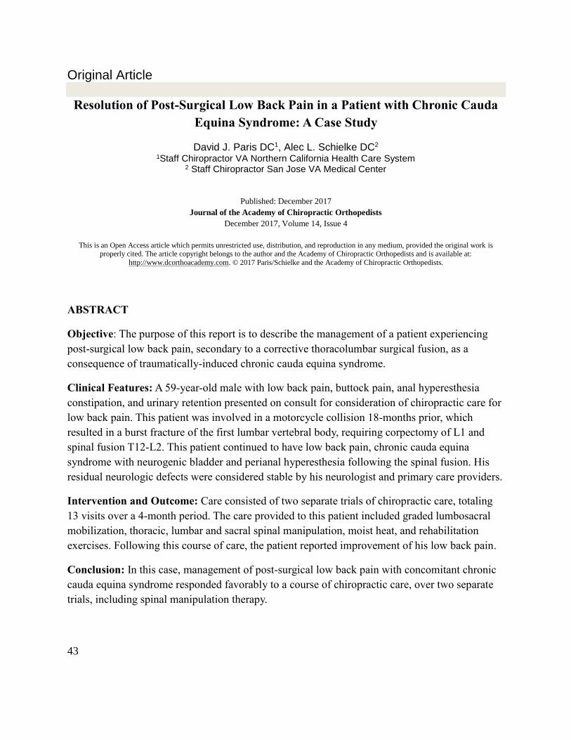
43
Original Article
Resolution of Post-Surgical Low Back Pain in a Patient with Chronic Cauda
Equina Syndrome: A Case Study
David J. Paris DC1, Alec L. Schielke DC2 1Staff Chiropractor VA Northern California Health Care System
2 Staff Chiropractor San Jose VA Medical Center
Published: December 2017 Journal of the Academy of Chiropractic Orthopedists
December 2017, Volume 14, Issue 4
This is an Open Access article which permits unrestricted use, distribution, and reproduction in any medium, provided the original work is properly cited. The article copyright belongs to the author and the Academy of Chiropractic Orthopedists and is available at:
http://www.dcorthoacademy.com. © 2017 Paris/Schielke and the Academy of Chiropractic Orthopedists.
ABSTRACT
Objective: The purpose of this report is to describe the management of a patient experiencing
post-surgical low back pain, secondary to a corrective thoracolumbar surgical fusion, as a
consequence of traumatically-induced chronic cauda equina syndrome.
Clinical Features: A 59-year-old male with low back pain, buttock pain, anal hyperesthesia
constipation, and urinary retention presented on consult for consideration of chiropractic care for
low back pain. This patient was involved in a motorcycle collision 18-months prior, which
resulted in a burst fracture of the first lumbar vertebral body, requiring corpectomy of L1 and
spinal fusion T12-L2. This patient continued to have low back pain, chronic cauda equina
syndrome with neurogenic bladder and perianal hyperesthesia following the spinal fusion. His
residual neurologic defects were considered stable by his neurologist and primary care providers.
Intervention and Outcome: Care consisted of two separate trials of chiropractic care, totaling
13 visits over a 4-month period. The care provided to this patient included graded lumbosacral
mobilization, thoracic, lumbar and sacral spinal manipulation, moist heat, and rehabilitation
exercises. Following this course of care, the patient reported improvement of his low back pain.
Conclusion: In this case, management of post-surgical low back pain with concomitant chronic
cauda equina syndrome responded favorably to a course of chiropractic care, over two separate
trials, including spinal manipulation therapy.
Journal of the Academy of Chiropractic Orthopedists
Volume 14, Issue 4
44
INTRODUCTION
The use of lumbar spinal manipulation and spinal rehabilitation for the management of low back
pain is not well documented for patients with concomitant chronic cauda equina syndrome
(CES). The authors of this report were able to identify only a single case report involving spinal
manipulation for the management of low back pain (LBP) in a patient with chronic CES (1).
In patients experiencing acute CES symptomatology, manipulation is an absolute
contraindication (2,3). Post-surgically, chronic CES is known to be associated with back pain and
neurologic deficits (4). Cauda equina syndrome may involve LBP, radicular pain in the lower
extremities, saddle anesthesia, urinary incontinence, or lower extremity motor and/or sensory
deficits (4,5). Acute CES is a medical emergency, however there is controversy regarding the risk
of immediate (within 48 hours) versus delayed surgical intervention, as surgery in an emergency
setting may involve suboptimal results and prolonged morbidity (6).
CASE STUDY
A 59-year-old male with low back pain of 18-months duration referred on consult by his primary
care provider (PCP) for chiropractic care. This patient experienced a motorcycle accident 18-
months prior which resulted in a burst fracture of the first lumbar vertebral body (L1),
necessitating emergency L1 corpectomy and T12-L2 fusion.
Following surgery, the patient was diagnosed with residual chronic CES and neurogenic bladder,
which necessitated the need for intermittent self-catheterization to void his bladder. The patient
also reported perianal hyperesthesia, which remained unchanged since the time of the surgery
and was primarily noticeable while wiping after passing a bowel movement. This patient was
able to defecate on his own via Valsalva maneuver on a 1-hour postprandial schedule. He also
reported transient radicular pain, extending to his feet, while performing the Valsalva maneuver
while voiding his bowels. No other lower extremity symptoms were noted by the patient.
This patient’s low back pain was described as a constant, “sharp ache” over the lumbosacral
region and was worse with all end-range lumbosacral ranges of motion. The patient rated his
average pain as a 4/10 on a Numeric Pain Rating Scale (NPRS) with range from 2/10 to 6/10,
depending on provocative and palliative factors. The patient completed the NPRS, but refused to
complete any other outcome measures. Patient described that his pain was relieved while laying
down supine, with moist heat (e.g. with a hot tub), but the pain was worsened during any activity
that involved prolonged lumbar flexion (e.g. sitting).
The patient had received chiropractic care for LBP previous to this injury, but denied any
chiropractic care since his accident and surgery 18 months ago. The patient also underwent
physical therapy for the management of his low back pain, previous to this injury, for
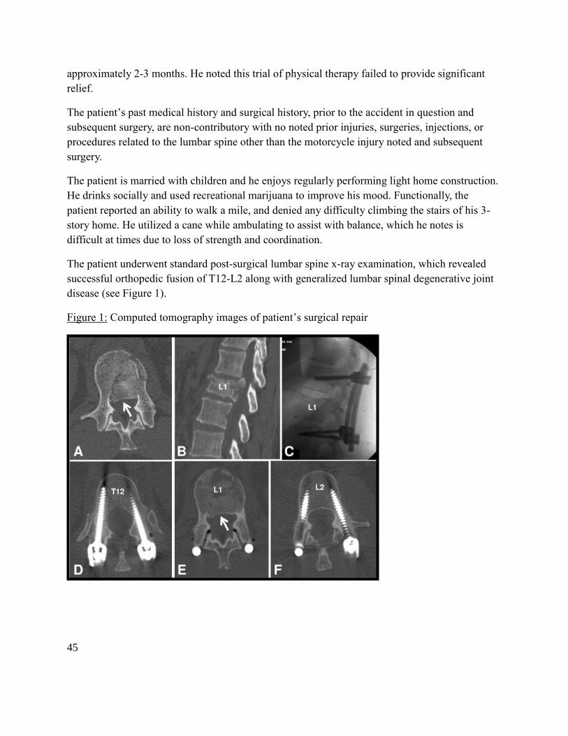
45
approximately 2-3 months. He noted this trial of physical therapy failed to provide significant
relief.
The patient’s past medical history and surgical history, prior to the accident in question and
subsequent surgery, are non-contributory with no noted prior injuries, surgeries, injections, or
procedures related to the lumbar spine other than the motorcycle injury noted and subsequent
surgery.
The patient is married with children and he enjoys regularly performing light home construction.
He drinks socially and used recreational marijuana to improve his mood. Functionally, the
patient reported an ability to walk a mile, and denied any difficulty climbing the stairs of his 3-
story home. He utilized a cane while ambulating to assist with balance, which he notes is
difficult at times due to loss of strength and coordination.
The patient underwent standard post-surgical lumbar spine x-ray examination, which revealed
successful orthopedic fusion of T12-L2 along with generalized lumbar spinal degenerative joint
disease (see Figure 1).
Figure 1: Computed tomography images of patient’s surgical repair
Journal of the Academy of Chiropractic Orthopedists
Volume 14, Issue 4
46
The physical examination revealed a 12-inch surgical scar over the patient’s left thoracolumbar
paravertebral gutter. Lumbosacral active range of motion were decreased by approximately 20%,
in all directions with no description of “pain” noted and only “soreness” if continued movement
pushing end-ranges. The patient reported pain following palpation of his lumbosacral
paravertebral muscles and moderate hypertonicity was noted, bilaterally. Bilateral lower
extremity manual muscle testing bilaterally graded as 4-4+/5, including extensor hallucis longus
with only exception plantar flexion graded 4/5 bilaterally. Deep tendon reflexes were rated as 3+
on the right quadriceps tendon, 2+ on the left quadriceps tendon, 0 on Achilles tendon bilaterally
with reinforcement. Babinski’s test was negative, bilaterally. Bilateral hypoesthesia to pinwheel
and light touch was noted along the posterior calves (S1/S2 dermatomes). No other sensory
deficits were detected in the regions of the L1-L5 dermatomes. Perianal hyperesthesia was
reported by the patient and was not examined, per patient’s request. Seated and supine Straight-
Leg Raise tests were negative, bilaterally, with note of localized low back pain when the legs
were raised above 45 degrees. When the Valsalva maneuver was performed that patient denied
lower extremity symptoms (i.e. pain or paresthesia). The patient was hesitant to perform an
aggressive Valsalva maneuver, due to a fear of fecal incontinence during the exam. On evaluation
patient also continues to report a history of low back pain and diffuse leg paraesthesias while
straining to void his bowels since accident (i.e. positive Dejerine’s triad).
The patient was diagnosed as follows: 1) status-post corpectomy and thoracolumbar fusion T12-
L2 with chronic CES and neurogenic bladder, 2) chronic low-back pain with myofascial
contribution as sequelae of injury, subsequent surgical intervention and deconditioning, and 3)
lumbar degenerative joint disease, per the radiology report.
INTERVENTION
Care consisted of two separate trials of care totaling 13 chiropractic visits over a 4-month period.
The first trial of care involved 6-visits and care was extended past the first trial of care, based on
improved LBP. Therapies included graded lumbosacral mobilization, thoracic, lumbar and sacral
spinal manipulation, moist heat, and rehabilitation exercises. Treatment began with graded
mobilization over the lumbosacral area (sacral extension) while prone, high velocity-low
amplitude (HVLA) chiropractic spinal manipulation (diversified technique) was applied to the
mid-thoracic spine along with hydrocollator therapy for 10-minutes, during each visit. As a
safely measure, pre-manipulative positional stress was utilized and symptoms were assessed
prior to each treatment. Rehabilitation exercises began with supine single-leg, knee to chest
stretching for 30 seconds in 3 separate positions (same shoulder, opposite shoulder and across
waist).
Improvement in low back pain was noted immediately post-treatment at each visit. After each of
the first 3 visits, the patient reported lasting improvement from his LBP. Drop table chiropractic
manipulation therapy was applied to the patient’s lumbosacral area midway into initial trial of
47
care (3rd visit), as was the introduction of Quadruped Reach (also known as “Bird Dog”), Side
Bridge and Curl-up exercises (also known as McGill’s Big 3). These were chosen as they are
considered to be safe exercises for low back pain patients (8,9). The patient continued to respond
positively and had no difficulties understanding or performing his home-based exercises.
Handouts were dispensed detailing his home exercises instructions with progressions. He was
instructed to progress when he was able to perform 3 sets of 12-15 repetitions with good control
of movement, performed and demonstrated on follow-ups visits at clinic, without worsening of
pain following performance of the exercises. The patient was instructed that mild post-exercise
soreness may be experiences and is considered to be a normal side-effect of this activity. Side
bridge was instructed initially at 8-10 slow repetitions with 2 breath holds in the elevated
position done 1-2 times daily. Quadruped Reach was instructed initially with 3-4 repetitions,
which were easily performed and demonstrated in-office. These exercises were progressed, on
initial visit, to 10-12 repetitions with 5-6 second hold, 1-2x daily. The patient was instructed to
perform Curl-Up exercise for 12 repetitions with a 2-breath hold, 1-2 times daily. All exercise
progressions were based on the patient’s ability to achieve a minimum of 2 times daily with
adequate comfort and good control as assessed by clinician. On the patient’s 6th visit, he was
shown an eyes-open, one-legged standing balance exercise. Initially he could not do this for
more than 2-3 seconds on either leg without a major correction or having to touch other foot to
ground due to poor balance. No pain, weakness or fear-avoidance was noted on performance of
this exercise. Of note, improving balance was a patient-specific goal of care as he noted he
would like to sail comfortably on his sail boat and he was avoiding this, due to his imbalance
issues.
Lumbosacral mobilization progressed to HVLA side-posture spinal manipulation (diversified
technique) during final two visits in initial trial. Manipulation targeted the L4-S1 segments, with
no adverse events reported. The patient noted a preference and greater pain relief from the drop-
assisted spinal manipulation, subsequent visits continued utilization of drop table technique to
the lumbosacral spine. Due to success with pain control and home exercise intervention, the
patient was offered, and declined, a physical therapy consultation for a more focused progression
and monitoring of home exercise plan.
The patient did not attend his follow-up re-examination and lost to follow-up, until he presented
as a walk-in 6-months later requesting care as need for exacerbations. On this visit he rated his
LBP as a 3/10, following recent snow shoveling and flare of LBP. He reported that he had
intentionally missed his post-trial follow-up appointment as his LBP had resolved and he “felt
great and didn’t feel he needed care.”
On his return following a 6-month absence, the patient presented without the need of a cane for
ambulatory assistance and reported rarely using his cane. He denied any LBP for the last 6-
months, rated as 0/10 NPRS, until this recent exacerbation as noted above. He further stated that
Journal of the Academy of Chiropractic Orthopedists
Volume 14, Issue 4
48
he was able to continue to build his house remaining very active, “more active than ever”, also
that he had taken up sailing again. He reported that his chronic CES symptoms were unchanged
and he continued to experience perianal hyperesthesia and continued to require urinary
catheterization and complies with follow-up evaluations from his neurologist to monitor his
chronic CES.
Currently, patient continues to follow-up on an as-needed basis for the management of his LBP
exacerbations. He has been advised to continue his activity level and other activities of daily
living and was instructed to continue his home exercises, as instructed. He declined any other
referrals or intervention and was happy to continue his home-based exercises.
DISCUSSION
This case is notable as cauda-equina syndrome (CES) represents a relative contraindication and
when acute a medical emergency. There are few case reports noting the multimodal care
chiropractors generally deliver in practice, along with spinal manipulative therapy involving
these conditions. This case describes the successful chiropractic treatment of 18-month chronic
low-back pain in a post-surgical patient with chronic cauda-equina syndrome. Multimodal care
consisted of graded lumbar mobilization, spinal manipulation, moist heat, and home-based
lumbar stability exercises.
There were no adverse events reported throughout this course of care and spinal manipulation
appeared to be safe and efficacious in the long-term management strategy for this patient’s LBP
with concomitant chronic CES. In this specific case, multimodal conservative chiropractic
treatment including spinal manipulation, resolved this patient’s low back pain.
Clinicians should be aware of the absolute contraindication of spinal manipulative therapy in
patients with acute CES. However, awareness of only acute CES guidelines and
recommendations may prevent referral of patients with chronic CES and spinal pain complaints
for conservative management, such as spinal manipulative therapy, spinal rehabilitation exercises
and other conservative measures.
LIMITATIONS
As a retrospective study, this case involves some limitations. First, only a NPRS and the patient’s
subjective interpretation of increased function were assessed as outcomes. The patient’s response
to care would have be better evaluated using additional outcomes assessments, particularly an
outcome measure involving the patient’s functional status. Secondly, although mobilization and
two chiropractic techniques were used (drop table and diversified side-posture HVLA) there can
be no statement on efficacy of those other than the stated individual patient preference. No
generalized effectiveness can be gained from this type of individual case-report.
49
CONCLUSION
Select patients with chronic CES may benefit from spinal manipulative therapy for the
management of LBP. However, further research in the form more rigorous testing, beyond case
reports, is needed to establish the safety and efficacy of this approach to care.
While the effectiveness and safety of this management strategy is not able to be applied to the
general population from this report, it does provide some insight into the clinical management of
patients with chronic CES via conservative care. It appears spinal manipulation therapy and
spinal rehabilitation exercises may be considered as a viable consideration in the treatment of
spinal pain complaints among select patients with chronic CES.
REFERENCES
1. Lisi A, Bhardwaj M. Chiropractic High-Velocity Low-Amplitude Spinal Manipulation in the
Treatment of A Case of Postsurgical Chronic Cauda Equina Syndrome. J Manipulative
Physiol Ther 2004; 27:574-578.
2. Haldeman S, Chapman-Smith D, Peterson D, editors. Guidelines for chiropractic quality
assurance and practice parameters. Gaithersburg, MD: Aspen Publishers; 1993. p. 175.
3. Bigos S, Bowyer O, Braen G, et al. Acute low back problems in adults. Clinical practice
guideline #14. AHCPR Publication #95-0642. Rockville, MD: Agency for Health Care
Policy and Research, Public Health Service, U.S. Department of Health and Human Services;
1994.
4. Memmo PA, Nadler S, Malanga S. Lumbar disc herniations: a review of surgical and non-
surgical indications and outcomes. J Back Musculoskel Rehab 2000;14:79-88.
5. Shapiro S, Kostuik JP. Medical realities of cauda equina syndrome secondary to lumbar disc
herniation. Spine 2000;25:348-352.
6. Gleave JR, MacFarlane R. Cauda equina syndrome: what is the relationship between timing
of surgery and outcome? Br J Neurosurg 2002;16:325-8.
7. Ahn UM, Ahn NU, Buchowski JM, Garrett ES, Sieber AN, Kotsuik JP. Cauda equina
syndrome secondary to lumbar disc herniation: a meta-analysis of surgical outcomes. Spine
2000;25:1515-22
8. Axler, C. and McGill, S.M. (1997). Low back loads over a variety of abdominal exercises:
Searching for the safest abdominal challenge, Med.Sci.Sports.Ex. 29(6): 804-811.
9. Kavcic, N., Grenier, S.G., and McGill, S.M. (2004a) Determining tissue loads and spine
stability while performing commonly prescribed stabilization exercises. Spine, 29(11): 1254-
1265.
Journal of the Academy of Chiropractic Orthopedists
Volume 14, Issue 4
50
Editorial Review
Biological Treatment Approaches for Degenerative Disk Disease: A Literature
Review of In Vivo Animal and Clinical Data
Yu Moriguchi, Marjan Alimi, Thamina Khair, George Manolarakis, Connor Berlin, Lawrence J. Bonassar, Roger Härtl
Global Spine J 2016;6:497–518
JACO Editorial Reviewer: Gregory C. Priest, DC, FACO
Published: December 2017
Journal of the Academy of Chiropractic Orthopedists
December 2017, Volume 14, Issue 4
The original article copyright belongs to the original publisher. This review is available from: http://www.dcorthoacademy.com
© 2017 Priest and the Academy of Chiropractic Orthopedists. This is an Open Access article which permits unrestricted use, distribution, and
reproduction in any medium, provided the original work is properly cited.
AUTHORS’ ABSTRACT
Study Design: Literature review
Objective: Degenerative disk disease (DDD) has a negative impact on quality of life and is a
major cause of morbidity worldwide. There has been a growing interest in the biological repair
of DDD by both researchers and clinicians alike. To generate an overview of the recent progress
in reparative strategies for the treatment of DDD highlighting their promises and limitations, a
comprehensive review of the current literature was performed elucidating data from in vivo
animal and clinical studies.
Methods: Articles and abstracts available in electronic databases of PubMed, Web of Science,
and Google Scholar as of December 2014 were reviewed. Additionally, data from unpublished,
ongoing clinical trials was retrieved from clinicaltrials.gov and available abstracts from research
forums. Data was extracted from the most recent in vivo animal or clinical studies involving any
of the following: (1) treatment with biomolecules, cells, or tissue-engineered constructs and (2)
annulus fibrosus repair.
Results: Seventy-five articles met the inclusion criteria for review. Among these, 17 studies
involved humans; 37, small quadrupeds; and 21, large quadrupeds. Findings from all treatments
employed demonstrated improvement either in regenerative capacity or in pain attenuation, with
the exception of one clinical study.
Conclusion: Published clinical studies on cell therapy have reported encouraging results in the
treatment of DDD and resultant back pain. We expect new data to emerge in the near future as
51
treatments for DDD continue to evolve in parallel to our greater understanding of disk health
and pathology.
Keywords: Intervertebral disk, disk regeneration, back pain, growth factor, cell therapy, platelet-
rich plasma, tissue engineering, annular repair
Clinical Relevance
The prevalence of degenerative disk disease is ubiquitous, reportedly occurring in more than
90% of people over 50 years of age. Degenerative disk disease can result in chronic back pain
and frequently results in surgical intervention in an attempt to alleviate the related pain and
disability. Unfortunately, the surgery itself may result in altered spinal biomechanics with
secondary adjacent segment degeneration. Current treatment options do not adequately address
the actual diseased tissue, the degenerated disk. This robustly-referenced literature review
explores biologic treatment options that may hold promise for direct treatment of the degenerated
disk.
JACO Editorial Summary:
• The intervertebral disk (IVD) is a complex of three tissues: the nucleus pulposus (NP),
the annulus fibrosus (AF), and the cartilaginous end plates.
• Degenerative disk disease is a multifactorial process that involves one or more of the
three tissues comprising the IVD complex.
• Intervertebral disk degeneration is said to occur in 40% of persons younger than 30 years
of age, and in more than 90% of those older than 50.
• Degenerative disk disease (DDD) can lead to chronic lower back pain and disability.
• Surgical intervention for DDD is reported to be performed in nearly 4 million patients
worldwide.
• Spinal fusion surgery presents risks for pseudarthrosis and adjacent segment
degeneration, resulting in higher rates of reoperation.
• This literature review evaluates in vivo animal and clinical data, with strong
inclusion/exclusion criteria, and gives consideration to published data as well as data
from ongoing, unpublished clinical studies.
• Biological repair of the degenerated IVD are classified into three categories:
biomolecular therapy, cell therapy, and tissue-engineered IVD construction. These
treatment strategies are further stratified into categories specific to the stage of
degeneration.
• The goal of biologic treatment of the degenerated IVD is to repair/reconstruct the actual
diseased tissue.
• Biomolecular treatment may include protein injection, gene therapy and platelet-rich
plasma.
• Cell therapy may include differentiated cells such as disk-relevant cells and articular
chondrocytes, and stem cells.
Journal of the Academy of Chiropractic Orthopedists
Volume 14, Issue 4
52
• Tissue-engineering strategies may include scaffold development and whole disk
transplantation with tissue-engineered construct.
• The authors acknowledge the difficulties associated with extrapolation of data from
animal studies to human applications, and are aware of model-based limitations such as
subjective assessment of pain and physical function.
• The authors feel that significant progress within the field of biologic therapies for DDD
has been made in the past decade, although there are a paucity of clinical studies. They
have attempted to consolidate and analyze a plethora of data from published and ongoing
research.
53
Editorial Review
Pain sensitization and osteoarthritis
Lars Arendt-Nielsen, Prof., DMSc, PhD
Clinical Experimental Rheumatology 2017; 35 (Suppl. 107): S68-S74
JACO Editorial Reviewer: Scott E Kilmer, DC, FACO, FACC
Published: December 2017
Journal of the Academy of Chiropractic Orthopedists
December 2017, Volume 14, Issue 4
The original article copyright belongs to the original publisher. This review is available from: http://www.dcorthoacademy.com
© 2017 Kilmer and the Academy of Chiropractic Orthopedists. This is an Open Access article which permits unrestricted use, distribution, and
reproduction in any medium, provided the original work is properly cited.
AUTHOR’S ABSTRACT:
Treating chronic musculoskeletal pain, and chronic joint pain (osteoarthritis (OA) in particular),
is challenging as the peripheral and central pain mechanisms are not fully discovered, and safe
and as effective analgesic drugs are not available. In general, the preclinical models of OA are
limited to provide fundamental understanding of the pain mechanisms involved in patients with
chronic joint pain (1). The pain associated with joint discomfort is highly variable, often
underestimated by clinicians, and shows only modest association with crude radiological
scorings. One reason for the disconnect between the extent of structural damage and pain is
neural plastic changes occurring in the peripheral and central nervous system resulting in pain
sensitization impacting the patient’s experience of pain. In recent years, a variety of human
quantitative and mechanistic pain assessment tools (Quantitative Sensory Testing, QST) have
been developed, providing new opportunities for diagnostic phenotyping of OA patients and the
associated degree of sensitization. Mechanistic phenotyping has revealed specific subgroups of
specifically sensitized OA patients, and been used as a predictive guideline to evaluate which
patients are most likely to experience continued chronic pain after an otherwise technically
successful knee replacement (chronic postoperative pain). Furthermore, such techniques may be
used to profile new or existing drugs together with other e.g. cognitive or behavioral therapies
with the potential to manage joint pain.
JACO Editorial Summary:
• This article was written by Lars Arendt-Nielsen, Prof.,DMSc.,PhD. Dr. Arendt-Nielsen is
on the Faculty of Medicine at Aalborg University, School of Medicine in Denmark.
Journal of the Academy of Chiropractic Orthopedists
Volume 14, Issue 4
54
• The purpose of this paper is to explain pain sensitization in osteoarthritis and show how
selected biomarkers are used to help predict those patients that may develop increased
pain after total joint arthroplasty (TJA).
• It is estimated that by 2030 total hip arthroplasty in the US will increase by 200% and
total knee arthroplasty will increase by 700%. Additionally, around 20% of total knee
arthroplasty patients will have continued pain after surgery and approximately 10% of
total hip arthroplasty patients will have continued pain after the surgery.
• Pain sensitization occurs as a result of “continuous and intense nociceptive input from
joints”. Studies have shown this type of nociceptive input occurs with osteoarthritis and is
a main driver of peripheral and central sensitization.
• There are two basic ways to clinically evaluate for pain sensitization. The first is
assessment using scores/questionnaires such as VAS, McGill Pain Questionnaire,
WOMAC and others. The second method is the use of quantitative mechanistic pain
biomarkers. There are two quantitative mechanistic biomarkers used in the study. 1.
Temporal Summation. This occurs when neurons in the dorsal horn become more
excitable due to repeated nociceptive input. Bedside testing using this method is
accomplished by using computer controlled pressure algometry. After repeated
stimulation, the patient reports an increase in severity and duration of pain which is not
supported by the radiographic findings. 2. Descending Pain Modulation. This type of pain
modulation occurs when nociceptive input is received in the brain from a segment remote
from the site of pain caused by the osteoarthritis. Clinical testing of descending pain
modulation is accomplished with the use of cuff algometry when one cuff delivers a
conditioning stimulus and the other delivers a test stimulus. This triggers a “pain inhibits
pain phenomenon”. This phenomenon can be diminished in patients with OA and can be
used to help identify those patients that may have continued pain after total joint
arthroplasty.
• Using the methods mentioned above to measure for pain sensitization, there is evidence
to show that there is a correlation of preoperative hyperalgesia and chronic postoperative
pain. Further, joint revision surgery based only on the patient’s report of pain can result
in 50% of those patients experiencing chronic/increased pain after the revision.
• This paper demonstrates how mechanistic phenotyping can be used to aid in predicting
which patients would likely develop chronic or increased pain after total joint
arthroplasty. Pain sensitization should be evaluated prior to surgery and pain alone should
not be used as the sole indicator for total joint arthroplasty.
55
Editorial Review
Structural Correlates of Pain in Osteoarthritis
T. Neogi
Clinical and Experimental Rheumatology 2017;35 (Suppl. 107):S75-S78
JACO Editorial Reviewer: Daniel P. Dock, DC, FACO
Published: December 2017
Journal of the Academy of Chiropractic Orthopedists
December 2017, Volume 14, Issue 4
The original article copyright belongs to the original publisher. This review is available from: http://www.dcorthoacademy.com
© 2017 Dock and the Academy of Chiropractic Orthopedists. This is an Open Access article which permits unrestricted use, distribution, and
reproduction in any medium, provided the original work is properly cited.
AUTHOR’S ABSTRACT:
Osteoarthritis (OA) is the most common form of arthritis, with knee OA itself being among the
most common conditions and a leading cause of disability among older adults worldwide. Pain is
a key symptom in the decision to seek medical attention, yet available therapies for managing
OA are limited with only minimal or moderate efficacy. Current approaches to pain management
in OA have been rather non-specific, limited to acetaminophen or NSAIDs primarily, without
targeting underlying structural lesions that may be contributing to pain in OA. With the advent of
MRI, a number of studies have noted the importance of bone marrow lesions and
synovitis/effusion to the pain experience in OA. These pathologic features are therefore
attractive treatment targets, with some proof-of-concept studies demonstrating the potential
efficacy of targeting these lesions. Another increasingly recognised important contribution to
pain in OA is sensitisation, which is associated with pain severity. Synovitis/effusion have been
identified as potentially leading to development and worsening of sensitisation. Much work
remains to be done in understanding the mechanisms by which structural pathology causes pain;
such insights are urgently needed to develop new treatment approaches to help millions of people
worldwide who are burdened by pain from OA.
Editorial Review:
Knee Osteoarthritis (OA) is among the most common conditions and leading cause of disability
among older adults. With the advent of MRI, a number of studies have noted the importance of
bone marrow lesions and synovitis effusion to the pain experience in OA. Another increasing
Journal of the Academy of Chiropractic Orthopedists
Volume 14, Issue 4
56
recognized important contribution to pain in OA is sensitisation. [Editor’s note: The original
article author’s spelling of sensitisation, rather than sensitization, is maintained in this Editorial
Review]
Results: Approximately 10-12% of the adult population have symptomatic OA of any joint.
Knee OA is a leading cause of disability among older adults, accounting for a greater risk of
mobility disability.
One barrier to understanding the genesis of pain in OA is the so-called “structure-symptom
discordance”, some individuals have radiographic changes with minimal symptoms, while others
have more significant pain with only minimal (if any) structural pathology noted on radiograph.
Findings of osteophyte with joint space narrowing characterized as radiographic OA, while its
combination of symptoms (i.e. pain, aching, stiffness) in the same joint attributable to OA is
considered to be symptomatic OA. The discordance diminishes with more severe stages of
disease. It has been recognized that radiographs are relatively insensitive to numerous
pathologic changes, and are better visualized on MRI.
Something within the knee must be contributing to symptoms. Joint replacement, intra-articular
lidocaine injection, and intra-articular corticosteroid injection give relief of pain.
As OA progresses, neurovascular invasion may disrupt the osteochondral junction, accompanied
by growth of sensory nerves and increased expression of nerve growth factor (NGF), which can
facilitate sensitisation. Neurobiologic mechanisms have been recognized, particularly of
sensitisation.
MRI studies give further insight into structural pathology contributing to OA pain. MRI studies
have shown subchondral bone changes (bone marrow lesions), and synovitis/effusion. The bone
marrow lesions and synovitis/effusion have been associated with pain and with pain fluctuation.
Use of a patellofemoral knee brace caused reduction in pain and reduction of bone marrow
lesions.
Meniscal lesions are common in OA. Meniscal tears were equally occurring among those with
knee pain as those without knee pain. MRI findings in knees of older adults can be challenging
as 89% of people without any radiographic evidence of knee OA have at least one MRI feature,
seen in both painful and pain-free knees.
The contribution of structural joint pathology to the pain experience in OA remains incompletely
understood.
MRI sheds light onto specific pathologic features that may play an important role in causing
pain.
57
Factors beyond structural pathology also contribute to the pain experience factors, including
psychologic factors.
Pain sensitisation and the overall efficiency of CNS pain modulation mechanisms, merit further
studies.
JACO Editorial Summary: Two points that jumped out at me when reading this article was the
reduction in pain and the reduction of bone marrow lesions when the patient used a
patellofemoral knee brace, and the strengthening of the lower extremity muscles helping with
proper patellar glide. This outlines the possibility of using arch supports to cause relief of pain
and relief of mobility disability (remember, the “foot goes flat, and the leg twists in” causing
twisting of the knee structures).
• Knee OA was pointed as a leading cause of disability in older adults, accounting for a
greater risk of mobility disability. This is important considering deep venous thrombosis,
pneumonia, cardiovascular conditions, and cerebrovascular conditions.
• Patients with knee pain may have normal appearing radiographs; osteoarthrosis may be
revealed on the radiographs of patients without knee pain. The article points out that there
can be knee pain with normal appearing x-rays.
• Sensitisation can explain why the patient can have more pain than is justified by the
exam.
• The future: Stem Cell Injections and Platelet-Rich Plasma (PRP) are now having success
in some cases for pain relief and even reduction of MRI visible bone marrow edema.
This is important as it may help take away some of the mobility disability (and the
adverse conditions that may occur with mobility disability).
Journal of the Academy of Chiropractic Orthopedists
Volume 14, Issue 4
58
Editorial Review
Strawberries Improve Pain and Inflammation in Obese Adults with
Radiographic Evidence of Knee Osteoarthritis
Jace Schell, R. Hal Scofield, James R. Barrett, Biji T. Kurien, Nancy Betts, Timothy J. Lyons,
Yan Daniel Zhao, and Arpita Basu
Nutrients 2017, 9, 949; doi:10.3390/nu9090949.
JACO Editorial Reviewer: Scott D. Banks, DC, MS
Published: December 2017
Journal of the Academy of Chiropractic Orthopedists
December 2017, Volume 14, Issue 4
The original article copyright belongs to the original publisher. This review is available from: http://www.dcorthoacademy.com
© 2017 Banks and the Academy of Chiropractic Orthopedists. This is an Open Access article which permits unrestricted use, distribution, and
reproduction in any medium, provided the original work is properly cited.
AUTHORS’ ABSTRACT:
Osteoarthritis (OA), the most common form of arthritis, is a significant public health burden in
U.S. adults. Among its many risk factors, obesity is a key player, causing inflammation, pain,
impaired joint function, and reduced quality of life. Dietary polyphenols and other bioactive
compounds in berries, curcumin, and tea have shown effects in ameliorating pain and
inflammation in OA, but few clinical studies have been reported. The purpose of the present
study was to examine the effects of dietary strawberries on pain, markers of inflammation, and
quality of life indicators in obese adults with OA of the knee. In a randomized, double-blind
cross-over trial, adults with radiographic evidence of knee OA (n = 17; body mass index (BMI):
(mean SD) 39.1 + 1.5; age (years): 57 + 7) were randomized to a reconstituted freeze-dried
strawberry beverage (50 g/day) or control beverage daily, each for 12 weeks, separated by a 2-
week washout phase (total duration, 26 weeks). Blood draws and assessments of pain and quality
of life indicators were conducted using the Visual Analog Scale for Pain (VAS Pain), Measures
of Intermittent and Constant Osteoarthritis Pain (ICOAP), and Health Assessment Questionnaire-
Disability Index (HAQ-DI) questionnaires, which were completed at baseline and at weeks 12,
14, and 26 of the study. Among the serum biomarkers of inflammation and cartilage degradation,
interleukin (IL)-6, IL-1, and matrix metalloproteinase (MMP)-3 were significantly decreased
after strawberry vs. control treatment (all p < 0.05). Strawberry supplementation also
significantly reduced constant, intermittent, and total pain as evaluated by the ICOAP
questionnaire as well as the HAQ-DI scores (all p < 0.05). No effects of treatment were noted on
serum C-reactive protein (CRP), nitrite, glucose, and lipid profiles. Dietary strawberries may
59
have significant analgesic and anti-inflammatory effects in obese adults with established knee
OA.
JACO Editorial Summary:
• The morbidity rates with drug treatments for chronic osteoarthritic pain have driven
interest in natural therapies noted for the far superior safety profile. Some of the more
studied include plant phenols such as derived from turmeric, ginger and other food items.
• Clinical benefit has been previously been demonstrated in metabolic syndrome with
phenolics from berries such as strawberries. As metabolic syndrome and osteoarthritis
share a major driving mechanism, inflammation, it was sought to measure their impact on
both inflammatory and clinical features in osteoarthritic patients.
• Inflammatory biomarkers (IL-6, IL1b), tissue degeneration markers (matrix
metalloproteinase (MMP)-3) and clinical outcome markers (ICOAP questionnaire and
HAQ-DI scores) significantly improved after 12 weeks of active supplementation
confirming the original hypothesis.
• No adverse effects were seen although the study population was only 17 subjects. Some
adverse effects would be anticipated over the 12 weeks of observation in this study with
NSAIDs.
• The applicability of the data to broader populations is difficult as the study population
was significantly obese. As obesity is highly associated with a background pro-
inflammatory state could be argued that the clinical effect may be greater in a non-obese
population.
• Although similar outcomes have been shown with many phenolic food sources, very little
is known about their comparative efficacy to guide current clinical application beyond
clinician experience. Some study has found synergistic combinations of phenolic food
sources to produce better outcomes that monotherapies and this should also be
considered.
Summary:
The study supports the idea of the use of plant phenolics such as strawberries are a beneficial
option perhaps better suited to long-term use as is needed in osteoarthritis. They may be
particularly helpful in the management of patients who have already had adverse effects with
drug therapies.

Journal of the Academy of Chiropractic Orthopedists
Volume 14, Issue 4
60
Radiology Corner
Retrosomatic Cleft: A Radiology Review
Jacinda D. Woods, DC1
1Radiology Resident, Palmer College of Chiropractic, Davenport, Iowa
Published: December 2017
Journal of the Academy of Chiropractic Orthopedists
December 2017, Volume 14, Issue 4
This is an Open Access article which permits unrestricted use, distribution, and reproduction in any medium, provided the original work is
properly cited. The article copyright belongs to the author and the Academy of Chiropractic Orthopedists and is available at:
http://www.dcorthoacademy.com. © 2017 Woods and the Academy of Chiropractic Orthopedists.
History
A 25 year old male presented with right sided low back pain and left hip pain for the past five months.
The patient had also reported history of left iliac fracture.
Plain Films
Figure 1: AP Lumbopelvic Figure 2: Lateral Lumbar
Report
The AP and lateral lumbopelvic radiographic examination revealed L5 left pedicle region
sclerosis with a suspected right-sided pars defect. A well-corticated osseous irregularity was
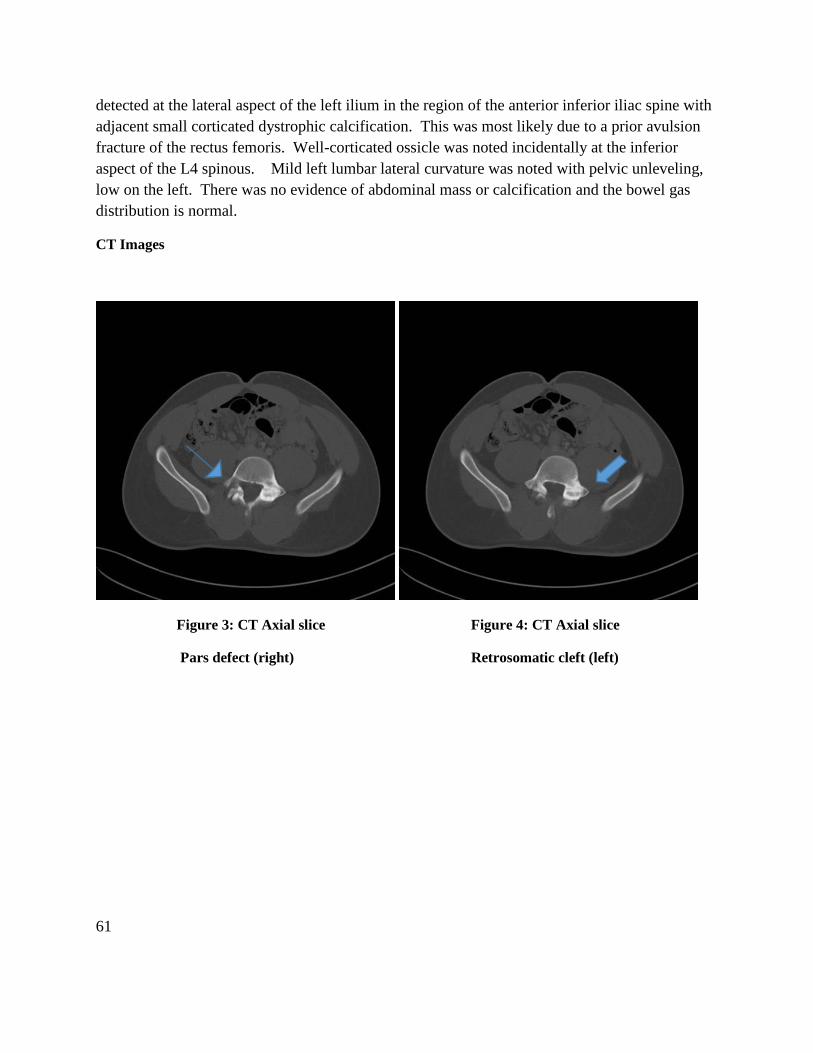
61
detected at the lateral aspect of the left ilium in the region of the anterior inferior iliac spine with
adjacent small corticated dystrophic calcification. This was most likely due to a prior avulsion
fracture of the rectus femoris. Well-corticated ossicle was noted incidentally at the inferior
aspect of the L4 spinous. Mild left lumbar lateral curvature was noted with pelvic unleveling,
low on the left. There was no evidence of abdominal mass or calcification and the bowel gas
distribution is normal.
CT Images
Figure 3: CT Axial slice Figure 4: CT Axial slice
Pars defect (right) Retrosomatic cleft (left)
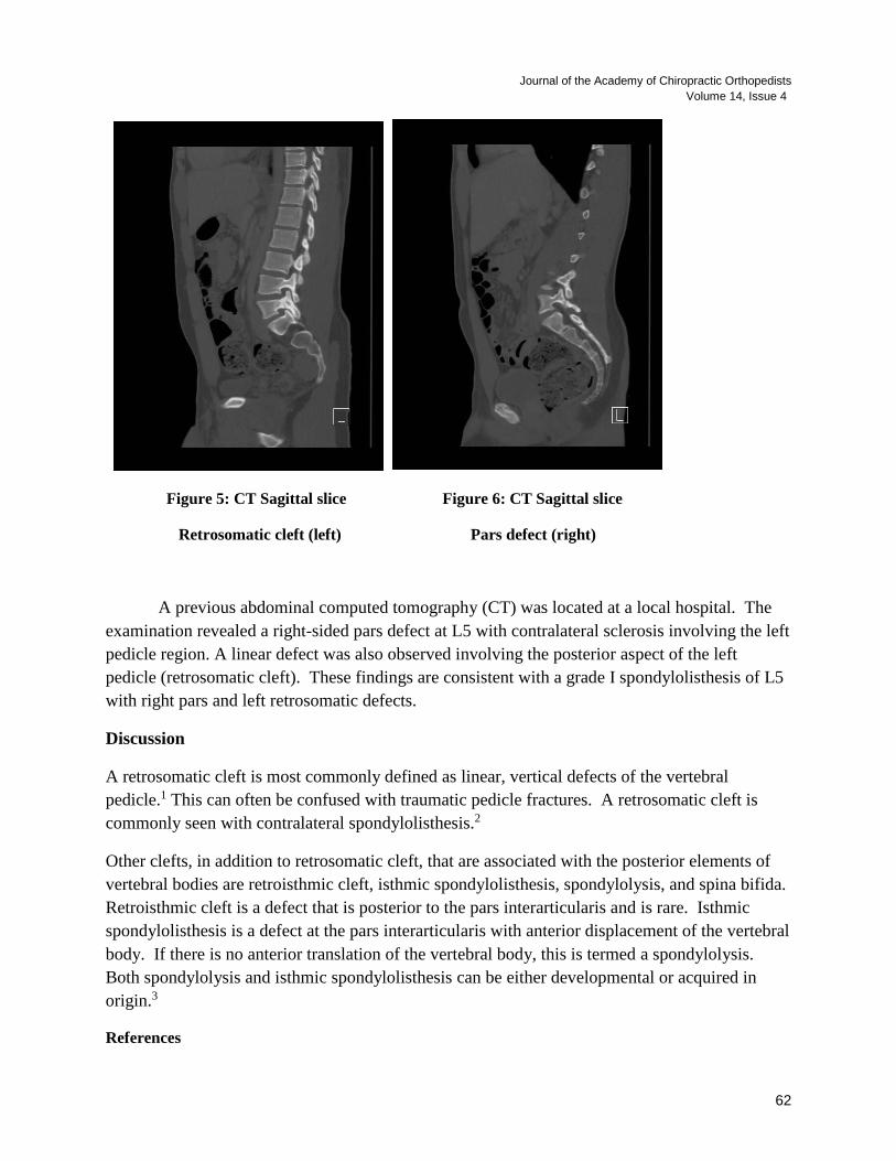
Journal of the Academy of Chiropractic Orthopedists
Volume 14, Issue 4
62
Figure 5: CT Sagittal slice Figure 6: CT Sagittal slice
Retrosomatic cleft (left) Pars defect (right)
A previous abdominal computed tomography (CT) was located at a local hospital. The
examination revealed a right-sided pars defect at L5 with contralateral sclerosis involving the left
pedicle region. A linear defect was also observed involving the posterior aspect of the left
pedicle (retrosomatic cleft). These findings are consistent with a grade I spondylolisthesis of L5
with right pars and left retrosomatic defects.
Discussion incidentally at the inferior aspect of the L4 spinous.
A retrosomatic cleft is most commonly defined as linear, vertical defects of the vertebral
pedicle.1 This can often be confused with traumatic pedicle fractures. A retrosomatic cleft is
commonly seen with contralateral spondylolisthesis.2
Other clefts, in addition to retrosomatic cleft, that are associated with the posterior elements of
vertebral bodies are retroisthmic cleft, isthmic spondylolisthesis, spondylolysis, and spina bifida.
Retroisthmic cleft is a defect that is posterior to the pars interarticularis and is rare. Isthmic
spondylolisthesis is a defect at the pars interarticularis with anterior displacement of the vertebral
body. If there is no anterior translation of the vertebral body, this is termed a spondylolysis.
Both spondylolysis and isthmic spondylolisthesis can be either developmental or acquired in
origin.3
References

63
1. MDCT of variations and anomalies of the neural arch and its processes: part 1--pedicles, pars
interarticularis, laminae, and spinous process. AJR Am J Roentgenol. 2011 Jul;197(1):W104-13. doi:
10.2214/AJR.10.5803.
2. M Soleimanpour, M L Gregg and R Paraliticci. Bilateral retrosomatic clefts at multiple lumbar levels.
American Journal of Neuroradiology. September 1995, 16 (8) 1616-1617.
3. Jarrah Ali Al-Tubaikh, Maximiliun F. Reiser. Congenital Diseases and Syndromes An Illustrated
Radiological Guide. 2009, 145-146.

Journal of the Academy of Chiropractic Orthopedists
Volume 14, Issue 4
64
Ortho Quiz
by Steven L. Kleinfield D.C., F.A.C.O.
1. Which of the following is not considered to be causational for olecranon bursitis?
a. Trauma
b. Infection
c. Gout
d. Prolonged Pressure
e. All of the above are considered causational
f. None of the above are considered causational
2. The Orthopedic Test “Dugas” is considered a classic test for evaluation for the presence of
which condition:
a. Carpal Tunnel Syndrome
b. Postural Compression Syndrome
c. Shoulder Dislocation
d. Rib Fracture
3. Which of the following orthopedic tests should be performed first when suspecting Thoracic
Outlet Syndrome:
a. Adson’s Test
b. Allen’s Test
c. Wright’s Test
d. Halstead Maneuver
4. Your patient is a 60 year old male. He is of Scottish heritage. His main complaints are as
follows: He has recently been noticing that he is developing bone pain, generally stiff all over
and that he tires easily. At present he seems to be getting headaches quite frequently and he
notices that he feels like his hat size is increased since he can't wear his old Stetson any longer.
a. Cervicogenic Encephalgia
b. Osteoarthrosis
c. Paget’s Disease
d. Osteogenesis Imperfecta
5. The most common type of headache is:
a. Cluster Headache
b. Classic Migraine Headache
c. Common Migraine Headache
d. Tension Headache

65
Current Events
❖ The dates for the 2018 Academy of Chiropractic Orthopedists online examination are as
follows:
o Friday, May 18, 2018
o Friday, July 20, 2018
Please contact the Academy as soon as you can with your notice of intent to sit the
Academy Board examination.
Jerrold R Wildenauer DC, FACO
1859 Warrior Drive
Mendota Heights, MN 55118
TEL: 612-454-1472
FAX: 651-846-5590
E-mail: [email protected]
❖ Apply for the Lipe Scholarship
Details at http://www.accoweb.org/lipescholarship.html
❖ The full hours of the following conventions have been accepted by the Academy as
qualifying for re-credentialing.
o American College of Chiropractic Orthopedists 2018 Annual Convention
19 Apr to 21 Apr 2018
Hilton Garden Inn Carlsbad Beach, Carlsbad CA
https://acoco.wildapricot.org/event-2436518
o CFS 2017 Annual Fall Convention
October 5-7, 2017
Chicago Marriott Oak Brook
1401 W 22nd Street | Oak Brook, IL 60523
630.573.8555 www.marriott.com/chiob
http://www.forensic-sciences.org/convention/
❖ ACC-RAC 2018 Association of Chiropractic Colleges 25 th Educational
Conference and Research Agenda Conference
March 8-10, 2018
InterContinental Dallas
15201 Dallas Parkway, Addison, TX 75001
http://chirocolleges.org/acc-rac-conference/
Journal of the Academy of Chiropractic Orthopedists
Volume 14, Issue 4
66
Answers to Ortho Quiz
1. e. All of the above can be considered causational
https://orthoinfo.aaos.org/en/diseases--conditions/elbow-olecranon-bursitis
2. c. Shoulder Dislocation
https://medisavvy.com/shoulder-dugas-test/
3. b. Allen’s Test
https://www.thestudentphysicaltherapist.com/allens-test.html
4. c. Paget Disease
https://www.mayoclinic.org/diseases-conditions/pagets-disease-of-bone/symptoms-causes/syc-
20350811
5. d. Tension Headache
https://www.healthxchange.sg/head-neck/brain-nervous-system/three-types-primary-
headaches