Embed Size (px)
Citation preview
JACO Journal of the Academy of Chiropractic Orthopedists
2018
Volume 15
Issue 1
March 2018
JACO Journal of the Academy of
Chiropractic Orthopedists
The Open Access, Peer-Reviewed and Indexed Publication of
the Academy of Chiropractic Orthopedists
March 2018 – Volume 15, Issue 1
Editorial Board Editor-In-Chief
Shawn M. Neff, DC, MAS, FACO
Managing Editor
Tracey A. Littrell, DC, DACBR, DACO, CCSP®
Associate Editors
James Demetrious, DC, FACO
David Swensen, DC, FACO
Alicia M. Yochum, RN, DC, DACBR, RMSK
Current Events Editor
James R. Brandt, DC, MS, FACO
Editorial Advisory Board
James R. Brandt, DC, MS, FACO
Ronald C Evans, DC, FACO
James Demetrious, DC, FACO
Michael Henrie, DO
Robert Morrow, MD
Bruce Gundersen, DC, FACO
Editorial Review Board Scott D. Banks, DC MS Ward Beecher, D.C., FACO
Thomas F. Bergmann, DC Gary Carver, DC, FACO
Jeffrey R. Cates, DC, FACO Rick Corbett, DC, DACBR, FCCO(C)
Donald S. Corenman, MD, DC, FACO Clinton Daniels, DC, MS, DAAPM
Anthony Vincent D'Antoni, MS, DC, PhD James Demetrious, DC, FACO
Daniel P. Dock, DC, FACO Neil L. Erickson, DC, DABCO, CCSP®
Simon John Forster, DC, DABCO Jaroslaw P. Grod, DC, FCCS(C)
Evan M. Gwilliam, DC, MBA Tony Hamm, DC, FACO
Nathan Hinkeldey, DC, DACRB Dale Huntington, DC, FACO
Charmaine Korporaal, M.Tech: Chiropractic Ralph Kruse, DC, FACO
Thomas Mack, DC, FACO Joyce Miller, DC, FACO
Loren C. Miller DC, FACO William E. Morgan, DC, DAAPM
Raymond S Nanko, DC, MD, DAAPM, FACO Deanna O'Dwyer, DC, FACO
Casey Okamoto, DC Joni Owen, DC, FACO
Gregory C. Priest, DC, FACO Christopher Roecker, DC, MS, DACO, DACSP
J Chris Romney, DC, FACO Roger Russell, DC, MS, FACO
Stephen M. Savoie, DC, FACO Alec Schielke, DC
Brandon Steele, DC John Stites, DC, DACBR, DACO
David Swensen, DC, FACO Cliff Tao, DC, DACBR
John M. Ventura, DC, FACO Michelle A Wessely BSc, DC, DACBR
Michael R. Wiles, DC, MEd, MS James A. Wyllie, DC DABCO
Steve Yeomans, DC, FACO Alicia M. Yochum, RN, DC, DACBR, RMSK
Articles, abstracts, opinions and comments appearing in this journal are the work of submitting authors, have been reviewed by
members of the editorial board and do not reflect the positions, opinions, endorsements or consensus of the Academy.
Journal of the Academy of Chiropractic Orthopedists March 2018- Volume 15, Issue 1
Journal of the Academy of Chiropractic Orthopedists
March 2018 – Volume 15, Issue 1
Editor’s Desk
❖ Shawn M. Neff, DC, MAS, FACO
Original Articles
❖ Warnecke RL: Management of Low Back and Hip Pain with Leg Weakness –
Choosing the Proper Technique: A Case Report: JACO 2018, 15(1):3-12
Abstracts and Literature Review
❖ Nilsson T, et al: Hand-arm vibration and the risk of vascular and
neurological diseases – A systematic review and meta-analysis; Reviewed
by Yeomans SG. JACO 20187, 15(1):13-15
Radiology Corner
❖ Yochum AM: Shoulder pain in a power lifter: JACO 2018, 15(1):16-18
Ortho Quiz
❖ Kleinfield SL: Ortho Quiz. JACO 2018, 15(1):19
Current Events
❖ Diplomate Examination Information ❖ Conferences
Answers to Ortho Quiz
❖ Check your knowledge on page 21
Journal of the Academy of Chiropractic Orthopedists
Volume 15, Issue 1
2
The Editor’s Desk
Shawn M. Neff, DC, MAS, FACO
Editor-in-Chief
Welcome to the March 2018 issue of the Journal of the Academy of Chiropractic Orthopedists.
Spring is slowly arriving and bringing new life and new beginnings. This issue marks the
beginning of the 15th year of the journal.
The picture this month is of a cane one of my
patients carved. He was kind enough to make a
trip over to the hospital to share his talent with
me. We at the journal and we your colleagues
in the specialty as well as our patients rely on
you sharing your talent with us in the form of
original research and case reports. If you have
an idea but need help bringing it to fruition,
contact me to be paired with an experienced
chiropractic researcher to coauthor.
We continue to work to remedy the issues we
are having with the archives of the Journal. If
you have copies of any of the archived journals
which are currently corrupted, please contact me
We hope that 2018 is the best year ever not only
for the journal but for all of you in your personal
and professional lives.
I hope you all enjoy this issue.
Sincerely,
-Shawn
3
Original Article
Management of Low Back and Hip Pain with Leg Weakness –
Choosing the Proper Technique: A Case Report
Rebecca Warnecke, DC1
1Private Practice – Grand Rapids, MI
Published: March 2018 Journal of the Academy of Chiropractic Orthopedists
March 2018, Volume 15, Issue 1
This is an Open Access article which permits unrestricted use, distribution, and reproduction in any medium, provided the original work is
properly cited. The article copyright belongs to the author and the Academy of Chiropractic Orthopedists and is available at:
http://www.dcorthoacademy.com. © 2018 Warnecke and the Academy of Chiropractic Orthopedists.
Abstract
Objective: The purpose of this study is to report the management of a patient with insidious onset
of low back pain and bilateral iliofemoral pain. There was no history of trauma reported or
evidence of trauma upon examination.
Clinical Features: A 26-year-old male presented to the clinic with low back pain, bilateral
iliofemoral pain, bilateral thigh weakness and pain, bilateral groin pain, and urinary
incontinence.
Intervention and Outcome: The patient was treated with two separate trials of chiropractic care
resulting in 20 visits over the course of 12 weeks. The chiropractic care consisted of Sacro-
Occipital Technique (SOT) treatment applied to the pelvic region, but traditional spinal
manipulation was also utilized during one visit. In addition to chiropractic care, the patient also
participated in exercise rehabilitation for a total of 7 visits and 4 sessions of kinesiology taping to
the lumbar spine.
Conclusion: This study demonstrates a favorable response to SOT of the pelvis as an alternative
to traditional techniques of spinal manipulation more commonly seen in chiropractic care. It also
reveals the importance of coupling spinal care with exercise rehabilitation.
Journal of the Academy of Chiropractic Orthopedists
Volume 15, Issue 1
4
Background
There are multiple ways to perform spinal manipulation. In chiropractic, the different variations
are known as “techniques”. Estimates vary regarding the number of chiropractic techniques
employed around the world. In a survey performed by both Australian Chiropractic Associations
involving 280 doctors of chiropractic, the most common techniques used in practice were
diversified, activator, flexion-distraction, and soft-tissue therapies.1 Three techniques for the low
back that are the most widely studied in research include side-posture high velocity low
amplitude (HVLA) manipulation, flexion-distraction, and general mobilization.2 In general,
traditional chiropractic treatment focuses on manual manipulation of the spine. Sacro-Occipital
Technique (SOT), another chiropractic technique, was developed in the 1920s by Dr. Major
Bertrand de Jarnette3 after appreciating a reduction in pain while lying on a traditional
chiropractic table with certain elements elevated. He reasoned that by applying specific
biomechanical forces to the pelvis, one could resolve pelvic asymmetry. SOT differs from
traditional chiropractic techniques because it does not involve manual manipulation, providing a
less invasive approach to the treatment of low back pain. De Jarnette performed many
experiments to identify the optimal instrument for application of SOT and ultimately selected
wooden wedges, also known as “blocks”, that are to be positioned beneath the patient’s pelvis. In
1964, he published the results of his experimentation.3 SOT has since been taught at various
chiropractic colleges. Several case studies have been published describing increased range of
motion and improved pelvic biomechanics following utilization of SOT.4
It is estimated that low back pain affects 80% of people over the course of a lifetime.5 Due to the
prevalence of this musculoskeletal complaint, research much be done to determine how best to
individualize treatment for each patient. Per the American College of Physicians, spinal
manipulation was strongly recommended as a form of non-pharmacologic treatment for acute,
subacute, and chronic low back pain.6 Classification of the most common chiropractic techniques
regarding varying patient presentation of low back pain is essential in providing congruent
evidence-based care to the estimated 700 million people world-wide affected by low back pain at
any given moment.7
Case Presentation
A 26-year-old Asian-American male presented to a private practice clinic with chief complaints
of iliofemoral pain, low back pain, lower extremity pain, foot pain, and thigh weakness, all
presenting bilaterally. His symptoms began a year and a half prior to the initial appointment but
had worsened over the past five months. It was then that he started to use a cane to assist with
ambulation. Upon deeper questioning, the patient reported struggling with urinary incontinence
that began at the start of this condition. With no known history of trauma, the patient attributed
his symptoms to long hours of working on his feet at a restaurant job, rarely having a day off to
5
rest. The patient stated that the original pain was confined to his left iliofemoral joint but had
recently begun to affect the contralateral side. In addition to the main complaints, he reported
upper back pain as well as neck pain. On the Numeric Pain Rating Scale (NPRS), he rated his
pain in both iliofemoral joints, low back, mid back, neck, bilateral feet, and bilateral thighs as an
8/10. The pain was temporarily relieved by massage, stretching, and ibuprofen but always
returned, especially after long work days. The patient stated that the pain at the initial
appointment was no different in character or intensity than it had been over the last five months.
Via patient history intake, the patient reported difficulty sleeping, sitting in a chair for more than
one hour, and prolonged car riding. He also reported experiencing numbness and tingling of the
thighs, muscle and joint pain, and fatigue. The family medical, surgical and pharmacological
histories were not pertinent to the treatment of this patient. The patient visited a primary care
physician five months prior due to the exacerbation of the condition and was then diagnosed with
piriformis syndrome. The patient also reported receiving several chiropractic treatments four
months prior with minimal results.
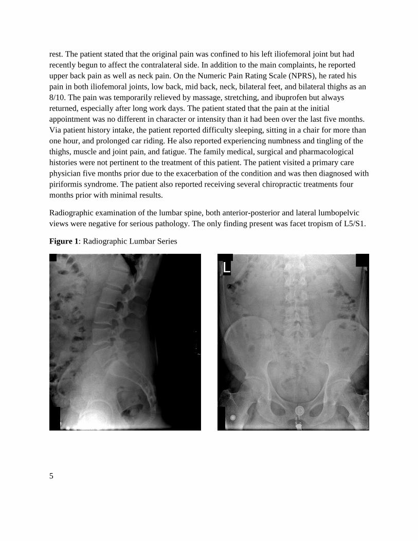
Radiographic examination of the lumbar spine, both anterior-posterior and lateral lumbopelvic
views were negative for serious pathology. The only finding present was facet tropism of L5/S1.
Figure 1: Radiographic Lumbar Series
Journal of the Academy of Chiropractic Orthopedists
Volume 15, Issue 1
6
The initial physical examination revealed a slightly elevated blood pressure of 134/99 mmHg
with a pulse of 76 beats per minute. Grip strength was recorded at 110 lbs. on the left and 105
lbs. on the right. Significant hypertonicity and spasms were noted at the left trapezius and levator
scapula, right thoracic erector spinae, and right gluteus medius and piriformis musculature.
Thoracolumbar range of motion was measured using dual inclinometers. Thoracolumbar flexion
was minimally decreased at 55 out of 60 degrees but provoked sharp left iliofemoral pain.
Thoracolumbar extension was decreased at 17 out of 25 degrees but did not cause pain or
discomfort at any location. Thoracolumbar right lateral flexion was decreased to 14 out of 25
degrees and caused sharp left iliofemoral pain. Thoracolumbar left rotation was within normal
limits of 30 degrees but also provoked the left iliofemoral joint pain. All other thoracolumbar
ranges of motion were non-provocative and were within normal limits. Restricted right sacro-
iliac fluid motion was noted upon examination with exquisite tenderness on palpation. Palpation
of the right lower posterior rib cage and right piriformis muscle provoked localized pain, as well.
Muscle testing of the hip flexors, hip abductors, hip adductors, and knee flexors was a 3/5 on the
right and 4/5 on the left. Muscle testing of the right lower extremity provoked right iliofemoral
pain but muscle testing of the left lower extremity did not.
Both patellar and achilles deep tendon reflexes were rated 2+ bilaterally. Nachlas test was
positive on the left for both thoracic and lumbar pain and negative on the right. The patient
experienced difficulty bilaterally with both heel and toe walking but pain only on the left.
Straight Leg Raise (SLR) was positive on the right with reproduction of low back and posterior
thigh pain radiating to the heel; SLR was negative on the left. Patrick Test was positive on the
left for iliofemoral pain and negative on the right. Prone Leg Extension Test was positive
bilaterally for imbalanced muscle activation and caused severe pain in the left sacro-iliac joint
while raising the left leg; however, the patient experienced greater difficulty elevating the right
leg (without pain). Valsalva maneuver was negative for pain and discomfort.
Following this examination, the patient was diagnosed with bilateral L2-L4 radiculopathy,
mechanical low back pain, sacro-iliac joint dysfunction, and muscle strain of the left hip flexor
musculature.
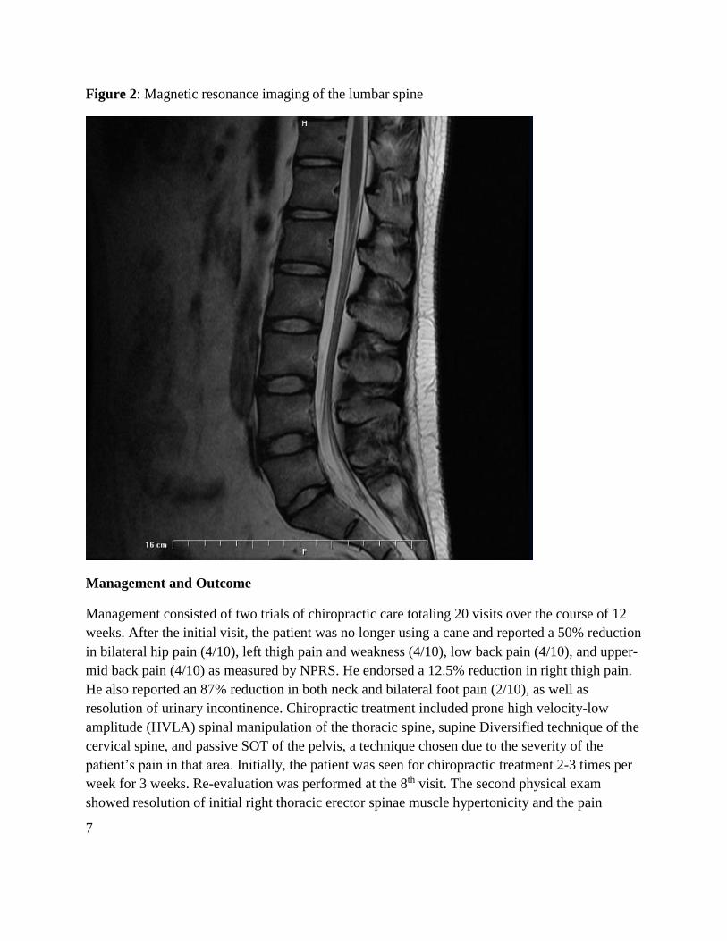
Patient was referred for MRI evaluation. The MRI was negative and revealed a normal lumbar
spine.
7
Figure 2: Magnetic resonance imaging of the lumbar spine
Management and Outcome
Management consisted of two trials of chiropractic care totaling 20 visits over the course of 12
weeks. After the initial visit, the patient was no longer using a cane and reported a 50% reduction
in bilateral hip pain (4/10), left thigh pain and weakness (4/10), low back pain (4/10), and upper-
mid back pain (4/10) as measured by NPRS. He endorsed a 12.5% reduction in right thigh pain.
He also reported an 87% reduction in both neck and bilateral foot pain (2/10), as well as
resolution of urinary incontinence. Chiropractic treatment included prone high velocity-low
amplitude (HVLA) spinal manipulation of the thoracic spine, supine Diversified technique of the
cervical spine, and passive SOT of the pelvis, a technique chosen due to the severity of the
patient’s pain in that area. Initially, the patient was seen for chiropractic treatment 2-3 times per
week for 3 weeks. Re-evaluation was performed at the 8th visit. The second physical exam
showed resolution of initial right thoracic erector spinae muscle hypertonicity and the pain
Journal of the Academy of Chiropractic Orthopedists
Volume 15, Issue 1
8
during palpation of the lower right posterior rib cage. The patient’s blood pressure was 138/98
mmHg with a pulse of 64 beats per minute. The following orthopedic examinations remained
positive for pain in the low back and/or hip area: Nachlas on the right, Ely on the right, Kemps
on the right, and SLR on the right causing radicular symptoms down the right thigh and leg.
SLR was positive on the left for low back pain only. The patient endorsed complete resolution of
left lower extremity pain, left hip pain, and left lower extremity weakness. He rated his right
lower extremity pain at 6/10, low back pain at 3-4/10, right hip pain at 6/10, neck pain at 1/10,
and mid-back pain at 3/10 on the NPRS. The patient subjectively reported that standing and
working had become easier since initiation of care, and that his gait had improved. He also
reported feeling stronger, especially in the left leg, and endorsed better sleep.
At this point of care, a new chiropractic technique was chosen for the treatment of the low back
and pelvis due to the improved but plateaued results in the low back and iliofemoral joints. For
one visit, the HVLA drop table mechanism (Thompson technique) was utilized instead of SOT
for the pelvis with instrument assisted manipulation (Activator instrumentation) utilized in the
lumbar spine. The patient returned two days later having regressed to symptomatic baseline,
rating his pain at 8/10 on the NPRS and endorsing a recurrence of bilateral lower extremity
weakness. Patellar and achilles deep tendon reflexes were again performed and remained +2
bilaterally. The patient denied recurrence of urinary incontinence and denied any new issues with
bowel function. After two additional visits with application of SOT, the patient reported 3/10 on
NPRS in all areas and feeling “90% better.” Pelvic SOT was continued throughout the remainder
of the treatment plan. Upon resolution of the exacerbation, the patient was referred to an exercise
rehabilitation specialist in the same private practice clinic for therapy involving lumbo-pelvic
stabilizing exercises and stretching in addition to kinesiology taping.
Rehabilitation exercise sessions occurred once per week for 4 weeks followed by every other
week for 8 weeks and included both strengthening exercises and stretches. The first phase of
exercises included calf raises, donkey kicks, and standing hip abduction. The first phase of
stretches included modified child’s pose, runner’s calf and achilles stretch, standing quadruped
stretch, sitting figure 4 stretch, and the sitting hamstring stretch. After the second visit of exercise
rehabilitation and two sessions of kinesiology taping of the lower back, the patient reported
greater ease during exercises and less pressure on the low back during work hours. During phase
two of exercise rehabilitation, new exercises and stretches were introduced including the
cat/camel stretch, single leg bridges, ball squats, flutter kicks, air bicycles, crossed-leg piriformis
stretch, side lunges, supine hip flexor stretch, and the knee hug stretch.
For the remaining 6 weeks, the patient was seen once per week. During the last three visits, drop
table spinal manipulation of the left iliofemoral joint was also administered. By the end of this
phase of care, the reported NPRS scores were as follows: 0/10 in bilateral legs and feet, 2/10 in
bilateral iliofemoral joints, 1/10 in both the mid back and neck, and a 2/10 in the low back.
Thoracolumbar flexion was decreased to 50/60 degrees and no longer provoked pain in the left
9
iliofemoral joint but merely affected tight bilateral hamstring and calf muscles. Thoracolumbar
extension caused minor right iliofemoral pain and had increased to 19/25 degrees.
Thoracolumbar right lateral flexion greatly increased to 24/25 degrees and only produced mild
thigh soreness. Thoracolumbar left lateral flexion was recorded at 20/25 degrees and produced
mild leg soreness and slight low back pain. Thoracolumbar bilateral rotation produced no pain
and were both within normal limits at 30/30 degrees. The patient’s blood pressure decreased to
126/85 with a pulse of 60 beats per minute. Nachlas and Ely were negative bilaterally for pain.
The patient’s grip strength had increased to 125 lbs. on the left and 117 lbs. on the right. SLR
was positive bilaterally for slight low back pain but no longer caused radicular symptoms.
Kemps was positive bilaterally for minor low back pain. Prone leg extension was positive on the
left for suboptimal muscle activation, but the patient continued to struggle to elevate the right leg
more. Patellar and achilles deep tendon reflexes remained a +2 bilaterally. The patient reported
NPRS scores of 2-3/10 in all previous areas. He also stated that riding in a car and sitting had
also become better tolerated.
After his final visit of the second trial of care, he denied pain in any thoracolumbar range of
motion. As of the time of publication, this patient continues to be seen every 2-3 weeks, as
needed, for management of his low back and iliofemoral complaints, as well as his muscle
weakness. He continues to perform his home exercises and stretches approximately 3-4 times per
week while keeping a six-day work schedule where he is on his feet for 8 hours or more at a
time.
Journal of the Academy of Chiropractic Orthopedists
Volume 15, Issue 1
10
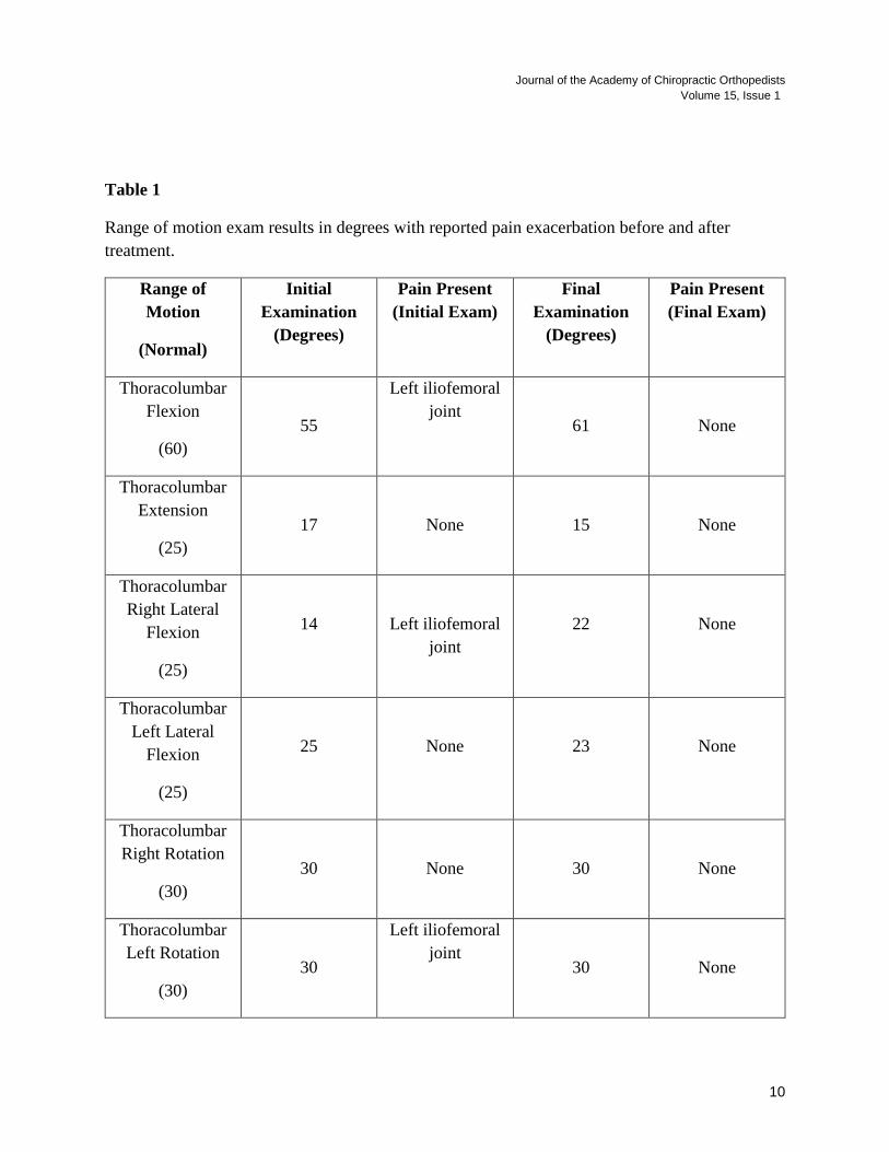
Table 1
Range of motion exam results in degrees with reported pain exacerbation before and after
treatment.
Range of
Motion
(Normal)
Initial
Examination
(Degrees)
Pain Present
(Initial Exam)
Final
Examination
(Degrees)
Pain Present
(Final Exam)
Thoracolumbar
Flexion
(60)
55
Left iliofemoral
joint
61
None
Thoracolumbar
Extension
(25)
17
None
15
None
Thoracolumbar
Right Lateral
Flexion
(25)
14
Left iliofemoral
joint
22
None
Thoracolumbar
Left Lateral
Flexion
(25)
25
None
23
None
Thoracolumbar
Right Rotation
(30)
30
None
30
None
Thoracolumbar
Left Rotation
(30)
30
Left iliofemoral
joint
30
None
11
Discussion
This study demonstrates success in the management of low back pain and iliofemoral pain with
concomitant bilateral lower extremity weakness. This course of treatment also resulted in
resolution of long-lasting urinary incontinence after the initial visit. Due to bilateral leg weakness
and urinary incontinence, imaging was necessary to rule out more serious pathology, such as a
space occupying lesion. The MRI revealed no contraindications to spinal manipulation.
The urinary tract is controlled by a complex neurological relationship involving sympathetic and
parasympathetic nerves of the autonomic nervous system.8 Nerves exiting from the
thoracolumbar region of the spinal cord are associated with the sympathetic function of the
urinary tract; parasympathetic and somatic innervation arise from the sacral segments of the
spinal cord.9 This well-established concept of neuroanatomy provides a valid hypothesis as to
how neurological interference or dysfunction at a particular spinal level could potentially cause
associated visceral symptoms in a patient. There is, however, very limited research currently on
the treatment of these symptoms with spinal manipulation.
Although this study produced many benefits for this patient, it is important to emphasize that
further research is necessary to determine the validity of the treatment rendered. Until a larger
body of evidence exists to suggest which chiropractic techniques should be provided for various
patient presentations of low back pain, the results of this study may not be generalized to the
overall population.
This case study suggests that not all chiropractic techniques are appropriate for all patients with
low back pain. Chiropractors must be diligent and thorough during initial examination to
determine the best course of treatment possible for each patient. Although other techniques, such
as side posture HVLA, have been more thoroughly researched, it is important to identify which
chiropractic techniques are more suitable for each patient being treated.
Limitations
Due to the uncontrolled nature of case report studies, this study had several limitations.
Causation may not be determined in this study as it could, in comparison, from a randomized
control trial. It is important to note that only NPRS, range of motion, and general patient
subjective measures were the main identifying factors in determining patient outcomes. More
objective measures, including a rated outcome assessment questionnaire such as the Back-
Bournemouth Questionnaire, would have generated more validity and objective results in this
study.
Journal of the Academy of Chiropractic Orthopedists
Volume 15, Issue 1
12
Consent
Written consent for this publication was obtained from the patient.
Competing Interests
The author declares no competing interests.
References
1. Clijsters M, Fronzoni F, Jenkins H. Chiropractic treatment approaches for spinal
musculoskeletal conditions: a cross-sectional survey. Chiropractic & Manual Therapies.
2014;22:33. doi:10.1186/s12998-014-0033-8.
2. Cooperstein, Robert et al. Chiropractic technique procedures for specific low back
conditions: Characterizing the literature. Journal of Manipulative & Physiological
Therapeutics, Volume 24, Issue 6, 407-424
3. Heese N. Sacro occipital technic block therapy: Origin and development.
Chiropr Hist. 2013 Winter;32(2):50-58
4. Hochman JI. The effect of sacro occipital technique category II blocking on spinal ranges
of motion: a case series. J Manipulative Physiol Ther. 2005 Nov-Dec;28(9):719-23.
5. Rubin Dl. Epidemiology and Risk Factors for Spine Pain. Neurol Clin. 2007;
May;25(2):353-71.
6. Qaseem A, Wilt TJ, McLean RM, Forciea MA, . Noninvasive Treatments for Acute,
Subacute, and Chronic Low Back Pain: A Clinical Practice Guideline From the American
College of Physicians. Ann Intern Med. 2017;166:514–530. doi: 10.7326/M16-2367
7. Hoy D, March L, Brooks P, et al. The global burden of low back pain: estimates from the
Global Burden of Disease 2010 study. Annals of the Rheumatic Diseases. Published
Online First: 24 March 2014. doi: 10.1136/annrheumdis-2013-204428
8. Fry CH, et al. In: Incontinence. Abrams P, Cardozo L, Khoury S, Wein A, editors. Health
Publications Ltd; Jersey: 2005. pp. 313–362.
9. Fowler CJ, Griffiths D, de Groat WC. The neural control of micturition. Nature reviews
Neuroscience. 2008;9(6):453-466. doi:10.1038/nrn2401.
13
Editorial Review
Hand-arm Vibration and the Risk of Vascular and Neurological
Diseases – A Systematic Review and Meta-analysis.
Tohr Nilsson, Jens Wahlstrom, Lage Burstrom
PLoS ONE 2017;12(7):e0180795 / https://doi.org/10.1371/journal.pone.0180795
Copyright: 2017 Nilsson et al. (Open access article)
JACO Editorial Reviewer: Steven G. Yeomans, DC, FACO
Published: March 2018
Journal of the Academy of Chiropractic Orthopedists
March 2018, Volume 15, Issue 1
The original article copyright belongs to the original publisher. This review is available from: http://www.dcorthoacademy.com
© 2018 Yeomans and the Academy of Chiropractic Orthopedists. This is an Open Access article which permits unrestricted use, distribution, and
reproduction in any medium, provided the original work is properly cited.
Author’s Abstract:
Background: Increased occurrence of Raynaud's phenomenon, neurosensory injury and carpal
tunnel syndrome has been reported for more than 100 years in association with work with
vibrating machines. The current risk prediction modelling (ISO-5349) for ªRaynaud's
phenomenonº is based on a few studies published 70 to 40 years ago. There are no corresponding
risk prediction models for neurosensory injury or carpal tunnel syndrome, nor any systematic
reviews comprising a statistical synthesis (meta-analysis) of the evidence.
Objectives: Our aim was to provide a systematic review of the literature on the association
between Raynaud's phenomenon, neurosensory injuries and carpal tunnel syndrome and hand-
arm vibration (HAV) exposure. Moreover the aim was to estimate the magnitude of such an
association using meta-analysis.
Methods: This systematic review covers the scientific literature up to January 2016. The
databases used for the literature search were PubMed and Science Direct. We found a total of
4,335 abstracts, which were read and whose validity was assessed according to pre-established
criteria. 294 articles were examined in their entirety to determine whether each article met the
inclusion criteria. The possible risk of bias was assessed for each article. 52 articles finally met
the pre-established criteria for inclusion in the systematic review.
Journal of the Academy of Chiropractic Orthopedists
Volume 15, Issue 1
14
Results: The results show that workers who are exposed to HAV have an increased risk of
vascular and neurological diseases compared to non-vibration exposed groups. The crude
estimate of the risk increase is approximately 4±5 fold. The estimated effect size (odds ratio) is
6.9 for the studies of Raynaud's phenomenon when including only the studies judged to have a
low risk of bias. The corresponding risk of neurosensory injury is 7.4 and the equivalent of
carpal tunnel syndrome is 2.9.
Conclusion: At equal exposures, neurosensory injury occurs with a 3-time factor shorter latency
than Raynaud's phenomenon. Which is why preventive measures should address this vibration
health hazard with greater attention.
Clinical Relevance:
JACO Editorial Summary:
• This article was written by authors from the Occupational and Environmental Medicine,
Department of Public Health & Clinical Medicine, Umea University, Umea, Sweden.
• The purpose was to reevaluate the association between Raynaud’s phenomenon,
neurosensory injury and carpal tunnel syndrome (CTS) by providing a systematic
literature review including risk of bias and, to estimate the risk exposure of vibratory tool
use based on meta-analysis.
• Relevant literature published from 1945-2016 resulted in 4335 abstracts, 294 articles,
with 52 making the final selection as only those including relative risk (odds ratio) were
included. This was further reduced to 24 articles included to complete the meta-analysis
portion of the study.
• Hand-arm-vibration syndrome (HAVS) is the internationally acknowledged condition
describing the symptom complex that occurs to the peripheral neurological, vascular and
musculoskeletal systems when prolonged, extensive power tool vibration exposure occurs
in manual work environments.
• The vascular component of HAVS includes vasospasm in the digital capillaries (causing
“white finger”/Raynaud’s Phenomenon).
• The neurological component includes both a diffuse peripheral neurosensory injury and
median nerve entrapment at the wrist (causing CTS)
• Skeletal injuries in HAVS include osteoarthritis and development of muscular
dysfunction (such as tendonopathies, tenosynovitis, fibrosis/Duputren’s contracture).
• The vascular and nerve manifestations in HAVS can occur together or separately.
• Workers who are exposed to HAV have a crude estimate of 4-5 fold increased risk of
developing vascular and/or neurological disease.
• The estimated effect size (odds ratio) of 6.9 for Raynaud’s phenomenon, 7.4 for
neurosensory injury, and 2.9 for CTS when utilizing only the low risk of bias studies (but
the number of CTS studies were few making the risk estimate for CTS less precise/more
sensitive to bias).
• Comparing and contrasting high vs. low exposure to vibration groups resulted in a pooled
risk estimate that varies between 2.5 and 5 for both Raynaud’s phenomenon and neuro-
sensory injury. There were too few studies found to calculate this for CTS.
15
• Possible outcome bias included shifts or changes in diagnostic approaches used in early
vs. more recent studies as early studies included collectively many “sub-categories” that
were reported in later studies (like large vs. small fibre neuropathy and entrapment
syndromes like CTS).
• Also, between 1945 and 2016, testing protocols changed (lab tests, electro-diagnostic
tests and various cold provocation tests are examples) as well as diagnostic precision.
This may be why earlier studies found more vascular injuries vs. a shift to more nerve
injuries found in the more recent studies.
• Further bias regarding the effect size was noted between the studies determined to have
low vs. high risk of bias. The corresponding effect size for Raynaud’s phenomenon was
6.8 vs. 3.6, and for neuro-sensory impairment 7.8 vs. 3.3, respectively.
• Possible exposure bias (how long the person was exposed to vibration) is an issue as well,
as differences in defining the degree of exposure was not consistent between the studies
reviewed.
• Risk of developing HAVS is also affected by co-morbidities (other concurrent health
conditions) as well as medication effects (such as BP meds). Interactions between
different diseases/comorbidities, medication effects as well as sleep, fitness and age-
related factors are currently lacking in these studies.
• With all the potential for bias described above, it remains clear that workers exposed to
vibration/HAV have an increased risk of developing vascular and neurological diseases
compared to non-vibration-exposed groups.
• At equal exposures, neurosensory injury occurs with a 3-time factor shorter latency than
Raynaud’s phenomenon.
• More preventive measures are needed to aggressively address the tool vibration health
hazard.
Summary:
This study describes the history of HAVS, compares and contrasts the early from the later studies
(1945-2016), reports odds ratios using both the studies with low risk of bias only vs. those that
have high risk of bias and, discuss in great detail the potential for other biases that should be
considered. This high level of transparency is commended and appreciated. In the end, it remains
clear that prevention must be aggressively addressed as it is key to minimizing the risk of
developing neurological and/or vascular injury caused by tool-induced vibration exposure.
Journal of the Academy of Chiropractic Orthopedists
Volume 15, Issue 1
16
Radiology Corner
Shoulder Pain in a Power Lifter
Alicia M. Yochum RN, DC, DACBR, RMSK
Published: March 2018 Journal of the Academy of Chiropractic Orthopedists
March 2018, Volume 15, Issue 1
This is an Open Access article which permits unrestricted use, distribution, and reproduction in any medium, provided the original
work is properly cited. The article copyright belongs to the author and the Academy of Chiropractic Orthopedists and is available at:
http://www.dcorthoacademy.com. © 2018 Yochum and the Academy of Chiropractic Orthopedists.
A 45 year old female competitive power lifter presented for chiropractic care of her left shoulder
pain. The pain was at the anterior superior shoulder and the patient did report the occasional
sensation of grinding.
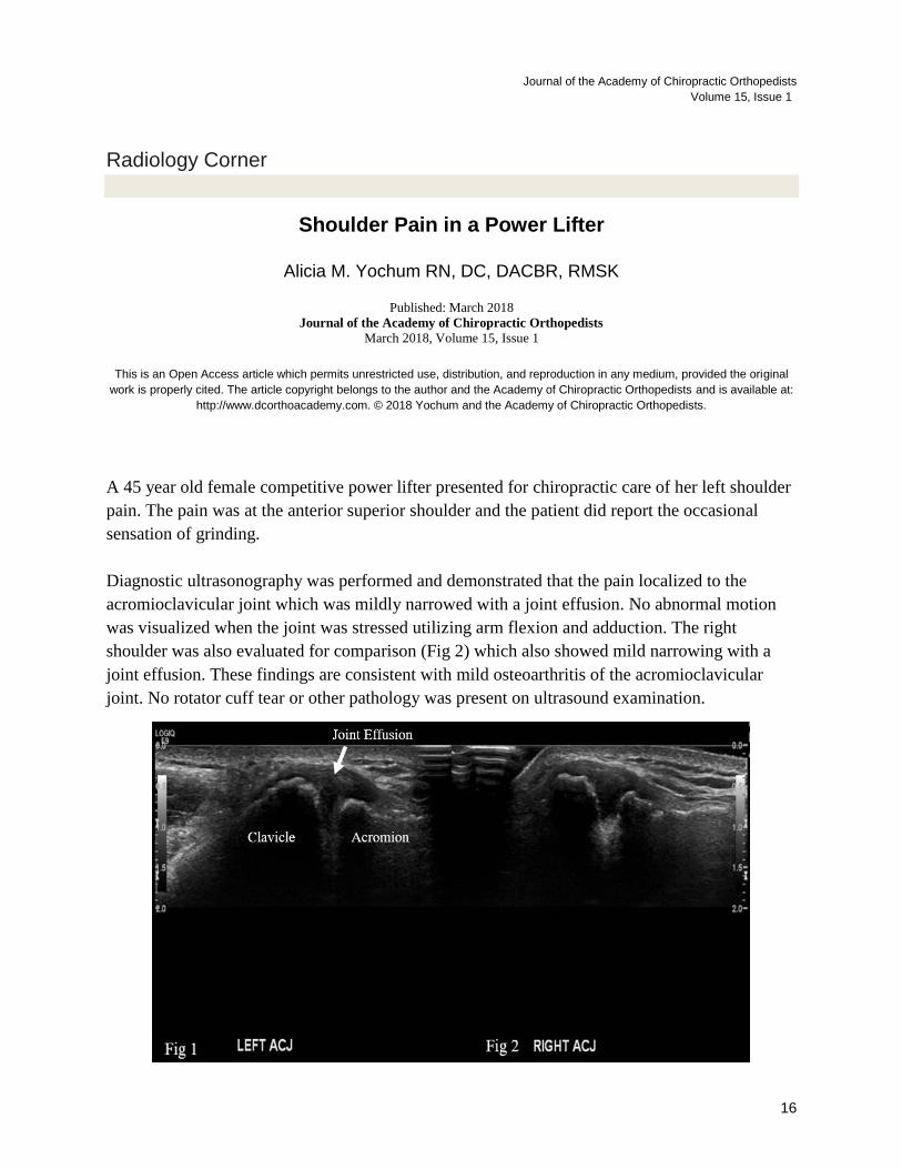
Diagnostic ultrasonography was performed and demonstrated that the pain localized to the
acromioclavicular joint which was mildly narrowed with a joint effusion. No abnormal motion
was visualized when the joint was stressed utilizing arm flexion and adduction. The right
shoulder was also evaluated for comparison (Fig 2) which also showed mild narrowing with a
joint effusion. These findings are consistent with mild osteoarthritis of the acromioclavicular
joint. No rotator cuff tear or other pathology was present on ultrasound examination.
17
Fig1 and 2: Long axis views of the acromioclavicular joint using diagnostic ultrasound
demonstrating a mildly narrowed joint with a joint effusion bilaterally.
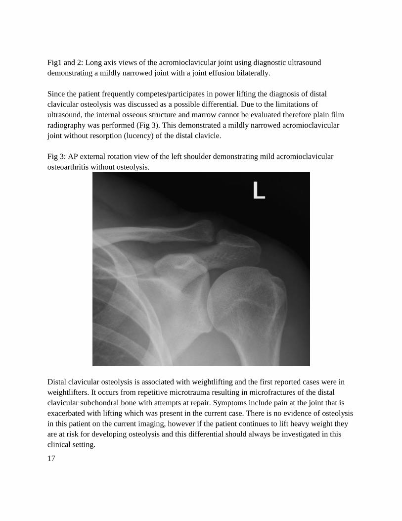
Since the patient frequently competes/participates in power lifting the diagnosis of distal
clavicular osteolysis was discussed as a possible differential. Due to the limitations of
ultrasound, the internal osseous structure and marrow cannot be evaluated therefore plain film
radiography was performed (Fig 3). This demonstrated a mildly narrowed acromioclavicular
joint without resorption (lucency) of the distal clavicle.
Fig 3: AP external rotation view of the left shoulder demonstrating mild acromioclavicular
osteoarthritis without osteolysis.
Distal clavicular osteolysis is associated with weightlifting and the first reported cases were in
weightlifters. It occurs from repetitive microtrauma resulting in microfractures of the distal
clavicular subchondral bone with attempts at repair. Symptoms include pain at the joint that is
exacerbated with lifting which was present in the current case. There is no evidence of osteolysis
in this patient on the current imaging, however if the patient continues to lift heavy weight they
are at risk for developing osteolysis and this differential should always be investigated in this
clinical setting.
Journal of the Academy of Chiropractic Orthopedists
Volume 15, Issue 1
18
Magnetic resonance imaging (MRI) would also be an imaging tool that could be utilized and
would show resorption of the clavicle cortex with bone marrow edema. Treatment includes
avoidance of provocative maneuvers, modification of weight training techniques such as a deep
bench press, ice, rest and rehab to improve joint stability.
References:
1. Schwartzkopf R. et. al. Distal clavicular osteolysis: a review of the literature. Bull of the
NYU hospital for Joint Diseases 2008; 66 (2): 94-101

19
Ortho Quiz
by Steven L. Kleinfield D.C., F.A.C.O.
1. Which statement regarding Talipes Equinovarus is incorrect:
a. The foot rotates inward and downward
b. The vast majority of these deformities are congenital in nature
c. This condition is bilateral about 50% of the time
d. Some patients may need to have their Achilles tendon lengthened
e. All of the above are correct
2. A flexion deformity of both the PIPJ and DIPJ in the toes is better known as:
a. Hammer Toe
b. Claw Toe
c. Charcot Marie Tooth Disease
d. Bunion
3. A flexion deformity of only the PIPJ with the DIPJ extended is better known as:
a. Hammer Toe
b. Claw Toe
c. Charcot Marie Tooth Disease
d. Bunion
4. Sesamoiditis in the foot has its pain typically felt where:
a. Base of Digit 1
b. Base of digit 2
c. Base of Digit 3
d. Base of Digit 4
e. Base of digit 5
5. Another name for a Bunion is:
a. Pump Bump
b. Hammer Toe
c. Hallux Valgus
d. Hallux Varus
Journal of the Academy of Chiropractic Orthopedists
Volume 15, Issue 1
20
Current Events
❖ The Part I online examination will be available for candidates to take on either Friday,
May 18th, or Saturday morning, May 19th, or Friday, July 20th, or Saturday morning
July 21st. Apply on the Academy website: http://dcorthoacademy.org/
❖ Apply for the Lipe Scholarship
Details at http://www.accoweb.org/lipescholarship.html
❖ The full hours of the following conventions have been accepted by the Academy as
qualifying for re-credentialing.
o American College of Chiropractic Orthopedists 2018 Annual Convention
19 Apr to 21 Apr 2018
Hilton Garden Inn Carlsbad Beach, Carlsbad CA
https://acoco.wildapricot.org/event-2436518
o CFS 2017 Annual Fall Convention
October 5-7, 2017
Chicago Marriott Oak Brook
1401 W 22nd Street | Oak Brook, IL 60523
630.573.8555 www.marriott.com/chiob
http://www.forensic-sciences.org/convention/
21
Answers to Ortho Quiz
1. Which statement regarding Talipes Equinovarus is incorrect:
e. All of the above are correct
http://www.footeducation.com/page/clubfoot-deformity-talipes-equinovarus
2. A flexion deformity of both the PIPJ and DIPJ in the toes is better known as:
b. Claw Toe
http://www.footeducation.com/page/claw-toes
3. A flexion deformity of only the PIPJ with the DIPJ extended is better known as:
a. Hammer Toe
http://www.footeducation.com/page/claw-toes
4. Sesamoiditis in the foot has its pain typically felt where:
a. Base of Digit 1
http://www.footeducation.com/page/sesamoiditis
5. Another name for a Bunion is:
c. Hallux Valgus
https://www.mayoclinic.org/diseases-conditions/bunions/symptoms-causes/syc-20354799


![Celula Urbana [Jaco]](https://img.pdfslide.net/doc/110x75/568c491d1a28ab491692e8fb/celula-urbana-jaco.jpg)