Embed Size (px)
Citation preview

FSH and Bone 2010: Evolving Evidence
Jameel Iqbal, Li Sun, and Mone Zaidi*
The Mount Sinai Bone Program,
Mount Sinai School of Medicine, New York, NY
*Correspondence: Mone Zaidi, MD, PhD
Tel: 212-241-8798; fax: 212-426-8312
Page 1 of 13 Accepted Preprint first posted on 6 May 2010 as Manuscript EJE-10-0397
Copyright © 2010 European Society of Endocrinology.

Abstract
Bone loss due to menopause, natural or artificial, has been attributed solely to low estrogen.
However, in a woman’s life, the most precipitous bone loss begins two years prior to the last
menstrual period, during which time estrogen levels are unperturbed whereas FSH is elevated.
Our cell-based and mouse genetic studies have shown that FSH stimulates bone resorption by
osteoclasts directly in a pituitary-bone axis, independently of the estrogen effect. On the basis of
this and evolving clinical and scientific evidence, we propose that elevated FSH contributes to
bone loss across the menopausal transition, particularly during late perimenopause. Redina and
coworkers strengthen the view for a primary role of FSH signaling in the regulation of bone mass
and bone remodeling in humans by demonstrating that an ‘activating’ polymorphism AA rs6166
causes low bone mass and high bone turnover.
Page 2 of 13

Osteoporosis is a devastating and relentless disease, resulting in a fracture in one out of
every two individuals over the age of 50. What causes this increased bone fragility and resulting
susceptibility to hip, spine and wrist fractures? In the early 1700’s the British surgeon John
Hunter hypothesized that bone undergoes remodeling—that is, bone is forever changing itself by
reorganizing into a newer, stronger configuration to resist ongoing stresses, such as physical
activity.1 Bone is lost when the cells degrading old bone, osteoclasts, outpace the cells re-laying
new collagen and mineral, the osteoblasts.
What would cause the osteoclasts to degrade bone at a higher rate than the body’s ability
to re-build bone? During the mid 1900’s, Fuller Albright at the Massachusetts General Hospital
noted that the most dramatic bone loss occurred after menopause, either natural or surgically
induced.2 Thus, he hypothesized a link between the loss of sex steroids and osteoporosis, a
corpus of observations that led to estrogen hormone replacement therapy becoming the first
successful treatment for osteoporosis.
Within the past decade, hormone replacement therapy has fallen out of favor because of
associated cardiovascular risks.3 Fortunately, the plot of bone density loss versus age in both
genders has been re-examined and certain key observations made. It was noted that women have
a precipitous drop in bone mass during the menopausal transition. Notably, bone loss is
profoundly accelerated in the late perimenopause.4, 5
In fact, nearly half of life time bone loss
occurs within the first five years of menopause; this loss of bone mass is associated with
microstructural degradation ultimately predisposing to fractures.6
Surprising, however, during this phase of rapid, late perimenopausal bone loss, estrogen
levels are normal. Thus, a conundrum: could the hypothesis – the loss of estrogen is the sole
Page 3 of 13

cause of osteoporosis – be wrong? There is clear genetic and pharmacological evidence for a
protective role of estrogen on the skeleton.7 In vitro studies show that estrogen directly inhibits
osteoclast formation.8, 9
Estrogen also acts on osteoblasts to indirectly inhibit osteoclast
formation by decreasing the secretion of the pro-osteoclastic cytokine RANK-L and increasing
the decoy receptor for RANK-L, osteoprotegerin (OPG).10
Indirect effects of estrogen on
osteoclast formation also occur through T-lymphocytes, where estrogen inhibits the production
of another pro-osteoclastic cytokine TNF-α.11-13
In addition to its inhibitory effect on osteoclast
formation, estrogen has established anabolic actions mediated via osteoblasts14
.
If estrogen protects the skeleton, why then is there striking bone loss occurring in the late
perimenopause if estrogen levels during this period are unperturbed? The answer to this question
was ultimately inspired by our earlier studies on hyperthyroidism. In 2003, we discovered that
the pituitary-derived thyroid-stimulating hormone (TSH) could bypass its primary endocrine
target, the thyroid, and act directly on osteoclasts and osteoblasts to modulate bone turnover.15
The results of those studies had profound implications for the bone loss associated with
subclinical hyperthyroidism in which thyroid hormones are normal and TSH is low. For the first
time, we could explain why bone loss occurred in these patients given their normal thyroid
hormone levels—low TSH appeared directly to cause their bone loss.16
Using a similar line of argument, we hypothesized in 2006 that changes in the circulating
levels of the pituitary-derived follicle-stimulating hormone (FSH) may contribute to the late peri-
menopausal bone loss when estrogen levels are unperturbed. This hypothesis was then bolstered
by several clinical studies on the menopausal transition, where bone loss was found to correlate
with dramatic increases in FSH. The Study of Women’s Health Across the Nations (SWAN), a
longitudinal, cross-sectional study of 2375 peri-menopausal women found a strong correlation of
Page 4 of 13

FSH levels to markers of bone degradation,17
and demonstrated that changes in the levels of FSH
over 4 years could predict decreases in bone mass.5 Similarly, Xu et al. found a significant
association between the incidence of osteoporosis and high serum FSH levels in a group of 689
native Chinese women.18
Likewise, Sowers et al. show that spine and femoral neck bone loss
accelerates in women between 47.6 and 51 years, i.e. during FSH stage 3 (34 to 54 mIU/mL),
which corresponds to 2 years prior to the final menstrual period.19
These strong correlations now
help to clinically stratify women at a high risk of bone loss using serum FSH.20
Endowed with the knowledge that FSH levels correlated strongly with bone loss during
late perimenopause, we investigated whether FSH could directly stimulate bone degradation by
osteoclasts. We found that FSH augmented the formation, function and survival of both human
and mouse osteoclasts.21
By activating the osteoclast FSH receptor, FSH triggered several of the
signaling pathways used by RANK-L to transduce its pro-resorptive effects.21, 22
Moreover,
others and we found that FSH can indirectly stimulate osteoclast formation by enhancing the
production of several pro-osteoclastogenic cytokines, TNFα, IL-6, and IL-1β.23, 24
Recently,
Pacifici and co-workers have shown that FSH increases CD40-ligand expression on T
lymphocytes, thereby triggering increased TNFα production.
Using genetically modified mice, we demonstrated a direct effect of FSH on the skeleton.
Notably, mice lacking the β subunit of FSH or the FSHR were protected from bone loss
associated with estrogen deficiency, although these mice have a compensatory rise in serum
androgens accounting for some of the skeletal phenotype.21
Importantly, however,
haploinsufficient FSHβ heterozygotes (animals having a 50% reduction in circulating FSH
levels, but with normal estrogen levels) displayed increased bone mass.21
This latter finding
Page 5 of 13

showed that, even in situations of normal estrogen, FSH was acting independently to decrease
skeletal mass.21
We can thus extrapolate that the bone loss occurring in the perimenopausal
period partly arises from elevated FSH levels.3
Three studies take us further towards establishing a cause-effect relationship between
FSH and bone loss in vivo. Firstly, amenorrheic women with similar estrogen levels having a
mean serum FSH of 35 mIU/mL had greater bone loss than those with a level of 8 mIU/mL.25
Second, exogenously administered FSH enhanced ovariectomy-induced alveolar bone loss in
rats.26, 27
The bone loss post-ovariectomy, as well as that induced by exogenous FSH, was
significantly reduced by an FSH antagonist, providing unequivocal evidence for a direct effect of
FSH on bone in vivo.26, 27
While the evidence for causality between high FSH and bone loss continues to expand,
several lingering questions remain. Osteoporosis is a multifactorial disorder with genetic
variation accounting for up to 80% variation in bone mineral density.28
Rendina et al. sought to
tie the role of FSH in causing osteoporosis to the genetic variability that exists among the human
population.29
To do so, they examined 289 postmenopausal women for FSHR polymorphisms at
two sites, rs1394205 and rs6166, and then analyzed the influence these polymorphisms had on
bone mass and bone turnover.29
The results were impressive: the authors found that the SNP rs6166 of the FSH receptor
gene significantly influenced bone mass in postmenopausal women.29
Prior studies on AA
rs6166 have associated this polymorphism with increased stimulation of the ovarian FSHR.
Based on knowledge that FSH acts to decrease bone mass, one would anticipate that women
bearing an ‘activating’ FSHR polymorphism will have lower bone density. That was exactly the
Page 6 of 13
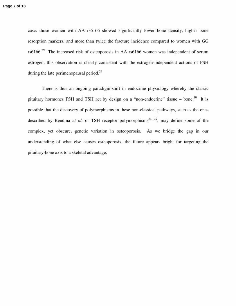
case: those women with AA rs6166 showed significantly lower bone density, higher bone
resorption markers, and more than twice the fracture incidence compared to women with GG
rs6166.29
The increased risk of osteoporosis in AA rs6166 women was independent of serum
estrogen; this observation is clearly consistent with the estrogen-independent actions of FSH
during the late perimenopausal period.29
There is thus an ongoing paradigm-shift in endocrine physiology whereby the classic
pituitary hormones FSH and TSH act by design on a “non-endocrine” tissue – bone.30
It is
possible that the discovery of polymorphisms in these non-classical pathways, such as the ones
described by Rendina et al. or TSH receptor polymorphisms31, 32
, may define some of the
complex, yet obscure, genetic variation in osteoporosis. As we bridge the gap in our
understanding of what else causes osteoporosis, the future appears bright for targeting the
pituitary-bone axis to a skeletal advantage.
Page 7 of 13

Acknowledgements
M.Z. and L.S. are supported by grants from the National Institutes of Health. J.I. acknowledges
the support of the American Federation for Aging Research.
Disclosures
M.Z. consults for Genentech, Amgen, and Warner Chilcott. M.Z. is also a named inventor of a
pending patent application related to osteoclastic bone resorption filed by the Mount Sinai
School of Medicine (MSSM). In the event the pending or issued patent is licensed, he would be
entitled to a share of any proceeds MSSM receives from the licensee. J.I. and S.L. have nothing
to disclose.
Page 8 of 13

References:
1. Evans CH. John Hunter and the origins of modern orthopaedic research. J Orthop Res
2007 25 556-560.
2. Reifenstein EC, Jr. & Albright F. The metabolic effects of steroid hormones in
osteoporosis. J Clin Invest 1947 26 24-56.
3. Iqbal J & Zaidi M. Understanding estrogen action during menopause. Endocrinology
2009 150 3443-3445.
4. Randolph JF, Jr., Sowers M, Bondarenko IV, Harlow SD, Luborsky JL & Little RJ.
Change in estradiol and follicle-stimulating hormone across the early menopausal
transition: effects of ethnicity and age. J Clin Endocrinol Metab 2004 89 1555-1561.
5. Sowers MR, Jannausch M, McConnell D, Little R, Greendale GA, Finkelstein JS, Neer
RM, Johnston J & Ettinger B. Hormone predictors of bone mineral density changes
during the menopausal transition. J Clin Endocrinol Metab 2006 91 1261-1267.
6. Akhter MP, Lappe JM, Davies KM & Recker RR. Transmenopausal changes in the
trabecular bone structure. Bone 2007 41 111-116.
7. Nakamura T, Imai Y, Matsumoto T, Sato S, Takeuchi K, Igarashi K, Harada Y, Azuma
Y, Krust A, Yamamoto Y, Nishina H, Takeda S, Takayanagi H, Metzger D, Kanno J,
Takaoka K, Martin TJ, Chambon P & Kato S. Estrogen prevents bone loss via estrogen
receptor alpha and induction of Fas ligand in osteoclasts. Cell 2007 130 811-823.
8. Shevde NK, Bendixen AC, Dienger KM & Pike JW. Estrogens suppress RANK ligand-
induced osteoclast differentiation via a stromal cell independent mechanism involving c-
Jun repression. Proc Natl Acad Sci U S A 2000 97 7829-7834.
9. Srivastava S, Toraldo G, Weitzmann MN, Cenci S, Ross FP & Pacifici R. Estrogen
decreases osteoclast formation by down-regulating receptor activator of NF-kappa B
ligand (RANKL)-induced JNK activation. J Biol Chem 2001 276 8836-8840.
10. Srivastava S, Weitzmann MN, Kimble RB, Rizzo M, Zahner M, Milbrandt J, Ross FP &
Pacifici R. Estrogen blocks M-CSF gene expression and osteoclast formation by
regulating phosphorylation of Egr-1 and its interaction with Sp-1. J Clin Invest 1998 102
1850-1859.
11. Armour KE, Armour KJ, Gallagher ME, Godecke A, Helfrich MH, Reid DM & Ralston
SH. Defective bone formation and anabolic response to exogenous estrogen in mice with
targeted disruption of endothelial nitric oxide synthase. Endocrinology 2001 142 760-
766.
12. Garcia Palacios V, Robinson LJ, Borysenko CW, Lehmann T, Kalla SE & Blair HC.
Negative regulation of RANKL-induced osteoclastic differentiation in RAW264.7 Cells
by estrogen and phytoestrogens. J Biol Chem 2005 280 13720-13727.
13. Roggia C, Gao Y, Cenci S, Weitzmann MN, Toraldo G, Isaia G & Pacifici R. Up-
regulation of TNF-producing T cells in the bone marrow: a key mechanism by which
estrogen deficiency induces bone loss in vivo. Proc Natl Acad Sci U S A 2001 98 13960-
13965.
14. Jagger CJ, Chow JW & Chambers TJ. Estrogen suppresses activation but enhances
formation phase of osteogenic response to mechanical stimulation in rat bone. J Clin
Invest 1996 98 2351-2357.
Page 9 of 13

15. Abe E, Marians RC, Yu W, Wu XB, Ando T, Li Y, Iqbal J, Eldeiry L, Rajendren G, Blair
HC, Davies TF & Zaidi M. TSH is a negative regulator of skeletal remodeling. Cell 2003
115 151-162.
16. Abe E, Sun L, Mechanick J, Iqbal J, Yamoah K, Baliram R, Arabi A, Moonga BS,
Davies TF & Zaidi M. Bone loss in thyroid disease: role of low TSH and high thyroid
hormone. Ann N Y Acad Sci 2007 1116 383-391.
17. Sowers MR, Greendale GA, Bondarenko I, Finkelstein JS, Cauley JA, Neer RM &
Ettinger B. Endogenous hormones and bone turnover markers in pre- and perimenopausal
women: SWAN. Osteoporos Int 2003 14 191-197.
18. Xu ZR, Wang AH, Wu XP, Zhang H, Sheng ZF, Wu XY, Xie H, Luo XH & Liao EY.
Relationship of age-related concentrations of serum FSH and LH with bone mineral
density, prevalence of osteoporosis in native Chinese women. Clin Chim Acta 2009 400
8-13.
19. Sowers MR, Zheng H, Jannausch ML, McConnell D, Nan B, Harlow S & Randolph JF,
Jr. Amount of Bone Loss in Relation to Time around the Final Menstrual Period and
Follicle-Stimulating Hormone Staging of the Transmenopause. J Clin Endocrinol Metab.
20. Zaidi M, Turner CH, Canalis E, Pacifici R, Sun L, Iqbal J, Guo XE, Silverman S, Epstein
S & Rosen CJ. Bone loss or lost bone: rationale and recommendations for the diagnosis
and treatment of early postmenopausal bone loss. Curr Osteoporos Rep 2009 7 118-126.
21. Sun L, Peng Y, Sharrow AC, Iqbal J, Zhang Z, Papachristou DJ, Zaidi S, Zhu LL,
Yaroslavskiy BB, Zhou H, Zallone A, Sairam MR, Kumar TR, Bo W, Braun J, Cardoso-
Landa L, Schaffler MB, Moonga BS, Blair HC & Zaidi M. FSH directly regulates bone
mass. Cell 2006 125 247-260.
22. Wu Y, Torchia J, Yao W, Lane NE, Lanier LL, Nakamura MC & Humphrey MB. Bone
microenvironment specific roles of ITAM adapter signaling during bone remodeling
induced by acute estrogen-deficiency. PLoS One 2007 2 e586.
23. Iqbal J, Sun L, Kumar TR, Blair HC & Zaidi M. Follicle-stimulating hormone stimulates
TNF production from immune cells to enhance osteoblast and osteoclast formation. Proc
Natl Acad Sci U S A 2006 103 14925-14930.
24. Cannon JG, Cortez-Cooper M, Meaders E, Stallings J, Haddow S, Kraj B, Sloan G &
Mulloy A. Follicle-stimulating hormone, interleukin-1, and bone density in adult women.
Am J Physiol Regul Integr Comp Physiol 298 R790-798.
25. Devleta B, Adem B & Senada S. Hypergonadotropic amenorrhea and bone density: new
approach to an old problem. J Bone Miner Metab 2004 22 360-364.
26. Liu S, Cheng Y, Fan M, Chen D & Bian Z. FSH aggravates periodontitis-related bone
loss in ovariectomized rats. J Dent Res 89 366-371.
27. Liu S, Cheng Y, Xu W & Bian Z. Protective effects of follicle-stimulating hormone
inhibitor on alveolar bone loss resulting from experimental periapical lesions in
ovariectomized rats. J Endod 36 658-663.
28. Ralston SH. Genetics of osteoporosis. Ann N Y Acad Sci 1192 181-189.
29. Rendina D, Gianfrancesco F, De Filippo G, Merlotti D, Esposito T, Mingione A, Nuti R,
Strazzullo P, Mossetti G & Gennari L. FSHR gene polymorphisms influence bone
mineral density and bone turnover in postmenopausal women. Eur J Endocrinol.
30. Iqbal J, Davies TF, Sun L, Abe E, Carpi A, Mechanick JI & Zaidi M. Skeletal
morphofunctional considerations and the pituitary-thyroid axis. Front Biosci (Schol Ed)
2009 1 92-107.
Page 10 of 13

31. van der Deure WM, Uitterlinden AG, Hofman A, Rivadeneira F, Pols HA, Peeters RP &
Visser TJ. Effects of serum TSH and FT4 levels and the TSHR-Asp727Glu
polymorphism on bone: the Rotterdam Study. Clin Endocrinol (Oxf) 2008 68 175-181.
32. Heemstra KA, van der Deure WM, Peeters RP, Hamdy NA, Stokkel MP, Corssmit EP,
Romijn JA, Visser TJ & Smit JW. Thyroid hormone independent associations between
serum TSH levels and indicators of bone turnover in cured patients with differentiated
thyroid carcinoma. Eur J Endocrinol 2008 159 69-76.
Page 11 of 13

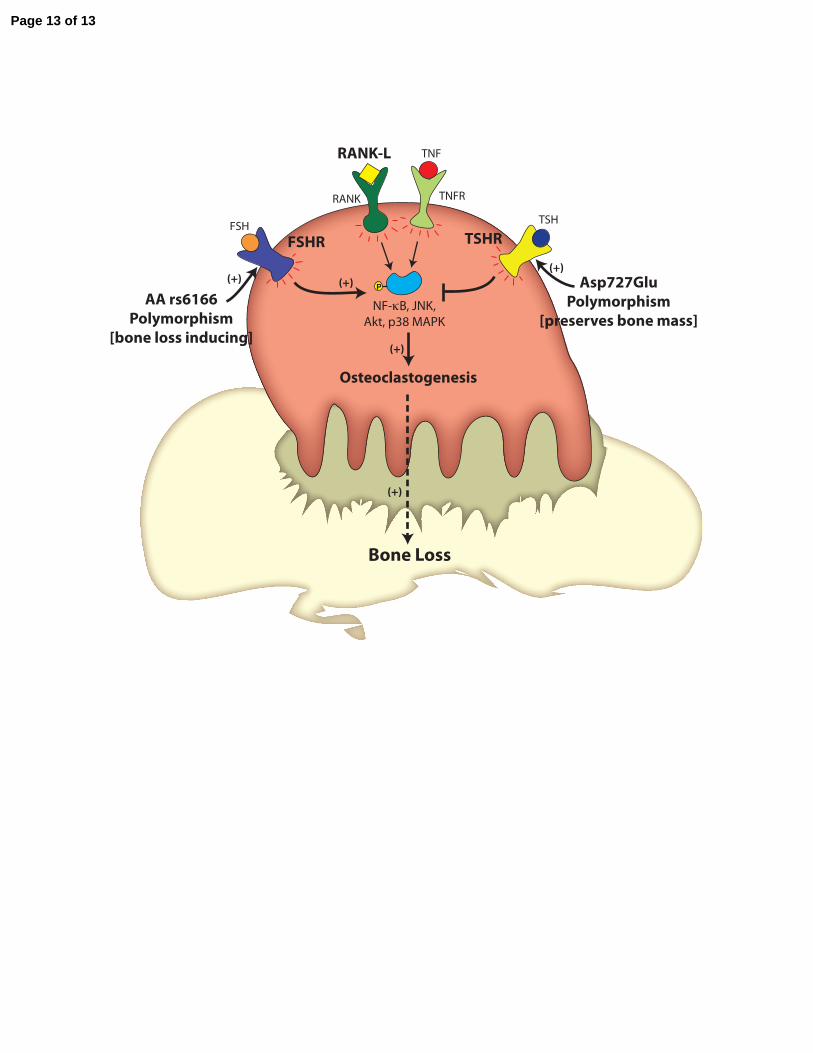
Figure Legend:
Figure 1: Polymorphisms in the Pituitary-Bone Axis Regulate Bone Mass and Bone
Turnover. The two anterior pituitary hormones FSH and TSH regulate osteoclastogenesis and
bone resorption reciprocally. “Activating” polymorphisms in the FSH receptor (AA rs6166) and
the TSH receptor (Asp727Glu) are therefore associated with low and high bone mass,
respectively.
Page 12 of 13
P
NF-κB, JNK,Akt, p38 MAPK
Osteoclastogenesis
RANK TNFR
RANK-L TNF
FSHR TSHRFSH TSH
(+)
Bone Loss
AA rs6166Polymorphism
[bone loss inducing]
Asp727GluPolymorphism
[preserves bone mass]
(+)(+)
(+)
(+)
Page 13 of 13