Embed Size (px)
Citation preview

Janica E. Walden, MDNeuroradiology
University of North Carolina

Holoprosencephaly (HPE)Spectrum of congenital structural forebrain
anomalies defined by different degrees of frontal lobe fusion
Impaired midline cleavage of the embryonic forebrain
“Face predicts brain”: severe midline anomaly = severe HPE
Clinical severity relates to degree of hemispheric and deep gray nuclei fusion

Etiology & PathologyNormal prosencephalic cleavage occurs at 4-6 weeksHPE: disruption in dorsoventral axis patterning of
secondary prosencephalon,Result of mutations affecting signaling genes (Sonic
hedgehog gene) which regulate neural tube patterning.Extreme hypoplasia of neocortexDorsal cyst (especially in association with non-
cleaved thalami) thought to represent expansion of partially blocked posterodorsal 3rd ventricle
Variable degree of fusion of diencephalon & basal ganglia/thalamus with incorporation into upper brainstem
EpidemiologyOccurs in 1 to 1.4 per 10,000 live births
More common in early embryogenesis with high spontaneous miscarriage rates
Maternal factors include alcohol use, diabetes, retinoic acid1% risk to infants of diabetic mothers (200-fold
increased risk than that of general population)Male: female ratio = 1.4: 1

Facial AnomaliesSevere facial anomalies correlate with severity
of HPE in 80%+/- midline cleftingpremaxillary agenesis if severeabsent superior frenulum+/- central incisorproboscissingle nare; single nasal bone/absent inter-nasal
suturescaudal metopic sutureinfants of diabetic mothers may have alobar HPE
with near-normal facies
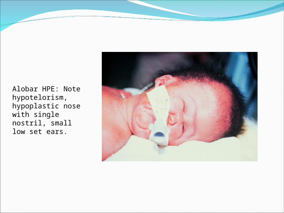
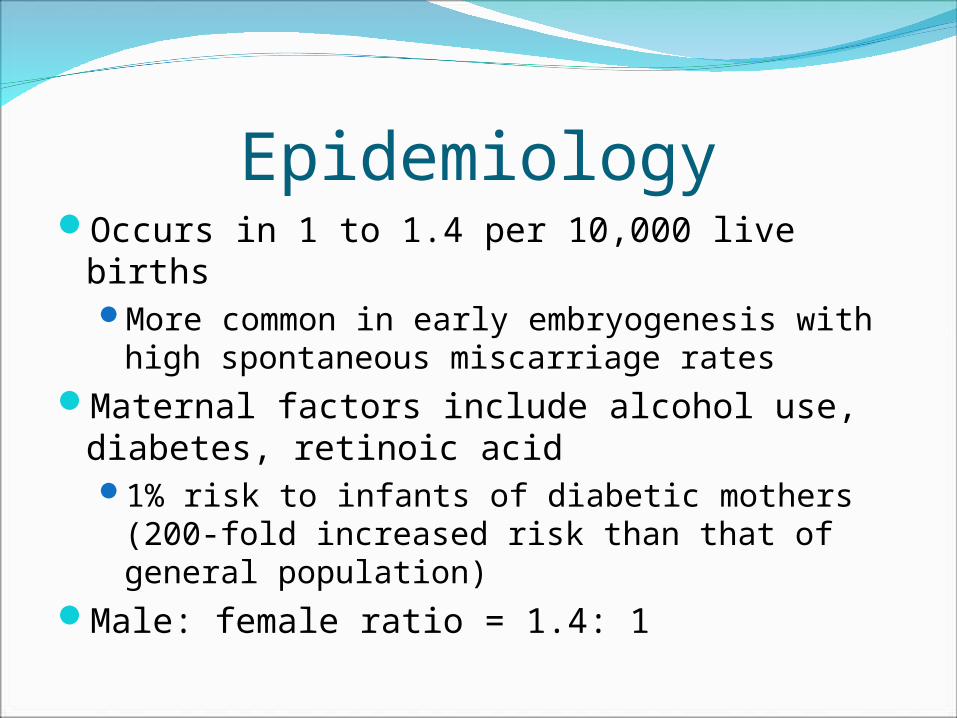
Alobar HPE: Note hypotelorism, hypoplastic nose with single nostril, small low set ears.

Clinical FeaturesMost severe (classic alobar HPE) features include:
cyclopia, proboscis, midline facial clefting, microcephaly
Severe of pituitary/hypothalamic dysfunction (75% especially diabetes insipidus) & disturbed body temperature regulationCorrelates with degree of hypothalamic non-separation
Seizures (50%) & mental retardation Most severe with cortical malformations
Dystonia & hypotoniaSeverity correlates with degree basal ganglia non-
separation

ClassificationDefined by degree of frontal lobe fusionSylvian angle (of Barkovich) = lines drawn
tangentially through Sylvian fissuresAnteriorly displaced Sylvian fissures results in
increased Sylvian angleThe larger the Sylvian angle is the more severe
frontal lobe hypoplasia is too3 types of HPE based on criteria (lobar,
semilobar, and alobar), as well as a middle interhemispheric variant, septooptic dysplasia, and single central incisor

Alobar Holoprosencephaly“Pancake” or “horseshoe” brainMonoventricleLarge dorsal cystFused diencephalonBasal ganglia & thalami may form gray
matter fusion massNo interhemispheric fissureNo olfactory nerves

Alobar HPE: note fused thalamic & hemispheres, monoventricle, absent interhemispheric fissure and venous sinsues, & azygous ACA.

Fetal MRI shows alobar HPE.

MR T1 images in alobar HPE.

Diagnosis of HPE by UltrasoundDiagnosis of HPE by ultrasound can be made
as early as 9 weeks gestational age.Development of forebrain can be delineated
in detail with ultrasound from 7 weeks on.Alobar HPE may be detectable as early as the
end of week 7Non-visualization of the butterfly sign is very
helpful in diagnosis

Semilobar HoloprosencephalyPartial occipital/temporal hornsModerate sized dorsal cystFused diencephalonPartial fusion of basal ganglia > thalamiInterhemipheric fissure present posteriorlyAbsent of hypoplastic olfactory tracts and
bulbsCorpus callosum is rudimentary

CT in semilobar HPE.

MRI in semilobar HPE.

Lobar HoloprosencephalyFormed lateral ventriclesSmall or no dorsal cystFused diencephalon and/or fornices+/- partial fusion of basal ganglia > thalamiInterhemispheric fissure nearly normalSmall or normal olfactory nerves

MRI in lobar HPE.
Middle Interhemispheric VariantSylvian fissures connect across midline over vertex
(86%)Interhemispheric fusion of posterior frontal/parietal
lobes, with normal separation of anterior frontal/occipital lobes
Non-cleavage of thalami > basal gangliaHeterotopias and cortical dysplasias common (86%)Thought to reflect abnormal induction of embryonic roof
plate Classic HPE = abnormal induction of embryonic floor plateMay explain absence of craniofacial malformations
Spasticity, hypotonia, seizures, developmental delay

MRI in midline intehemispheris variant of HPE.

ReferencesSepulveda Waldo, Dezerega Victor, Be Cecilia. First-
Trimester Sonographic Diagnosis of Holoprosencephaly. Journal of Ultrasound in Medicine 23: 761-765.
Hahn Jin, Barnes Patrick. Neuroimaging Advances in Holoprosencephaly: Refining the Spectrum of the Midline Malformation. American Journal of Medical Genetics 154C: 120-132.
Blaas H., Eriksson A., Salvesen K., et al. Brains and faces in holoprosencephaly: pre- and postnatal description in 30 cases. Ultrasound Obstet Gynecol 2002; 19: 24-38.
Takanashi Jun-ichi, Barkovich A. James, Clegg Nancy, Delgado Mauricio. Middle Interhemispheric Bariant of Holoprosencephaly Associated with Diffuse Polymicrogyria. AJNR 2003; 24: 394-397.
Simon Erin, Hevner Robert, Pinter Joseph, et al. The Middle Interhemispheric Variant of Holoprosencephaly. AJNR 2002; 23: 151-155.