Embed Size (px)
Citation preview

Jittipan Chavadej, Ph. D. Anatomy Department,Fac. of Science
Mahidol University
yr.2000
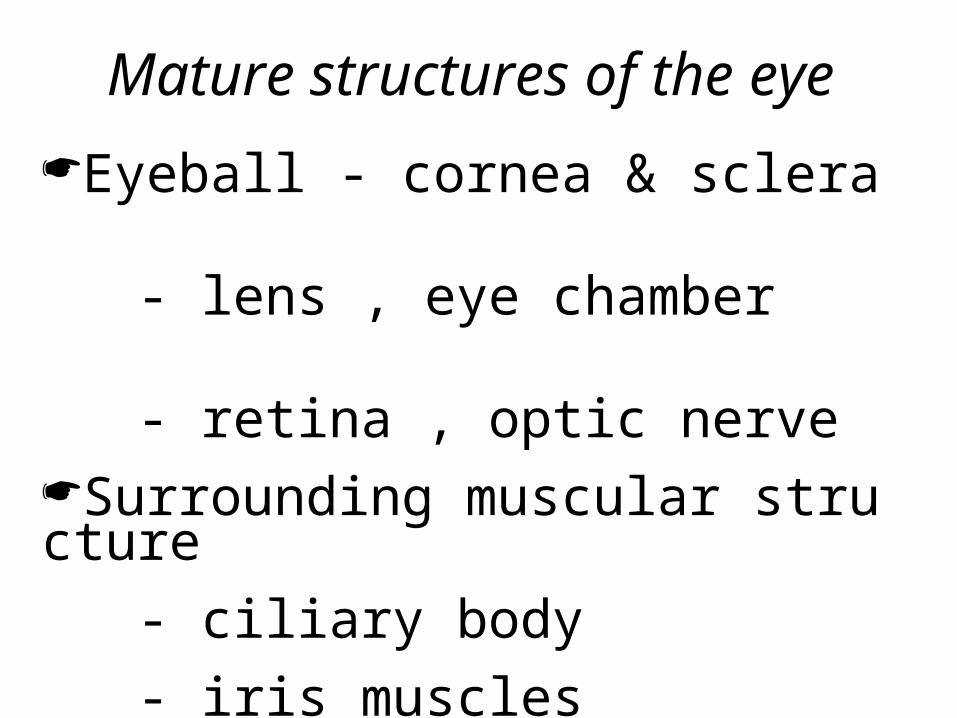
Mature structures of the eye
- Eyeball cornea & sclera
- lens , eye chamber
- retina , optic nerve Surrounding muscular structure
- ciliary body
- iris muscles
- extraocular muscles&conjunctiva
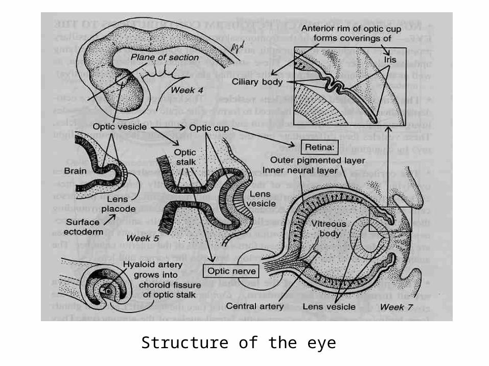
Structure of the eye
Formation of the eye
Are formed by 3 different germ layers.
1 . Neuroectoderm
2 . Surface ectoderm
3. Mesoderm
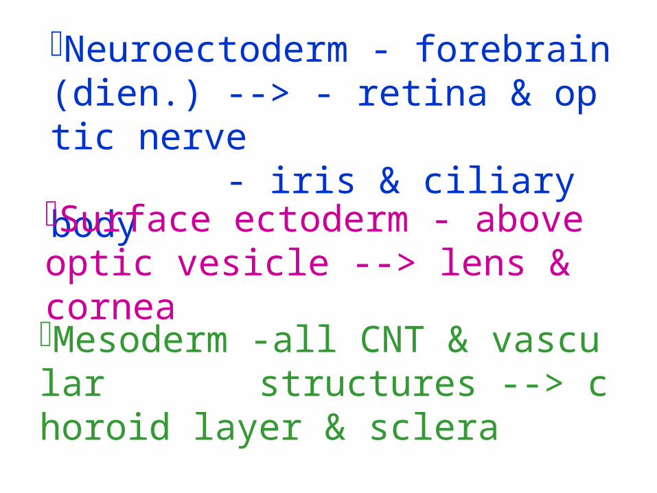
- Neuroectoderm forebrain(di -- - en.) > retina & optic nerve
- iris & ciliary body
Surface ectoderm - above optic vesicle --> lens & cornea
- Mesoderm all CNT & vascular -- structures > choroid layer
& sclera

Retina & Optic nerve form from optic vesicle
Diagram showing the formation of optic vesicle & the le
ns placode
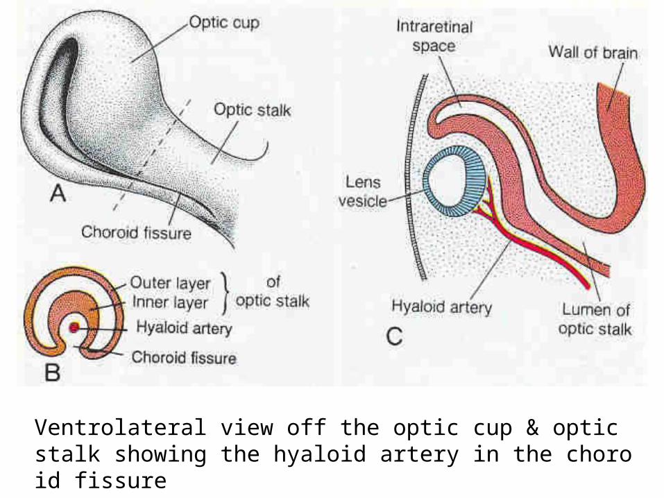
Ventrolateral view off the optic cup & optic stalk showi ng the hyaloid artery in the choroid fissure

Retina formationChanges of inner layer of optic cup (lens & cornea)
neural retina
Epith. cells neurons & light ph otoreceptor cells (rods&cones)
Thickening
differentiation
Outer layer of optic cup (thin) pigment layer of the retina

Neural retina (multilayers)
- consists of rods & cones
- bipolar neurons in
inner nuclear layer
- ganglion cells in ganglion cell layer
•1st-ganglion cell--> optic nerve
•2nd-bipolar neurons & rods - cones
Differentiation

Diagram showing the differentiation of layers of the neural retina.
Optic nerve/stalk

Diagram showing the development of the layers of the neural retina
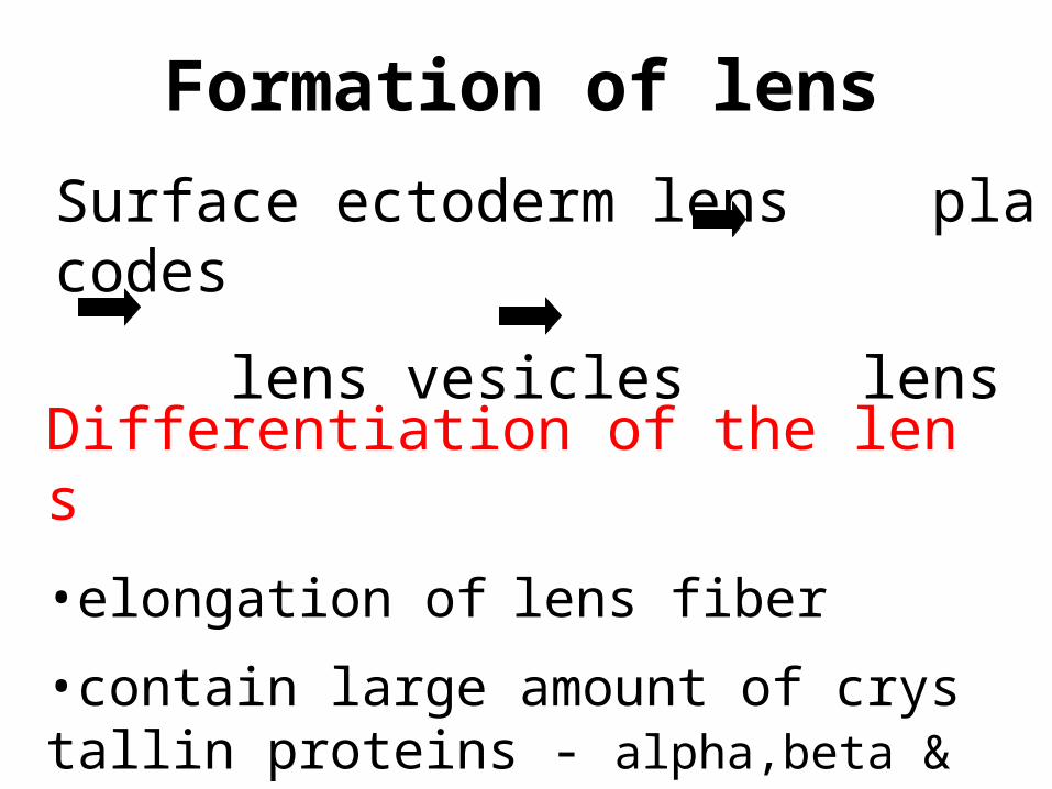
Formation of lens
Surface ectoderm lens placodes
lens vesicles lens
Differentiation of the lens
• elongation of lens fiber
• contain large amount of crystallin - proteins alpha,beta & gamma(mol.leve
l)

Invagination
Lens cup
Lens vesicle
Elongation of 10 fiber
Embryonic lens
Alpha(+/-)
Crystallin(no)
Alpha(++)Beta(+/-)Gamma(+/-)
Alpha(+++)Beta(+++)Gamma(+++)
Alpha(++)Beta(++)Gamma(+)
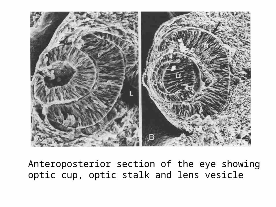
Anteroposterior section of the eye showing optic c up, optic stalk and lens vesicle

Formation of cornea
- 2 *sources surface ectoderm Inductive influence of the lens
- - surface ectoderm primary stroma :
collagen type I, II, IX
- -neural crest cells corneal endotheliu -m & secondary stroma (hyaluronic acid hyalur
onidase)
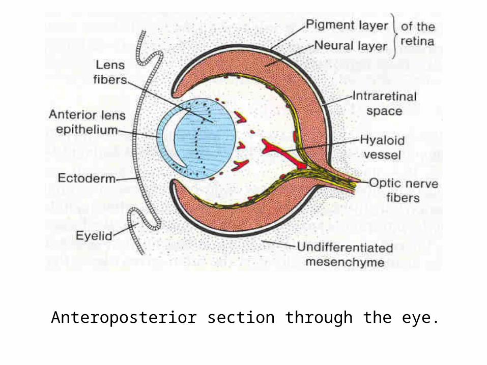
Anteroposterior section through the eye.
Final developmental changes of cornea= Formation of transparency- by removing water from seconda
ry stroma 40( %)
- degradation of hyaluronic acid
-thyroxine-- mmmmmmm mmmmmmmmmm>m - by pumping sodium into antr cha
mber of the eye
Iris & Ciliary body formation-at the lip of optic cup
* Ciliary body - muscle containing structure + suspensory ligament
of the lens --(radial set of muscle) >modul ate shape of the lens
*Iris - 2 sets of muscle (sphincter &
dilator pupillae) -- >control the amt of light passing through the lens

Development of the iris and the ciliary body.The rim of th e optic cup is covered by the mesenchyme, in which the s
phinter and dilator pupillae develop from the underlyingectoderm.

Choroid coat and sclera
Origin m mmmmm mm m mmmmmmmm= al cells + neural crest cells outs
- ide the optic cup under influence of the pigmented epith. of the retin
a Choroid coat- a highly vascular tunicSclera-dense collageneous covering
- tough outer coat of the eye

Diagram showing the development of the choroid & the scl
era
Vitreous body & Hyaloid artery
Vitreous body- loose mesenchyme fo rming a loose fibrillar mesh along with a
gelatinous substance in optic cup
Hyaloid artery- enter the eyeball thro ugh the choroid fissure of the optic stalk
-- -->retina,vitreous body >postr wall of t he lens
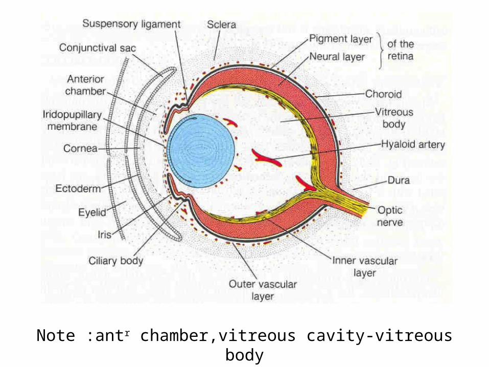
Note :antr - chamber,vitreous cavity vitreous body

Regression of hyaloid artery
(in the vitreous body)
Persistance of prox. part of hyalo id artery as central artery of the re
tina
Changes of Hyaloid artery

A B
C
Stages in developme nt and regression of t he hyaloid artery in t he embryonic eye

Eyelid and Lacrimal gland7th - wk. fold of skin9th - wk. meet & temporary fusion6th - mo. loosening of epith. union7th - mo. reopening of the eyelid

Conjunctival sac - space betw. the front of eyeball & eyelid
mmm. - multiple epithelial bu d from lateral surface ectoderm
- nasolacrimal duct
- begin to function~6 wk. after birth
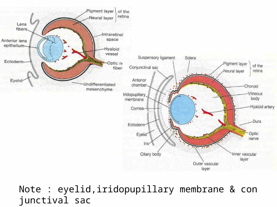
Note : eyelid,iridopupillary membrane & conjunctiva
l sac

Congenital malformations of the eye
Anophthalmos - absence of an eym Microphthalmos - smaller than normal
Coloboma iridis - nonclosure of ch oroid fissure of the iris