Embed Size (px)
Citation preview

Jittipan Chavadej,Ph.D.Dept. of Anatomy,Fac. of Science,
Mahidol University
September,2000

Diagrams showing formation of the neural groove, neural tube and neural crest
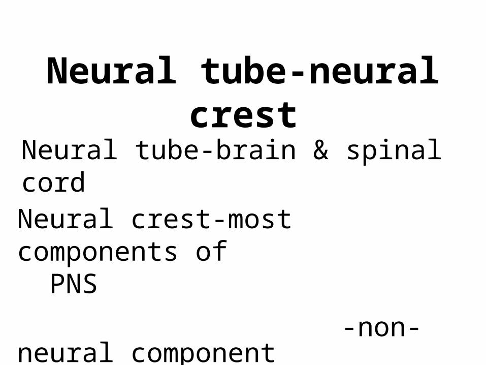
Neural tube-neural crest
Neural tube-brain & spinal cord
Neural crest-most components of PNS
-non-neural component in the body and head.

Fundamental processes in Nervous system
formation
•Induction-Proliferation
•Migration-Differentiation
•Pattern formation
•Intercellular communication
•Stabilization or Elimination
•Development of integrated pattern
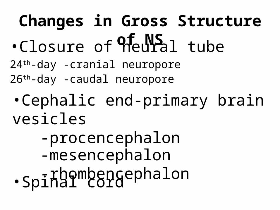
Changes in Gross Structure of NS•Closure of neural tube
24th-day -cranial neuropore 26th-day -caudal neuropore
•Cephalic end-primary brain vesicles
-procencephalon-mesencephalon-rhombencephalon•Spinal cord
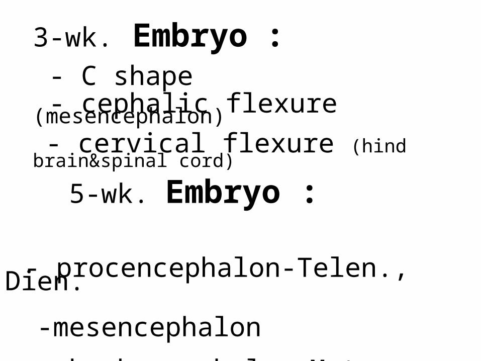
3-wk. Embryo : - C shape - cephalic flexure (mesencephalon) - cervical flexure (hind brain&spinal cord)
5-wk. Embryo : - procencephalon-Telen., Dien.
-mesencephalon
-rhombencephalon-Meten., Myelen.

A-lateral view of the brain vesicles and part of spinal cord. B-diagram showing the cavities of the three brain vesicles and spinal cord

Diencephalon - optic vesicles
Mesencephalon - sharply bend by cephalic flexureRhombencephalon - Pontine flexure thin roof

Basic anatomy of the five-part human brain
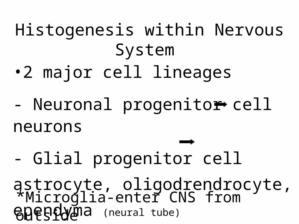
Histogenesis within Nervous System
•2 major cell lineages
- Neuronal progenitor cell neurons
- Glial progenitor cell
astrocyte, oligodrendrocyte,
ependyma (neural tube)*Microglia-enter CNS from outside

Cell lineages in the developing central nervous system
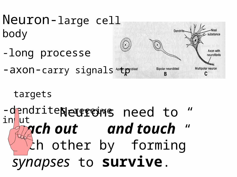
Neuron-large cell body
-long processe
-axon-carry signals to
targets
-dendrites-receive input
Neurons need to “ reach out and touch “ each other by forming synapses to survive.
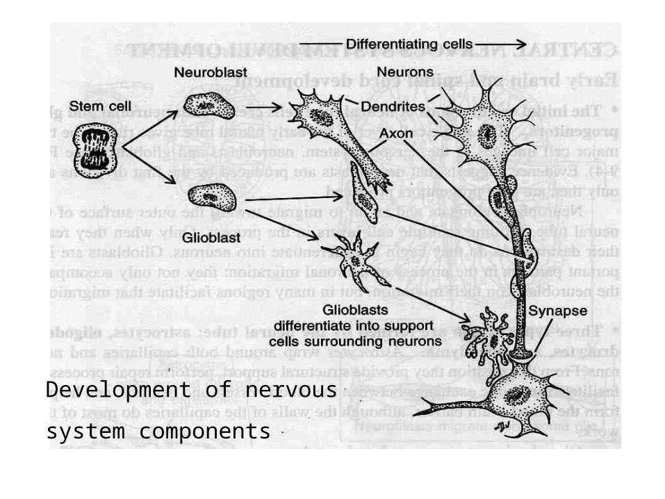
Development of nervous
system components
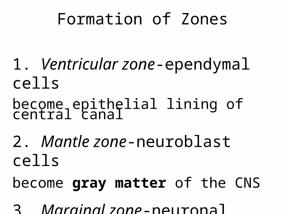
Formation of Zones
1. Ventricular zone-ependymal cellsbecome epithelial lining of central canal
2. Mantle zone-neuroblast cellsbecome gray matter of the CNS
3. Marginal zone-neuronal processesbecome white matter of the CNS
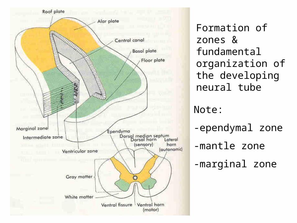
Formation of zones & fundamental organization of the developing neural tube
Note:
-ependymal zone
-mantle zone
-marginal zone
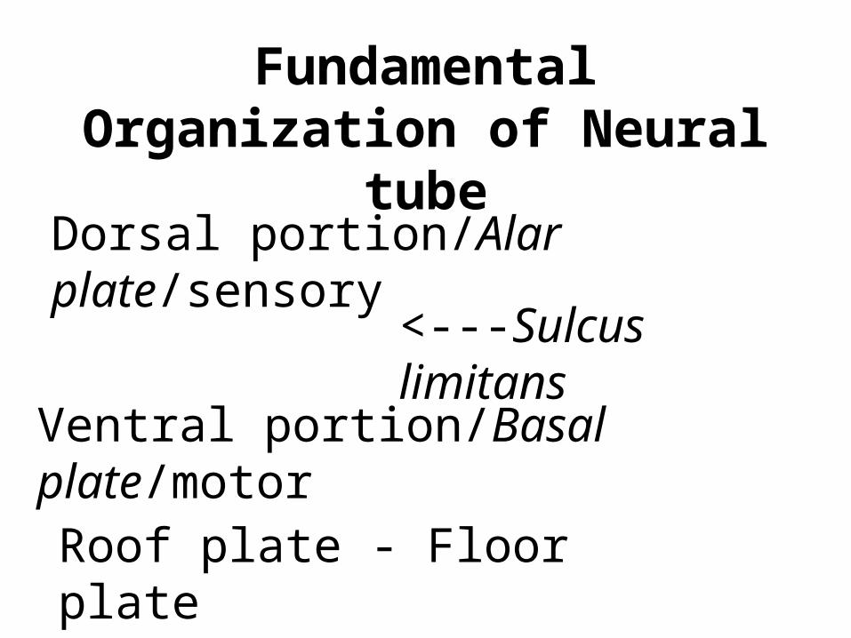
<---Sulcus limitans
Fundamental Organization of Neural
tubeDorsal portion/Alar plate/sensory
Ventral portion/Basal plate/motorRoof plate - Floor plate

A-development of regional specialization across the neural tube. B-formation of the spinal cord
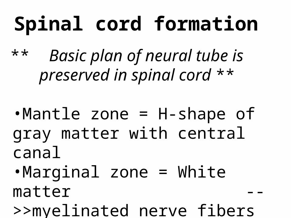
Spinal cord formation
** Basic plan of neural tube is preserved in spinal cord
**•Mantle zone = H-shape of gray matter with central canal
•Marginal zone = White matter -->>myelinated nerve fibers
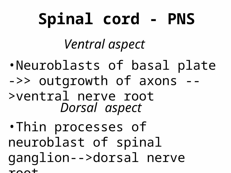
Spinal cord - PNS
•Neuroblasts of basal plate ->> outgrowth of axons -->ventral nerve root
•Thin processes of neuroblast of spinal ganglion-->dorsal nerve root
Dorsal aspect
Ventral aspect

Development of a peripheral nerve
A-showing motor axon, growing fiber of nerve cell in DRG. B-showing ventral motor and dorsal sensory root joining to form the trunk of spinal nerve
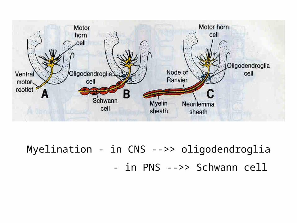
Myelination - in CNS -->> oligodendroglia
- in PNS -->> Schwann cell

Gross change of spinal cord•6-wk.-->full length of vertebral
column•8-wk.-->end ~Co4•14-wk.-->end ~S1•23-wk.-->end ~L4•Birth-->end ~L3•Adult--> end~L2-3-filum terminale &cauda equina
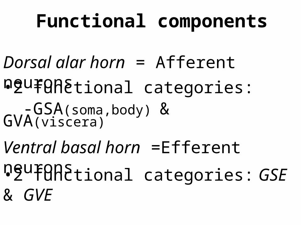
Functional components
•2 functional categories: -GSA(soma,body) & GVA(viscera)
Dorsal alar horn = Afferent neurons
Ventral basal horn =Efferent neurons•2 functional categories: GSE & GVE

Dorsal alar plate-sensory horn of spinal cord
Ventral basal plate-motor horn of spinal cord
Brain Formation
•The original organization of neural tube is altered in the formation of many regions of the brain.•Nerve cells form concentrated collections called nuclei in the brain
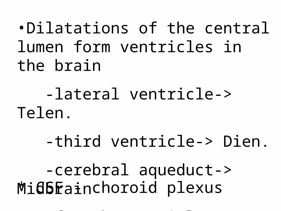
•Dilatations of the central lumen form ventricles in the brain
-lateral ventricle-> Telen.
-third ventricle-> Dien.
-cerebral aqueduct-> Midbrain
-fourth ventricle-> Hindbrain* CSF - choroid plexus
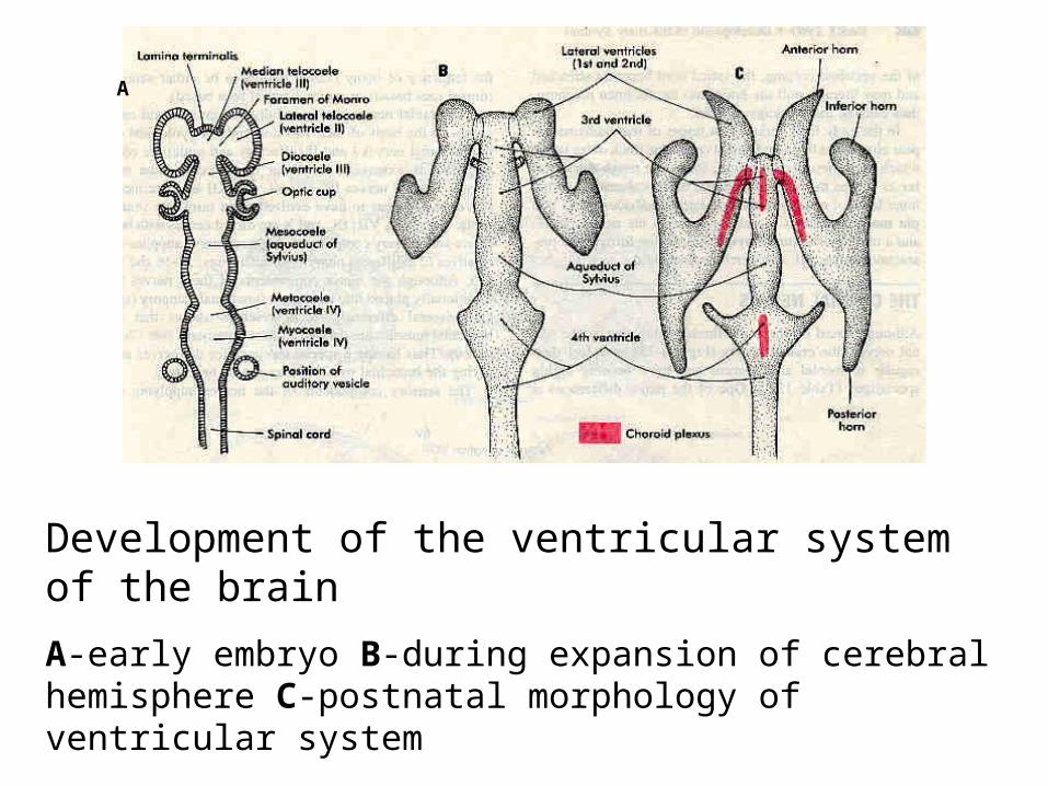
Development of the ventricular system of the brain
A-early embryo B-during expansion of cerebral hemisphere C-postnatal morphology of ventricular system
A
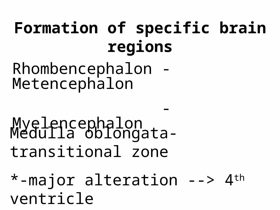
Formation of specific brain regions
Rhombencephalon - Metencephalon
- MyelencephalonMedulla oblongata-transitional zone
*-major alteration --> 4th ventricle
Basal plate&Alar plate in brainstem
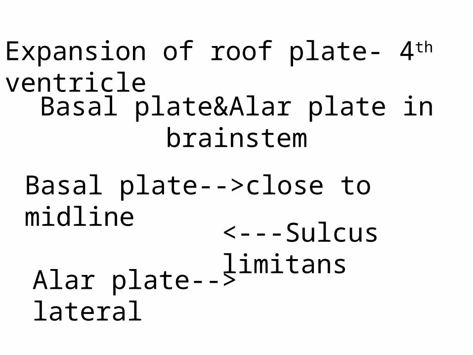
Expansion of roof plate- 4th ventricle
Basal plate-->close to midline
Alar plate--> lateral
<---Sulcus limitans
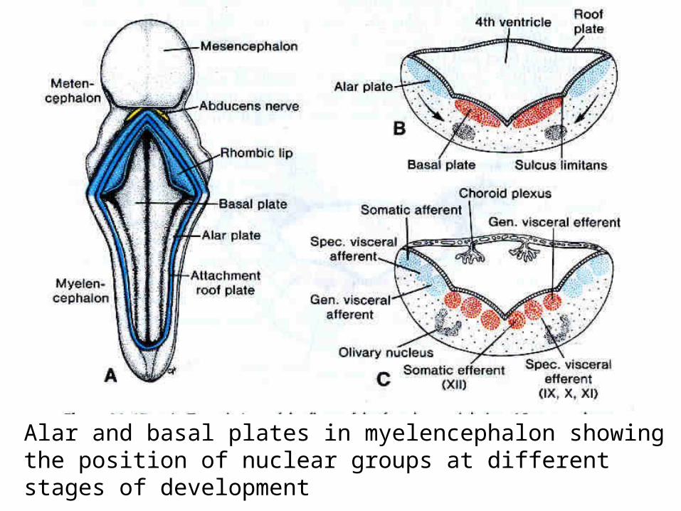
Alar and basal plates in myelencephalon showing the position of nuclear groups at different stages of development
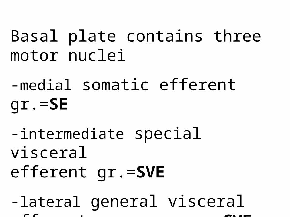
Basal plate contains three motor nuclei
-medial somatic efferent gr.=SE
-intermediate special visceral efferent gr.=SVE
-lateral general visceral efferent gr.=GVE

Alar plate contains sensory nuclei*-most lateral
special somatic afferent gr.=SSA
general somatic afferent gr.=GSA *-intermediate
special visceral afferent gr.=SVA *-medial general visceral afferent gr.=GVA
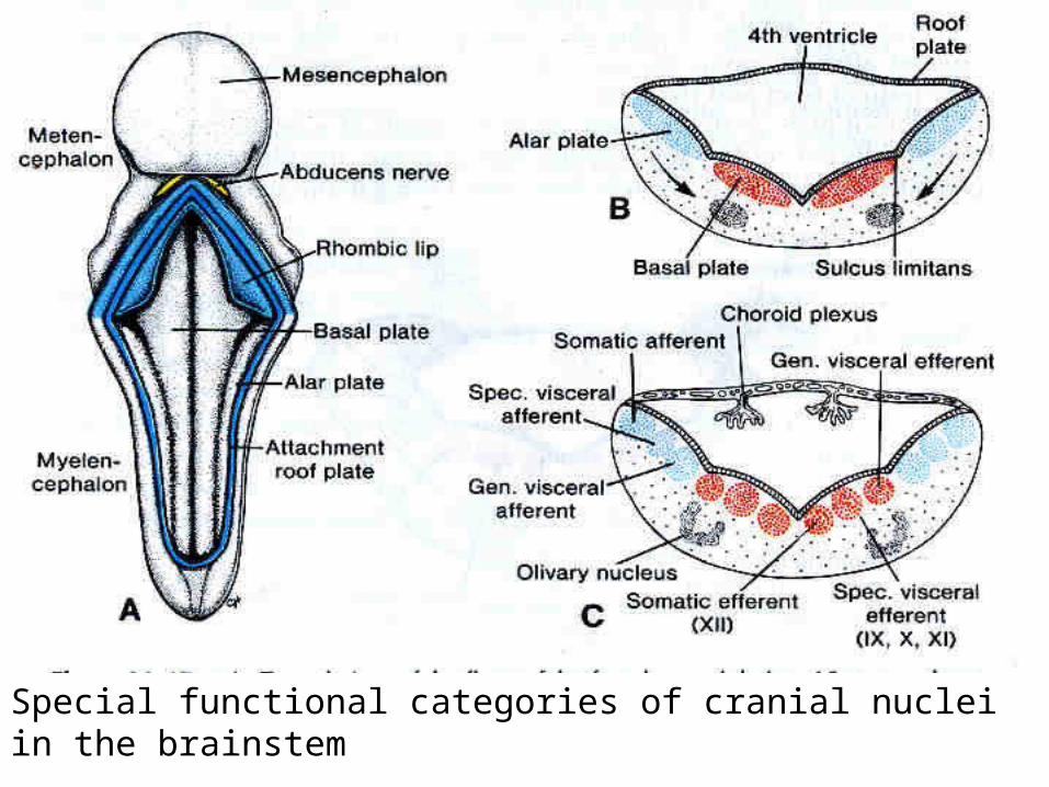
Special functional categories of cranial nuclei in the brainstem
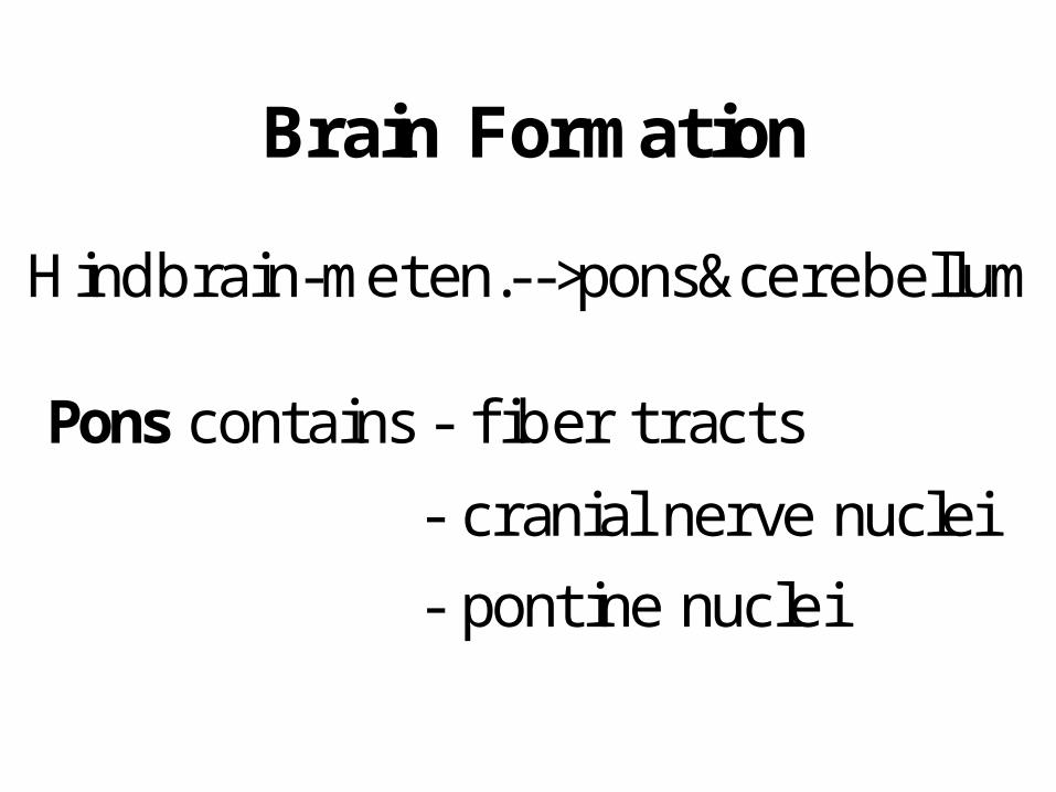
Brain Formation
Hindbrain-meten.-->pons&cerebellum
Pons contains - fiber tracts
- cranial nerve nuclei
- pontine nuclei

Cranial end of hindbrain (meten.)expands outwards to f ormcerebellum.
Cerebellum - specialization of alarplates --> Rhombic lips
Two cerebellar hemispheres+Vermis
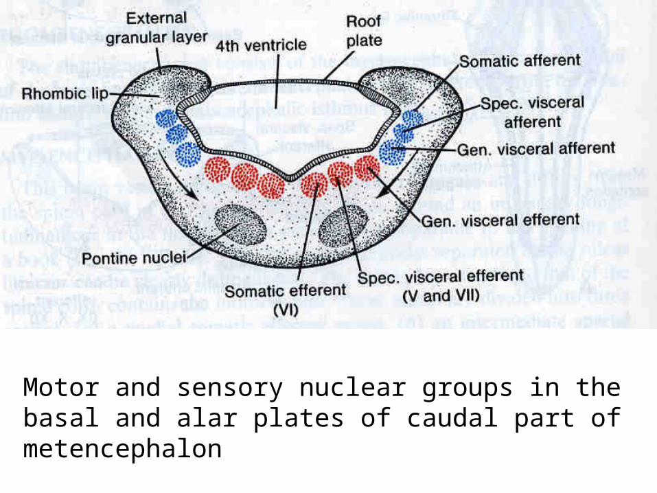
Motor and sensory nuclear groups in the basal and alar plates of caudal part of metencephalon
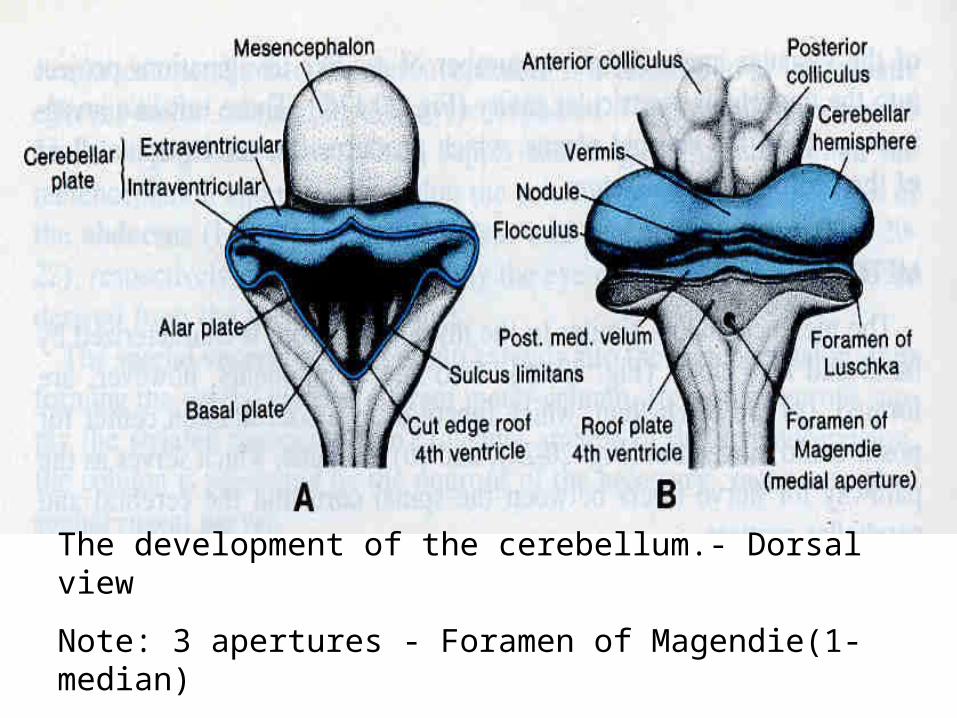
The development of the cerebellum.- Dorsal view
Note: 3 apertures - Foramen of Magendie(1-median)
-Foramen of Luschka(2-lateral)
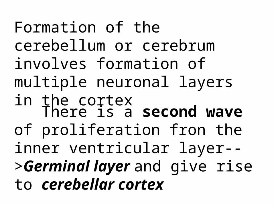
Formation of the cerebellum or cerebrum involves formation of multiple neuronal layers in the cortex
There is a second wave of proliferation fron the inner ventricular layer-->Germinal layer and give rise to cerebellar cortex
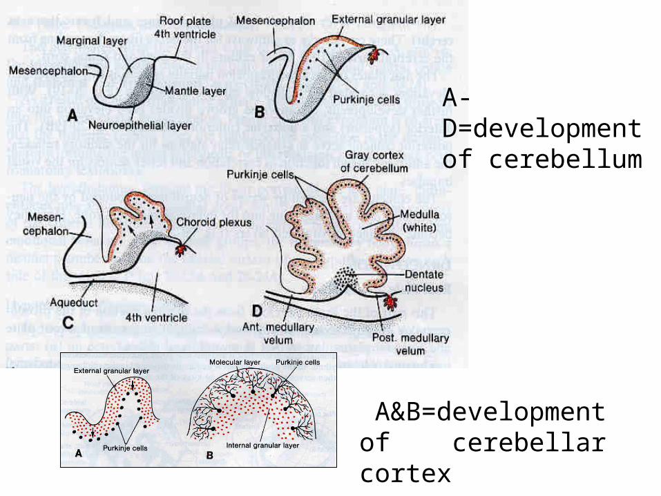
A-D=development of cerebellum
A&B=development of cerebellar cortex

Glia play an important role in migration of cortical neurons
Radial glia-->for guidance neurons to their destination
The external cortical neurons-layers * The cerebellar hemispheres undergo extensive folding = Folia
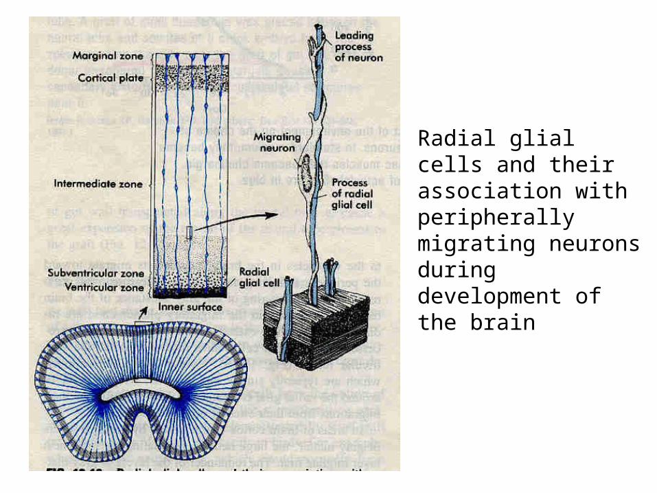
Radial glial cells and their association with peripherally migrating neurons during development of the brain

DEVELOPMENT of
Nervous System (cont.)
Dr. Jittipan Chavadej
Anatomy Department
yr,2000
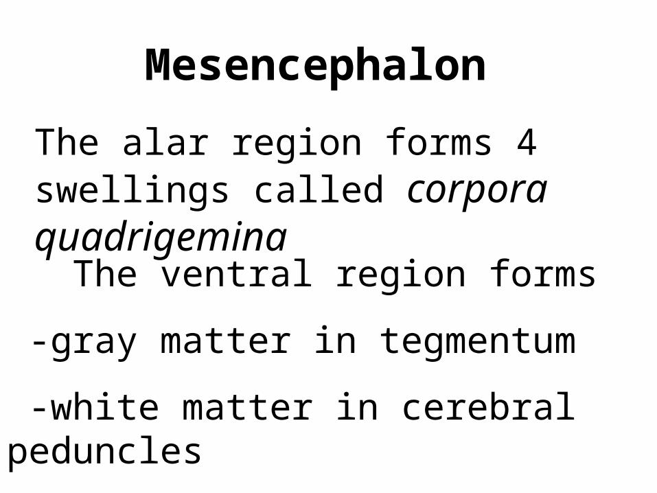
Mesencephalon
The alar region forms 4 swellings called corpora quadrigemina
The ventral region forms
-gray matter in tegmentum
-white matter in cerebral peduncles

Dorsal view of the midbrain & hindbrain

Tegmentum -cranial motor nuclei
-2 prominent relay nuclei=red nucleus
& substantia nigra
Colliculi =relay nuclei for auditory & visual systems
Cerebral peduncle=fiber tracts from cerebrum > cerebellum > spinal cord

Cross section-early&later developing mesencephalon (blue-sensory, red-motor)
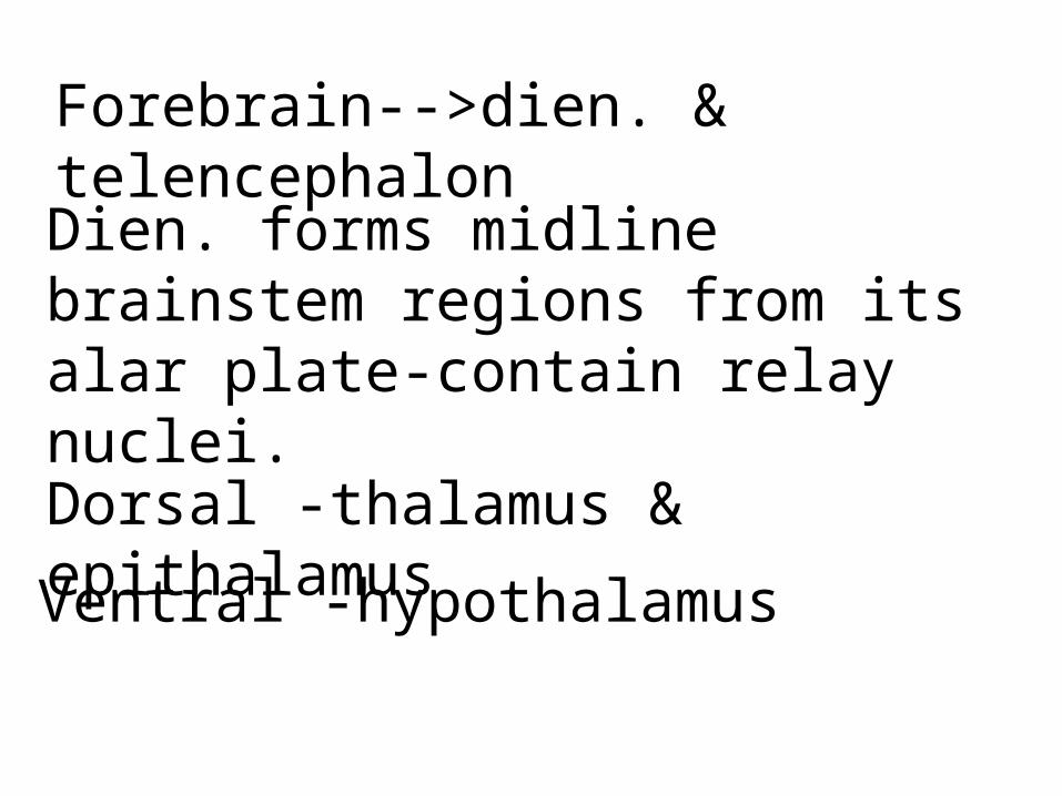
Forebrain-->dien. & telencephalonDien. forms midline brainstem regions from its alar plate-contain relay nuclei.
Dorsal -thalamus & epithalamus
Ventral -hypothalamus

Medial surface & X-section of prosencephalon. Note:corpus striatum bulge from the floor of lateral ventricle
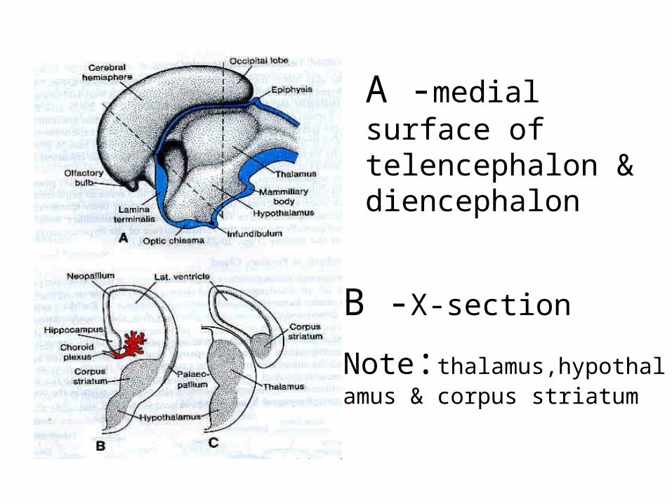
A -medial surface of telencephalon & diencephalon
B -X-section
Note:thalamus,hypothalamus & corpus striatum
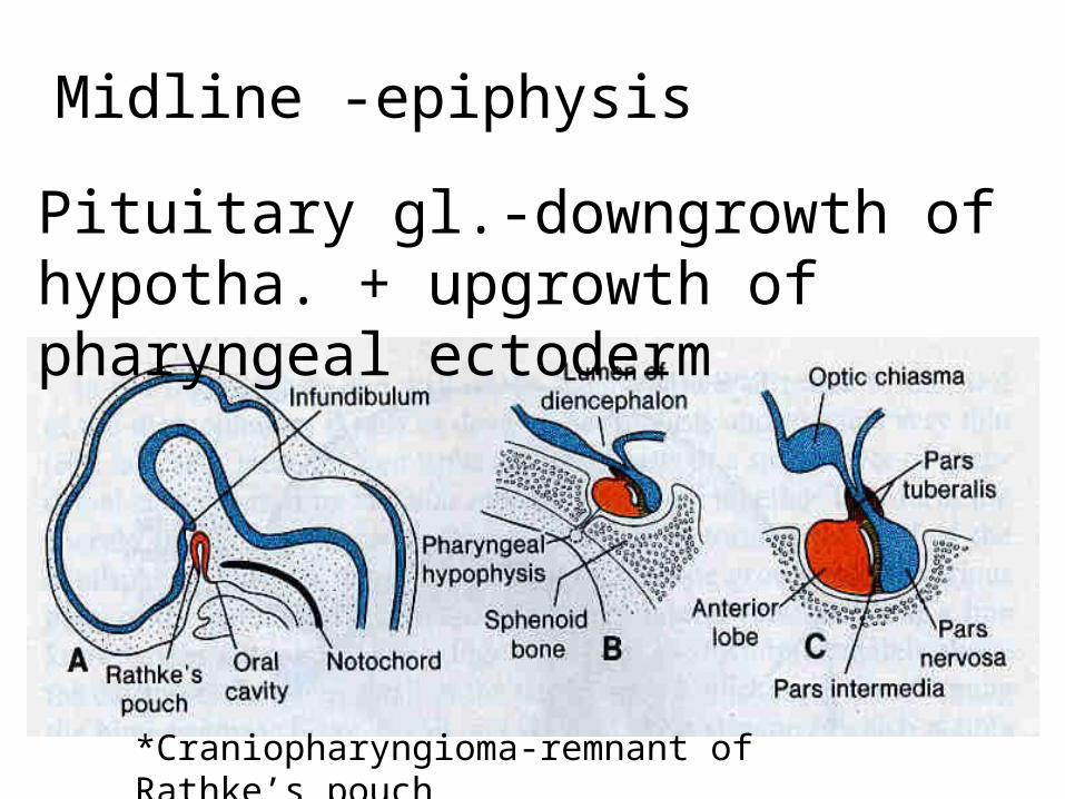
*Craniopharyngioma-remnant of Rathke’s pouch
Midline -epiphysis
Pituitary gl.-downgrowth of hypotha. + upgrowth of pharyngeal ectoderm
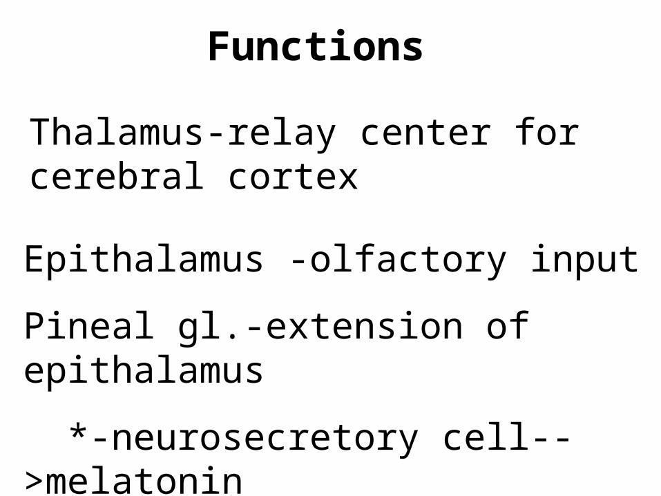
Functions
Thalamus-relay center for cerebral cortex
Epithalamus -olfactory input
Pineal gl.-extension of epithalamus
*-neurosecretory cell-->melatonin
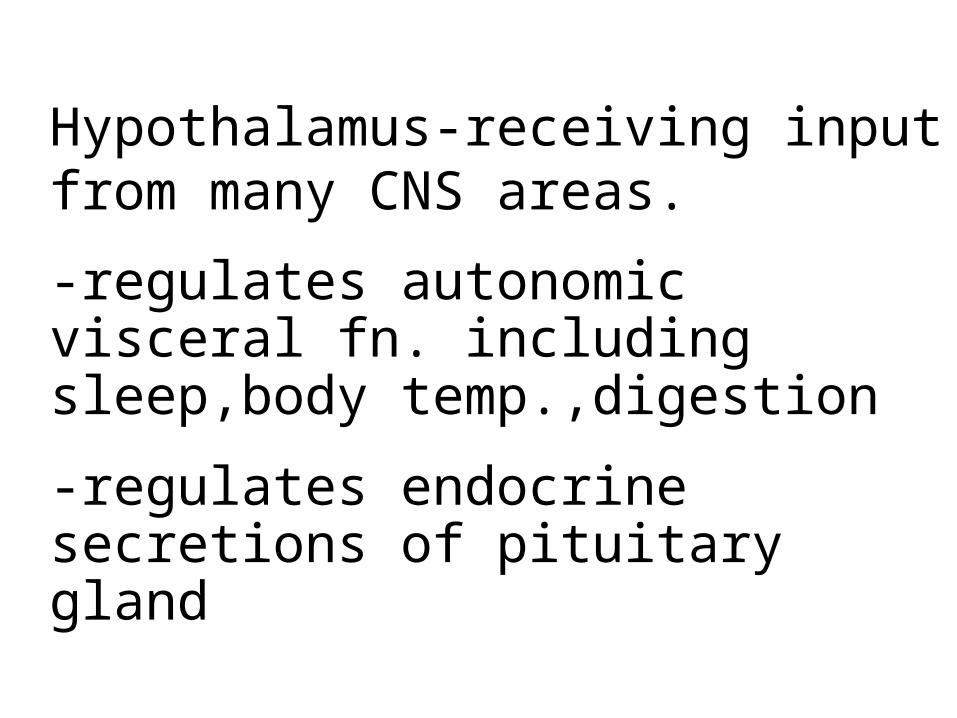
Hypothalamus-receiving input from many CNS areas.
-regulates autonomic visceral fn. including sleep,body temp.,digestion
-regulates endocrine secretions of pituitary gland

Telencephalon forms the cerebral hemispheres by bilateral expansionCerebral cortex-waves of migration to form cerebral cortex=neocortex
-intermediate zone-->white matter *Forming multiple synaptic connections is important to the development of the brain*

*The cerebral hemispheres fold into lobes and gyri->begin 14 weeks
Fetal period-frontal,parietal, temporal and occipital lobes
Sulcus/sulci separate some lobesConvolutions - gyrus/gyri
-begin betw.6&8months
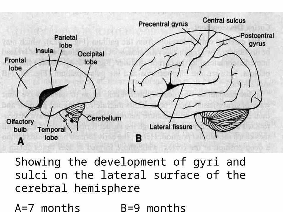
Showing the development of gyri and sulci on the lateral surface of the cerebral hemisphere
A=7 months B=9 months
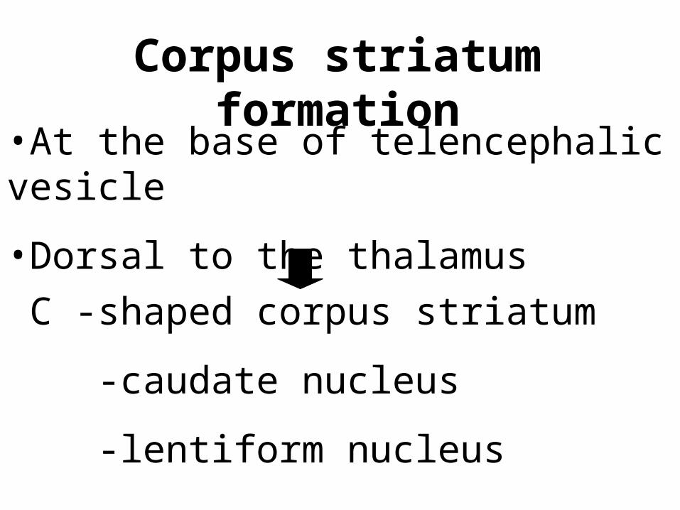
Corpus striatum formation
•At the base of telencephalic vesicle
•Dorsal to the thalamus
C -shaped corpus striatum
-caudate nucleus
-lentiform nucleus
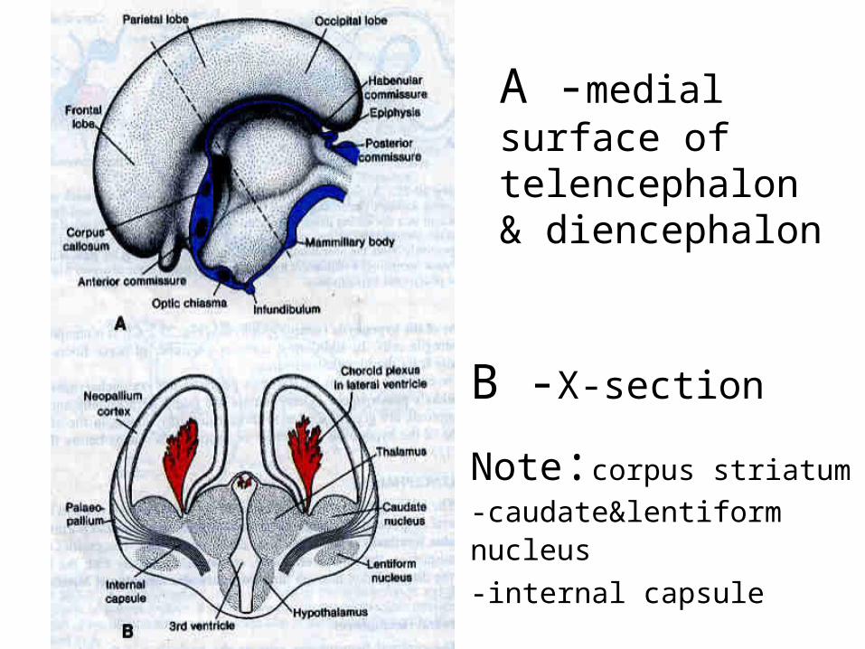
A -medial surface of telencephalon & diencephalon
B -X-section
Note:corpus striatum -caudate&lentiform nucleus
-internal capsule

Commissures
**Lamina terminalis** •1st - antr commissure
•2nd- hippocampal commissure
•3rd - corpus callosum-biggest
-postr & habenular commissures
(pineal gland) -optic chiasm
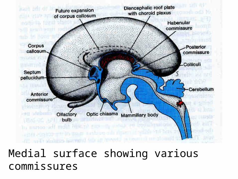
Medial surface showing various commissures
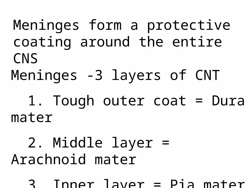
Meninges form a protective coating around the entire CNS
Meninges -3 layers of CNT
1. Tough outer coat = Dura mater
2. Middle layer = Arachnoid mater
3. Inner layer = Pia mater
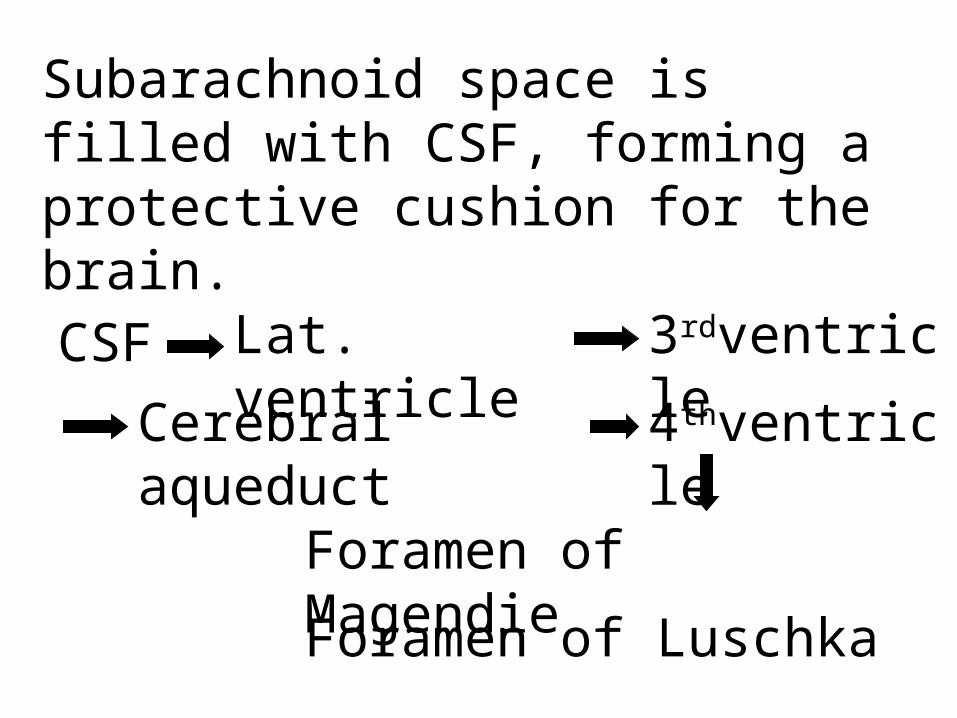
Subarachnoid space is filled with CSF, forming a protective cushion for the brain.
CSF Lat. ventricle
3rdventricle
Cerebral aqueduct
4thventricle
Foramen of Magendie
Foramen of Luschka
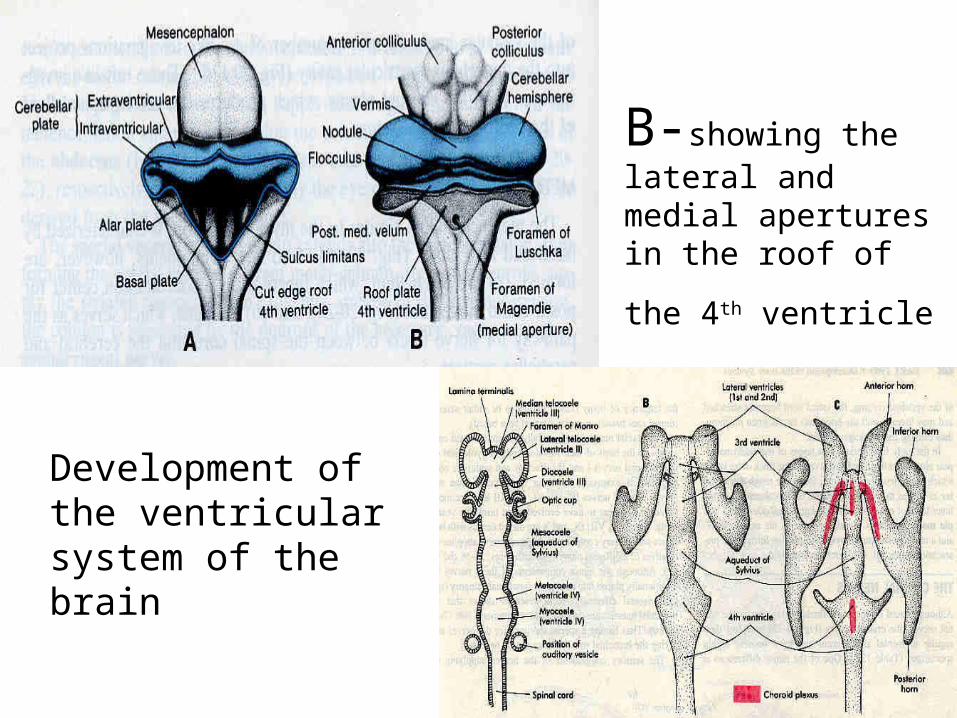
B-showing the lateral and medial apertures in the roof of the 4th
ventricle
Development of the ventricular system of the brain
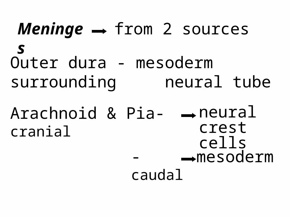
Outer dura - mesoderm surrounding neural tube
Arachnoid & Pia-cranial
neural crest cells
-caudal
mesoderm
Meninges
from 2 sources

Congenital defects of the CNS
•Mental retardation-no detectable brain abnormality
•Gross morphological defects
-spina bifida/occulta/cystica-meningocele/
meningomyelocele /meningoencephalocele-rachischisis

Congenital malformations of the NS
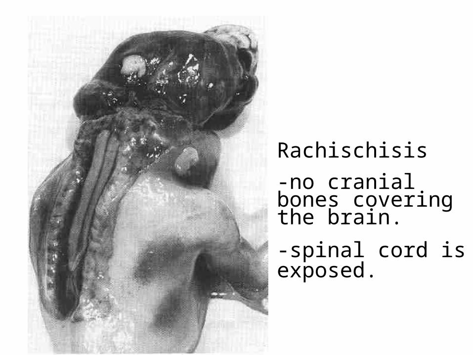
Rachischisis
-no cranial bones covering the brain.
-spinal cord is exposed.
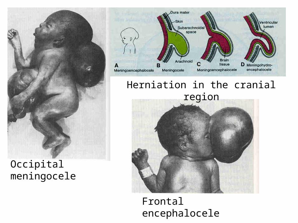
Occipital meningocele
Frontal encephalocele
Herniation in the cranial region
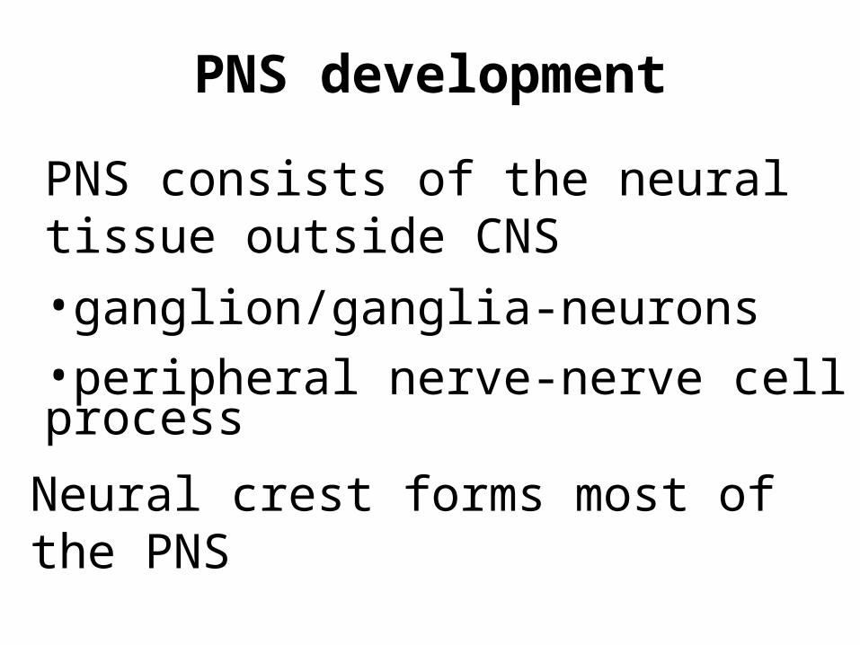
PNS development
PNS consists of the neural tissue outside CNS•ganglion/ganglia-neurons•peripheral nerve-nerve cell process
Neural crest forms most of the PNS
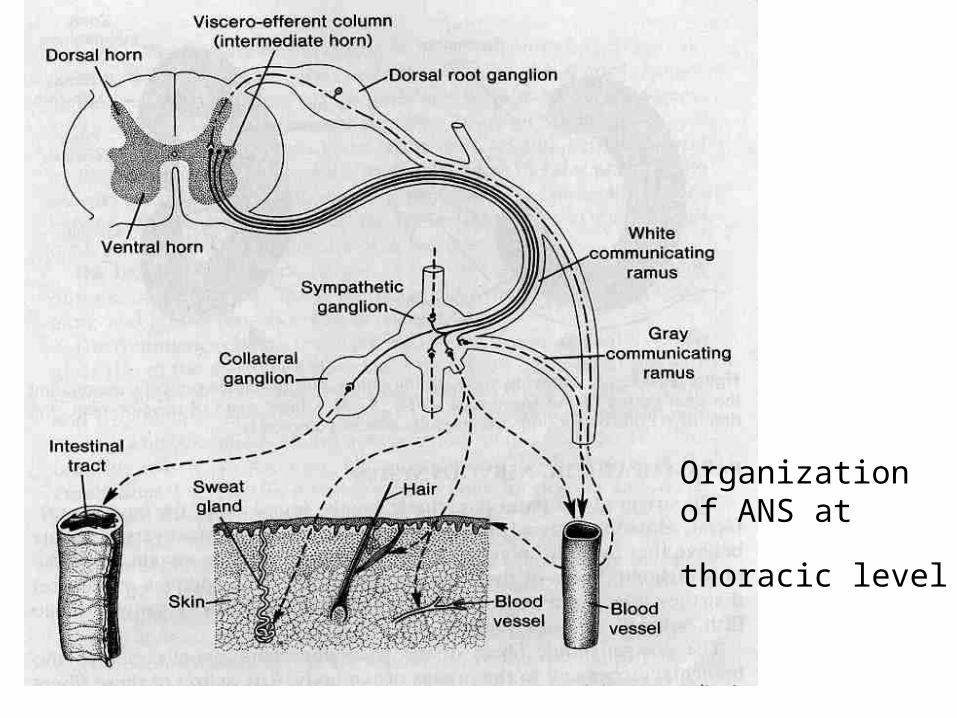
Organization of ANS at thoracic
level

Neural crest
Neural crest forms most of the PNS
Two types of ganglia
sensory ganglia
autonomic motor ganglia
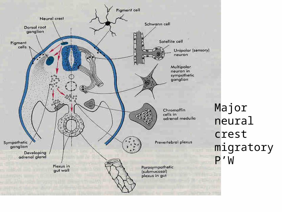
Major neural crest migratory P’W