Embed Size (px)
Citation preview

Joint National Ph.D. Meeting
2012
Rimini11-13 October 2012
http://DOTT2012.azuleon.org
ABCD · Associazione di Biologia Cellulare e del DifferenziamentoSIBBM · Società Italiana di Biofisica e Biologia Molecolare
� Joint National Ph.D. Meeting
Scientific Organisers
Senior Committee
Gennaro Ciliberto (Naples)Antonella De Matteis (Naples)
Giulia Piaggio (Rome)Ruggero Pardi (Milan)
Junior Committee
Giulia Antoniali (Udine)Giulia Bon (Rome)Erik Dassi (Trento)
Riccardo Rizzo (Naples)Tommaso Selmi (Modena)
Giusy Tornillo (Turin)
Organising SecretariatAzuleon srl
With the support of

�Rimini • 11-1� October �01�
prog
ra
mm
e
Programme

� Joint National Ph.D. Meeting
pro
gr
am
me

�Rimini • 11-1� October �01�
prog
ra
mm
e
Thursday, 11 october13:00-14:30 Lunch
14:00-15:00 Registration
Session1:CellBiologyChairs: Riccardo Rizzo (Naples), Tommaso Selmi (Modena)
15:00-15:30 Maria Giovanna De Leo (Rome) Defining the function of OCRL and ClC5 in membrane trafficking in proximal tubular cells
15:30-16:00 Matteo Fossati (Milan) A novel role of the VSV-G export signal at the Golgi complex
16:00-16:30 Roberta Misaggi (Catanzaro) Expression profiling and gene silencing techniques: identification of heavy chain ferritin-dependent and independent genes in k562 cells
16:30-19:00 PosterSessionI(oddnumbers)[with coffee]
19:00-20:00 KeynoteLecture1Chairs: Giusy Tornillo (Turin), Giulia Antoniali (Udine)
Andrea Ballabio (Naples) Signals from the lysosome
20:30 Dinner

� Joint National Ph.D. Meeting
pro
gr
am
me
Friday, 12 october Session2:Mitochondriainhealthanddisease
Chairs: Riccardo Rizzo (Naples), Tommaso Selmi (Modena)
8:30-9:00 Justina Sileikyte (Padua) Outer membrane regulation of the permeability transition pore in mitochondria lacking TSPO
9:00-9:30 Salvatore Rizza (Rome) Characterization of mitochondrial homeostasis upon S-nitrosoglutathione reductase (GSNOR) deficiency: implication in cancer therapy
9:30-10:00 Luca Giordano (Bari) What can modulate the penetrance in Leber’s Hereditary Optic Neuropathy?
10:00-10:30 Giulia Guzzo (Padua) The mitochondrial chaperone TRAP1 promotes neoplastic growth by modifing tumor metabolism
10:30-11:00 Coffee Break
Session3:TranslationalresearchChairs: Giulia Bon (Rome), Giusy Tornillo (Turin)
11:00-11:30 Silvia Saragozza (Novara) Antibody on demand array
11:30-12:00 Gnana Prakasam Krishnamoorthy (Naples) Therapeutic strategy to target the Receptor Tyrosine Kinase AXL in Thyroid Cancer
12:00-13:00 KeynoteLecture2Chairs: Giulia Bon (Rome), Giusy Tornillo (Turin)
Gianni Cesareni (Rome) Systems Biology: the solution to everything or rather the latest addition to information overflow?
13:00-14:30 Lunch
14:30-17:30 PosterSessionII(evennumbers)[with coffee]
�Rimini • 11-1� October �01�
prog
ra
mm
e
Session4:GeneregulationChairs: Giulia Antoniali (Udine), Erik Dassi (Trento)
17:30-18:00 Simona Pilotto (Pavia) Catalysis and combinatorial assembly of histone demethylase LSD1 complexes
18:00-18:30 Assunta Saide (Naples) Identification and functional characterization of heterodimeric complex between transcription factor EB and the related helix-loop-helix zipper factors, TFE-3 and MITF
18:30-19:00 Krishna Parsi (Rome) A genome wide role for nuclear Dicer1 and Ago1 in transcriptional regulation in human cells
19:00-19:30 Silvia Careccia (Rome) Transcriptional regulation of the oncosuppressor microRNA let-7c in acute myeloid leukemia
19:30-20:00 ABCDAssembly
20:30 Dinner
� Joint National Ph.D. Meeting
pro
gr
am
me
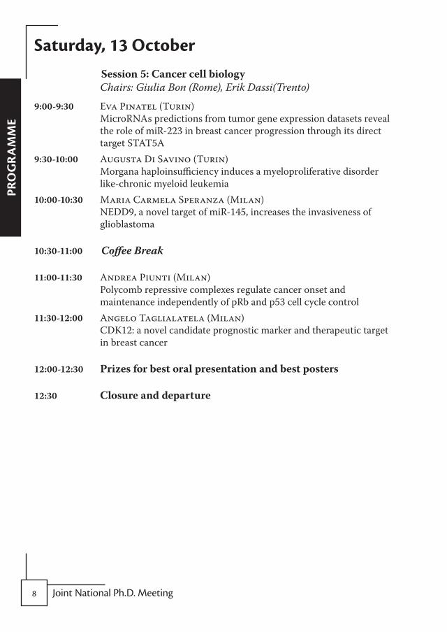
Saturday, 13 october Session5:Cancercellbiology
Chairs: Giulia Bon (Rome), Erik Dassi(Trento)
9:00-9:30 Eva Pinatel (Turin) MicroRNAs predictions from tumor gene expression datasets reveal the role of miR-223 in breast cancer progression through its direct target STAT5A
9:30-10:00 Augusta Di Savino (Turin) Morgana haploinsufficiency induces a myeloproliferative disorder like-chronic myeloid leukemia
10:00-10:30 Maria Carmela Speranza (Milan) NEDD9, a novel target of miR-145, increases the invasiveness of glioblastoma
10:30-11:00 Coffee Break
11:00-11:30 Andrea Piunti (Milan) Polycomb repressive complexes regulate cancer onset and maintenance independently of pRb and p53 cell cycle control
11:30-12:00 Angelo Taglialatela (Milan) CDK12: a novel candidate prognostic marker and therapeutic target in breast cancer
12:00-12:30 Prizesforbestoralpresentationandbestposters
12:30 Closureanddeparture

�Rimini • 11-1� October �01�
or
alS
Oral Presentationsin chronological order of presentation
(presenting authors are shown underlined)

10 Joint National Ph.D. Meeting
or
alS

11Rimini • 11-1� October �01�
or
alS
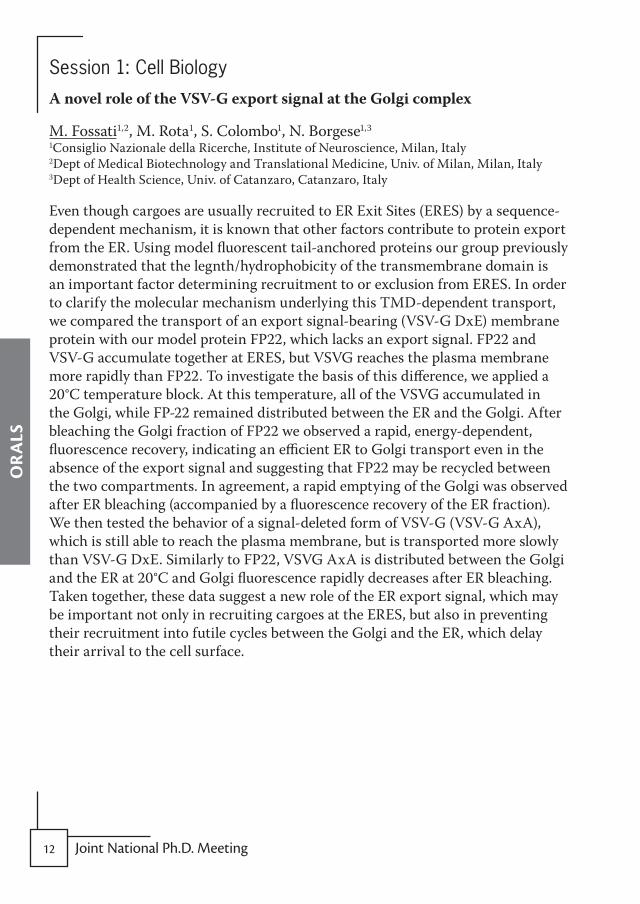
Session 1: Cell Biology
DefiningthefunctionofOCRLandClC5inmembranetraffickinginproximaltubularcells
M.G. De Leo, M. Santoro, M. Vicinanza, S. Iacobacci, A. Carissimo, D. Di Bernardo, M.A. De Matteis TIGEM - Telethon Institute of Genetics and Medicine- Naples, Italy
Inherited renal Fanconi syndromes are a group of genetically heterogeneous disorders characterized by the inability of Renal proximal tubular cells (PTCs) to reabsorb salts, nutrients and low molecular weight (LMW) proteins from the ultrafiltrate and by their loss in the urine. PTCs reabsorb approximately 70% of filtered sodium and water, more than 85% of the filtered phosphate, HCO3 and LMW proteins including albumin, vitamin-binding proteins and hormones. The reabsorptive function of the PTCs is mainly carried out by receptors, transporters and channels that cycle between the apical plasma membrane and the endosomes. Not surprisingly, among the genes that are defective in Fanconi syndrome are those encoding endosomal proteins such as the 5-phosphatase OCRL and the Cl-/H+ antiporter CLC5. Although the function of these proteins in endocytic membrane trafficking of PTCs remains to be defined, the common kidney phenotype caused by mutations of the two genes points to the possibility that OCRL and CLC5 act within the same molecular pathway. We exploited the overlap of the clinical outcome caused by mutations of OCRL and CLC5 to delineate the molecular networks regulating the trafficking pathways responsible for the reabsorptive function of PTCs. We compared the localisation of CLC5 and OCRL in human kidney cells (HK2) and found that CLC5 distributed between early and late endosomes and that some of these structures also contained OCRL. In addition, we found that CLC5 undergoes mistrafficking in OCRL KD cells. To elucidate the pathways in which these genes are involved, we performed microarray gene expression profiling studies of HK2 cells depleted for OCRL or CLC5, and found that a large subset of genes undergoes similar changes in response to OCRL- and ClC5-KD, confirming that OCRL and ClC5 impact on common molecular pathways.
1� Joint National Ph.D. Meeting
or
alS
Session 1: Cell Biology
AnovelroleoftheVSV-GexportsignalattheGolgicomplex
M. Fossati1,2, M. Rota1, S. Colombo1, N. Borgese1,3
1Consiglio Nazionale della Ricerche, Institute of Neuroscience, Milan, Italy 2Dept of Medical Biotechnology and Translational Medicine, Univ. of Milan, Milan, Italy 3Dept of Health Science, Univ. of Catanzaro, Catanzaro, Italy
Even though cargoes are usually recruited to ER Exit Sites (ERES) by a sequence-dependent mechanism, it is known that other factors contribute to protein export from the ER. Using model fluorescent tail-anchored proteins our group previously demonstrated that the legnth/hydrophobicity of the transmembrane domain is an important factor determining recruitment to or exclusion from ERES. In order to clarify the molecular mechanism underlying this TMD-dependent transport, we compared the transport of an export signal-bearing (VSV-G DxE) membrane protein with our model protein FP22, which lacks an export signal. FP22 and VSV-G accumulate together at ERES, but VSVG reaches the plasma membrane more rapidly than FP22. To investigate the basis of this difference, we applied a 20°C temperature block. At this temperature, all of the VSVG accumulated in the Golgi, while FP-22 remained distributed between the ER and the Golgi. After bleaching the Golgi fraction of FP22 we observed a rapid, energy-dependent, fluorescence recovery, indicating an efficient ER to Golgi transport even in the absence of the export signal and suggesting that FP22 may be recycled between the two compartments. In agreement, a rapid emptying of the Golgi was observed after ER bleaching (accompanied by a fluorescence recovery of the ER fraction). We then tested the behavior of a signal-deleted form of VSV-G (VSV-G AxA), which is still able to reach the plasma membrane, but is transported more slowly than VSV-G DxE. Similarly to FP22, VSVG AxA is distributed between the Golgi and the ER at 20°C and Golgi fluorescence rapidly decreases after ER bleaching. Taken together, these data suggest a new role of the ER export signal, which may be important not only in recruiting cargoes at the ERES, but also in preventing their recruitment into futile cycles between the Golgi and the ER, which delay their arrival to the cell surface.

1�Rimini • 11-1� October �01�
or
alS
Session 1: Cell Biology
Expressionprofilingandgenesilencingtechniques:identificationofheavychainferritin-dependentandindependentgenesink562cells
R. Misaggi1, M. Di Sanzo1, H.M. Bond1, C. Cosentino1, C. Stellato2, G. Giurato2, A. Weisz2, B. Quaresima1, T. Barni1, F. Amato1, G. Cuda1, G. Viglietto1, G. Morrone1, M.C. Faniello1, F. Costanzo1
1Dept of Experimental and Clinical Medicine, “Magna Graecia” University of Catanzaro, ”Salvatore Venuta” Campus, Catanzaro, Italy 2Molecular Medicine Laboratory, Faculty of Medicine and Surgery, Univ. of Salerno, Baronissi, Italy
Ferritin is the major intracellular iron storage protein. It is composed of 24 subunit of two types, ferritin H chain (FHC) and ferritin L chain (FLC). The heavy subunit is primarily responsible for the ferroxidase activity of the ferritin complex, whereas the light subunit facilitates the storage of iron into the ferritin core. FHC, moreover, has been implicated in the control of erythroid differentiation. In this study we sought to define the repertoire of genes whose expression is controlled by FHC during the hemin-induced differentiation of erythromyeloid K-562 cells. We performed mycroarray analysis in five different sets of cells: i) untreated wild type K562 (K562WT), ii) FHC-silenced K562 (K562shFHC), iii) FHC-reconstituted K562 (K562shFHC/pc3FHC), iv) hemin-treated K562WT, v) hemin-treated K562shFHC. We identified about 600 RNAs differentially expressed during hemin-induced differentiation of K562 cells, independently from the presence of FHC. Moreover, we found that a set of 100 mRNA appears to be modified in FHC-silenced K562 cells and in differentiated cells; furthermore 11 mRNA appears are modified by FHC-silencing but do not appear to respond to hemin treatment. This study confirms that the role of FHC is not limited to iron metabolism, suggesting potential implications of this protein in critical biological processes.
1� Joint National Ph.D. Meeting
or
alS
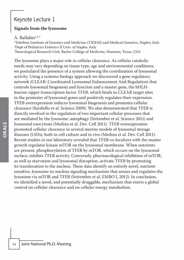
Keynote Lecture 1
Signalsfromthelysosome
A. Ballabio1,2,3
1Telethon Institute of Genetics and Medicine (TIGEM) and Medical Genetics, Naples, Italy 2Dept of Pediatrics Federico II Univ. of Naples, Italy 3Neurological Research Unit, Baylor College of Medicine, Houston, Texas, USA
The lysosome plays a major role in cellular clearance. As cellular catabolic needs may vary depending on tissue type, age and environmental conditions, we postulated the presence of a system allowing the coordination of lysosomal activity. Using a systems-biology approach we discovered a gene regulatory network (CLEAR: Coordinated Lysosomal Enhancement And Regulation) that controls lysosomal biogenesis and function and a master gene, the bHLH-leucine zipper transcription factor TFEB, which binds to CLEAR target sites in the promoter of lysosomal genes and positively regulates their expression. TFEB overexpression induces lysosomal biogenesis and promotes cellular clearance (Sardiello et al. Science 2009). We also demonstrated that TFEB is directly involved in the regulation of two important cellular processes that are mediated by the lysosome: autophagy (Settembre et al. Science 2011) and lysosomal exocytosis (Medina et al. Dev. Cell 2011). TFEB overexpression promoted cellular clearance in several murine models of lysosomal storage diseases (LSDs), both in cell culture and in vivo (Medina et al. Dev. Cell 2011). Recent studies in our laboratory revealed that TFEB co-localizes with the master growth regulator kinase mTOR on the lysosomal membrane. When nutrients are present, phosphorylation of TFEB by mTOR, which occurs on the lysosomal surface, inhibits TFEB activity. Conversely, pharmacological inhibition of mTOR, as well as starvation and lysosomal disruption, activate TFEB by promoting its translocation to the nucleus. These data identify an entirely novel, nutrient-sensitive, lysosome-to-nucleus signaling mechanism that senses and regulates the lysosome via mTOR and TFEB (Settembre et al. EMBO J, 2012). In conclusion, we identified a novel, and potentially druggable, mechanism that exerts a global control on cellular clearance and on cellular energy metabolism.
1�Rimini • 11-1� October �01�
or
alS
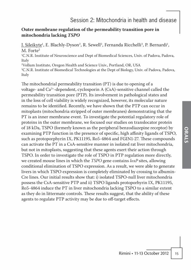
Session 2: Mitochondria in health and disease
OutermembraneregulationofthepermeabilitytransitionporeinmitochondrialackingTSPO
J. Sileikyte1, E. Blachly-Dyson2, R. Sewell2, Fernanda Ricchelli3, P. Bernardi1, M. Forte2
1C.N.R. Institute of Neuroscience and Dept of Biomedical Sciences, Univ. of Padova, Padova, Italy 2Vollum Institute, Oregon Health and Science Univ., Portland, OR, USA 3C.N.R. Institute of Biomedical Technologies at the Dept of Biology, Univ. of Padova, Padova, Italy
The mitochondrial permeability transition (PT) is due to opening of a voltage- and Ca2+-dependent, cyclosporin A (CsA)-sensitive channel called the permeability transition pore (PTP). Its involvement in pathological states and in the loss of cell viability is widely recognized, however, its molecular nature remains to be identified. Recently, we have shown that the PTP can occur in mitoplasts (mitochondria stripped of outer membranes) demonstrating that the PT is an inner membrane event. To investigate the potential regulatory role of proteins in the outer membrane, we focused our studies on translocator protein of 18 kDa, TSPO (formerly known as the peripheral benzodiazepine receptor) by examining PTP function in the presence of specific, high affinity ligands of TSPO, such as protoporphyrin IX, PK11195, Ro5-4864 and FGIN1-27. These compounds can activate the PT in a CsA-sensitive manner in isolated rat liver mitochondria, but not in mitoplasts, suggesting that these agents exert their action through TSPO. In order to investigate the role of TSPO in PTP regulation more directly, we created mouse lines in which the TSPO gene contains loxP sites, allowing conditional elimination of TSPO expression. As a result, we were able to generate livers in which TSPO expression is completely eliminated by crossing to albumin-Cre lines. Our initial results show that: i) isolated TSPO-null liver mitochondria possess the CsA-sensitive PTP and ii) TSPO ligands protoporhyrin IX, PK11195, Ro5-4864 induce the PT in liver mitochondria lacking TSPO to a similar extent as they do in littermate controls. These results suggest, that the ability of these agents to regulate PTP activity may be due to off-target effects.

1� Joint National Ph.D. Meeting
or
alS
Session 2: Mitochondria in health and disease
CharacterizationofmitochondrialhomeostasisuponS-nitrosoglutathionereductase(GSNOR)deficiency:implicationincancertherapy
S. Rizza1, G. Di Giacomo2, C. Montagna2, S. Cardaci1, M.R. Ciriolo1,2, G. Filomeni1,2
1Dept of Biology, University of Rome “Tor Vergata”, Rome, Italy 2IRCCS San Raffaele Pisana, Rome, Italy
S-nitrosylation of cysteine residues is a post-translational modification that reversibly affects protein function and stability, and deeply impacts on signal transduction. The amount of S-nitrosylated proteins (Prot-SNOs) is indirectly regulated by S-nitrosoglutathione reductase (GSNOR), a Class III alcohol dehydrogenase that catalyzes the degradation of S-nitrosoglutathione (GSNO), the most abundant cellular low molecolar weight S-nitrosothiol, which is in equilibrium with Prot-SNOs. It has been reported that GSNOR deficiency leads to an excessive accumulation of Prot-SNOs, which finally results in an impairment of DNA repair machinery and the spontaneous formation of hepatocellular carcinoma (HCC) in about 50% of GSNOR-/- aged mice. In particular, GSNOR deficiency causes S-nitrosylation and subsequent degradation of the DNA-repair enzyme O6-alkylguanine DNA-alkyltransferase (AGT), sensitizing GSNOR-/- mice to spontaneous DNA mutations. Moreover, many lines of evidence indicated that nitrosative stress due to NO (over)production affects mitochondrial homeostasis interfering with: i) the efficiency of electron transport chain (ETC) that leads to alterated ATP production; ii) Ca2+ homeostasis and apoptotic response; iii) physiological organelle turnover; iv) regulation of mitochondrial network morphology. Recently, many studies demonstrated that cancer cells exhibit mitochondrial dysfunctions and metabolic alterations suggesting new therapeutic approaches aimed at specifically targeting mitochondria of cancer cells. In the light of these lines of evidence and the prominent impact of S-nitrosylation on mitochondrial homeostasis, we hypothesize that mitochondrial impairment reasonably caused by GSNOR deficiency could be deeply involved in HCC onset in GSNOR-/- mice, thereby suggesting innovative mitochondrial therapeutical approaches in the development of chemotherapeutic strategies against HCC.

1�Rimini • 11-1� October �01�
or
alS
Session 2: Mitochondria in health and disease
WhatcanmodulatethepenetranceinLeber’sHereditaryOpticNeuropathy?
L. Giordano1, M. Fonsdituri1, S. Deceglie1, M. Roberti1, F. Fracasso1, P. Loguercio Polosa1, V. Carelli2, P. Cantatore1 1Dept of Biosciences, Biotechonologies and Pharmacological Sciences, Univ. of Bari “Aldo Moro”, Bari, Italy 2Dept of Neurological Sciences, Univ. of Bologna, Bologna, Italy
Leber’s hereditary optic neuropathy (LHON) is a maternally inherited blinding disease due to point mutations in complex I subunit genes of mtDNA. In most LHON families the mtDNA mutation is homoplasmic, but only a limited number of subjects becomes affected. Thus, the mtDNA mutation is necessary but not sufficient to cause optic neuropathy. Environmental triggers and genetic modifying factors have been considered to explain the variable penetrance in LHON. With the purpose to identify the factors responsible for the variable penetrance in LHON we studied several functional and molecular parameters in fibroblast grown in the presence of galactose, that forces the cell to rely on mitochondrial respiration for ATP synthesis, thus highlighting the respiratory defect in LHON subjects. We found that unaffected mutation carriers had consistent phenotypic differences compared to affected subjects since they exhibited a similar growth rate of controls, whereas a slower growth rate characterized the affected subjects. Furthermore carriers had a higher intracellular ATP content and produced lower L-lactate compared with affected cell lines. Investigations on the molecular bases underlying the carrier phenotype showed that carriers had a significantly higher mtDNA copy number and mitochondrial mass compared to affected. These results were paralleled by the data of western blots analysis of some key proteins involved in mitochondrial biogenesis and mtDNA replication, such as NRF1, TFAM and mtSSB. Moreover we found that carriers fibroblasts displayed the highest capacity in repopulating mtDNA after mtDNA depletion with ethidium bromide. All these data clearly indicate that carriers have a more efficient mitochondrial biogenesis than affected patients which seems sufficient to compensate the LHON mutation; this might explain the variability of penetrance, and open an avenue towards the development of predictive genetic tests on disease risk and therapeutic strategies.

1� Joint National Ph.D. Meeting
or
alS
Session 2: Mitochondria in health and disease
ThemitochondrialchaperoneTRAP1promotesneoplasticgrowthbymodifingtumormetabolism
G. Guzzo1, M. Sciacovelli1, V. Morello2, C. Frezza3, L. Zheng3,4, N. Nannini5, F. Calabrese5, G. Laudiero6, F. Esposito6, M. Landriscina7, P. Defilippi2, P. Bernardi1, A. Rasola1
1CNR Institute of Neuroscience, Dept of Biomedical Sciences, and Venetian Institute of Molecular Medicine, University of Padova, Padova, Italy 2Molecular Biotechnology Centre, Dept of Genetics, Biology and Biochemistry, Univ. of Torino, Torino, Italy 3Cancer Research United Kingdom, The Beatson Institute for Cancer Research, Glasgow, United Kingdom 4Strathclyde Institute of Pharmacy and Biomedical Sciences, Univ. of Strathclyde, Glasgow, United Kingdom 5Dept of Diagnostic Medical Sciences and Special Therapies, Univ. of Padova, Padova, Italy 6Dept of Biochemistry and of Medical Biotechnologies, Univ. of Napoli Federico II, Napoli, Italy 7Dept of Medical Sciences, Univ. of Foggia, Foggia, Italy
Cancer is a highly heterogeneous and complex disease, whose development requires a reorganization of cell metabolism. Most tumor cells downregulate mitochondrial oxidative phosphorylation and increase the rate of glucose consumption and lactate release, independently of oxygen availability (the Warburg effect). This metabolic rewiring is believed to favour tumor growth and survival. However the molecular mechanisms that inhibit oxidative phosphorylation (OXPHOS) during neoplastic progression are only partially understood. Within this context, we studied TRAP1, a conserved chaperone of the Heat Shock Protein 90 (HSP90) family, localized mainly in the mitochondrial matrix and whose expression is induced in the majority of tumor types. We found that TRAP1 is associated to succinate dehydrogenase (SDH), the Complex II of the respiratory chain. We observed in different tumor cell models that TRAP1 diminished in vivo tumor cell respiration by inhibiting the succinate:coenzyme Q reductase (SQR) activity of Complex II. This Complex II inhibition was further enhanced in TRAP1-expressing cells that progressed through a focus forming assay (in vitro tumorigenesis assay), causing an accumulation of succinate that led to the stabilization of the pro-neoplastic transcription factor HIF1α?? thus favouring the metabolic switch necessary for tumor growth and progression.

1�Rimini • 11-1� October �01�
or
alS
Session 3: Translational research
Antibodyondemandarray
S. Saragozza, D. Cotella, D. Sblattero, C. Santoro Dept Health Science
The technology protein array allows the measurement of multiple proteins in parallel and in a miniaturized format. This high-throughput (HT) technology involves several steps including cloning, expression and purification of the proteins, spotting on microarray surface and downstream analyses. Even in a low throughput applications this approach is time- and cost-intensive. A new type of array, where the cDNA is spotted onto a slide with an anti-tag antibody that allow the capture of the protein produced with in vitro transcription/translation (IVTT), were produced in the last years. In this project we want exploit this idea producing protein array where the protein is capture onto the slide using a dsDNA fragment. This approach include the: i) DNA spotting on the array; ii) protein in situ production by IVTT; iii) proteins binding to the coding DNA and downstream assay. The protein array is obtained just when needed starting from spotted DNA. To allow the binding of proteins to their DNA template we have used the E. coli protein TUS that can couple itself with very high affinity to the double stranded DNA sequence ter. As a first step we have find the best IVTT system that allow the production of all the proteins tested; secondary we have confirmed that all this proteins fused with TUS protein are able to bind efficiently to ter sequence on ELISA plate and on array; finally we have perform an antibody array based on these system. In conclusion we can state that our system is very promising.

�0 Joint National Ph.D. Meeting
or
alS
Session 3: Translational research
TherapeuticstrategytotargettheReceptorTyrosineKinaseAXLinthyroidcancer
G.P. Krishnamoorthy, T. Guida, C. Visciano, F. Liotti, M. Santoro, F. Carlomagno, RM. Melillo Univ. of Naples-Federico II, Dipartimento di Biologia e Patologia Cellulare e Molecolare/Istituto di Endocrinologia ed Oncologia Sperimentale del CNR ”G.Salvatore”, Napoli, Italy
The AXL receptor tyrosine kinase is overexpressed and active in various cancer types including thyroid carcinoma, and several preclinical studies suggest that targeting AXL is an effective therapeutic strategy in AXL-positive cancers. Heat shock protein 90 (HSP90) acts as a molecular chaperone to regulate the conformation, activation, function and stability of many cancer-related kinases. Inhibition of HSP90 by Geldanamycin and its derivative 17-Allyl-Ammino-17-demethoxygeldanamycin (17-AAG), leads to simultaneous combinatorial depletion of a wide range of its clients through the induction of their misfolding and proteosome-mediated degradation. The Quality control E3 ligase CHIP ubiquitinates misfolded proteins and favours their degradation. Here we show that treatment of AXL-expressing thyroid cancer cells with 17-AAG induces its proteosome-mediated degradation. Specifically, 17-AAG induces the downregulation of the fully glycosylated, mature form of the receptor that is exposed on the plasma membrane and responds to ligand stimulation. Degradation is preceded by AXL ubiquitination by CHIP. Endogenous and overexpressed AXL protein co-immunoprecipitated with CHIP and HSP90, and this complex is modulated by 17-AAG treatment. By using different AXL mutants and AXL small molecule inhibitors, we demonstrate that AXL sensitivity to 17-AAG requires AXL kinase domain, but is not dependent on AXL kinase activity. Overall our data elucidate the biological basis of AXL downregulation by HSP 90 inhibition and suggest that Hsp90 inhibition could be effective in treating AXL dependent thyroid cancer.

�1Rimini • 11-1� October �01�
or
alS
Keynote Lecture 2
SystemsBiology:thesolutiontoeverythingorratherthelatestadditiontoinformationoverflow?
G. Cesareni Dept of Biology, Univ. of Rome Tor Vergata, Rome, Italy
Over the past decade the frequency of the string “Systems Biology” has increased dramatically in grant applications and manuscript titles. This buzzword expresses the hope that an integrative approach to the modeling of cell signaling may support us while attempting to overcome the limits that we encounter when we try to infer the behavior of a cell or an organism after perturbation. This approach has helped some of us to obtain a few grants and to publish a number of papers. Its contribution to the progress of our understanding of complex biological responses is less clear. I will discuss what Systems biology can do to help answer our biological questions and I will warn you against possible risks. Finally I will present the results of the project of Francesca Sacco one of my PhD students who used a high content phenotypic screening combined with modeling to investigate the contribution of the family of phosphatase proteins to the modulation of growth pathways.

�� Joint National Ph.D. Meeting
or
alS
Session 4: Gene regulation
CatalysisandcombinatorialassemblyofhistonedemethylaseLSD1complexes
M. Tortorici, S. Pilotto, V. Speranzini, A. Mattevi Dept Biology and Biotechnology, Univ. of Pavia, Italy
The main line of my research project is to investigate two human multiprotein complexes involved in chromatin remodelling. We have studied a nuclear complex formed by the association of histone deacetylase, co-repressor protein CoREST and lysine-specific histone demethylase LSD1 that specifically acts on Lys4 of histone H3. This LSD1/HDAC/CoREST multi-enzyme module first eliminates the acetyl groups from acetylated Lys residues and then removes methyl groups from Lys4. Our structural studies of LSD1/CoREST highlighted a specific binding-site for the H3 N-terminal tail and a catalytic machinery that is closely related to that of other flavin-dependent amine oxidases. These insights have been critical for our efforts towards structure-based development of demethylase inhibitors. These newly designed inhibitors were evaluated with a cellular model of acute promyelocytic leukemia chosen since its pathogenesis includes aberrant activities of several chromatin modifiers. Marked effects on cell differentiation and an unprecedented synergistic activity with anti-leukemia drugs were observed. It has been recently discovered that the transcription factor Snail1 binds to LSD1/CoREST and that the three proteins are over-expressed in cancer cell lines and breast tumors. Snail1 controls the epithelial-mesenchymal transition, which is essential for numerous developmental processes (including metastasis). Structure determination of the ternary complex LSD1/CoREST/Snail1 peptide has revealed that the N-terminal residues of Snail1 bind in the active site of LSD1 mimicking the H3 tail. Therefore, Snail1 is a potential endogenous inhibitor of LSD1. Furthermore, this finding predicts that other members of the Snail1-related transcription factor family associate to LSD1 through a similar histone-mimicking mechanism. Our efforts are now focused on the structural investigations of nucleosome binding to LSD1-containing protein complexes through biophysical methods and crystallography.

��Rimini • 11-1� October �01�
or
alS
Session 4: Gene regulation
IdentificationandfunctionalcharacterizationofheterodimericcomplexbetweentranscriptionfactorEBandtherelatedhelix-loop-helixzipperfactors,TFE-3andMITF
A. Saide1,2, G. Mansueto1, D.L. Medina1, A.Ballabio1,3
1Telethon Institute of Genetics and Medicine, Naples, Italy 2Seconda Univ. degli Studi di Napoli. Caserta, Italy 3Dept of Pediatrics, Univ.degli Studi di Napoli Federico II, Naples, Italy
The bHLH-leucine zipper Transcription Factor EB (TFEB) is part of the MiT transcription factor family whose members share significant homology and recognize the same DNA elements. Functionally, TFEB binds to CLEAR element a consensus sequence that overlaps the E-Box (CANNTG) and coordinates the expression of a “lysosomal gene network”, regulating lysosomal biogenesis and function. We demonstrated that the expression of TFEB in cultured cells enhances the lysosomal system by increasing the number of lysosomes and enhancing the clearance complex molecules such as glycosaminoglycans, known to accumulate in lysosomal storage diseases. We show that TFEB and its related family members TFE3 and MITF heterodimerize with each other so we decide to analyze the functionally consequences of this complexes and the role of these proteins in the biological processes in which TFEB is involved.

�� Joint National Ph.D. Meeting
or
alS
Session 4: Gene regulation
AgenomewiderolefornuclearDicer1andAgo1intranscriptionalregulationinhumancells
K.M. Parsi*1, K. Hideya*2, N. Hornig1, A. Maxwell Burroughs2, A. Saxena2, C. Daub2, Y. Hayashizaki2, P. Carninci2, V. Orlando1
1Dulbecco Telethon Institute, Epigenetics and Genome Reprogramming, Rome, Italy 2RIKEN Omics Science Center, RIKEN Yokohama Institute, Tsurumi-ku, Yokohama, Kanagawa, Japan *These authors contributed equally to this work
Small RNAs and RNA interference (RNAi) components have emerged as key effectors of pathways that control gene expression. The role of RNAi in the cytoplasm is well documented, but the role of small RNA and RNAi components in the nucleus, especially in animals, remains largely obscure. Here we sought to study genome wide function of RNAi factors Dicer1 and Ago1 proteins in human HepG2 cells. By chromatin immunoprecipitation coupled to high throughput sequencing (ChIP-seq) and RNA sequencing (RIP-seq) we found that human RNAi components Dicer1 and Ago1 strongly associate with promoters and enhancers of active gene loci. Furthermore, by co-immunoprecipitation experiments we found that Dicer1 interacts in vitro with RNA Polymerase II (Pol II) and TFIIH in an RNA dependent manner, and also it interacts with enhancer binding proteins, p300 and HDAC2 factors. Finally, loss of function experiments show that Dicer1 and Ago1 function affect the in vivo Pol II chromatin binding and transcriptional output. Our findings suggest that the chromatin associated RNAi components Dicer1 and Ago1 take part in enhancer driven transcriptional activation and homeostasis.

��Rimini • 11-1� October �01�
or
alS
Session 4: Gene regulation
TranscriptionalregulationoftheoncosuppressormicroRNAlet-7cinacutemyeloidleukemia
S. Careccia1, A. Pelosi1, M. Levrero2, G. Piaggio1, MG. Rizzo1
1Dept of Experimental Oncology, Laboratory of Molecular Oncogenesis, Regina Elena National Cancer Institute, Rome 2Dept of Internal Medicine Gene expression Laboratory, Univ. “Sapienza” of Rome
We have recently shown that a small subset of microRNAs (miRNAs), whose expression is frequently deregulated in human malignancies, is differentially expressed in acute promyelocytic leukemia (APL), an acute myeloid leukemia (AML) subtype bearing the leukemia promoting PML/RARα fusion protein. In particular, APL patients display lower levels of miRNA let-7c than normal promyelocytes and its expression increases after all-trans-retinoic acid (ATRA) treatment. The let-7c, a member of the let-7 family, is located in an intron of the long non-coding gene LINC00478. We also demonstrated a coordinated regulation of let-7c expression with that of the host gene, suggesting that let-7c transcription was controlled by the host gene promoter whose canonical RARE elements are bound by PML/RARα in an ATRA-sensitive manner. Notably, we have identified for let-7c a new transcriptional start site in an intron of host-gene and up-stream of the pre-miRNA. This new promoter has an own transcriptional activity strongly supporting the hypothesis that in the let-7c transcription at least two promoters have to be considered: a distal host-gene promoter and a proximal intronic promoter. Moreover, we analysed epigenetic enzymes and modifications, as histone acetylations and methylations, on either intronic and host gene let-7c promoter. Our data show that ATRA treatment induces an open chromatin configuration only on the host gene promoter, with an enrichment of p300 and an increase of epigenetic marks that correlate with a more active transcriptional state. Moreover, we also show that the intragenic promoter drives transcription of let-7c depending on the tumor hystotype. These results strongly support the model that ATRA-induced up-regulation of let-7c in APL cells may be mediated by epigenetic events thus illustrating how an aberrantly formed chromatin remodelling complex may control the transcriptional silencing of a differentiation-associated-miRNA upon the onset of APL.

�� Joint National Ph.D. Meeting
or
alS
Session 5: Cancer cell biology
MicroRNAspredictionsfromtumorgeneexpressiondatasetsrevealtheroleofmiR-223inbreastcancerprogressionthroughitsdirecttargetSTAT5A
E. Pinatel1,2, F. Orso1,3,4, C. Damasco1,2, P. Provero1,2, D. Taverna1,3,4
1MBC, 2Dept of Genetics, Biology and Biochemistry, 3Dept of Oncological Sciences, 4Center for Complex Systems in Molecular Biology and Medicine, at the Univ. of Torino, Torino, Italy
MicroRNAs are single strand non-coding RNAs that simultaneously down-modulate the expression of multiple genes post-transcriptionally binding to the 3’UTRs of target mRNAs. This sequence-based binding can be bioinformatically predicted however it is not possible to know when or in which cell-context these interactions are functional. Our goal is to understand if and how predicted microRNAs regulate key-genes involved in breast cancer progression. Targetscan, Miranda, Diana-microT and Mirbase algorithms and an exact Fisher test were used to statistically predict microRNAs enriched in targets among differentially expressed protein-coding genes obtained from 4 breast cancer datasets and 6 potential candidates were identified. Among them miR-223, mostly expressed in tumor microenvironment cells and reported to be actively transferred in breast cancer cells through microvescicles. We overexpressed miR-223 in breast cancer cells and observed increased sensitivity to chemotherapy and cell death in anoikis conditions but no effects on adhesion and proliferation. Increased migration was also noted. The analysis of miR-223 predicted targets revealed the enrichment in pro-survival genes such as IGFR, STAT5A, and MEF2C. We proved that STAT5A is a direct target of miR-223 and has a relevant role in the apoptotic response mediated by this small non-coding RNA. We are now searching for other targets possibly involved in cell movement. We also would like to investigate miR-223 effects directly in animals. Our investigation revealed a prominent role of miR-223 in the therapeutic approach of breast cancer.

��Rimini • 11-1� October �01�
or
alS
Session 5: Cancer cell biology
Morganahaploinsufficiencyinducesamyeloproliferativedisorderlike-chronicmyeloidleukemia
A. Di Savino1, A. Morotti2, C. Panuzzo2, U. Familiari3, A. Camporeale1, B. Miniscalco4, R. Ferretti1, F. Fusella1, M. Papotti3, G. Saglio2, G. Tarone1, M. Brancaccio1 1Dipartimento di Genetica, Biologia e Biochimica, Molecular Biotechnology Center, Univ. di Torino, Torino, Italy 2Dipartimento di Scienze Cliniche e Biologiche, Univ. di Torino in Orbassano, Torino, Italy 3Divisione di Patologia, Univ. di Torino in Orbassano, Torino, Italy 4Dipartimento di Patologia Animale, Univ. di Torino, Grugliasco, Torino, Italy
Morgana is a ubiquitously expressed protein that behaves like an HSP90 co-chaperone and protects cells from cell death induced by different stress stimuli. Null mutation in morgana result in centrosome amplification and lethality in both drosophila and mouse. In mouse cells, morgana forms a complex with Hsp90 and ROCK I and ROCK II, and directly binds ROCK II, inhibiting its kinase activity and leading to centrosome amplification. morgana +/- primary cells display an increased susceptibility to neoplastic transformation and morgana +/- mice are more prone to tumor development after treatment with a chemical carcinogen. To further characterize the role of morgana haploinsufficiency in tumor onset, we assess the susceptibility of morgana +/- mice to spontaneous tumor formation during their lifespan. Here we show that about 40% of morgana +/- mice get sick and die mostly between 12 and 16 months of age. Morgana haploinsufficiency in mice induces with age a fatal myeloproliferative disorder resembling chronic myeloid leukemia (MPD like-CML) manifested by severe anemia, significant leukocytosis with neutrophilia in the peripheral blood along with myeloid hyperproliferation in the bone marrow and spleen and myeloid cell infiltration of liver. In diseased heterozygous mice the number of cells showing multiple centrosomes and aberrant metaphases is significantly higher than wild type littermates. Moreover increased ROCK II kinase activity is observed in morgana +/- bone marrows cells. In order to link mouse phenotype with human disease, we evaluate morgana gene expression in chronic myeloid leukemia (CML) patient samples in chronic phase. We find that the 80% of atypical CML patients and 25% of Philadelphia chromosome positive CML patients show reduced morgana mRNA level respect to controls. Taken together, our data propose morgana as a bona fide suppressor of MPD in mice and raise the possibility that morgana downregulation contributes to the development of hematologic malignancies in humans.

�� Joint National Ph.D. Meeting
or
alS
Session 5: Cancer cell biology
NEDD9,anoveltargetofmiR-145,increasestheinvasivenessofglioblastoma
M.C. Speranza1,3, V. Frattini1,3, F. Pisati1,3, D. Kapetis2, P. Porrati1, M. Eoli1, S. Pellegatta1,3, G. Finocchiaro1,3
1Unit of Molecular Neuro-Oncology, Fondazione I.R.C.C.S. Istituto Neurologico C. Besta, Milan, Italy 2Bioinformatics, Fondazione I.R.C.C.S. Istituto Neurologico C. Besta, Milan, Italy 3Dept Experimental Oncology, Campus IFOM-IEO, Milan, Italy
miR-145 is an important repressor of pluripotency in embryonic stem cells and a tumor suppressor in different cancers. Here, we found that miR-145 is strongly down-regulated in glioblastoma (GB) specimens and corresponding glioblastoma-neurospheres (GB-NS, containing GB stem-like cells) compared to normal brain (NB) and to low-grade gliomas (LGG). We observed a direct correlation between miR-145 expression and the progression-free survival (PFS) in LGG patients and overall survival (OS) in GB patients. Using microarray analysis, we identified relevant differences in gene expression profiles between GB-NS over-expressing miR-145 (miRover-NS) and GB-NS Empty (Empty-NS). We focused our attention on HEF1/Cas-L/NEDD9, a scaffold protein involved in invasion in several types of cancer. We confirmed a significant down-regulation of NEDD9 in miRover-NS and we found a higher expression in GB and GB-NS compared to NB. Approximately 50% of LGG patients expressed higher levels of NEDD9 than NB, and the PFS of such patients was shorter than in patients expressing lower levels of NEDD9. We observed that intracranial injection of GB-NS over-expressing miR-145 delays significantly tumor development :deriving tumors showed a significant down-regulation of NEDD9. In addition, we demonstrated a significant inhibition of invasion in silencing experiments with GB-NS shNEDD9 (shNEDD9), and an up-regulation of miR-145 in shNEDD9, suggesting a double-negative feedback loop between miR-145 and NEDD9. Our results demonstrate the critical role of miR-145 and NEDD9 in regulating glioblastoma invasion and suggest a potential role of NEDD9 as a biomarker for glioma progression.

��Rimini • 11-1� October �01�
or
alS
Session 5: Cancer cell biology
PolycombrepressivecomplexesregulatecanceronsetandmaintenanceindependentlyofpRbandp53cellcyclecontrol
A. Piunti1, A. Rossi1, A. Mereike2, S. Jammula1, G. Fragola1,3, S. Casola3, G. Testa1, K. Helin2, D. Pasini1
1Dept of Experimental Oncology, European Institute of Oncology, Milan, Italy 2Biotech Research and Innovation Centre and Centre for Epigenetics, Univ. of Copenhagen, Denmark 3IFOM Foundation—FIRC Institute of Molecular Oncology Foundation, Milan, Italy
The Polycomb Group proteins (PcGs) are present in cells nuclei as two main repressive complexes named Polycomb Repressive Complex 1 (PRC1) and 2 (PRC2). Both have been involved in several cellular functions among which the ability to promote cellular proliferation is the main PcG feature that links their activity to cancer development. Both complexes are directly involved in repressing the transcription of the Ink4aArf locus that encodes for the tumor suppressive proteins p16 and p19Arf (p14Arf in humans), potent inhibitors of cell growth via the negative regulation of pRB and p53 functions. Thus, since the activity of both PRC1 and PRC2 complexes is frequently enhanced in different type of human tumors, inhibition of PcG functions has been proposed for many years as a potential strategy for cancer treatment. Yet, the fact that the pro-proliferative role of PcG proteins depends on the repression of the pRB and p53 pathways, of which most if not all tumors are defective, generates a scientific paradox for the effectiveness of PcG inhibition in cancer treatment. Here we show that PRC2 depletion dramatically impairs cellular proliferation independently of the expression of the Ink4a/Arf locus as well as of cells defective of p53 or pRb activities. Moreover, we will show that immortalization of cells, via inhibition of both pRb and p53 functions, still requires PcG activities for proper proliferation. Finally, we demonstrate that the activity of the PRC2 complex is required for both the transformation and the maintenance of the transformed phenotype obtained by expression of potent oncogenes such as H-RasV12 or c-myc in cells defective for the pathways of p53 and pRb. Collectively these data strongly suggest that PRC2 is a master regulator of cellular proliferation independently of the impairment of main tumor suppressive pathways, thus supporting PRC2 as a druggable target in tumors where oncosuppressive pathways are de-regulated and proliferation is enhanced

�0 Joint National Ph.D. Meeting
or
alS
Session 5: Cancer cell biology
CDK12:anovelcandidateprognosticmarkerandtherapeutictargetinbreastcancer
A. Taglialatela3, M. Vecchi3, D. Tosoni1, I. Colaluca1, M. Coazzoli1, B. Giulini1, S. Pece1,2,3 and P.P. Di Fiore1,2,3
1Istituto Europeo di Oncologia, Milan, Italy 2Dip. di Scienze della Salute, Università di Milano, Milan, Italy 3IFOM, Fondazione Istituto FIRC di Oncologia Molecolare, Milan, Italy
Breast cancer heterogeneity, that constitutes a major hurdle to the personalized management of patients, demands new reliable prognostic and therapeutic biomarkers. Accumulated knowledge on the implication of different types of kinases in cancer holds great promise for the development of novel targeted therapies. Here, we describe CDK12 as a novel candidate oncogene and a putative prognostic/therapeutic biomarker in breast cancer. We identified CDK12, a protein putatively involved transcription and splicing regulation, in a high-throughput screening for serine/threonine kinases (STK) aberrantly regulated in different types of human tumors. In a large cohort of breast cancer patients we found, by in situ hybridization and immunohistochemistry on tissue microarrays, that CDK12 overexpression was associated with higher risk of disease recurrence, and with histological parameters of poor prognosis (tumor grade, Ki67, HER2 and ER status). Mechanistically, we found that gene amplification underpinned CDK12 overexpression. To prove the actual oncogenic role of CDK12 in breast cancer, we selected the breast cell line BT474 as a model of cancer cells harboring CDK12 amplification/protein overexpression. Compared to HCC1569, used as control breast tumor cells without CDK12 amplification, and to the normal breast MCF10A cells, BT474 displayed a significantly higher colony-forming ability in 3D-Assays, which was reverted by lentiviral shRNA silencing of CDK12. CDK12 ablation had no effect on HCC1569 and MCF10A cells; however, CDK12 overexpression in these cells dramatically increased their clonogenic ability in vitro and, more importantly, the tumorigenic potential of HCC1569 cells xenografted in the mammary gland of immuno-compromised NOD/SCID mice. Altogether, our data implicate CDK12 amplification/protein overexpression in breast carcinogenesis, and identify CDK12 as a putative novel therapeutic target in the subpopulation of breast cancer patients with CDK12 amplification.

�1Rimini • 11-1� October �01�
poSter
S
Posters
Odd-numbered posters will be on display during Poster Session I, even-numbered ones
during Poster Session II

�� Joint National Ph.D. Meeting
poSt
erS
��Rimini • 11-1� October �01�
poSter
S
P.1TheV648IRETvariantmayfunctionasadiseasephenotypemodifierinafamilywithinheritedmedullarythyroidcarcinomaassociatedtoRETV804Lmutation
C. Allocca1, M.D. Castellone1, A.M. Cirafici1, M. Muthu1, R. Bellelli1, S. Filetti 2, M. Santoro1
1Istituto di Endocrinologia ed Oncologia Sperimentale “G. Salvatore” (IEOS), C.N.R. c/o Dipartimento di Biologia e Patologia Cellulare e Molecolare, “L. Califano”, Univ. “Federico II”, Napoli, Italy 2Dipartimento di Scienze Cliniche, Univ. di Roma “Sapienza”, Roma, Italy
Germline RET mutations are responsible for > 95% cases of multiple endocrine neoplasia type 2 (MEN2) syndromes. RET is a transmembrane receptor, whose intracellular region contains the catalytic tyrosine kinase (TK) domain and tyrosine residues that when phosphorylated activate intracellular signaling. Mutations in codon 804 in the N-terminal half of RET TK domain (exon 14), replacing valine with methionine (V804M) or leucine (V804L) are relatively common non-cysteine RET mutations. Though disease heterogenity characterizes V804 RET mutations carriers, most of these patients present low penetrance disease, with late onset and a relatively indolent MTC and unfrequent occurrence of pheochromocytoma. Accordingly, V804 mutations are classified in the lowest level risk category (level A) of American Thyroid Association (ATA). Valine 648 (exon 11) maps in RET transmembrane domain (TM). Mutations in codon 648 have no tranforming ability in NIH3T3 fibroblasts and are not penetrant pathogenetic mutations per se, therefore their disease association may be related to unknown genetic conditions. In this study we show that, in one MEN2 family, two germline missense mutations at valine 804 (V804L) and valine 648 (V648I) targeting in trans the two RET alleles are associated with MTC and pheochromocytoma. Mechanistically, V648I mutation did not activate RET kinase but facilitated oligomerization and recruitment to lipid rafts of V804 mutant RET protein. This was associated to increased signaling to phosphatidylinositol 3-kinase (PI3K)/AKT and mitogenic potency. These findings suggest that MEN2 disease phenotype may be modified by rarely occurring RET variants that may cooperate with other RET mutations occurring in cis or in trans. In conclusion, rare RET alleles not oncogenic per se, such as V648I, may modify transforming potency of RET TK mutants, such as those affecting V804. Thus, their presence should be considered in MEN2 families presenting with an unusual phenotype.

�� Joint National Ph.D. Meeting
poSt
erS
P.2IdentificationofmechanismsresponsibleforNumbdegradationinhumanbreasttumors
L. Amadori1,2, S. Pece1,2,3, PP. Di Fiore1,2,3
1European Institute of Oncology, Milan, Italy 2IFOM, the FIRC Institute for Molecular Oncology Foundation, Milan, Italy 3Dipartimento Scienze della Salute, Univ. degli Studi di Milano, Milan, Italy
NUMB is a cell fate determinant endowed with several pleiotropic functions required for tissue morphogenesis and homeostasis, and its alteration has been implicated in different types of human cancers. Indeed, NUMB expression is lost in ∼40% of breast and in ~30% of lung cancers. In breast cancer, loss of NUMB results in increased oncogenic NOTCH activity and decreased TP53 tumor suppressor function. Mechanistically, loss of NUMB in human breast cancers is due to its deregulated ubiquitination and ensuing proteasomal degradation, as witnessed by the restoration of physiological NUMB expression in NUMB-deficient primary breast tumor cells upon proteasome inhibition with MG-132. Therefore, the molecular mechanisms underlying NUMB degradation in cancer most likely involve deregulation of components of the cellular machinery normally regulating the ubiquitination and/or phosphorylation status of the NUMB protein, such as E3-ubiquitin ligases, deubiquitinases (DUBs), kinases and phosphatases. To identify the molecular determinants responsible for NUMB loss, we devised a high-throughput phenotypic screening, based on the restoration of NUMB expression upon siRNA-mediated silencing of candidate enzyme families, in a suitable NUMB-deficient model-system. The model-system is represented by two established cell lines, namely MDA-MB361 and BT474, selected for their ability to recapitulate the NUMB-deficient phenotype of NUMB-deficient primary tumor cells: indeed, the basally low NUMB expression of these cells is promptly restored by MG132 treatment. As a high-throughput phenotypic assay, we have developed a capture ELISA. Positive candidates from the first-tier RNAi screening in established cell lines are being investigated through overexpression and/or silencing experiments in primary tumor cells, to preclinically validate their use as potential targets to restore NUMB dysfunction in the biologically aggressive NUMB-deficient breast tumors.

��Rimini • 11-1� October �01�
poSter
S
P.3NewmolecularinsightsinAPE1bindingtonCAREelementsofgenepromoters:identificationofSIRT1asnoveltargetgene
G. Antoniali1, C. D’Ambrosio2, L. Lirussi1, F. Dal Piaz3, M. Poletto1, C. Vascotto1, D. Marasco4, A. Scaloni2, F. Fogolari1, G. Tell1
1Dept of Biomedical Sciences and Technologies, Univ. of Udine, Udine, Italy 2Proteomics & Mass Spectrometry Laboratory, ISPAAM, National Research Council, Naples, Italy 3Dipartimento di Scienze Farmaceutiche, Univ. of Salerno, Fasciano, Italy 4Dept of Biological Sciences, Univ. of Naples “Federico II”, Naples, Italy
The apurinic/apyrimidinic endonuclease 1 (APE1) is a multifunctional protein contributing to genome stability through its central role in BER pathway of DNA lesions, caused by oxidating and alkylating agents and playing also a role in gene expression regulation, as a redox co-activator of several transcription factors. Recently, we demonstrated a role of APE1 in RNA metabolism, opening new perspectives for this essential protein. Another interesting and yet poorly characterized function for this non-canonical DNA repair protein is associated to its ability to bind to the negative calcium responsive elements (nCaRE) of some gene promoters thus acting as a transcriptional repressor. Since nCaRE are conserved sequences located within ALU repeats, which are widespread throughout the genome, many other functional nCaRE elements could exist playing a role in transcriptional regulation of genes. We performed bioinformatic analysis for the systematic searching of functional nCaRE sequences on human genome by filtering expression profile data of genes resulting disregulated upon APE1 knockdown. Among the list of 57 genes, whose expression is potentially regulated by APE1, we focused on the human deacetylase SIRT1, due to its relevant involvement in cell stress including senescence, apoptosis, tumorigenesis and, in particular, thought to play a role in cell response to genotoxic agents through its deacetylating activity on APE1 N-domain. We showed that the human SIRT1 promoter possesses two nCaRE elements. Through a multidisciplinary approach, based on SPR, limited proteolysis, Chip and gene reporter assays, we found that APE1 N-domain is required for the stable binding of nCaRE elements and that APE1 is part of a multi-protein complex which plays a central role in the regulatory function on SIRT1 gene especially after genotoxic stress. These findings opens new perspectives in understanding the role of nCaRE sequences on transcriptional regulation of mammalian genes.

�� Joint National Ph.D. Meeting
poSt
erS
P.4Identificationofnoveltumor-associatedantigens(TAAs)inovariancancer
F. Antony1, C. Deantonio1, D. Cotella1, P. Macor2, C. Santoro1, D. Sblattero1
1Dept of Health Sciences, University of Eastern Piedmont, Novara, Italy 2Dept of Life Sciences, Univ. of Trieste, Trieste, Italy
Background-aims: Circulating antibodies directed against self-antigens are a hallmark of several chronic diseases including autoimmune diseases and cancer. There is a growing need for the discovery of new autoantigens useful as biomarkers for the diagnosis, the prognosis and to guide therapeutic strategies. Here we present a high-throughput and unbiased approach to profile the immune responses in a model disease by identifying those antigens recognized by autoantibodies. Experimental design: Our technological platform combines selection of ORF (Open Reading Frame) filtered cDNA phage display libraries and peptide microarray analyses of readouts. As an initial step, we characterised ascitic fluids from patients for their antibody response targeted against soluble, insoluble intracellular proteins as well as cell surface expressed antigen present in OVCAR cells using various immunological assay. cDNA phage display library selection was performed to isolate ORFs recognised by antibody purified from ascitic fluid of primary ovarian cancer patients which were identified as most reactive. Hundreds of selected peptides were produced and used to construct proteins microarray. These were tested with a “test panel” of ascitic antibodies from cancer patients. Candidate antigens were validated by ELISA screening on ascitic fluids from 35 primary ovarian cancer patients, 13 secondary ovarian cancer, 18 other cancer and 28 non-cancerous controls. Result and discussion: We have identified and validated a set of different tumour-associated autoantigens that, together, define a “molecular signature” of the disease. These prospective biomarkers present in biological fluids are capable to distinguish ovarian cancer from non cancerous samples. Therefore, our platform has a great potential for the development of innovative tools for the diagnosis, prognosis and therapeutic guidance of autoantibodies-related pathologies.

��Rimini • 11-1� October �01�
poSter
S
P.5TargetingBcellanergyinchroniclymphocyticleukemia
B. Apollonio1,2, C. Scielzo1, S. Bertilaccio1, E. ten Hacken1,2, L. Scarfò1, P. Ghia1,2, M. Muzio1, F. Caligaris Cappio1,2 1Division of Molecular Oncology, San Raffaele Scientific Institute, Milano, Italy 2Vita-Salute San Raffaele Univ., Milano, Italy
Chronic Lymphocytic Leukemia (CLL) is due to the clonal proliferation and accumulation of CD5+ B lymphocytes in the peripheral blood, bone marrow and secondary lymphoid organs. Among stimuli originating from the microenvironment, B-cell receptor triggering has a crucial role in the survival and expansion of CLL cells. Our previous data demonstrated that one subset of CLL patients shows constitutive activation of NF-ATc1 and ERK1/2, a biochemical signature typical of anergic B lymphocytes. Based on these results, our aim was to further characterize the anergic subset in CLL and to test the potential therapeutic activity of signalling inhibitors in this group of patients. The anergic subset is characterized by the expression of low levels of surface IgM, increased expression of negative regulator receptors and impairment of calcium mobilization after BCR engagement in vitro. In addition, we demonstrated that ERK1/2 constitutive phosphorylation is associated a more stable clinical course of the disease. The use of small chemical MAPK signalling inhibitors and of a NF-AT inhibitory peptide, demonstrated that they specifically induced apoptosis in the selected group of anergic patients. Apoptosis induction is preceded by an initial phase of reversal of B cell responsiveness to antigen-like stimulation. We extended our studies also in in vivo models by using a xenograft model of CLL. Daily intraperitoneal administration of VIVIT peptide (an specific NF-AT inhibitor) was able to significantly reduce the growth of leukemic cells and to improve mice survival. In conclusion, we functionally characterized B cell anergy in human and we demonstrated that this cellular program (that in CLL is responsible for leukemic cell survival) could be efficiently targeted for therapeutic purpose.
�� Joint National Ph.D. Meeting
poSt
erS
P.6AnalysisofthecontributionoflysosomesinintracellularCa2+homeostasis
F. Baldassari1, M. Bonora1, C. Giorgi1, D.L. Medina2, A. Ballabio2, P. Pinton1
1Dept of Experimental and Diagnostic Medicine, Section of General Pathology, Interdisciplinary Center for the Study of Inflammation (ICSI), Laboratory for Technologies of Advanced Therapies (LTTA), Univ. of Ferrara, Ferrara, Italy 2Telethon Institute of Genetics and Medicine, Napoli
Lysosomes are primarily organelles involved in degradation and re-cycling processes that require calcium (Ca2+) dependent and independent phases. Deficiencies in lysosomal protein activity induce accumulation of non-recycled material, a general condition for lysosomal storage (LSD) appearance like, mucolipidosis (ML). In such condition a mutation in the Mcoln1 gene, that encode for a Lysosomal Transient Receptor Potential Channel (TRPC1), induce impairment of intraluminal Ca2+ concentration and lysosomal exocytosis. Based on this hypothesis, the rationale of this work is the analysis of the contribution of lysosomes in intracellular Ca2+ homeostasis. The stable induction of transcription factor EB induced increase in lysosome content associated with alterations in the endoplasmic reticulum Ca2+ content and in agonist induced Ca2+ responses in mitochondria and cytoplasm suggesting an active role for lysosomes in maintenance of intracellular Ca2+ homeostasis.

��Rimini • 11-1� October �01�
poSter
S
P.7Ambra1indeathandsurvivalofregulatoryTcells
J. Becher1,2, E. Volpe3, S. Pourpirali2, G. Ruocco3, G. Grasselli4, F. Ruffini5, R. Furlan5, G. Matarese6, D. Centonze4, L. Battistini3, F. Cecconi1,2
Induction of autophagy is known to be crucial upon T cell stimulation, thereby mediating the clearance of apoptotic factors, such as caspase-3. Infact, T cell specific inactivation of Beclin 1 leads to cell death after TCR stimulation, and protects mice against experimental autoimmune encephalomyelitis, due to a fail in the induction of autoreactive T cells. In this work, we are defining a novel role of autophagy in T cells, depending on Ambra1. In contrast to Beclin 1, which is found to be ubiquitously expressed in both resting and stimulated T cells, Ambra1 is expressed at very low levels in resting T cells but strongly increases 16 h after stimulation, this suggesting a role different than that mediated by Beclin1. Furthermore, such an increase in Ambra1 expression is even more pronounced when naïve T cells were stimulated under regulatory T cell polarizing conditions, accompanied by stronger autophagy induction; by contrast, under Th17 polarizing conditions, after an initial Ambra1 upregulation, both Ambra1 and autophagy decrease upon differentiation. Interestingly, Ambra1 upregulation correlates with the rising of Foxp3, the master transcription factor of regulatory T cells, suggesting a transcriptional effect of Foxp3 on the Ambra1 gene. These findings suggest a novel role of autophagy and Ambra1 in T cell development with particular focus on regulatory T cells.

�0 Joint National Ph.D. Meeting
poSt
erS
P.8MyosinVI,ClathrinandUbiquitin:newrelationships
M. Biancospino1, H-P. Wollscheid1, S. Polo 1,2
1IFOM Fondazione Istituto FIRC di Oncologia Molecolare, Milan, Italy 2Dipartimento di Scienze della Salute, Univ. degli Studi di Milano, Milan, Italy
Myosin VI is a unique member of the Myosin motor protein superfamily. Like all the other members it uses ATP hydrolysis to move along actin microfilaments, but it is the only one, so far identified, able to move towards the minus end of the fibers. This peculiarity makes of this protein an already demonstrated actor in endocytosis and trafficking as well as in cellular polarity determination and membrane junction formation (1,2). Splicing variants of the protein exhibit different roles in the cell within these processes. In our lab we found two Ubiquitin Binding Domains (UBDs) positioned between the cargo binding site and the dimerization region of Myosin VI, where the different splicing occurs. The first is a canonical MIU (Motif Interacting with Ubiquitin) domain (3), which is located in close proximity to a second motif that shows no sequence similarity to any other UBDs and we named Novel Binding Domain (NBD). We attempted to dissect the possible biological functions on these novel findings through extensive interaction screenings (YTH and MS) using these two domains as a bait. Among all the interactors, Clathrin Heavy Chain turned out to be a quantitatively prominent one, found in both the analysis (YTH and MS). Previous reports linked Clathrin-dependent endocytosis and Myosin VI through the intermediate Disabled 2 (Dab2) (4). Our findings pointed to a direct interaction between myosin and clathrin. Interestingly, different isoforms showed alternative interaction with Ubiquitin and Clathrin. Data will be presented.
References: 1 - Chibalina MV, et al. Biochem Soc Trans. 2009 Oct; 37(Pt 5):966-70. 2 - Sweeney HL, Houdusse A. Curr. Opin. Cell Biol. 2007 Feb;19(1):57-66. 3 - Penengo L, et al. Cell 2006; 124:1183-95. 4 - Morris SM, et al. Traffic. 2002 May;3(5):331-41

�1Rimini • 11-1� October �01�
poSter
S
P.9BiologicalroleofPIK3CAmutantsinErbB-2overexpressingbreastcancer
G. Bon1, R. Loria1, I. Manni1, M. Iezzi2, V. Toto2, P. Musiani2, S. Rossi1, G. Forni3, F. Cavallo3, G. Piaggio1, R. Falcioni1 1Dept of Experimental Oncology, Regina Elena Cancer Institute, Rome, Italy 2Aging Research Center, G. d’Annunzio Univ.Foundation, Chieti, Italy 3Molecular Biotechnology Center, Univ. of Turin, Turin, Italy
Among breast cancers, 80% of metastatic tumors show mutations of the p110 catalytic subunit α of PI3K (PIK3CA). It has been shown that mutations of PIK3CA E545K and H1047R, the most frequent in tumors, increase the activation of Akt phosphorylation and induce neoplastic transformation in vitro. It is unknown whether PIK3CA mutation is an early or a late event in the process of transformation of breast cancer. To investigate the role of PIK3CA kinase mutations in vivo, we generated transgenic mice carrying E545K or H1047R mutants of PIK3CA under the MMTV promoter. Whole mount analysis clearly evidenced that mammary glands derived from PIK3CA mutant mice show hyperplasia and an increase in tubulogenesis during lactation compared to mammary glands of control mice. PIK3CA mutant mice show an increase of ErbB-3 expression level in the mammary glands compared to controls, explaining how PIK3CA mutants could indirectly activate the PI3K pathway. With the aim to explore how mutated PIK3CA and ErbB2 cooperate in the process of tumorigenesis, PIK3CA transgenic mouse was bred with the neuT transgenic mouse model expressing the activated form of the rat homolog of HER2. We found that mammary glands from neuT/PIK3CA double transgenic mice develop hyperplasia at a previous stage compared to neuT transgenic mice. At this stage, these mammary glands show higher expression levels of HER2, HER3 and phospho-ERK compared to neuT single transgenic mice, suggesting that PIK3CA could accelerate the HER2-induced neoplastic process. To investigate the role of PIK3CA mutations in tumor formation and progression in a more detailed manner, we are now breeding our PIK3CA, neuT and neuT/PIK3CA double transgenic models with the transgenic reporter mouse MITO-Luc, in which an NF-Y-dependent promoter controls luciferase expression. In these mice, bioluminescence imaging of NF-Y activity visualizes areas of physiological cell proliferation and regeneration.
�� Joint National Ph.D. Meeting
poSt
erS
P.10RETkinaseinhibitorsforthetreatmentofthyroidcancer:molecularmechanismsofresistance
P. Buonocore, V. De Falco, L. Mele, V. Massa, M. Santoro Istituto di Endocrinologia ed Oncologia Sperimentale “G. Salvatore” (IEOS), C.N.R. c/o Dipartimento di Biologia e Patologia Cellulare e Molecolare, “L. Califano”, Univ.’ Federico II, Napoli, Italy
Medullary thyroid carcinoma (MTC) represents a challenging clinical problem, as most patients show distant metastases at time of diagnosis and radiotherapy and chemotherapy have no efficacy. Oncogenic conversion of the RET tyrosine kinase is a frequent feature of MTC, this making RET blockade a suitable therapeutic strategy for MTC. Many RET inhibitors are in current trials for thyroid cancer. Among these, based on the results of the Phase III “ZETA” clinical trial, vandetanib has been recently approved by US FDA for the treatment of surgically untreatable locally advanced or metastatic MTC. Cancer cells escape therapies with tyrosine kinase inhibitors; this may be due either to amplification or mutations in the target causing impaired binding of the drug, or to oncogenic switch of cancer cells to alternative signaling pathways thus bypassing drug-mediated block. No information is as yet available about the possibility MTC may escape chronic exposure to the drug by developing secondary resistance to the treatment. Therefore we have established an in vitro model system whereby to study secondary resistance to vandetanib. We chronically treated with vandetanib TT cells, a human MTC cell line, and obtained cells resistant to vandetanib (TT ZD/R). TT ZD/R cells showed transformed morphology, increased growth rate and capability to form tumors in nude mice. Moreover we demonstrated that TT ZD/R have overexpressed RET and stored RET-addicted growth. Thus, we looked at activation of RET, as a potential mechanism of activated signaling and vandetanib escape of resistant cells. RET was more robustly phosphorylated in TT ZD/R than in parental TT cells. Importantly, RET responded less efficiently to vandetanib in TT ZD/R than in parental TT cells. Finally we demonstated that a RET kinase inhibitor more potent, like ponatinib, is able to block the growth of TT ZD/R cells, indeed ponatinib concentrations sufficient to inhibit RET are “in vivo” achievable.
��Rimini • 11-1� October �01�
poSter
S
P.11RoleofestrogensinMDM4mediated-p53dependentapoptosis
M. Buttarelli1,2, L. Fici1,2, M. Pellegrino1,3, R. Masetti2, S. Xiong4, G. Lozano4, F. Moretti1
1Cell Biology and Neurobiology Institute, National Research Council of Italy (CNR), Rome, Italy 2Dept of Surgical Science, Catholic University, Rome, Italy 3Dept of Endocrinology, Catholic University, Rome, Italy 4The Univ. of Texas, M.D.Anderson Cancer Center, Houston, Texas USA
MDM4 is an important regulator of the oncosuppressor p53 function. In normal growth conditions, MDM4 binds to p53 and prevents its transcriptional activity. Furthermore, it cooperates with MDM2 in ubiquitination and degradation of p53. Conversely, upon DNA damage, MDM4 acts as a positive regulator of p53; cytoplasmic MDM4 promotes stabilization of p53 and increases its protein levels. Moreover, MDM4 facilitates p53 phosphorylation at Ser46 (a proapoptotic modification of p53) mitochondrial localization of p53, binding between p53Ser46P and BCL2, release of cytochrome C, and apoptosis. To confirm in vivo the positive regulation of MDM4 towards p53-mediated apoptosis, a mouse model expressing a Mdm4 transgene (Mdm4-Tg) was used. Two different strains of transgenic mice expressing different amount of MDM4 were subjected to g-irradiation and after 4-6 h thymocyte apoptosis analyzed. Male Mdm4-Tg mice showed indeed a significant increase of cell death in comparison to littermates CTR mice. Conversely, this was not observed in female mice. Ovariectomization of female mice partly reversed these effects, suggesting that estrogens may modulate MDM4-mediated p53 dependent apoptosis. These findings prompted us to investigate the molecular mechanism of hormone activity towards MDM4 and MDM4-mediated p53 dependent apoptosis. Chemical inhibition of estrogen receptors as well as cells growth in hormone deprived medium was able to increase MDM4 protein levels whereas treatment with 17-β estradiol antagonized these effects. Further experiments demonstrate that estrogens regulate MDM4 protein levels; indeed, MDM4 transcription was not modified by estrogens. Accordingly, block of protein degradation by MG132 was able to stabilize MDM4 whereas not in the presence of estrogens. Overall, these data evidence an additional level of regulation of p53 proapoptotic activity through modulation of its regulator MDM4. Of note, these data may have relevance in hormone-sensitive human tumors.

�� Joint National Ph.D. Meeting
poSt
erS
P.12ChangesinglucoseuptakeaftererlotinibtreatmentinNSCLCcelllines:apredictiveandprognosticvalue?
C. Caffarra, C. Fumarola, A. Cavazzoni, R. Alfieri, PG. Petronini Dept of Clinical and Experimental Medicine Univ., Parma, Italy
Background Erlotinib, an epidermal growth factor receptor (EGFR) tyrosine-kinase inhibitor (TKI), is an effective agent used in the management of advanced NSCLC. Nevertheless, there is no predictive factor available to identify the EGFR wild-type population that could benefit from erlotinib therapy. In support of clinical data that demonstrated the utility of FDG-PET to identify early resistant patients (after 2 days of erlotinib treatment) and to predict clinical outcome in unselected population with pretreated advanced NSCLC, we performed in vitro experiments in a panel of NSCLC cell lines. Here we evaluated the uptake of radio-labeled deoxy-D-Glucose 2-[1,2-3H(N)] in order to mimic the assessment of tumor glucose utilization. Results In this study we demonstrated that a 16h erlotinib treatment reduced 2DG uptake and glucose consumption in erlotinib sensitive cell lines, either EGFR wild-type (H322, H292 and Calu3) or EGFR mutated (HCC827), while producing no effect in resistant EGFR wild-type (H460, H1299 and Calu-1) cell lines or even an increase in EGFR mutated (HCC827GR5) cells. Unexpectedly a reduction of 2DG uptake was also observed in two resistant EGFR wild-type cell lines. Moreover, erlotinib induced a down-regulation of GLUT-1 expression presumably as a consequence of the inhibition of AKT signaling. Conclusions In accordance with the clinical data, our preclinical results suggest that conditions where erlotinib treatment fails to reduce or increase glucose uptake can be associated with resistance, whereas a decrease of glucose uptake does not necessarily indicate drug sensitivity. Moreover, the inhibition of AKT signalling pathways seems to play a role in erlotinib-mediated down-regulation of glucose transport activity. Therefore FDG-PET assessment, after 2 days of erlotinib treatment, could be clinically useful to identify early resistant patients and to predict clinical outcome in unselected population with pretreated advanced NSCLC.
��Rimini • 11-1� October �01�
poSter
S
P.13DissectionofTFEBfunctioninthekidney
A. Calcagnì, M. Mansueto, C. Spampanato, A. Ballabio TIGEM, Napoli, Italy
TFEB is a member of the Mi/TFE transcription factors together with MITF, TFE3 and TFEC. Previous data collected in our lab revealed a TFEB involvement in cellular clearance. This seems to be mediated through three different mechanisms, which are lysosomal biogenesis, lysosomal exocytosis and autophagy. Literature data evidenced the existence of a t(6;11) translocation involving the transcription factor EB (TFEB) and Alpha genes, also known as MALATI, leading to a TFEB increased expression and subsequently to the development of a Renal cell carcinoma (RCC) . Considering this, our interest points on the characterization of the role of TFEB in the kidney through the use of specific transgenic CRE mouse model under the control of the KSP-cadherin16 promoter. KSP-cadherin16 is a kidney specific member of the cadherin family. Its unique renal expression admits an activation of the CRE system and subsequently a tissue-specific over-expression of a Tcfeb3xFLAG transgene or alternatively silencing of the endogenous gene depending from the mouse lines crossed. In addition a Tamoxifen inducible KSP-cdh16 CRE-ER system will be used to analyze the effects of a renal TFEB over-expression or knocking-down also in a time-specific manner. Preliminar results underline a crucial role of TFEB in the tissue maturation and organization. Kidneys from ksp-cdh16 TFEB over-expressing mice were recovered. Over-expression of the Tcfeb3XFLAG transgene has been confirmed both at RNA and protein level. Kidneys show an evident increase in their size together with a vacuolar appearance. Histological analysis underline an enlargement of renal tubules and an alteration in the renal architecture. Aim of my project is to define TFEB function in the renal system and to characterize new molecular pathways in which TFEB is involved.

�� Joint National Ph.D. Meeting
poSt
erS
P.14microRNAspromotingmutant-p53gain-of-functioninbreastcancer
V. Capaci1,2, A.Bisso1,2, G. Del Sal1,2 1Laboratorio Nazionale Consorzio Interuniversitario Biotecnologie (LNCIB) - Area Science Park, Trieste, Italy 2Dipartimento di Scienze della Vita, Univ. of Trieste, Trieste
Triple-negative breast cancer (TNBC) is a subtype of breast cancer (BC) characterized by highly aggressive phenotype, correlated with unfavorable prognosis and lack of specific therapy. At the molecular level TNBC are characterized by highly prevalence of p53 mutant expression. Mutations in the TP53 gene are one of the most frequent genetic events in human cancers and generate mutant-p53 proteins that not only have lost wild-type tumor suppressive functions, but also paradoxically acquired oncogenic properties (“gain-of-function”, GOF). Here we investigated whether part of mut-p53 GOFs exerted in TNBC could be mediated by microRNAs - small RNAs able to finely regulate gene expression, involved in many physiological processes frequently disrupted during tumorigenesis - which expression is promoted mut-p53. We will describe the identification and functional characterization of a miRNA regulated by mut-p53 that contributes to acquisition of mut-p53-dependent cellular migration ad invasion. We speculate that understanding pathways affected by mutp53/microRNAs axis will unveil new mechanisms that can be exploited to develop new therapeutic strategies to tackle TNBC aggressiveness.
��Rimini • 11-1� October �01�
poSter
S
P.15MetabolicfateandcellularfunctionsofCeramides
S. Capasso, D. Russo, G. D’Angelo Istituto di Biochimica delle Proteine, Consiglio Nazionale per le Ricerche (IBP-CNR), Napoli
Being the common metabolic precursor of all sphingolipids (SLs) and exerting fundamental signaling functions on its own right, Ceramide (Cer) is considered a key molecule in a number of biological processes in eukaryotes. A recent breakthrough in the understanding of Cer biology has been the discovery that six different enzymes (CerS1-6) are responsible for the synthesis of specific Ceramide species with different acyl-chain composition. The metabolic fates and specific cellular functions of the different Cer species are only partially understood. Here, by the use of epistatic knock-down and over-expression approaches we aim at gaining new information on the role of specific Ceramides in cell fate decisions and on their metabolic fate. The results of our research might lead to a reconsideration of Cer role in cell stress conditions, to the discovery of more specific drug targets in anti-proliferative treatments and, more in general, to a redrawing of the sphingolipid metabolic chart.

�� Joint National Ph.D. Meeting
poSt
erS
P.16ADAM10contributestoMICBsheddingfrommultiplemyelomacellsinresponsetogenotoxicstress
A. Zingoni, F. Cecere, R. Molfetta, A. Soriani, M.L. Iannitto, R. Paolini, M. Cippitelli, A. Santoni Dept of Molecular Medicine, “Sapienza” Univ. of Rome, Italy
The MHC class I-related chain MICA and MICB ligands for the activating receptor NKG2D can be shed from tumor cells, and the presence of these soluble molecules in sera is often associated with impaired NK and CTL anti-tumor effector functions and disease progression. In a previous study, we showed that multiple myeloma (MM) cells up-regulate the surface expression of NKG2D ligands after treatment with low doses of chemotherapeutic drugs and become more susceptible to NK cell lysis. Here, we investigated whether drug treatment of a panel of MM cell lines could result in MICB release. Soluble MICB was strongly up-regulated at 24 hours after drug treatment and accumulated at 72 hours. The mechanisms that regulate the shedding of MICA/B molecules involve different members of ADAM (A Disintegrin And Metalloproteinase) family, thus we asked whether the activity or the amount of these enzymes could be modulated upon pharmacological treatment. Interestingly, ADAM10 expression was substantially upregulated by chemotherapeutic agents either in different MM cell lines or in ex vivo malignant CD138+/CD38+ plasma cells both at mRNA and at protein level. In addition, we found that the ADAM10 specific inhibitor GI254023X dramatically decreased the shedding of MICB from drug-treated cells in a dose-dependent manner and was accompanied by a concomitant increase of cell-surface MICB. Similar results were obtained by shRNA-mediated ADAM10 gene silencing. Finally, the inhibition of metalloproteinase activity in drug-treated cells resulted in an increased susceptibility to NK-cell mediated cytotoxicity. Our findings indicate that low doses of chemotherapeutic drugs not only enhance MICB surface expression on MM cells, but also promote its shedding. The relative contribution of membrane associated vs soluble MICB in NKG2D dependent mechanisms of NK cell recognition of MM cells, should be considered to design NK cell-based immunochemotherapeutic approaches.

��Rimini • 11-1� October �01�
poSter
S
P.17Definingthecomplexrelationshipbetweenegfrendocytosis,signalingandcancer
A. Conte1, L. Vereohef1, F. Capuani1, A. Ciliberto1, PP. Di Fiore1,2,3, S. Sigismund1
1IFOM, the FIRC Institute for Molecular Oncology, Milan, Italy 2Dipartimento di Scienze della Salute, Univ. degli Studi di Milano, Milan, Italy 3European Institute of Oncology, Milan, Italy
Epidermal growth factor receptor (EGFR)-dependent signaling is involved many physiological processes, and its deregulation leads to many cellular disfunctions and pathologies, commonly related to cancer. Endocytosis has a crucial impact on downstream EGFR signaling response and it is regulated by ligand concentration. Indeed, depending on the EGF dose, the EGFR can be internalized through clathrin-mediated endocytosis (CME) or non-clathrin endocytosis (NCE). The switch between CME and NCE occurs over a narrow range of EGF concentrations (1-10 ng/ml). Importantly, receptor ubiquitination shows a threshold response over the same range of EGF doses and it indeed is responsible for the commitment of EGFR to NCE. Importantly, the switch between CME and NCE has a crucial impact on signaling, since CME is mainly for recycling and signal sustaining, while NCE causes signal extinction through EGFR degradation. In collaboration with System Biology group in our Institute, we have designed an early mathematical model of EGFR activation that quantitatively accounts for the ubiquitination threshold observed at 2 minutes of EGF stimulation in vivo. Importantly, the model is also able to predict the behavior of the receptor upon different perturbations. A crucial aspect for the biological validation of the model was to obtain quantitative data to be integrated in mathematical formalism. To this aim we have set-up quantitative 125I-EGF and ELISA-based assays to follow EGFR surface levels as well as ubiquitination and phosphorylation. A crucial step for this project is the validation of the early model predictions in different cellular systems, that represents the starting point for the construction of a time-resolved model that will be later generated. This extended mathematical model will take into account events downstream of EGFR activation, namely integration of CME and NCE, sorting and intracellular signaling. Data will be presented.
�0 Joint National Ph.D. Meeting
poSt
erS
P.18Acommonprogenitorgivesrisetohepaticepithelialandmesenchymalderivatives:newinsightsindevelopment,regenerationandliverfibrosis
A. Conigliaro1, L. Amicone1, V. Costa1, M. De Santis1, C. Mancone2, B. Sacchetti3, M. Riminucci3, D.A. Brenner4, T. Kisseleva4, P. Bianco3, M. Tripodi1,2 1Dept Cellular Biotechnologies and Haematology, Sapienza Univ., Rome, Italy 2L. Spallanzani National Institute for Infectious Diseases, IRCCS, Rome, Italy 3Dept Molecular Medicine, Sapienza Univ., Rome,Italy 4Dept of Medicine, California Univ, San Diego, La Jolla, California, USA
Tissues of the adult organism maintain homeostasis and respond to injury by means of progenitor/stem cell compartments capable to give rise to the appropriate progeny . In organs composed by cell types of different embryological origins (e.g. the liver), the normal tissue turnover may in theory involve different stem/precursor cells able to respond coordinately to physiopathological stimuli. The unique feature of the liver to regenerate acutely (e.g., following partial hepatectomy) has been mainly ascribed to proliferation of terminally differentiated parenchymal liver cells (hepatocytes and cholangiocytes) in response to mitogenic stimuli. However, it has also been shown that, in response to chronic injury or to toxics inhibiting hepatocytes proliferation, a hepatic progenitor cells compartment giving rise to hepatocytes and cholangiocytes is activated. Regarding the Hepatic Stellate Cells (named also Ito cells or fat storing cells), a unique and elusive peri-sinusoidal cell implicated with the origin of liver fibrosis, the precursor compartment guaranteeing their turnover in adult organ is yet unveiled. We show here that both epithelial and mesenchymal liver cell types including hepatocytes, cholangiocytes and Hepatic Stellate Cells arise from a common progenitor. Orthotopic transplantation of cells of clonal origin generated epithelial liver specific and also, surprisingly, hepatic mesenchymal derivatives suggesting the existence of a “bona fide” organ-specific meso-endodermal precursor cell and thus profoundly modifying current models of progenitor commitment. Heterotopic transplantation and in vitro experiments suggest that the precursor fate is controlled in a cell autonomous fashion; live imaging observation allowed for re-conducting this dimorphism to a single mother cell.

�1Rimini • 11-1� October �01�
poSter
S
P.19Erlotinibenhancesantibody-dependentcellularcytotoxicityofwild-typeerlotinib-sensitiveNSCLCcelllines
D. Cretella, A. Cavazzoni, F. Saccani, R. Alfieri, P.G. Petronini Dept of Clinical and Experimental Medicine, Univ. of Parma, Italy
Background: The Epidermal Growth Factor Receptor (EGFR) is an important target for cancer therapy and results overexpressed in 40-80% of non-small cell lung cancer (NSCLC) patients. Two main classes of ErbB family receptor inhibitors are currently in practical use: small-molecule EGFR tyrosine kinase inhibitors (TKI), such as gefitinib or erlotinib and monoclonal antibodies such as cetuximab and trastuzumab. ADCC, one of the killing mechanism of the immune system mediated by Natural Killer cells, plays a pivotal role in the anti-cancer effects exerted by mAbs. Therefore, increasing the ADCC activity is an important objective in the development of novel therapeutic approaches. Results: In this study we explored the potential of combining erlotinib with cetuximab or trastuzumab to improve the efficacy of EGFR targeted therapy in EGFR wild-type NSCLC cell lines. Erlotinib induces an accumulation of EGFR and/or HER2 protein at the plasma membrane level only in TKI sensitive NSCLC cell lines. The anti-tumour effect of drug combination was more evident in ADCC experiments compared with cell viability experiments. In the Calu-3 xenograft model, the combined treatment significantly inhibited tumour growth when compared to both the single agent treatments. Conclusion: Our results indicate that erlotinib increases surface expression of EGFR and/or HER2only in EGFR-TKI sensitive NSCLC cell lines and, in turns, leads to increased susceptibility to ADCC both in vitro and in a xenograft models. The combination of erlotinib with monoclonal antibodies represents a potential strategy to improve the treatment of wild-type EGFR NSCLC patients sensitive to erlotinib.

�� Joint National Ph.D. Meeting
poSt
erS
P.20AURA:AtlasofUTRRegulatoryActivity
E. Dassi, A. Malossini, A. Re, T. Mazza, T. Tebaldi, L.Caputi, A. Quattrone Laboratory of Translational Genomics - Centre for Integrative Biology, Univ. of Trento, Mattarello (TN), Italy
The Atlas of UTR Regulatory Activity (AURA) is a manually curated and comprehensive catalog of human mRNA untranslated regions (UTRs) and UTR regulatory annotations. Through its intuitive web interface, it provides full access to a wealth of information on UTRs that integrates phylogenetic conservation, RNA sequence and secondary structure data, single nucleotide variation, gene expression and gene functional description. Several thousands of interactions between UTRs and regulatory elements such as RBPs, miRNAs and non-coding RNAs, or cis-elements such as AREs, are reported from literature and specialized databases. Different search modes allow the user to focus on the features of a particular UTR rather than on the set of UTRs controlled by a specific trans-acting factor. To continuosly improve its catalog, AURA welcomes the submission of post-transcriptional events data by the research community, offering a dedicated submission form to ease the task. AURA website is freely available at http://aura.science.unitn.it.

��Rimini • 11-1� October �01�
poSter
S
P.21RegulationofhumanMENAexpressionbyNF-Ytranscriptionfactor
L. de Latoulière1, I. Manni1, S. Artuso1, F. Di Modugno2, P. Nisticò2, G. Piaggio1 1Experimental Oncology Dept, Laboratory of Molecular Oncogenesis, Regina Elena National Cancer Istitute, Rome, Italy 2Experimental Oncology Dept, Laboratory of Immunology, Regina Elena National Cancer Istitute, Rome, Italy
One of the key upregulated genes in invasive human mammary tumors encodes the actin regulatory protein hMena, hMena is a member of the Ena/VASP family, proteins that control the geometry of assembling F-actin networks and play a role in cell migration in a number of cell types and organisms. The MENA gene encodes the 570aa hMena protein and different alternative splicing derived isoforms, often expressed in a tissue specific manner. Up today different splice variants of hMena have been identified. The hMENA transcript contains 15 exons while by including of exon 11a and by skipping of exon 6, hMena11a and hMenaΔV6 isoforms are generated. Interestingly, hMena11a and hMenaΔv6 are alternatively expressed and identify an “epithelial” or “mesenchymal” phenotype, respectively. In particular, the protein hMena is absent in the healthy breast epithelium and appears in benign lesions that will develop tumors. Thus it is a candidate to be a marker of early detection for breast cancer as well as an important therapeutic target. The transcription factor NF-Y supports proliferation regulating transcription of genes responsible for cell cycle progression. Previous results from the lab in which I am working provided evidences that NF-Y is involved in aberrant proliferation in cancer. Based on this consideration, the aim of my PhD project is to investigate the possible role of NF-Y on the regulation of different isoforms of hMena. Preliminary data show that NF-Y regulates the levels of hMena11a isoform reported as involved in proliferative activity of breast cancer cells. We are considering three different hypotheses through which NF-Y regulates hMena11a levels: transcriptional, cotranscriptional splicing or post-translational hypothesis. Preliminary experiments aimed at investigating which one of the three hypothesis is right will be presented.

�� Joint National Ph.D. Meeting
poSt
erS
P.22MCUinvolvementincellcycle
E. De Marchi1, C. Giorgi1, M. Bonora1, P. Pinton1
1Dept of Experimental and Diagnostic Medicine, Section of General Pathology, Interdisciplinary Center for the Study of Inflammation (ICSI), Laboratory for Technologies of Advanced Therapies (LTTA), Univ. of Ferrara, Ferrara, Italy
Mitochondrial calcium uptake by mitochondrial calcium uniporter (MCU) has a great importance for the regulation of cell life and energy production. It will be particularly interesting to investigate if MCU could be a pivotal regulator of cell cycle. After an optimization of synchronization’s protocol for cells derived from primary culture (mouse embryonic fibroblast, MEFs), we started to study the expression of MCU in different phases of cell cycle in synchronized MEFs cells. Moreover, we measured mitochondrial and cytosolic calcium response after stimulation with agonist in each phase of cell cycle. Preliminary data show an increased expression of MCU in the late phases of cell cycle associated with increased agonist-dependent mitochondrial calcium responses.
��Rimini • 11-1� October �01�
poSter
S
P.23ThetranscriptionalfactorsSnailandHNF4α,respectivelyEMTandMETmastergenes,oppositelyregulatestheanti-fibrogenicmicroRNA-29familymembers
C. Cicchini1, V. de Nonno1, S. A. Ciafrè2, L. Amicone1, M. Tripodi1
1Istituto Pasteur-Fondazione Cenci Bolognetti, Dept of Cellular Biotechnologies and Haematology, Sapienza Univ. of Rome, Italy 2Dept of Experimental Medicine and Biochemical Sciences, Univ. of Rome Tor Vergata, Italy
Current models hypothesize that cellular transitions between epithelial and mesenchymal states, via Epithelial-to-Mesenchymal Transition (EMT) and its reverse Mesenchymal to Epithelium Transition (MET), allow tissue repair and that these processes may be subverted in chronically inflamed tissues causing fibrosis or neoplasias. We previously demonstrated that the transcriptional repressor and EMT master gene Snail is the main effector of TGFβ-induced EMT in hepatocytes and identified HNF4α, master gene of hepatocyte differentiation, as a Snail direct target. This implyes that Snail in hepatocytes plays a key role in both morphogenesis and differentiation. Furthermore, we provided evidences that HNF4α acts as MET master gene in hepatocytes by negatively regulating Snail. Thus, HNF4α transcriptional activation and repression of critical EMT genes is pivotal for the maintenance of a stable epithelial phenotype as well as for the MET regulation. Recently, we extended our studies to regulator microRNAs founding that the stemness inhibiting miRs-200a, b, c and miR-34a are transcriptionally regulated by Snail and HNF4α in opposite manner to respectively control stem cell maintenance or differentiation. We now integrated the HNF4α/Snail minicircuitry with miR29 family members, central players in liver fibrogenesis and tumorigenesis. MiRs-29 regulate the synthesis of extracellular matrix proteins and hepatic collagen homeostasis and the profibrogenic mediator TGFβ acts inducing their reduction. Starting from our bioinformatic analysis revealing putative Snail and HNF4 binding sites on miRs-29 promoters, we investigated a possible regulation of these miRs by both these transcriptional factors. ChIP analysis showed the recruitment of HNF4 as a transcriptional activator on promoters of these miRNAs in normal hepatocytes in vitro and in vivo while Snail is recruited as a repressor after TGFβ-induced EMT. Functional miRs-29 roles in hepatocytes are currently under investigation.
�� Joint National Ph.D. Meeting
poSt
erS
P.24LINE1activityregulationandmobilizationaccompaniestransdifferentiationofMEFsintodopaminergicneurons
F. Della Valle1, M. Caiazzo2, B. Bodega1, V. Broccoli2, V. Orlando1 1Dulbecco Telethon Institute, IRCCS Fondazione S.Lucia, Rome 2San Raffaele Scientific Institute, Division of Neuroscience, Milan
LINE1 class of repetitive elements is composed by sequences capable of active retrotrasposition and by elements carrying deletion in the 5’ UTR or mutation inside the ORF sequence, the latter being unable to be mobilized (Moutri et al Nature 2005). Despite several reports suggesting a role for repetitive elements in gene regulation, their mechanisms and their global impact on tissue specific gene expression programs and cell identity are largely unknown. Recently, it was reported that full-lenght LINE1 mRNAs are expressed in iPSC and can retrotraspose even better in iPSC than in differentiated somatic cells (HDF), as iPSC reprogramming reinstates LINE1 trascription and creates the environment required for their reintegration in the genome (Wissing et al Human molecular genetics 2011). We hypothesized that L1 retrotrasposition activity could be a fundamental physiological event for the establishment of gene expression patterns, hence to remobilize these repetitive sequences may be a key necessary step to allow transdifferentiation. The transdifferentiation of embryo fibroblast into induced dopaminergic neurons (iDA), by the overexpression of three neural transcription factors and using a neural colture media (Caiazzo et al Nature 2011), represents a suitable model for studying the involvement of LINE1 in the cell fate decision. We already analized L1 copy number during myogenic differentiation and we observed a different activity and copy number in Duchenne muscular dystrophy patients respect to healthy donors. For those reasons we performed the analysis of full length L1 elements transcription and copy number variation during transdifferentiation process. By using a specific taqman assay we found a gradual increase of LINE1 expression and number of copy of those elements. Our data indicate that LINE1 can be remobilized under a specific signaling pathway when a new cell fate is established during differentiation.
��Rimini • 11-1� October �01�
poSter
S
P.25ThegameofSox6transcriptionfactoranditspartnersinglobinswitching
S. Elangovan1, G. Barbarani1, A. Farinato1, G. Cova1, I. Font Monclus1, G. Ferrari2, S. Ottolenghi1, A. Ronchi1 1Dept of Biotechnology & Biosciences, Univ. of Milano-Bicocca, Milan, Italy 2San Raffaele Telethon Institute for Gene Therapy (HSR-TIGET), San Raffaele Scientific Institute, Milan, Italy
Human globin genes regulation is the most characterized in the human genome at the mRNA and protein level. The switch from human γ to β-globin gene expression (fetal haemoglobin HbF- to adult haemoglobin HbA- production) is a major event in haemoglobin biosynthesis. Mutations of the beta globin gene lead to diseases such as ‘’sickle cell anemia’’ and ‘’β- thalassemia’’. In these pathologies, the reactivation of HbF synthesis in the adult to compensate for the lack of normal β-globin production is potentially therapeutic and represents an important clinical goal. The human β-globin locus on chromosome 11 contains a set of enhancer elements called β- locus control region (LCR). Recent discoveries demonstrate physical interaction between βLCR region and the structural γ and β-globin genes are governed by transcriptional factors and chromatin remodeling complexes. Several transcription factors are essential for erythroid commitment and for differential globin genes expression during development. Sox6 is one of the important Transcription Factor that plays a major role in definitive erythropoiesis. In particular, Sox-D family proteins are known to interact with other transcription factors and to promote the assembly of biologically active multiprotein complexes thanks to their ability to act as “architectural proteins” (Dumitriu B, et al. 2006). However, the mechanism by which these interactions occur is still unclear. Sox6 acts as a repressor of epsilon and gamma globins genes, where it cooperates with BCL11a in controlling gamma globin expression. Coup-TFII is another transcription factor, which is identified as a repressor of the gamma globin gene by binding to the double CCAAT box region.. By using a unique erythroleukemic cell line expressing both gamma and beta genes we are studying how Sox6 might cooperate with different candidate gamma repressor proteins (Coup-TFII and BCL11a) to differentially regulate fetal versus adult globin genes.
�� Joint National Ph.D. Meeting
poSt
erS
P.26miRNAsactasregulatorsoftranscriptionaldynamicsandbiologicalnoise:asystemandsyntheticbiologyapproach
V. Siciliano1,*, I. Garzilli1,*, C. Fracassi1, S. Ventre1, S. Criscuolo1, D. di Bernardo1,2 1Telethon Institute for Genetics and Medicine, TIGEM, Naples, Italy 2Dept of Computer and Systems Engineering, Federico II University, Naples,Italy *These people contributed equally to this work
miRNAs are small non-coding RNA able to modulate mRNA transcription and translation, usually by repressing expression of target genes. The role of miRNAs is still under debate, since unlike transcription-factors, the phenotype induced by their perturbation (i.e. knock-down or overexpression) is usually subtle. Here we use a synthetic biology approach to explore the role of a miRNA-mediated feedback loop in the dynamics of gene expression and in its ability to buffer fluctuations in gene expression: we experimentally constructed a synthetic circuit constituted by a positive feedback loop (PFL) and a negative feedback loop (NFL) stably integrated in a mammalian Chinese Hamster Ovary (CHO) cell line (PNFL clones). We performed experimental analysis of the PNFL clones to study: (1) cell-to-cell variability by quantifying the fluorescence distribution across the PNFL cell population by Fluorescence Activated Cell Sorting (FACS); (2) transcription dynamics in expression of the fluorescence protein following the activation of the transcription factor over more than 2000 min by time-lapse fluorescence microscopy and a dedicated microfluidics device. Experimental data obtained from PNFL clones and PFL clones, were complemented by detailed mathematical analysis, able to simulate transcription dynamics and cell-to-cell variability. Integrated analysis of experimental and modeling results demonstrate that both the PFL and PNFL clones behave as “toggle switches”, i.e. they can either be in high or low levels of expression. microRNA in NFL greatly speeds up the transcriptional response of PNFL clones and buffers fluctuations in gene expression, causing the fluorescent protein level to be uniform across the population.
��Rimini • 11-1� October �01�
poSter
S
P.27HIPK2splicingvariants:adifferentialroleintumorigenesis?
V. Gatti, F.Siepi, G. Di Rocco, S. Soddu Dept of Experimental Oncology, Regina Elena National Cancer Institute, Rome, Italy
HIPK2 is a S/T kinase involved in the regulation of various cellular functions including development, cell growth, proliferation, and apoptosis. It is well known that HIPK2 activity is regulated by several post-translational modifications such as phosphorylation, ubiquitylation and sumoylation as well as by the interaction with specific cofactors like axin, HMGA1 and PML. Nevertheless, at present time, relatively little is known about HIPK2 post-transcriptional regulation, so we focused our attention on alternative splicing. In silico analysis showed that at least three different mRNAs are transcribed from the HIPK2 gene. Besides the full length mRNA, there are two shorter isoforms. One encodes a protein lacking part of the homeodomain-interacting domain and the other one a protein with its C-terminal region truncated near the site of caspase cleavage. Thus, the latter one should be potentially more active because not ubiquitylatable. RT-PCR experiments confirmed the presence of both alternative splicing events in different cell types. Moreover preliminary results obtained by WB suggest that there are two isoforms localized in the cytoplasm and two in the nucleus. In tumor cells one of the two nuclear isoforms is delocalized into the cytoplasm.

�0 Joint National Ph.D. Meeting
poSt
erS
P.28Purificationandanalysisofextracellularmatrixesandsecretedmoleculesfromhealthycolonmucosaandcolorectalcancer
L. Genovese1,2, M. Nebuloni3, L. Zawada3, A. Tosoni3, A. Bachi4, A. Andolfo5, C. Magagnotti5, M. Alfano2
1Vita-Salute San Raffaele Univ., School of Medicine, Milan, Italy 2AIDS Immunophatogenesis Unit, Division of Immunology, Transplantation and Infectious Diseases, San Raffaele Scientific Institute, Milan, Italy 3Pathology Unit, Dept of Clinical Sciences, Univ. of Milan, Milan, Italy 4Biological Mass Spectrometry Unit, Division of Genetics & Cell Biology, San Raffaele Scientific Institute, Milan, Italy 5ProMiFa, Protein Microsequencing Facility, Division of Cell Biology and Genetics, San Raffaele Scientific Institute, Milan, Italy
Background. The extracellular microenvironment is representative of all changes occurring in the stromal and neoplastic cells, providing the final signature of healthy and tumor-bearing tissue. Aim. Goal of this study was to develop a protocol and strategy for the purification and analysis of ECM and secreted molecules from healthy colon mucosa and colorectal cancer (CRC). Methods. Mucosae were decellularized or cultured for purification and analysis of ECM or secreted molecules, respectively. ECM and secreted proteins were analyzed by histochemistry, immuno-histochemistry, confocal microscopy and high resolution mass spectrometer and multiplex analysis. Results. ECM was free of nuclei, nucleic acids and cellular antigens, but with stromal components well preserved and distributed as in the untreated tissue. Moreover, ECM maintained proper 3D structure typical of healthy or neoplastic tissue as revealed by z-stack confocal reconstruction of FITC stained proteins and carbohydrates by 2-Aminoacridone. Nonetheless, 3D distribution and proteomic analysis of ECM revealed qualitative and quantitative differences of proteins, glycoproteins and carbohydrates between healthy, perilesional and tumor mucosae. Analysis of secreted proteins also revealed qualitative differences between healthy mucosa and tumor mass. Conclusions. We have designed a protocol and strategy for the study of extracellular microenvironment of CRC, with the aim to establish i) the selective expression of proteins (tumor mass or perilesional area vs. healthy colon mucosa) useful as therapeutical targets, ii) but also localizing protein distribution with relevant biological events, such as invasion and metastasis.
�1Rimini • 11-1� October �01�
poSter
S
P.29smpxexpressionduringzebrafishembryonicdevelopment
A. Ghilardi, L. Del Giacco Dipt Biosciences, Milan Univ., Milano, Italy
As the most common sensory disorder in humans, hearing loss affects about 1 in 1000 newborns. It is assumed that at least half of the cases have a genetic basis, and more than two-thirds of this subset is classified as nonsyndromic hearing loss (NSHL) because of the absence of additional symptoms. The vast majority of NSHL is caused by mutations in autosomal genes. Previous work has identified two distinct nonsense mutations, associated with hearing deficiency, in the small muscle protein, X-linked (SMPX) gene, and a loss-of-function mechanism underlying this form of impairment has been proposed. SMPX encodes a cytoskeleton-associated protein that has been suggested being responsive to mechanical stress. The presence of Smpx in hair and supporting cells of the murine cochlea reinforces the thesis of its importance in ear functioning. Firmly established as a model organism for embryonic developmental studies, zebrafish is now emerging as an effective system to elucidate the fundamental aspects of several human diseases. Despite the significant evolutionary distance, gene functions and pathways are often highly conserved between zebrafish and humans. Specifically, zebrafish is an excellent model for studying inner ear development and function. Indeed, the development of the zebrafish ear is similar to that observed in other vertebrates, and the signaling pathways required for inner ear development and functions are strikingly conserved. Our project aims to gain insight into the mechanisms underlying the deafness involving SMPX using zebrafish as a disease model. We first defined an overview of the expression of smpx during zebrafish embryonic development. smpx was transcribed in the embryo starting from 12 hours of development and, as in mammals, expressed in the inner ear, among other territories. We are currently performing gain- and loss-of-function experiments in order to define smpx function in inner ear development and functioning.

�� Joint National Ph.D. Meeting
poSt
erS
P.30Anti-21Aasatooltopromoteproliferationandhumanstemcellsdecommitmentinacontrolledmanner
A. Gigoni1, M. Gaetani1,3, R. Cancedda1,2, A. Pagano1,2 1Dept of Experimental Medicine, Univ. of Genoa, Genoa, Italy 2IRCSS-AOU SAN Martino-National Institute for Cancer Research (IST) Genoa, Genoa, Italy 3Unit of Rigenerative Medicine of Biomedical Technologies, IsMETT, Palermo, Italy
In a recent study we demonstrated an inverse correlation between the synthesis of the RNA polymerase (pol) III-transcribed non-coding (nc)-RNA 21A and cell proliferation. These results suggested the use of anti21A to promote a controlled increase of cell proliferation. To test this hypothesis we transfected a cocktail of three different chemically-stabilized anti21A RNAs in Neuroblastoma cells and measured the possible decrease of cell doubling time. After 48h we obtain approximately a two-fold increased number of cells with respect to the control. The cell population did not show alterations in vitro of apoptosis rate, morphology, colon formation and invasive potential suggesting the possible use of Anti-21A as a tool to promote cell decommitment/proliferation. Conversely, the further analysis in vivo of possible changes in tumor formation capacity showed a possible increase of their malignant potential possibly associated to a different susceptibility to microenvironmental signals in vivo. In agreement with an increased susceptibility to microenvironmental signals is the further observation that anti-21A-transfected human bone marrow stromal cells are affected in their osteogenic differentiation potential in vivo, suggesting a decommitment of MSCs and an enhanced response to pro-osteogenic signals. In this light Anti-21A might take great interest in tissue engineering and cell based therapies Due to the possible interest of this phenomena for tissue engineering purposes a more complex experimental evaluation in vivo of these preliminary observations is in progress.

��Rimini • 11-1� October �01�
poSter
S
P.31MolecularmechanismsoftheCtBP1-S/BARS-dependentmembranefissionprocessesinvolvedinmembranetrafficking
L.L. Giordano, C. Valente, A. Pagliuso, A. Luini, D. Corda Institute of Protein Biochemistry, National Research Council, Naples, Italy
Membrane curvature and the membrane fission that follows are essential events in the regulation of intracellular membrane trafficking. They are required for the formation of membranous transport carriers and are controlled by a cooperative contribution of both lipids and proteins. A fission machinery including the protein CtBP1-S/BARS is required for the formation of basolaterally directed post-Golgi carriers (PGCs) and COPI vesicles, and for the fission of isolated Golgi membranes. CtBP1-S/BARS was reported to act as a lysophosphatidic acid acyltranferase (AGPAT). Later work, however, showed that this activity is due to a tightly associated protein. We thus reasoned that CtBP1-S/BARS can interact with an AGPAT, and we report here that indeed, CtBP1-S/BARS can bind two Golgi-localised AGPAT isoforms, AGPAT3 and AGPAT4. Using COS7 cells expressing the temperature-sensitive mutant VSVG as a cargo reporter, we show that inhibition of AGPAT4 by injection of an anti-AGPAT4 antibody or addition of CI976, a general AGPAT inhibitor, or depletion of AGPAT4 by RNA interference, inhibit the formation of PGCs. Here, the decrease in PGCs was accompanied by the formation of long VSVG-containing tubules from the trans-Golgi network (TGN), which do not undergo fission to generate free carriers. Thus, AGPAT4 is involved in CtBP1-S/BARS mediation of TGN-to-plasma membrane transport through the control of the formation/ fission of constitutive PGCs. Similar results were obtained for AGPAT3, which is required for CtBP1-S/BARS-dependent COPI vesicle formation and tubule formation. Collectively, these results shed light on the molecular basis of how CtBP1-S/BARS and its specific interactions with these two lipid metabolic enzymes, AGPAT3 and AGPAT4, controls membrane fission during the formation of intracellular transport carriers.

�� Joint National Ph.D. Meeting
poSt
erS
P.32Roleofthyroidhormonesandepigeneticdrugsincardiacdifferentiationofmouseembryonicstemcells
A. Re1,2, S. Granata1,3, S. Nanni1,3, A. Aiello1,3 V. Pantisano1,3, G. Campostrini4, A. Rossini4, F. Trimarchi1,2, A. Barbuti4, C. D’Angelo5, A. Biroccio5, C. Gaetano6, A. Pontecorvi1,3, S. Mattiussi1,7, A. Farsetti1,7
1Dipartimento di Oncologia Sperimentale, Istituto Nazionale Tumori Regina Elena, Roma 2Dipartimento di Medicina Clinica-Sperimentale e Farmacologia, Univ. di Messina, Messina 3Cattedra di Endocrinologia, Ist. Patologia Medica, Univ. Cattolica del Sacro Cuore, Roma 4Univ. degli Studi di Milano, Milano 5Laboratorio di Chemioterapia Sperimentale, Istituto Nazionale Tumori Regina Elena, Roma 6Goethe Univ., Frankfurt, Germany 7Istituto di Biologia Cellulare e Neurobiologia, Consiglio Nazionale delle Ricerche (CNR), Roma
Heart disease often leads to cardiomyocyte death and pathological remodelling. Heart transplantation is usually the solution, although limited by donors number and restrictive inclusion criteria. These considerations prompted research into stem cell–based alternatives which however is restricted by a limited source of adult stem cells and their relative inefficient contribution to heart regeneration. Embryonic stem cells (ESC) retain great promise as an unlimited source of pluripotent progenitors for myocardial regeneration, however their therapeutics use is still impaired by the incomplete understanding of factors governing cardiomyocytes differentiation from embryonic or endogenous adult stem cells. We aimed at creating ESC-derived cardiomyocytes suitable for experimental cell transplantation therapies. We treated murine ESC with Triiodothyronine (T3), an active thyroid hormone form, and/or with anacardic acid (AA), a naturally occurring epigenetic drug that inhibits the histone acetylases (HATs), and investigated whether cardiac cell differentiation occurred and at which efficiency. To facilitate identification of differentiated cells, engineered R1 cells expressing a red fluorescent protein (RFP) gene under the NCX1 promoter, an early cardiac differentiation marker, were used. The hanging-drop embryoid body (EB) technique was used to reproduce in vitro an embryo-like architecture. ESC derived RFP-positive cardiomyocytes were collected and analysed by RT-PCR, western blot, electrophysiology and fluorescence activated cell sorting. The results show that both AA and T3 promote cardiac differentiation but with apparently distinct mechanisms: AA acts by decreasing lysine acetylation including histone H3 Lysine 9 (H3K9Ac), while T3 anticipates EBs beating increasing H3K9Ac levels. These findings underlie the possible presence of multiple epigenetically controlled signalling pathways leading to cardiomyocyte differentiation.
��Rimini • 11-1� October �01�
poSter
S
P.33TheroleofCa2+homeostasis,autophagyandapoptosisinhumancomplexIdeficiency
V. Granatiero, G. Pallafacchina, R. Rizzuto Dept of Biomedical Sciences, Univ. of Padua, Padua, Italy
Mitochondria are the primary site for cellular energy production in eukaryotic cells since they host the enzymes of respiratory chain. Besides the fundamental function in respiration, mitochondria have also an important role in Ca2+ homeostasis, cell death and autophagy. In line with this, we are investigating the mitochondrial Ca2+ dynamics as well as apoptosis and autophagy response in complex I deficient cells. In particular, we are measuring the mitochondrial Ca2+ response after synergistic stimulation in primary fibroblasts from patients with complex I mutation. In order to correlate mitochondrial Ca2+ signals to mitochondria-induced autophagy and cell death, we are analyzing the effect of pro-apoptotic stimuli and of starvation in mutant fibroblasts. Our results demonstrate that there is a significant decrease of mitochondrial Ca2+ uptake in cells with specific ND5 mutations (1) (2). These mutations cause only minor alteration in cytosolic and endoplasmic reticulum Ca2+ level, thus pointing to an effect localized to mitochondria. In addition, we find an interesting correlation between the decrease in mitochondrial Ca2+ uptake and the induction of autophagy. Finally, we find that these patient fibroblasts seem to be less sensitive to apoptotic stimuli. Our hypothesis is that the decrease in mitochondrial Ca2+ uptake and the related increase in the autophagy flux could be a protective mechanism against the induction of apoptosis and cell degeneration that characterize cells with impaired mitochondrial function. Understanding the origin and transduction of these signals will be crucial for the development of therapeutic approaches to mitochondrial diseases.
References: (1) Malfatti E., Novel mutations of ND genes in complex I deficiency associated with mitochondrial encephalopathy, Brain, 2007 (2) Bugiani M., Clinical and molecular findings in children with complex I deficiency, Biochimica and Biophysica Acta, 2004_review

�� Joint National Ph.D. Meeting
poSt
erS
P.34StudyoftheroleoftheendocyticproteinEpsin3inbreastcancer
C. Iavarone1, G. Serio2, N. Micali1, A. Disanza1, S. Sigismund1, M. Vecchi1, PP. Di Fiore1,3,4
1IFOM, the FIRC Institute for Molecular Oncology, Milan, Italy 2Dipartimento di Scienze Oncologiche, Università degli Studi di Torino, Candiolo (To), Italy 3European Institute of Oncology, Milan, Italy 4Dipartimento di Scienze della Salute, Universita’ degli Studi di Milano, Milan, Italy
Epsin3 (Epn3) belongs to the Epsin family of endocytic proteins. Unlike other Epsin members which are ubiquitously expressed, Epn3 expression has been reported to be restricted to migrating keratinocytes and down-regulated following cell differentiation, suggesting that its expression may be spatially and temporally regulated. Furthermore, Epn3 has been found specifically up-regulated in pathological conditions, including human cancer. We have analyzed the expression of EPN3 in human breast cancers by tissue microarray analysis and we have shown that it is, indeed, overexpressed in approximately 30% of the breast tumors. Based on this, we propose now to study the function of Epn3 and its involvement in human tumors, employing both in vitro and in vivo approaches. To characterize in vitro the role of Epn3 we initially set-up stable knock down (KD) of Epn3 in BT474 cells overexpressing Epn3, and we performed a series of functional studies, including classical tumorigenic assays. Our preliminary data suggest that ablation of Epn3 impairs anchorage-independent growth, as assessed by soft-agar assays. We are planning to perform xenograft experiments by injecting subcutaneously Epn3-KD and control cells in immunodeficient mice to score for differences in tumor formation and/or growth. As complementary experiments, we overexpressed Epn3 in MCF10A, a human normal mammary cell line, showing low levels of the protein. Our results show that Epn3 overexpression induces transcriptional and morphological changes, which are typical of an epithelial-to-mesenchymal transition (EMT). Furthermore, Epn3 in these cells seems to increase the capacity to form mammospheres in vitro, suggesting a possible role of Epn3 in the breast cancer stem cells. This result will be further consolidated in vivo. In addition, different strategies are on going in order to look into the molecular mechanisms involved.

��Rimini • 11-1� October �01�
poSter
S
P.35PKC-βactivationisrelatedtoweightgainduringatypicalantipsychoticstreatment
E. Ioannidi1, A. Rimessi1, C.Pavan2, B. Zavan3, P. Pinton1 1Dept of Experimental and Diagnostic Medicine, Univ. of Ferrara,Italy 2Psychiatric Clinic, Univ. of Padova, Italy 3Dept of Biomedical Sciences, Univ. of Padova, Italy
Atypical antipsychotics (APDs) are currently used in clinical practice for a variety of mental disorders (schizophrenia, bipolar disorder, severe behavioral disorder) presenting as common side effects the weight gain, obesity, lipid abnormalities and diabetes. Our previous studies in vitro on adipogenic events, show that in cell cultures of human pre-adipocytes and rat muscle-derived stem cells, where APDs (clozapine, olanzapine, quetiapine, risperdione and aripiprazole) were added in the presence of high glucose, presented an increase in lipid accumulation and an enhancement of pre-adipocyte differentiation. Our data show that Protein Kinase C isoform β (PKC β) has a crucial role in the metabolic pathways leading to the neo-differentiation of adipose cells during APDs treatment. The pharmacological inhibition and the molecular silencing of PKC-β prevent the APDs-induced lipid accumulation, providing its direct involvement into this process.
�� Joint National Ph.D. Meeting
poSt
erS
P.36Methylation-BERcyclesdriveestrogeninducedtranscription
R. Landi1, A. Pezone1, C. Zuchegna2, G. Russo1, A. Porcellini2, V. E. Avvedimento1
1Dept Molecular and Cellular Biology and Pathology, Univ. of Naples Federico II, Italy 2Dept Structural and Functional Biology, Univ. of Naples Federico II, Italy
CpG methylation is not only a stable epigenetic mark but it is also associated with transcription. The effectors of DNA methylation are DNA methyltransferases (DNMTs) that catalyze either de novo or maintenance methylation of hemimethylated DNA during replication. Demethylation of DNA has recently been linked to BER enzymes, which remove mismatched or alkylated bases. To analyze the association between transcription and methylation, we used an estrogen-inducible system and analyzed the chromatin DNA changes of several genes following minutes after estrogen induction. We report that DNMT1 methylates specific CpGs on the enhancer (ERE) and DNMT3a in association with BER enzymes (OGG1-APE1-TDG) substitutes the modified methylCs, leading to loss of methyl groups. We show the recruitment of DNMT1 and DNMT3a on the chromatin is tightly associated to the recruitment and activity of BER and oxidative enzymes, suggesting that methylation and oxidation cycles are linked. We find a DNA methylation-demethylation cycle of 15-30 minutes which corresponds to the cycle of demethylation-methylation of histone H3 lysine 9 and lysine 4 and to the first cycle of transcription. Inhibition of these cyclical processes profoundly affects the normal transcriptional events, indicating that the DNMTs may have a critical role in the shape and speed of these cycles.

��Rimini • 11-1� October �01�
poSter
S
P.37Identificationandmappingofphosphatasesthatmodulateautophagy
F. Langone1, F. Sacco1, P. F. Gherardini1, S. Paoluzi1, A. Ragnini-Wilson1,2, K. Boldt 3, M. Ueffing 3, L. Castagnoli1, G. Cesareni1 1Dept Biology, Tor Vergata Univ., Rome, Italy 2High-throughput Microscopy facility, Dept Translational and Cellular Pharmacology, Consorzio Mario Negri Sud, SM. Imbaro, Italy 3Institute Ophthalmic Research Eberhard-Karls, Tuebingen Univ., Germany
The project aims at characterizing the functional role of protein phosphatases in the autophagic process. Autophagy is highly regulated by more than 30 AuTophaGic (ATG) proteins that interact with several kinases and phosphatases. I will present preliminary results from a siRNA screening, based on the down-regulation of the 298 human phosphatase genes by RNA interference. Several readouts, including the formation of LC3 dots, were measured by a high content fluorescence microscopy approach. The multiparametric analysis allowed to map the perturbation caused by down regulation of a phosphatase onto specific nodes of a “cell growth” model that was optimized for epithelial cells. This combined experimental and bioinformatic analyses suggest a role of eight phosphatases in the regulation of the autophagic process. To investigate the role of these phosphatases in autophagy we plan to detect their epistatic interaction with ATG proteins by downregulating the phosphatases and ATG proteins by RNA interference.

�0 Joint National Ph.D. Meeting
poSt
erS
P.38The Oxtr-/-mousemodelofautism:molecularbasisofneuronalexcitation/inhibitionunbalanceandcorrelationbetweenspinedensityandbehavioralphenotype
M. Leonzino1, 2, I. Ceresini2, M. Busnelli2, M. Sala1, 2, I. Barajon3, B. Chini2
1Dipartimento di Biotecnologie e Medicina Traslazionale, Univ. of Milan 2Institute of Neuroscience, CNR, Milan 3Dipartimento di Morfologia Umana, Univ. of Milan
Oxytocin, a hypothalamic neuropeptide, plays a crucial role in neuroendocrine, cognitive and social behaviors. Indeed the Oxtr-/- mice display autistic-like deficits in social behavior and cognitive functions. An impairment in the neuronal excitation/inhibition (E/I) balance has been hypothesized to account for the increased susceptibility to seizures and autistic-like symptoms observed in these animals. In primary hippocampal neurons of Oxtr-/- an increased ratio of glutamatergic versus GABAergic synapses was indeed reported (Sala et al., 2011. Biol Psy). We are now working to understand in detail the molecular basis of this E/I unbalance of neurotransmission. Firstly, we’re evaluating, in primary neuronal culture from Oxtr+/+ and Oxtr-/- mice, the expression of ion channels involved in regulating resting membrane potential (Cation Chloride Cotransporters and Inward Rectifying Potassium Channels) and the developmental expression profile of oxytocin and vasopressin receptors. Secondly, we are investigating if there’s a correlation between spine density and the behavioral phenotype of the Oxtr-/- mice. In particular, we’re using Golgi-Cox impregnation to analyze how spine density changes after exposition of animals to cognitive stimuli, and if there’s a difference between Oxtr-/- and Oxtr+/+ mice. We focused on specific brain areas involved in learning and memory processes, such as CA1 region of hippocampus, orbitofrontal cortex and dorsolateral striatum. Preliminary results indicate that, in basal condition, the two genotypes have similar spine densities in all considered areas. In dorsolateral striatum (DLS), however, Oxtr-/- neurons have higher dendritic lenght, indicating a higher overall number of spines. Moreover, Oxtr-/-, but not Oxtr+/+ mice, seem to use extensively the DLS in all the phases of a learning task. This suggest that Oxtr-/- have a more pronounced procedural approach to experience and learning, which is consistent with their higher cognitive rigidity.
�1Rimini • 11-1� October �01�
poSter
S
P.39Roleofredoxstateinstarvingadipocytes
D. Lettieri Barbato1, K. Aquilano1, S. Baldelli1, B. Pagliei1, MR. Ciriolo1,2 1Dept of Biology, Univ. of Rome “Tor Vergata” 2IRCSS San Raffaele Pisana
Adipose tissue (AT) shows similar alterations in obesity and aging with an increased fibrosis compared to lean or young subjects. Moreover, similar to obese adipose tissue, aged adipose tissue becomes starved due to the inability of the vasculature to keep pace with tissue changes. In relation to this aspect, we investigated the metabolic alterations following nutrient deprivation in 3T3-L1 adipocytes. Starved adipocytes showed a prompt increase of mitochondrial reactive oxygen species (ROS) production that has a causative role in the induction and shuttling of FoxO1 protein into nuclear compartment. As consequence to these changes, a parallel increase in adipose tryglicerides lipase (ATGL) expression was observed. FoxO1 inhibition obtained by antioxidants supplementation or RNA interference blunted ATGL induction. Herein, the results obtained demonstrate that FoxO1-induced ATGL expression is related to a mitochondrial ROS production in starving adipocytes. We next explored the sequential events leading to the adipocytes adaptations during nutrient stress. AMPK is strongly activated and in turn a phospho-inhibition of hormone sensitive lipase (HSL) was also revealed. Consequentially, a powerful reduction in free fatty acids (FFA), glycerol release and induction of lipid oxidation genes was elicited. AMPK inhibition counteracts these adaptive responses in energetically stressed adipocytes. Hence, AMPK activation acts as a metabolic checkpoint primarily blocking ATGL-mediated FFA release and simultaneously stimulating their catabolism necessary to prevent energy stress.

�� Joint National Ph.D. Meeting
poSt
erS
P.40Characterizationofthetranscriptioncoremachineryinseaurchinmitochondria
C. Lionetti, S. Deceglie, M. Roberti, P.Cantatore, P. Loguercio Polosa Dept of Bioscenze, Biotechnology and Pharmacological Sciences, Bari, Italy
The peculiar gene organization of mitochondrial DNA (mtDNA) in invertebrates prompted us to characterize the mitochondrial transcription mechanism in these organisms, using sea urchin as a model system. Basal transcription initiation in sea urchin is carried out by the mitochondrial RNA polymerase (POLRMT) and by the transcription initiation factors TFB2M and TFAM. These proteins are homologous to the human counterparts; however, TFAM lacks the canonical C-terminal tail, which in human is needed for stable association to DNA and transcription activation. We intend to dissect sea urchin transcription mechanism by assembling in vitro the recombinant basal transcription system, comprising the recombinant purified POLRMT, TFAM and TFB2M, in the presence of a proper DNA template. We have expressed and highly purified TFAM with a His tag at its N-terminal, using the baculovirus expression system in insect cells. By DNA binding assays we observed that TFAM binds specifically, albeit with low stability, DNA sequences located at a fixed distance from the six AT-rich consensus sequences placed along the mitochondrial genome. Expression of POLRMT and TFB2M in the baculovirus system, alone or in combination, was not successful since both proteins formed aggregates. We are in the process of co-expressing POLRMT and TFB2M in bacteria using the pDUET plasmid, which is designed for the co-expression of two target genes. Bacteria are more versatile than insect cells, as they can grow at low temperature, a condition that usually favours recombinant protein solubility. The production of soluble TFB2M, which is a known interacting partner of POLRMT, should help to increase the solubility of the polymerase and facilitate protein purification. We will employ the recombinant purified transcription factors and POLRMT to reconstitute in vitro the basal mitochondrial transcription apparatus, using as template fragments of mtDNA containing putative promoter sequences.

��Rimini • 11-1� October �01�
poSter
S
P.41MesenchymalStemCells(MSC)inducethehomingofendogenousstem/progenitorcellsinanectopicboneformationmodel
R. Tasso1,2, C. Lo Sicco1,2, R. Cancedda1,2
1DIMES, Univ. of Genova,2 A.O.U. San Martino–IST, National Cancer Research Institute, Genova, Italy
MSC are effective therapeutic agents in a variety of clinical situations of tissue injury acting as cellular modulators. The in vivo therapeutic effects of MSC depend on their anti-inflammatory potential, as well as on their capacity to stimulate functional mobilization of host cells. Using an ectopic model of bone regeneration, we demonstrated that MSC activate endogenous mechanisms leading to new tissue formation by host cells. The aim of the project is to identify the host compartmental origin of the mobilized cells and to deepen the molecular mechanisms leading to the recruitment process. Combinations of Red Fluorescent Protein-positive (RFP+) MSC/scaffold were implanted in syngeneic WT mice that were lethally irradiated and reconstituted with a Green Fluorescent Protein-positive (GFP+) bone marrow (BM) (chimeric mice). Implants were extracted at different times and endogenous cells, harvested through enzymatic digestions, were characterized. Using the BM transplantation model, we demonstrated that MSC triggered the mobilization toward the implanted scaffold of both mature locally resident CD31+GFP- cells and BM-derived TLR2+CD133+VEGFR2+ GFP+ endothelial progenitor cells (EPC), indicating that the vasculature of the cell-seeded scaffolds originated from both the sprouting of pre-existing vessels and the recruitment of circulating bone marrow progenitor cells. Moreover, BM-derived CD146+CD105+GFP+ (pericyte like) cells with osteogenic potential were recovered from the MSC-seeded scaffold 11 days after implantation. The activation of endogenous stem cells from either the blood or a tissue-specific niche is a promising approach for therapeutic success that could be translated to disparate tissue injury models, such as bone fracture healing. The aim of our future research will be to understand which host’s progenitor cells are mobilized into peripheral blood and recruited at the site of injury in order to characterize and define their role in the bone healing process.
�� Joint National Ph.D. Meeting
poSt
erS
P.42UnravelingtheimpactofmicroRNAonAmyotrophicLateralSclerosispathogenesis
A. Loffreda1, M-D. Ruepp2, S.M.L. Barabino1
1Dept of Biotechnology and Biosciences, Univ. of Milano-Bicocca, Milan, Italy 2Dept of Chemistry and Biochemistry, Univ. of Bern, Bern, Switzerland
ALS is a neurodegenerative disease that specifically affects upper and lower motor neurons leading to progressive paralysis and death. There is currently no effective treatment. Thus, identification of the signaling pathways and cellular mediators of ALS remains a major challenge in the search for novel therapeutics. Recent studies have shown that non-coding RNA molecules have a significant impact on normal CNS development and onset and progression of neurological disorders. Based on this evidence this study will specifically test the hypothesis that misregulation of miRNA expression is a common feature in familiar ALS. Hence, we will exploit human neuroblastoma cell lines expressing SOD(G93A) mutation as tools to investigate the role of miRNAs in familiar ALS. To this end we performed a genome-wide scale miRNA expression on these cells, using whole-genome small RNA deep-sequencing followed by quantitative real time validation (qPCR). This strategy allowed us to find a group of up and down regulated miRNA, which are predicted to play a role in the motorneurons physiology and pathology. I verified this group of misregulated miRNA by qPCR on cDNA derived from ALS mice models at early stage of the disease and furthermore, on cDNA derived from lymphocytes from a small group of ALS patients. Thus I chose an miRNA that was up-regulated in cells, mice and in patients. I am also validating the bioinformatic-predicted targets by UTR-report luciferase. Finally, I would perform the mutagenesis of miRNAs binding site to verify the specificity of interaction. Thereafter I will analyze the effect of misregulated targets on pathogenesis or progression of ALS by loss of functions or gain of functions experiments. In the future, I would also define the mechanisms responsible for the miRNAs level misregulation, by silencing or stimulating the signal transduction pathways putatively involved in miRNA regulation.
��Rimini • 11-1� October �01�
poSter
S
P.43AnovelroleoftheendocyticadaptorproteinsEps15andEps15L1intheregulationofNotchsignaling
C. Lucano1, PP. Di Fiore1, 2, 3, N. Offenhäuser1
1IFOM, The FIRC Institute for Molecular Oncology Foundation, Milano 2Dept of Experimental Oncology, European institute of Oncology, Milano 3Dipartimento di Scienze della Salute, Univ. degli Studi, Milano
Eps15 and Eps15L1 are two endocytic adaptors, involved in both clathrin-dependent and clathrin-independent internalization via their AP2 binding sites and ubiquitin interaction motives (UIM), respectively. Double Knockout (DKO) mice for Eps15/Eps15L1 die between 9.5 and 11.5 dpc. The morphological analysis suggested a partial Notch loss of function phenotype, confirmed by QPCR of Notch target genes. Genetic evidence in Drosophila melanogaster supports a model whereby endocytic control of the ligand Delta is essential for Notch signaling. This process is dependent on the Ubiquitin E3 ligase mindbomb and the fly homolog of Epsin. Also in mammals the Epsins are shown to be essential for Notch signaling by affecting ligand endocytosis. Given that Epsin and Eps15 physically interact, that both proteins posses UIM domains, and that Eps15/15L1 DKO mice phenocopy Epsin1/Epsin2 DKO, we set out to test whether Eps15 and Eps15L1 regulate Notch signaling by regulating Notch ligand trafficking. To study this, we set up an in vitro coculture/transactivation assay. We observed a ca. 40-50% reduction in Notch activity after KD of Eps15 or Eps15L1 in the signal sending cell, but no further increase after the combined KD. Similarly, single KD of Epsin1 or 2 reduced Notch activation by around 50% and combined KD of both had no further effect. Instead, the combined KD of Eps15 and Epsin1 leads to a complete block of Notch signaling. At the moment we are setting up a Dll1 internalization assays to address the underlying molecular mechanism and to start to untangle the complexity of the Eps15/L1/Epsin1/2 network in Notch ligand endocytosis.
�� Joint National Ph.D. Meeting
poSt
erS
P.44TheroleofUHRF1ine-cadherinepigeneticregulation:amodulationviapromoterassociatednon-codingRNAs?
E. Magnani1, C. Pistore1, F. Babbio1, G. Carbone2, C. Catapano2, IM. Bonapace1 1Dept of Theoretical and Applied Sciences, Univ. of Insubria, Busto Arsizio (VA), Italy 2Institute of Oncology Research (IOR), Oncology Institute of Southern Switzerland (IOSI), Bellinzona, Switzerland
The epigenetic silencing of tumour suppressor genes is a key step during tumorigenesis. This process involves several mechanisms including DNA methylation and histone modifications. UHRF1 is a multi-domain protein able to recognize both DNA and histone silencing marks, coupling the preservation of histone-modification through the cell cycle with maintenance of DNA methylation. Recently it has been demonstrated that UHRF1 is over-expressed in several tumours and that its over-expression correlates to tumour progression. In the last years it has been demonstrated that also non-coding RNAs play a pivotal role in the epigenetic regulation through different mechanisms, including the transcriptional gene silencing. Here we demonstrated that, in prostate cancer cell lines, UHRF1 mediates epigenetic silencing of many tumour suppressor genes including CDH1, through the binding with its promoter and the recruitment of the histone-methyltransferase Suv39H1. Our data demonstrated that the aggressive androgen-independent cells (PC3) express high levels of UHRF1, while the low tumorigenic androgen-dependent cells (LNCaP) show low levels of UHRF1, according to the low and high level of CDH1, respectively. Then, we demonstrated that PC3 and LNCaP express two non coding RNAs, sense and antisense directed (paRNA-S and paRNA-AS), transcribed from CDH1 promoter. In particular, LNCaP showed both paRNA-S and paRNA-AS, whereas PC3 expressed only paRNA-S. RNA-ChIP analysis demonstrated that UHRF1 is able to bind both the paRNAs. In particular in LNCaP UHRF1 binds paRNA-AS, whereas in PC3 UHRF1 binds paRNA-S, probably recruiting the others epigenetic factors involved in CDH1 silencing, such as SuV39H1. In fact in PC3 the silencing of UHRF1 impaired Suv39H1 binding to the CDH1 promoter and induced CDH1 and paRNA-AS re-expression. Supported by these data we can hypothesize that in these cells UHRF1 and paRNAs are involved in the CDH1 silencing, presumably part of the same regulatory network.

��Rimini • 11-1� October �01�
poSter
S
P.45TheCOP9Signalosomeasamasterrepressoroftheoncogene-inducedresponse
L. Maiorino4, M. Panattoni1, F. Sanvito2, L. Zentilin5, M. Giacca5, C. Doglioni2, L.G. Guidotti3, R. Pardi1,4 1Leukocyte Biology Unit, San Raffaele Scientific Institute, Milan, Italy 2Dept of Pathology, San Raffaele Scientific Institute, Milan, Italy 3Immunopathogenesis of Liver Infections Unit, San Raffaele Institute, Milano, Italy 4Vita-Salute San Raffaele Univ. Medical School, Milano, Italy 5Molecular Medicine Laboratory, ICGEB, Univ. of Trieste, Trieste, Italy
The COP9 signalosome (CSN) is a highly conserved protein complex, that was identified in plants on the basis of altered light response in loss of function mutants. The most extensively studied function of the CSN is as a regulator of protein ubiquitination through the activity of its catalytic subunit CSN5/JAB1 that removes the modifier Nedd8 from cullin component of E3 ubiquitin ligases. The overall goal of this study is to understand the in vivo role of the CSN. To this purpose we conditionally deleted CSN5/JAB1 in the liver, both constitutively and acutely, as a useful model to perform cell cycle and survival analysis during post-injury regeneration. Mice carrying constitutive deletion of CSN5/JAB1 developed a severe liver disease that showed features of an abortive regenerative process, with atypical nuclei, necrotic foci and signs of progenitor cell expansion. In regenerating livers, at molecular level, the depletion of CSN5/JAB1 resulted in accumulation of proto-oncogene products and coordinate upregulation of DNA repair-associated genetic programs, evocative of oncogene-induced responses. Mutant phenotypes were rescued by crossing CSN5/JAB1 null mice with cdkn2a null mice, lacking both p19/ARF and p16/Ink4a. Targeting of CSN5/JAB1 function during the adult life is lethal within the fifth week of deletion, through a dramatically rapid loss of hepatic metabolic function, which is compatible with an acute liver failure. In our working hypothesis the CSN is a repressor of oncogene induced DNA damage response by controlling the protein levels of proto-oncogenes and effectors of the response upstream of p19/ARF. Collectively, our findings suggest that CSN5/JAB1 and in general the CSN, by controlling rapid post-translational turnover of critical substrates, exerts a pivotal role in the coordination of developmental programs entailing proliferation, differentiation, survival and DNA damage in response to environmental signals.

�� Joint National Ph.D. Meeting
poSt
erS
P.46Title:RAB5inmammaryepithelialmorphogenesisandtumorigenesis
C. Malinverno1 and G. Scita1,2
1Fondazione IFOM, Milano, IT 2Dipartimento di Scienze della Salute, Univ. degli Studi di Milano, Milano, Italy
The small GTPase RAB5 is essential for endosome biogenesis and hepatocyte polarity in vivo (1). RAB5 is also emerging as a key player in tumor progression and metastatic dissemination. Consistently, our laboratory showed that the expression of RAB5 in human tumors cells is sufficient to promote the formation of migratory and invasive protrusions leading to enhance mesenchymal cell motility both in 2D and 3D (2). Furthermore, elevated RAB5A expression correlates with the metastatic potential of human lung and gastric cancer, increased migration of hepatocellular carcinomas and with lymph nodes metastasis in breast cancer patients (3). Collectively, this evidence supports the notion that RAB5 promotes tumor dissemination. In Drosophila, however, loss-of-function mutant of RAB5 transforms imaginal disc epithelia into highly proliferative tissues (4), pointing to a tumor suppressor function of RAB5. To rationalize the complex role of RAB5 in tumor development and metastasis, we initially investigated its physiological function in normal mammary epithelial MCF10A cells grown on 3D matrix. Under these conditions, these cells recapitulate features of normal breast epithelium morphogenesis, further allowing us to dissect the impact of RAB5 in normal and in oncogene-altered epithelial behavior and architecture organization (5). We generated stable and inducible MCF10A cells expressing either RAB5-WT or its dominant negative form (RAB5-S34N). We found that the expression of RAB5-S34N is sufficient to promote MCF10A acini formation even in the absence of EGF. Conversely, RAB5-WT-expressing cells formed a significantly reduced number of acini, but the few RAB5-WT that grow had a dramatic increased size and altered morphology. Using this model, we thus propose to identify critical pathways and mechanisms through which RAB5 altered function or expression impact on mammary gland morphology and, eventually, tumorigenesis.

��Rimini • 11-1� October �01�
poSter
S
P.47STAT3playsacentralpathogenicroleinauto-immunemyocarditis,viaenhancedTh17lymphocytesdifferentiationandcomplementactivation
F. Marino1, F. Cavallo2, A. Camporeale1, S. Bandini2, S. Fornero3, R. Levi3, P. Gunning4, V. Poli1
1Dept of Genetics, Biology and Biochemistry, Molecular Biotechnology Center, Univ. of Torino, Torino, Italy 2Dept of Clinical Biology Sciences, Molecular Biotechnology Center, Univ. of Torino, Torino, Italy 3Dept of Life Sciences and Systems Biology, Univ. of Torino, Torino, Italy 4Dept of Chemistry, Univ. of Toronto, Mississauga Road North, Mississauga, ON, Canada
Knock-in mice systemically expressing the constitutively active mutant STAT3C develop lethal autoimmune myocarditis from 3 weeks of age, with massive cardiac infiltration of leukocytes, high levels of cardiac IL-17 and complement activation, as shown by high expression levels of C3 and other complement components and by cardiac deposition of C3 and Membrane Attack Complex (C5b-C9). The fully-fledged disease can only develop when both hematopoietic and non-hematopoietic cells display constitutive STAT3 activity. CD4+ Th17 cells play a prominent pathogenic role, being more abundant in vivo and more prone to in vitro polarization. This is the first demonstration that in vivo aberrantly constitutive STAT3 activity is sufficient to alter CD4+ T lymphocytes differentiation towards the Th17 lineage. Among the non-hematopoietic cell types involved, a central role is played by hepatocytes. These, in agreement with the previously observed up-regulation of several liver acute phase mRNAs, express enhanced levels of both IL-6R and C3 RNA before disease development. Indeed, both C3 and IL-6 play pathogenic roles in Experimental Autoimmune Myocarditis (EAM), where we could show that STAT3 pharmacological inhibition is protective. Both C3 genetic deletion in Stat3C/C mice and liver C3 depletion by means of in vivo siRNA treatments are only moderately effective in delaying and reducing disease symptoms. Interestingly, sick Stat3C/C; C3-/- mice still display C5b-C9 cardiac deposition, potentially suggesting a C3-independent activation of the complement cascade. We propose that pre-existing inflammatory conditions involving IL-6/STAT3-mediated activation of the liver acute phase response and complement activation may represent an important factor determining the progression of myocarditis to severe heart disease and failure, and that circulating C3 may represent a predictive marker for susceptibility to progressive myocarditis and dilated cardiomyopathy.

�0 Joint National Ph.D. Meeting
poSt
erS
P.48DEPDC1,aPin1/mutantp53modulatedgene,canimpactonaggressivenessinbreastcancer
C. Marotta, D. W. Walerych, R. Mendoza-Maldonado, G. Del Sal Laboratorio Nazionale CIB (LNCIB), Area Science Park, Trieste, Italy
Cancer progression from primary to metastatic sites is a multistep and complex process. Though it is still largely unknown and regulated by many factors, there are key players that govern the complexity of tumor growth and metastasis. We have previously reported the fundamental role of the propyl isomerase Pin1 in mutant p53 oncogenic functions, one of major tumorigenesis regulators and we have identified a 10-gene mutant p53-Pin1 expression signature associated with poor prognosis in breast cancer. In this signature, we have identified DEPDC1 that had been found to be overexpressed in various cancer types and it specifically correlates with more aggressive phenotype. We have demonstrated that DEPDC1 depletion has a strong effect in decreasing migration and invasiveness in MDA-MB-231 triple negative breast cancer (TNBC) cells. We found that DEPDC1 can promote aggressive cancer phenotypes in vitro cooperating with Ras inducing anchorage-independent growth. Moreover, silencing of DEPDC1 impinges on cell proliferation of breast cancer cells, inducing G1-arrest in mutant p53 background context. Considering that DEPDC1 is highly expressed in human breast cancer, especially in TNBC, our preliminary data suggest a critical role for DEPDC1 in disease progression and the establishment of distant metastases suggesting that DEPDC1 would be a promising molecular-target for novel therapies.

�1Rimini • 11-1� October �01�
poSter
S
P.49Bioenergeticcharacterizationofaneurofibromatosistype-1cellmodel
I. Masgras1, G. Guzzo1, F. Chiara2, R. Stein3, P. Bernardi1, A. Rasola1
1Dept of Biomedical Sciences and CNR Institute of Neurosciences, Univ. of Padova, Italy 2Dept of Molecular Medicine, Univ. of Padova, Italy 3Dept of Neurobiochemistry, Tel Aviv Univ., Ramat Aviv, Israel
Neurofibromatosis type-1 is an autosomal dominant genetic disorder affecting one in 2500/3500 persons worldwide. It is caused by loss-of-function type mutations in the NF1 gene which encodes for the protein neurofibromin (Nf1) and predisposes patients to tumor development following additional mutations on the remaining normal allele. Neurofibromin is a large polypeptide, ubiquitously expressed, with a functional GTPase-activating protein (GAP) domain by which it negatively regulates the activity of the proto-oncoprotein p21-Ras. Therefore, NF1 acts as a typical tumor suppressor gene. However, the phenotype of NF1 patients is complex and variable, and efforts to clearly define the molecular pathology of the disease need a complete characterization of the biochemical functions of the Nf1 protein. Our group has previously shown that the Ras/ERK signalling axis has a mitochondrial branch (Rasola A. et al., PNAS 2010), whose biological function is only partially understood. Here we have investigated whether the hyperactivation of the Ras signalling pathway induced by neurofibromin inactivation can affect mitochondria bioenergetics. We report that NF1-/- mouse embryonic fibroblasts (MEFs) have a decreased Oxygen Consumption Rate (OCR) and lower Complex I (NADH dehydrogenase) and Complex II (succinate dehydrogenase) activities compared to wild type MEFs, suggesting that silenced cells have a more glycolytic metabolism. This metabolic switch (Warburg effect) is a typical marker of cancer cells that favours tumor progression. Accordingly, we observe that the absence of neurofibromin confers to MEFs the capability to form colonies in an in vitro tumorigenesis assay, and that the use of an ERK inhibitor completely abrogates colony formation. We hypothesize that Ras/ERK signalling is upstream to the regulation of mitochondrial bioenergetics, and that the metabolic rewiring prompted by Ras/ERK activation can contribute to the transformed phenotype that we observe in NF1-/- MEFs.
�� Joint National Ph.D. Meeting
poSt
erS
P.50RNF11bindsGGAproteinsandregulatestheintracellulartrafficbylinkingtheubiquitinationmachinerytotheendosomalsystem
E. Santonico1 , A. Mattioni1, S. Panni2, F. Belleudi3, M. Mattei1, M.R. Torrisi3,4, G. Cesareni1,5, L. Castagnoli1
1Dept of Molecular Biology, Tor Vergata Univ. of Rome 2Dept of Cell Biology, Univ. of Calabria 3Dept of Clinical and Molecular Medicine, Institute Pasteur-Fondazione Cenci Bolognetti, Sapienza Univ. of Rome 4Azienda Ospedaliera S. Andrea, Rome 5IRCCS Fondazione S. Lucia, Rome
RNF11 (Ring Finger Protein 11) is a RING-H2 E3-ligase that is overexpressed in several human tumor tissues. A double acylation at the N-terminal end anchors the mature protein primarily to the membranes of early endosome and recycling compartments. Even though the membrane anchoring mechanism has been established, the subcellular targeting signals responsible for RNF11 localization to specific vesicle compartments remain to be elucidated. Here, we show that two acidic-cluster-di-leucine (Ac-LL) motifs, that are recognized by the VHS domains of the GGA adaptors, act as determinants of TGN sorting and internalization from the plasma membrane. We also show that RNF11 and GGAs colocalize at vesicles that are close to the plasma membrane and that this class of vesicles is markedly depauperated in cells overexpressing an RNF11 mutant that is inactivated in the RING domain. We propose that RNF11 is a GGA cargo that actively participates in regulating the intracellular trafficking machinery. Accordingly, we demonstrate that RNF11 acts as an adaptor that recruits ITCH and drives the ubiquitination of GGAs by acting as a WW- and VHS-binding E3-ligase.
��Rimini • 11-1� October �01�
poSter
S
P.51Abioinformaticsapproachtoelucidatenetworksofmolecularinteractionsinahumanmodelofphysiologicalinflammation
E.M.C. Mazza1, P. Italiani2, S. Valsoni1, C. Battaglia3, D. Boraschi2, S. Bicciato1
1Center for Genome Research, Dept. of Life Sciences, University of Modena and Reggio Emilia, Modena, Italy 2Laboratory of Cytokines, Unit of Immunology, Institute of Biomedical Technologies (ITB), National Research Council (CNR), Pisa, Italy 3Dept of Biomedical Sciences and Technologies, Univ. of Milano, Milano, Italy
In healthy organisms, inflammation is the first defense mechanism and monocytes and macrophages are among the key players of this process. Since a deregulation of the activity of these cell populations is at the base of several pathological conditions, elucidating the molecular mechanisms of their activation represents a major step to study inflammatory disorders and, eventually, develop new therapeutic strategies. However, these mechanisms and their interplay during monocyte/macrophage activation remain poorly characterized. Here, we report the set up of a physiological inflammation model and of a bioinformatics approach that allow studying the inflammatory reaction during its entire course and to elucidate networks of molecular interactions which are at the basis of this process. Specifically, human monocytes of healthy donors have been exposed to a combination of factors reproducing physiological inflammatory conditions and monitored during a time courses of 48 hours. The time-course experiment has been complemented with publicly available data from steady-state experiments already performed on monocytes and macrophages. Algorithms for the integration of raw expression data obtained in different experiments and using different microarray platforms have been developed and applied to study time-course signals and for the integrative analysis of expression profiles. Furthermore, we designed a bioinformatics pipeline to reconstruct gene regulatory modules. The computational process starts with the identification of those genes whose expression changes during the time course and that, through enrichment analysis, appear to be involved in inflammatory processes. These genes can be considered as controllers of the process and thus are further used as regulators to identify regulatory modules. This approach allowed the characterization of several members of the interleukin-1 family as master regulators of specific regulatory modules linked to the inflammation process.
�� Joint National Ph.D. Meeting
poSt
erS
P.52Insightintomechanismofinvitroinsulinsecretionincreaseinducedbyantipsychoticclozapine:roleofFOXA1andmitochondrialcitratecarrier
A. Menga1, V. Infantino2 , V. Iacobazzi1
1Dept of Biosciences, Biotechnology and Pharmacological Sciences, Univ. of Bari, Bari, Italy 2Dept of Chemistry/Sciences, Univ. of Basilicata, Potenza, Italy
The use of clozapine and other antipsychotic drugs is known to be associated with a number of adverse metabolic side effects, including diabetes mellitus. These side effects could be, at least in part, the result of impaired islet cell function and abnormal insulin secretion, although the underlying mechanisms are unknown. The aim of this study is the identi?cation of targets for clozapine related to the abnormal insulin secretion. We identify a speci?c activation of the transcriptional factor FOXA1, but not FOXA2 and FOXA3, by clozapine in HepG2 cells. Clozapine enhances FOXA1 DNA-binding and its transcriptional activity, increasing mitochondrial citrate carrier gene expression, which contains a FOXA1 site in its promoter. Haloperidol, a conventional antipsychotic drug, does not determine any increase of FOXA1 gene expression. We also demonstrate that clozapine upregulates FOXA1 and CIC gene expression in INS-1 cells only at basal glucose concentration. In addition, we ?nd that abnormal insulin secretion in basal glucose conditions could be completely abolished by FOXA1 silencing in INS-1 cells treated with clozapine. The identi?cation of FOXA1 as a novel target for clozapine may shed more light to understand molecular mechanism of abnormal insulin secretion during clozapine treatment.

��Rimini • 11-1� October �01�
poSter
S
P.53S-nitrosylationisassociatedwiththeonsetofanageing-relatedmitochondrialphenotypeinskeletalmuscle:possibleinvolvementincachexia
C. Montagna1, S. Rizza2, G. Di Giacomo1, S. Cardaci2, M. R. Ciriolo1,2, E. Ferraro1, G. Filomeni1,2
1IRCCS San Raffaele Pisana, Rome, Italy 2Dept of Biology, Univ. of Rome TorVergata, Rome,Italy
Aging is a multi-factorial process that compromises the whole body biological functions. It has been frequently related to several pathological conditions, e.g., cancer, neurodegeneration and cachexia, a general physical wasting characterized by loss of weight and muscle mass. One of the primary determinants of its occurrence is the deleterious effects of reactive oxygen and nitrogen species (ROS and RNS, respectively). Indeed, a progressive and irreversible accumulation of oxidative damage caused by ROS/RNS negatively affects mitochondrial metabolism and dynamics, resulting in the impairment of cellular physiology, increase in incidence of disease, and reduction in life span. Moreover, it has been shown that autophagy, particularly mitophagy, declines during aging, thereby leading to the accumulation of intracellular waste products and damaged organelles, such as fragmented mitochondria. In this study we aimed at analyzing the correlation between S-nitrosylation unbalance and aging, taking into particular account the detrimental effects on skeletal muscle. As experimental model, we used knock out (KO) transgenic mice for S-nitrosoglutathione reductase (GSNOR), the enzyme that catalyzes the complete reduction of the main low molecular weight S-nitrosothiol within the cell, S-nitrosoglutathione (GSNO). As GSNO is in equilibrium with protein S-nitrosothiols (SNOs), GSNOR indirectly regulates the levels of protein-SNOs. Interestingly, preliminary data show that GSNOR deficiency underlies in mitochondrial dysfunction because of a general impairment of mitochondrial respiration, dynamics and mitophagy. Therefore, we hypothesize that the high level of protein-SNOs due to GSNOR deficiency, by negatively affecting autophagy and mitochondrial dynamics, could play a key role in the onset of age-related disorders, such as cachexia.
�� Joint National Ph.D. Meeting
poSt
erS
P.54HIPK2phosphorylateshistoneH2Batthemidbody:characterizationofanextra-chromosomalactivityofH2B
L. Monteonofrio1, C. Rinaldo1,2, F. Magi1,2, S. Soddu1 1Dept of Experimental Oncolgy, Regina Elena National Cancer Institute, Rome, Italy 2Institute of Molecular Biology and Pathology, CNR, Rome, Italy
HIPK2 (Homeodomain-Interacting Protein Kinase 2) contributes to cell proliferation by controlling abscission, the final step of cytokinesis, and prevents tetraploidization. The molecular characterization of this event allowed us to discover that HIPK2 exerts this function by phosphorylation of histone H2B at S14. Though HIPK2 and S14-phophorylated H2B are involved in DNA damage response, we found that during cytokinesis, both proteins co-localize at the midbody independently of the presence of chromosomal DNA at the cleavage furrow or other types of damaged DNA. In particular, we observed that H2B localizes at the midbody independently of HIPK2 but the absence of the kinase impairs histone phosphorylation at S14 that results in cytokinesis failure. These data show that H2B is a target of HIPK2 in cytokinesis, but the activity of H2B at the midbody is still unknown. Histones are the constitutive components of chromosomal nucleosomes. However, a few extra-chromosomal activities of histones have been described, including a role for H1.2 in cytochrome C release from mitocondria, H2B in innate antiviral immune response, and H3 in cytokinesis. Different isoforms of core and linker histones have been described; however, their functional significance is still unclear. Thus, we began to investigate the extra-chromosomal activity of H2B at the midbody by asking whether nucleosomal and extra-chromosomal functions of H2B are played by different H2B isoforms. HeLa cells were depleted of different H2B isoforms by specific RNA interference (siRNA) and assessed for the appearance of cytokinesis defects. Prevention of cell cleavage, abscission defects, and accumulation of bi- and multi-nucleated cells were observed, though at a different extent, with the majority of the isoform-specific siRNAs. These results support a crucial role of H2B in the last step of cell division and indicate that it is independent of the type of isoforms.

��Rimini • 11-1� October �01�
poSter
S
P.55IdenticationofamolecularnetworkcontrolledbythehomeoboxgeneHOXB7over-expressedinmetastaticstageIlungadenocarcinoma
S. Monterisi, F. Bianchi, PP. Di Fiore Molecular Medicine for Care Program, Dept of Experimental Oncology, European Institute of Oncology, Milan, Italy
HOXB7 is a member of the Antp homeobox family that encode transcription factors playing essential roles in embryo development and involved in homeostasis maintenance in adulthood. HOXB7 increased expression was identified in breast and oral cancer, melanoma, leukemia and ovarian carcinoma, and more recently increased expression of HOXB7 was found to render breast cancer cells resistant to tamoxifen through activation of the EGFR pathway (Jin K. et al. 2012). We recently identified HOXB7 to be one of the gene composing a signature able to predict prognosis in patients with stage I lung adenocarcinoma. Interestingly, when we forced HOXB7 expression in the not expressing H358 lung adenocarcinoma cell line we observed a considerable increase in cell migration and proliferation rate even in non-adhesion conditions, which was accompanied by the induction of the epithelial to mesenchymal transition. On the contrary, HOXB7 knockdown in A549 lung adenocarcinoma cell line with high endogenous levels of HOXB7 induced anti-proliferative and pro-apoptotic effects. Through a bioinformatics approach we identify a gene (ID_001) which promoter was predicted to be regulated by HOXB7, and indeed either qRT-PCR analysis of primary tumors and luciferase assay confirmed our prediction. Intriguingly, we observed that survival of H358 HOXB7 overexpressing cells was dependent on both HOXB7 and ID_001 expression. Indeed, lentiviral mediated shRNAs against the two genes resulted in an impaired proliferation and increased apoptosis only in H358 HOXB7 overexpressing cells or in A549 (both positives for ID_001) but not in HOXB7 wild type cells (negative to ID_001). We are now extending the characterization of downstream targets of ID_001 which will allow us to better understand the molecular network controlled by HOXB7 and its role in the lung cancer progression.

�� Joint National Ph.D. Meeting
poSt
erS
P.56Abioinformaticsframeworktoidentifyprognosticandpredictivemolecularaxesintriplenegativebreastcancer
S. Nuzzo1, M. Cordenonsi2, S. Bicciato1, S. Piccolo2
1Center for Genome Research, Dept of Life Sciences, Univ.of Modena and Reggio Emilia 2Dept of Biomedical Sciences, Univ. of Padova
Breast cancers (BCs) are a clinically heterogeneous group of diseases. Histologically similar tumors have different prognoses and response to therapy. This heterogeneous clinical behavior can in part be ascribed to the activation or de-activation of diverse molecular pathways. Microarray-based gene expression profiling allowed the stratification of BCs into molecularly and clinically different subtypes based on the activity of specific signaling cascades and mechanisms. This technique has become a working model for the molecular classification of BCs and for effective prognostic tools. In contrast, Triple Negative Breast Cancer (TNBC) still lacks not only of prognostic and therapeutic options, but also of a solid understanding of the molecular mechanisms at the base of its metastatic proclivity. TNBC constitutes 10%–20% of all BCs and is characterized by the lack of expression of estrogen receptor (ER), progesteron receptor (PgR), and of the human epidermal growth factor receptor 2 (HER2). Urgent clinical priorities for TNBC patients are the identification of the molecular drivers of this highly metastatic disease and the elaboration of this knowledge to develop diagnostic/prognostic tools and novel therapeutic strategies. The goal of this work was to construct a bioinformatics procedure to identify prognostic and predictive gene sets from the analysis of a large compendium of TNBC gene expression profiles with associated clinical data and information on response to neo-adjuvant chemotherapy. In this endeavor, we populated two integrated databases comprising publicly available expression profiles of more than 3500 non-redundant breast cancer samples from several cohorts. Starting from these data we investigated the ability to predict prognosis and chemotherapy response of a panel of about 50 gene expression modules describing relevant signaling cascades and oncogenic pathways and identified some molecular axes as prognostic and predictive signatures.

��Rimini • 11-1� October �01�
poSter
S
P.57RoleofSp1inthetranscriptionalinductionoftheRhoUGTPasegeneviathenon-canonicalWnt1/PCPpathway
V. Orecchia, D. Schiavone, I. Molineris, P. Provero, V. Poli Dept of Genetics, Biology and Biochemistry, Molecular Biotechnology Center, Univ of Turin, Italy
RhoU is a Cdc42-like GTPase originally identified as a Wnt1 target gene and implicated in Wnt1-mediated cellular migration. We have shown that RhoU is also a transcriptional target of STAT3 contributing to its pro-migratory functions. Although Wnt1-mediated RhoU induction is independent of STAT3, this GTPase represents a point of convergence between the STAT3 and Wnt oncogenic pathways. Wnt1-mediated RhoU transcriptional induction does not involve the canonical β-catenin-dependent pathway but requires JNK activity instead, which is typical of the non-canonical Wnt/Planar Cell Polarity (PCP) pathway (Schiavone et al, Biochem. J.,2010). We found that also Wnt4 and Wnt5a, respectively involved in canonical and non-canonical Wnt signaling, induce JNK-dependent RhoU transcription. In order to identify transcription factors involved in the still ill-defined PCP pathway, we analyzed serial 5’ deletions of the Wnt1-responsive TATA-less RhoU promoter, identifying a region between -366 and -200 required both for basal and inducible promoter activity. However, detailed linker scanning mutagenesis did not uncover any specific sequence responsible for Wnt1-mediated induction. The transcription factor Sp1 has been implicated not only in regulating transcription of housekeeping genes but also in inducible promoter involving its phosphorylation mediated by kinases such as JNK and/or MAPK. On the basis of the high total affinity of the RhoU promoter region for Sp1 binding, we assessed Sp1 in vivo binding by ChIP experiments. We detected Wnt1-dependent Sp1 binding to the RhoU promoter, which is abolished by treatment with the Sp1-inhibitor mithramycin, suggesting Sp1 as an important transcription factor in Wnt1-mediated RhoU regulation and in the PCP pathway.
�0 Joint National Ph.D. Meeting
poSt
erS
P.58IdentificationoftargetsofTwist1transcriptionfactorinanaplasticthyroidcancer
F.M. Orlandella1, G. Di Maro1, P. Salerno1, T. C. Bencivenga 1, M. Santoro1, G. Salvatore2
1Dipartimento di Biologia e Patologia Cellulare e Molecolare c/o Istituto di Endocrinologia ed Oncologia Sperimentale CNR, Univ. “Federico II”, Naples, Italy 2Dipartimento di Studi delle Istituzioni e dei Sistemi Territoriali, Univ. “Parthenope”, Naples, Italy
OBJECTIVES- Anaplastic thyroid carcinoma (ATC) is one of the most aggressive human tumors; it is characterized by chemoresistance, local invasion and distant metastases. While the prognosis of well-differentiated thyroid carcinoma is generally good, ATC is invariably fatal. Through, a cDNA microarray analysis we have isolated Twist1 as a gene upregulated in ATC. Twist1 is a basic helix-loop-helix transcription factor that plays an important role in the development and progression of human cancer. Here, we aimed to study the role of Twist1 in ATC. METHOD AND RESULTS- Knockdown of Twist1 by RNA interference in ATC cells reduced cell migration and invasion and increased sensitivity to apoptosis. The ectopic expression of Twist1 in thyroid carcinoma cells induced resistance to apoptosis and increased cell migration and invasion. To uncover the molecular mechanisms underlying Twist1 biological effects, we have performed a gene expression profile of Twist1 ectopically expressing thyroid cancer TPC cells in comparison to vector control cells. We found 37 genes upregulated and 43 downregulated by more than -5 fold in TPC-Twist1 transfectants compared to control cells. Twist1 gene signature was enriched for genes involved in apoptosis, migration and invasion, consistent with the biological function of Twist1. We selected for further validation 20 genes, among the top ranked up- and down-regulated genes. Expression levels of these genes was confirmed to be affected by Twist1 levels. Knockdown of HS6ST2, ID4, PDZK1, PDZK1IP, PAPLN, TACSTD1 Twist-upregulated target genes impaired cell viability. Knockdown of ID4 impairs also cell migration and invasion of TPC- Twist1 cells. CONCLUSIONS- Our data demonstrate that Twist1 plays a key role in determining malignant features of anaplastic thyroid cancer cells. These effects are mediated by a set of genes whose expression in under Twist1 control. The identified target genes are potential novel molecular determinants of ATC.
�1Rimini • 11-1� October �01�
poSter
S
P.59Definethelinkbetweenbioenergetic,PTPopeningandsurvivalinB-CLLcellmodels
V. Orlandi, A. Rasola, P. Bernardi Dept Biomedical Sciences, Univ. of Padua
B-cell chronic leukemia is the most frequent form of leukemia in adulthood. This tumor is caused by defects in apoptosis regulation and it is an incurable disease. Malignant B cells progressively accumulate in lymph nodes and bone marrow as a consequence of constitutive activation of B-cell receptor (BCR), alterations in protein degradation, impairment in the response to DNA damage, unbalance of Bcl-2 family proteins leading to survival enhancement. Patient survival is not prolonged by chemotherapy as B-CLL cells disable the pro-apoptotic pathways targeted by drugs. It is therefore essential to synergistically attack distinct components in the neoplastic cell in order to elicit its death. The aim of this project is the development of apoptosis-inducing strategies acting on mitochondria to induce selective killing of B-CLL cells. We are focusing our study on the definition of new B-CLL cell models through the characterization of the expression of mitochondrial kinases, such as ERK and GSK3, as we have recently demonstrated that they form a mitochondrial pathway which shields neoplastic cells from death stimuli. We aim at characterizing whether these proteins induce cell survival by the regulation of the permeability transition pore (PTP). We will define features of respiratory chain complexes and ROS production. We are therefore investigating how mitochondrial chaperones such as TRAP-1 and CyP-D contribute to the PTP locking in cancer cells, and we are defining the impact of respiratory chain modulation on their altered redox equilibrium. Taken together these findings will help us to understand the functional connections among mitochondrial kinases, chaperones, respiratory activity, ROS production and the ensuing PTP modulation, and their implication in B-CLL cell survival. As a further step, we plan to identify drugs that induce PTP opening and death in B-CLL cell models.
�� Joint National Ph.D. Meeting
poSt
erS
P.60DissectingtheroleofCblinEGFRendocytosis
R. Pascolutti1, V. Algisi1, E. Maspero1, S. Polo1, PP. Di Fiore1,2,3, S. Sigismund1
1IFOM, the FIRC Institute for Molecular Oncology, Milan, Italy 2Dipartimento di Scienze della Salute, Univ. degli Studi di Milano, Milan, Italy 3European Institute of Oncology, Milan, Italy
Cbl is the major E3 ligase involved in ubiquitination of Epidermal Growth Factor Receptor (EGFR). Ubiquitination by Cbl has a critical role in EGFR endocytosis by targeting receptors to lysosomal degradation. In addition to its role as an E3 ligase, Cbl functions also as an adaptor, by recruiting several proteins involved in the early phases of clathrin mediated endocytosis. Importantly, Cbl has been found mutated in different disorders, from myeloproliferative disease to Noonan syndrome and non-small lung cancer (NSCLC). Most of these mutations are located within the Ring finger domain and in the regulatory linker region, and are therefore predicted to affect E3 ligase activity. However, a series of mutations have been found also outside this region, suggesting that they might affect the adaptor function without altering E3 ligase activity. None of these mutations were characterized in detail at the mechanistic level. In order to have a more precise molecular picture of Cbl activity in EGFR ubiquitination and endocytosis, we plan to study different set of cancer-relevant mutants, combining two distinct approaches: 1) RNAi-based functional assays in vivo and 2) in vitro ubiquitination assays. Results will be presented.

��Rimini • 11-1� October �01�
poSter
S
P.61HepaticgenetransferofTFEB,amasterautophagyregulator,resultsinclearanceofmutantalpha-1-antitrypsin
N. Pastore1, K. Blomenkamp2, P. Piccolo1, F. Annunziata1, R. M. Sepe1, F. Vetrini3, D. Palmer3, P. Ng3, E. Polishchuk1, S. Iacobacci1, R. Polishchuk1, J. Teckman2, A. Ballabio1,3,4,5, N. Brunetti-Pierri1,5 1Telethon Institute of Genetics and Medicine, Naples, Italy 2Dept of Pediatrics, Saint Louis Univ. School of Medicine, Cardinal Glennon Children’s Medical Center, Saint Louis, MO, USA 3Dept of Molecular and Human Genetics, Baylor College of Medicine, Houston, TX, USA 4Jan and Dan Duncan Neurological Research Institute, Texas Children’s Hospital, Houston, TX, USA 5Dept of Pediatrics, Federico II Univ., Naples, Italy
Classical form of α-1-antitrypsin is the most common genetic disease of the liver and is due to a missense mutation that alters folding in the mutant protein (ATZ). The ATZ accumulates in hepatocyte endoplasmic reticulum and causes liver injury and toxicity. The transcription factor EB (TFEB) is a master gene that regulates lysosomal function and autophagy, and promotes cellular clearance. We investigated the efficacy of hepatic gene transfer of TFEB for clearance of hepatotoxic ATZ in the PiZ mouse model. PiZ mice were injected intravenously with a helper-dependent adenoviral vector expressing TFEB under the control of a liver-specific promoter (HDAd-TFEB) or, as controls, with a HDAd expressing an unrelated reporter gene (HDAd-AFP) or saline. Compared to controls, mice injected with HDAd-TFEB showed a dramatic reduction in hepatic ATZ globules and polymers. Ultrastructural studies revealed increased ATZ signals within autophagolysosomes in HDAd-TFEB injected mice. Taken together, these results demonstrate that hepatic gene transfer of TFEB reduces accumulation of ATZ by enhancement of hepatic autophagy. A marked reduction of hepatic monomeric ATZ was due to down-regulation of ATZ mRNA and was associated to a reduction of liver inflammation, as shown by decreased NFκB activation and hepatic IL-6 expression. PiZ mice injected with HDAd-TFEB showed significant decrease of hepatocyte apoptosis and hepatic fibrosis, which are key features of the hepatic disease. In summary, TFEB-mediated hepatocyte expression resulted in clearance of ATZ, improvement of the liver phenotype and therefore, is a novel and attractive gene-based strategy for the treatment of alpha-1-antitrypsin deficiency hepatic disease.
�� Joint National Ph.D. Meeting
poSt
erS
P.62TheproteinkinaseCβandthemitochondrialaxisaskeyregulatorsofautophagy
S. Patergnani, A. Rimessi, S. Marchi, M. Bonora, C. Giorgi, P. Pinton Dept of Experimental and Diagnostic Medicine, Section of General Pathology, Interdisciplinary Center for the Study of Inflammation (ICSI), Laboratory for Technologies of Advanced Therapies (LTTA), Univ. of Ferrara, Ferrara, Italy
Autophagy is the major intracellular system of degradation, and it plays an essential role in various biological events. Recent observations indicate that autophagy is modulated in response to the energy status of the mitochondrial compartment. However, the exact signaling mechanism that controls autophagy under these conditions remains unclear. In this study, we report that the activation of protein kinase C β (PKCβ), a member of the classical PKCs, negatively modulates the mitochondrial energy status and inhibits autophagy. Furthermore, cells treated with a pharmacological PKCβ inhibitor and PKCβ knock-out MEFs showed an increase in autophagy both in vitro and in vivo, as well as an increased mitochondrial membrane potential (ψm), suggesting a strong involvement of mitochondrial energy in the modulation of the autophagy machinery. Finally, we show that factors that increase the Ψm oppose the PKCβ-dependent inhibition of autophagy. Altogether, these data underscore the importance of PKCβ in the regulation of autophagy; moreover, the finding that a pharmacological modulation of the Ψm modifies autophagy levels may be useful in fighting the pathological contexts (including various types of cancer and neurodegenerative disorders) that are characterized by reduced levels of autophagy.
��Rimini • 11-1� October �01�
poSter
S
P.63Nutrientdependentpathwayofmitochondrialcalciumsignalling
M. Patron, A. Raffaello, D. De Stefani, R. Rizzuto Dept of Biomedical Sciences, Univ. of Padua, Padua, Italy
Ca2+ is a fundamental second messenger that enters the cytosol upon the opening of a variety of plasma membrane and ER/SR channels, and controls numerous cell functions, also at the mitochondrial site (1). Recently, Foskett’group identified a new role for constitutive Ca2+ transfer from ER to mitochondria that they demonstrated to be a crucial intracellular signal for AMPK activation and autophagy induction. Still unknown is the physiological signal inside the cell that can translate fluctuation in metabolite concentration with a specific regulation of mitochondrial Ca2+ content (2). Here we measured mitochondrial Ca2+ uptake using targeted recombinant aequorin (3). We found that in HeLa cells, after 2 hours of glucose deprivation, the mitochondrial Ca2+ uptake is reduced. This physiological response appears to be transient and reversible: indeed, after glucose deprivation, cells reduced mitochondrial Ca2+ uptake up to 4 hours, but then it returns to the levels measured in normal feeding condition. We investigate the possible involvement of a newly identified regulator of mitochondria Ca2+ uptake, MICU1, and we found that after 2 hours of glucose deprivation it is degraded. Based on its short half-life we wondered whether during glucose deprivation it could be ubiquitinated and rapidly degraded. We generated a MICU1 ubiquitination incompetent mutant (MICU1K296R) and we found that its overexpression partially abolished the effect of glucose deprivation on mitochondrial Ca2+ uptake. Further experiments will allow us to understand how MICU1 influences the modulation of the activity of mitochondrial Ca2+ transport system. Understanding this mechanism would point that mitochondria could be a direct link between glucose availability and the modulation of physio-pathological processes such as autophagy. References: (1) Rizzuto R. and Pozzan T., Physiol Rev. 2006 (2) Càrdenas C. et al., Cell 2010 (3) Pinton P. et al., methods Cell biol. 2007
�� Joint National Ph.D. Meeting
poSt
erS
P.64LocalDNAoxidationandDNAmethylationsetchromatinloopsanddrivethetranscriptioncyclesinducedbyestrogen
A. Pezone1, R. Landi1, G. Russo1, C. Zuchegna2, A. Porcellini2, V.E. Avvedimento1
1Dept Molecular and Cellular Biology and Pathology, Univ. of Naples Federico II, Italy 2Dept Structural and Functional Biology, Univ. of Naples Federico II, Italy
To investigate the mechanism underlying the formation of DNA-chromatin loops associated to transcription, we analyzed the temporal changes of epigenetic marks during transcription induced by estrogens. The active transcription factor (estrogen receptor), bound to the target DNA, induces demethylation of histone H3-lysine 9 or 4 (K9 or K4) and DNA oxidation. Simultaneously, on the same location DNMT1 and 3a or 3b, the DNA methyltransferase enzymes, are recruited and stimulate a wave of cyclical methylation of CpGs on the target DNA. We have mapped precisely the oxidized Gs and the methylated CpGs on the regulatory regions during transcription initiation. The oxidized Gs are processed by BER enzymes (OGG1 and APE1) leaving a transient nick. The relaxed DNA chromatin bends bringing in close proximity distant regions of gene: the transcription start site, ERE target and the polyA addition site(s). The DNA loop promotes the association of the receptor-hormone and RNA polymerase II. This enzyme normally is poised for activation of transcription at 5’ end of genes across the genome. As soon as the RNA polymerase touches the receptor, the enzyme receives directions and initiates transcription. The chromatin-DNA loops are not permanent but are formed and dissolved during transcription elongation in a cyclical fashion, depending on histone and DNA methylation-demethylation.Our data suggest that a cyclical oxidation-reduction wave drives the oscillations of chromatin-DNA loops. The recruitment and activation of BER and NER enzymes and the RNA polymerase II on the regulatory sites control the faithful repair of oxidized bases and ultimately the completion of the transcription cycles.

��Rimini • 11-1� October �01�
poSter
S
P.65DifferentialTLR9expressioninhumantransitionalBcells:anearlydiagnostictooltotraceIgMmemoryformationinsmallchildrenandimmunodeficientpatients
A. Aranburu, E. Piano Mortari, E. Giorda, R. Carsetti Research Center, Bambino Gesù Children’s Hospital, Rome, Italy
In peripheral blood there are three major B cell type: mature B cells (CD24low CD38low), memory B cells (CD24bright CD38low) and transitional B cells (CD24bright CD38bright). This last one are also find in the cord blood (CB) and they are phenotypically undistinguishable. They gradually diminish during childhood, differentiating into mature-naïve and memory B cells. Memory B cells are absent in the CB, but start to appear at low frequency in the neonate, ultimately reaching the frequencies observed in adults. In the memory pool the IgM memory B cells become detectable earlier and class-switched memory B cells are produced more gradually by the adaptive immune response. Ultimately, these two main types of memory converge in numbers and become equally abundant in young children, as well as in adults. Importantly, IgM memory B cells have a unique function in the first line of defence, by producing natural antibodies. We have demonstrated previously that transitional B cells are the precursors of IgM memory B cells. TLR9 expressing transitional B cells, when stimulated with CpG DNA (TLR9 agonist) in vitro, proliferate and differentiate to cells phenotypically identical to IgM memory B cells, and to IgM-secreting plasma cells. The remaining non-proliferating fraction resembles mature B-cells. Our working hypothesis is that this is a result of differential TLR9 expression within the transitional B cell pool, which may confer a diverse ability to proliferate. By using qPCR we have shown that phenotypically identical transitional B cells express different levels of TLR9.

�� Joint National Ph.D. Meeting
poSt
erS
P.66ThetranslationfactoreIF6isaNotch-dependentregulatorofcellmigration
M. Pinzaglia1, D. Benelli1, S. Cialfi3, C. Talora2, P. Londei1 1Dept of Cellular Biotechnologies and Haematology 2Dept of Molecular Medicine 3Dept of Pediatrics and Infantile Neuropsychiatry, Univ. of Rome Sapienza, Rome, Italy
A growing body of evidence indicates that protein factors controlling translation play an important role in tumorigenesis. The protein known as eIF6 is a ribosome anti-association factor that has been implicated in translational initiation and in ribosome synthesis. Over-expression of eIF6 is observed in many natural tumours, and causes developmental and differentiation defects in certain animal models. Here we show that the transcription of the gene encoding eIF6 is modulated by the receptor Notch-1, a protein involved in embryonic development, cell differentiation and in many neoplasms. Inhibition of Notch-1 signalling by γ-secretase inhibitors slowed down cell-cycle progression and reduced the amount of eIF6 in lymphoblastoid and ovarian cancer cell lines. Cultured ovarian cancer cell lines, transiently and stably over-expressing eIF6, did not show significant changes in proliferation rate, but displayed an enhanced motility capacity. On the whole, the results suggest that eIF6 is one of the downstream effectors of Notch-1 in the pathway that controls cell motility.

��Rimini • 11-1� October �01�
poSter
S
P.67TranscriptionalregulationofAmbra1incelldeathandsurvival
S. Pourpirali1, D. De Zio1,2, S. Di Bartolomeo1,2, F. Cecconi1,2
1Dulbecco Telethon Institute at th department of Biology, Univ. of Rome, Tor vergata, Rome, Italy 2Laboratory of Molecular Neuroembryology, IRCCS Santa Lucia Foundation, Rome, Italy
(Macro)autophagy is the process of self-digestion of cellular components through the lysosomal pathway. Disruption and deregulation of autophagy plays a significant role in various human pathologies, including neurodegeneration, cancer, and infectious diseases. Ambra1 (Activating Molecule in Beclin 1-Regulated Autophagy) is one of the genes involved in autophagy and plays a role in neurodevelopment. To analyze the effect of starvation on Ambra1 expression, HEK293 and HeLa cells were treated with EBSS and blocked at different time points. Ambra1 upregulation was detected at protein and mRNA levels following starvation. Ambra1 promoter sequence was obtained from the contig containing the Ambra1 gene sequence and was used for in silico analysis to find the possible transcription factor (TF) binding sequences and putative conserved regulatory elements inside the promoter. HEK293 and HeLa cells were transfected by these deletion constructs. The highest luciferase signal was obtained with the G fragment, while it seems that E and H fragments repress expression. G fragment contains three binding sites for C/EBP alpha, which could probably induce expression upon binding of this factor. On the other hand, there are several c-Myc and c-Jun binding sites in E and F fragments, which could serve as repressor binding elements. To prove the involvement of these sites in regulation of Ambra1 expression, new constructs lacking the sequences of interest will be created to for subsequent luciferase assays. To further confirm the role of these TFs, we will up- and down-regulate them using vectors containing TF and with anti-TF siRNAs, respectively. Also, we will construct mutant TFs deficient in DNA binding and will analyze their effect on Ambra1 mRNA levels and also on autophagy regulation.
100 Joint National Ph.D. Meeting
poSt
erS
P.68Down-regulationoftheNKG2DreceptorisdifferentiallycontrolledbyMICAandULBP2ligands
L. Quatrini1, R. Molfetta1, F. Gasparrini1, R. Galandrini2, C. Capuano2, A. Zingoni1, A. Santoni1, R. Paolini1
1Dept of Molecular Medicine and 2Dept of Experimental Medicine, “Sapienza” Univ. of Rome, Italy
NK cells are a distinct subset of lymphocytes that recognize and lyse a variety of tumor cells without deliberate stimulation. The specificity of NK cells mainly depends on the interplay between inhibitory receptors for MHC class I molecules, and a wide array of activating receptors that act in concert to induce efficient elimination of target cells.The activating NKG2D receptor on human NK cells mediates “induced self recognition” in that its ligands (NKG2DLs) are up-regulated by stressed or diseased cells. Evidence collected in the past years demonstrated that chronic exposure to NKG2DLs induces receptor down-modulation, thus providing a mechanism of escape from NK cell reactivity. The aim of this study was to evaluate whether different NKG2DLs, namely MICA and ULBP2, are equivalent in their capacities to down-modulate the surface receptor expression on human NK cells.We analyzed by FACS and fluorescence microscopy the rate and kinetics of NKG2D down-modulation in primary cultured NK cells and in the NKL NK cell line upon transient stimulation with target cells stably over-expressing comparable levels of MICA or ULBP2. Although both ligands were able to induce receptor down-modulation, NKG2D internalization and lysosomal degradation was more rapid and efficient in MICA-experienced cells that also showed a higher tyrosine phosphorylation of the ubiquitin ligase c-Cbl.All together these results demonstrate that NKG2D down-regulation is influenced by the nature of its ligand and suggest a different contribution of the ubiquitin pathway in the control of NKG2D internalization and degradation in MICA- versus ULBP2-experienced cells.

101Rimini • 11-1� October �01�
poSter
S
P.69Analysisofthesenescence-associatedsecretoryphenotypeofdoxorubicin-treatedMultipleMyeloma(MM)cellsaffectingNKcellmigrationandfunctions
B. Ricci, A. Soriani, M.L. Iannitto, A. Ponzetta, G. Bernardini, A. Santoni Dept of Molecular Medicine, Sapienza Univ. of Rome, Rome, Italy
Senescent cells exhibit a peculiar “Senescence-Associated Secretory Phenotype” (SASP) characterized by the release of a wide array of cytokines and chemokines. We have previously demonstrated that low doses of doxorubicin chemotherapeutic drug up-regulate the expression of NK cell activating ligands on senescent MM cells rendering them more sensitive to NK cell-mediated killing. Thus, we decided to analyze the doxorubicin-induced SASP of MM cells focusing on chemokines and cytokines that modulate NK cell migration and functions. Our data, obtained using mRNAs-arrays, indicate that treatment of SKO-007(J3) MM cells with doxorubicin enhances the mRNA expression of the NK cell chemotactic molecules CCL4, CCL5, CXCL10, CX3CL1 and of IL-15, a cytokine known to affect NK cell proliferation/differentiation. Successively, we have employed Luminex technology to characterize the senescent secretome in several MM cell lines and we observed that cytokines of our interest increase, where present, in supernatants of drug-treated cells. It has been previously shown that DNA damage response (DDR) and ROS are implicated in the induction of senescence. Moreover, it has been known that autophagy, a process regulated by mTOR, is an effector mechanism that mediate the onset of senescence and correlate with SASP. In this context, to clarify their involvement in the generation and composition of doxorubicin-specific SASP we have employed the ATM/ATR inhibitor caffeine, the ROS scavenger NAC, and the mTOR inhibitor Rapamycin. These inhibitors particularly reduced the release of CCL4, CCL5 and CX3CL1. qRT-PCR performed on the same samples shows also a decrease on their mRNA levels. Finally, we have demonstrated that conditioned supernatants from drug-treated SKO-007(J3) cells are able to increase NK cell migration. In conclusion, this study aimed at characterizing the chemotherapy-induced senescent phenotype can define its potential pathological/therpeutical effects in clinical management of MM.
10� Joint National Ph.D. Meeting
poSt
erS
P.70NewevidenceforGolgimaturationinmammals:couplingofcargotransportwiththerecyclingofGolgiresidents
R. Rizzo1, S. Parashuraman1, C. Puri1, A. Egorova1, J. Lucocq2, A. Luini1,3
1TIGEM - Telethon Institute of Genetics and Medicine, Naples, Italy 2School of Medicine, Medical & Biological Sciences, North Haugh, St. Andrews, United Kingdom 3The Institute of Protein Biochemistry, CNR, Naples, Italy
The Golgi apparatus is a ‘ribbon’ of interconnected stacks of membranous cisternae with characteristic enzymatic compositions, where cargo proteins are glycosylated during transport towards the plasma membrane. There are two classes of Golgi transport schemes, one based on stable, and the other on ‘maturing’, cisternae. Under the former, Golgi resident proteins (e.g., glycosylating enzymes) remain always in the same cisterna and secretory cargo is transported forward by vesicles moving from one cisterna to the next . According to the maturation scheme, new cisternae form at the cis Golgi face and then move forward through the stack; at the same time, cisternae ‘mature’ by acquiring, and then losing, glycosylating enzymes that recycle backward in lockstep with progression. Here, cargo is transported within advancing cisternae . While the maturation mechanism enjoys significant experimental support , the crucial evidence that Golgi enzymes recycle through the stack in mammalian cells remains missing . In this study, we have designed experiments based on the use of Golgi resident enzyme constructs that can be reversibly polymerized to distinguish maturing versus stable cisternae models. The results unequivocally support the role of enzyme recycling in the maturation mechanism.
10�Rimini • 11-1� October �01�
poSter
S
P.71Identificationbyexomesequencingandfunctionalcharacterizationofnoveldeafness-causingmutationsinPRPS1
M. Robusto1, R. Asselta1, S. Lancellotti2, P. Primignani3, P. Castorina4, S. Caccia1, U. Ambrosetti4, S. Duga1, G. Soldà1 1Dipartimento di Biotecnologie Mediche e Medicina Traslazionale, Univ. degli Studi di Milano, Milano, Italia 2Institute of Internal Medicine & Geriatrics, Haemostasis Research Center, Catholic Univ. School of Medicine, Rome, Italy 3S.O. Genetica Medica, Ospedale Niguarda Ca’ Granda, Milano, Italia 4UO Audiologia, Fondazione IRCCS, Ospedale Maggiore Policlinico, Mangiagalli e Regina Elena, Milano, Italia
Next-generation sequencing is being widely applied for gene discovery in rare inherited disorders, allowing the study of the genetic basis of previously intractable diseases. We used whole-exome sequencing (WES) to identify causative mutations underlying inherited nonsyndromic sensorineural hearing loss (NSHL), a disease with an extremely high level of genetic heterogeneity. WES of a single Italian proband affected by recessive NSHL led to the identification of a novel missense mutation within PRPS1, segregating with pre-lingual profound deafness in the proband’s family. Defects in this gene, which codes for the ubiquitously expressed phosphoribosylpyrophosphate synthetase 1 (PRS-I) enzyme, determine either X-linked syndromic conditions associated with hearing impairment (e.g. Arts syndrome and Charcot-Marie-Tooth disease-5), or NSHL (DFNX1 locus). A subsequent screening of the entire PRPS1 coding region by Sanger sequencing in 13 additional unrelated probands from NSHL families showing a likely X-linked inheritance pattern led to the discovery of a second missense mutation segregating with pre-lingual hearing impairment. The two novel variants were absent in a cohort of 126 Italian audiologically-tested normal-hearing controls. Both amino-acid substitutions are predicted to cause a destabilization of the enzyme structure and result in a marked reduction (>60%) of PRS-I activity in the patients’ erythrocytes compared to controls, as assessed by functional assays. In conclusion, we provide evidence of the usefulness of WES for the genetic diagnosis of NSHL, and highlight the recurrence of genetic defects in PRPS1, suggesting that it may represent a major locus for X-linked NSHL to be prioritised in genetic screenings.
10� Joint National Ph.D. Meeting
poSt
erS
P.72RepositioningofFDA-approveddrugsforbreastcancer
G. Sorrentino1,2,, N. Ruggeri1,2, M.l Mano3, G. Del Sal1,2
1Laboratorio Nazionale Consorzio Interuniversitario Biotecnologie (LNCIB) - Area Science Park, Trieste, Italy 2Dipartimento di Scienze della Vita, Univ. of Trieste, Trieste 3International Centre for Genetic Engineering and Biotechnology, Padriciano 99, Trieste, Italy
In a recent paper we provided molecular insights into the causal link between oncogenic signaling and the acquisition by mutant-p53 of a tumor promoting behavior in breast cancer. Indeed, we demonstrated that oncogenic signaling, as mimicked by H-RasV12 overexpression, causes phosphorylation of mutant-p53 on critical sites that are recognized by the phosphorylation-dependent prolyl isomerase Pin1. Only upon Pin1 mediated isomerization mutant-p53 is enabled to fully exert its malignant activities. Moreover we have demonstrated that the Pin1/mutant p53 axis must induce a specific subset of genes to fully establish malignant phenotypes. Among this group we identified 10 mutant-p53 direct target genes (“10 genes signature”), as evidenced by ChIP experiments. Kaplan-Meier survival analysis showed that expression of these genes significantly correlated with clinical outcome. In this project we aim at identifing FDA-approved drugs able to inhibit the expression of the “10 genes signature” by reducing the protein levels of mutant p53 in triple negative breast cancer cells. For this reason we performed a High Content Screening monitoring the levels of mutant p53 by immunofluorescence after treatment with 700 FDA-approved drugs. We identified several drugs capable to counteract the expression of the signature by impairing the levels of mutant-p53. Moreover some of these drugs showed strong antitumoral activity in different models of breast cancer.
10�Rimini • 11-1� October �01�
poSter
S
P.73Chemotherapyofskullbasechordomatailoredonresponsivenessofpatientderivedtumorcellstorapamycin
L. Ricci-Vitiani1, D. Runci1, G. D’Alessandris2, T. Cenci3, M. Martini3, F. Bianchi2, G. Maira2, R. De Maria1, L. M. Larocca3, R. Pallini2 1Dept of Haematology, Oncology, and Molecular Medicine, Istituto Superiore di Sanità, Rome, Italy 2 Institute of Neurosurgery , Univ. Cattolica del Sacro Cuore, Rome, Italy 3Institute of Pathology, Univ. Cattolica del Sacro Cuore, Rome, Italy
Purpose: Chordomas of the skull base are challenging tumors because of their deep surgical location and resistance to conventional radiotherapy. Chemotherapy plays a marginal role in the treatment of chordomas due to the difficulty in establishing tumor cell lines and valuable in vivo models. Experimental Design: Cell cultures were established from surgical specimens of a recurrent chordoma of the cranial base. The cells were cultured for more than 30 passages and the expression of the specific chordoma marker brachyury was monitored using both immunohistochemistry and western blot. Sensitivity of chordoma cells to the inhibition of specific signal transduction pathways was assessed by systematic high-throughput screening. Mutational analysis of KRAS and BRAF was performed. In vivo tumorigenicity was evaluated by grafting the chordoma cells in the flanks of immunocompromised mice. Mice bearing chordoma tumor xenografts were treated with rapamycin. Rapamycin was administered to the donor patient and its efficacy was assessed on follow-up neuroimaging. Results: Chordoma cell line maintained the expression of brachyury at late passages in culture and generated tumor xenografts closely mimicking the histology and phenotype of parent tumor. Systematic high-throughput screening with a library of compounds selected for specific pathway inhibition and/or cytotoxic activity demonstrated sensitivity of the chordoma cells to rapamycin. Based on these results, our patient received rapamycin therapy with substantial reduction of the tumor doubling time on follow-up MR images. Rapamycin was also effective in reducing the growth of chordoma tumor xenografts. Conclusion: This is the first chordoma case in whom chemotherapy was tailored on the sensitivity of patient derived tumor cells.

10� Joint National Ph.D. Meeting
poSt
erS
P.74UnderstandingtheGlycosphingolipidpatternestablishment
D. Russo, S. Capasso, G. D’Angelo Istituto di Biochimica delle Proteine, Consiglio Nazionale per le Ricerche (IBP-CNR), Napoli
Glycosphingolipids (GSLs) are ubiquitous lipid constituents of the eukaryotic plasma membranes involved in various aspects of cell and developmental biology. The balance between different GSLs is crucial to determine cells responsiveness to external environment and define their trascriptional state. The mechanisms responsible for GSL pattern establishment in specific cells and tissues is not understood. Taking advantage of the property of Shiga and Cholera toxins B subunits (ShTxB, and ChTxB) to bind specifically the globoside Gb3 and ganglioside GM1 respectively, we have devised a fluorescence based assay for the evaluation of GSLs expression profile in Hela cells. Single cell analysis showed a nearly complete mutual exclusion of Gb3 and GM1 expression in Hela cells. We have noticed that both ShTxB and ChTxB positive cells tent to appear in clusters whose size grows with time as a function of cell division, suggesting that cells inherit their GSLs profile through cell generations. qPCR approches revealed that GSL synthases mRNA expression is sensitive to GSLs either endogenously produced or exogenously added to cells. The resulting picture suggests a complex circuitry in GSL metabolism (based on cross-inhibitory feedback loops) whose topology accounts for bistability (mutual exclusion between specific GSL expression patterns) and self-sustained persistence of a given GSL pattern across cell generations. Alltogeter these observations might account for an unprecedented property of GSLs to specify for inheritable non-genetic information and thus to partecipate to the epigenetic control of fundamental biological processes such as differentiation and tissue patterning.
10�Rimini • 11-1� October �01�
poSter
S
P.75TargetedDNAmethylationinducedbyhomology-directedrepairinmammaliancells
G. Russo1, C. Zuchegna2, A. Pezone1, R. Landi1, A. Porcellini2, E.V. Avvedimento1
1Dept Molecular and Cellular Biology and Pathology, Univ. of Naples Federico II, Italy 2Dept Structural and Functional Biology, Univ. of Naples Federico II, Italy
Genomic DNA can be covalently modified by methylation, which is layered on the primary genetic information and can alter gene expression. There are two patterns of DNA methylation: the first, stable methylation, which is the base of imprinting, it is inherited in a sex-specific fashion and is invariant among individuals and cell types; the second, unstable or metastable methylation is variable among individuals and cell types and is associated with cancer and aging. Using a defined genetic system, we show that homology-directed repair of DNA double-strand break in a reference gene leaves methylation marks, as “scars”, on one strand of the repaired segment, which are transmitted to half of the daughter cells. Two types of cells are generated in a 1:1 ratio during repair, low (L) and high (H) GFP expressors, carrying hypermethylated and hypomethylated GFP DNA, respectively. DNMT1 and two regulatory proteins, Np95 and GADD45A (G45a), are recruited to the site of repair and are responsible for the selective methylation of the repaired DNA at the 3’ end of the break relative to transcription orientation. The initial methylation pattern of the locus is not stable. It is modified in a transcription-dependent fashion during the 15 - 20 days following repair. At this time the chromatin domain of the repaired gene stabilizes, generating clones with wide ranges of GFP expression.Collectively, our data indicate that somatic DNA methylation arises from homologous repair and is remodeled by local transcription in a precise time window during and after the damage. We propose that DNA methylation of repaired genes represents a DNA damage code and is source of variation of gene expression.
10� Joint National Ph.D. Meeting
poSt
erS
P.76IdentificationandmolecularcharacterizationofcrypticchromosomalrearrangementsinMentalRetardationpatients
G. Savio, G. Piovani, C. Magri, M. Traversa, A Salvi, G. De Petro, S. Barlati Dept Biomedical and Biotechnological Sciences, Univ. of Brescia, Brescia, Italy
BACKGROUND. Mental retardation (MR) is a generalized disorder appearing before adulthood, characterized by significantly impaired cognitive functioning and deficits. This pathology affects about 2-3% of young people. Our study aims to identify the molecular defects underlying MR in a cohort of 99 idiopathic MR paediatric patients for which chromosomal involvements were excluded by cytogenetic analyses. MATERIAL AND METHODS. Chromosome preparations were obtained on cultured peripheral lymphocytes using QFQ and GTG-banding, (according to ISCN). DNA’s patients were processed according to the instruction provided in the Affymetrix GeneChip Human Mapping 500K Array. Detection of Copy Number Variation (CNVs) was performed with Affymetrix GeneChip Chromosome Copy Number Analysis Tool (CNAT4) and CNAG 2.0 software. To confirm Array’s analysis, BAC clones, with Fluorescence In Situ Hybridisation (FISH) technique, are used to characterize and verify the rearrangements. Bioinformatics analysis of the sequences were carried out using (UCSC) Genome Browser, on the NCBI. RESULTS.. We found cryptic potentially pathogenetic aberrations, ranging in size from 0.5 to 8.3 Mb, in 31 out of 99 patients analysed. CONCLUSION. Array study provides a molecular diagnosis with criptic rearrangements in 30% of MR patients. Our report sustains the utility of the technology of SNP array for the identification of criptic duplications and deletions; and the combined use of FISH and SNP array in our work was crucial for better understanding the molecular mechanism at the basis of the rearrangements. Among the genes mapping in these CNV regions some MicroRNA (miRNA) genes were identified. MiRNAs are endogenous, single-stranded, small non coding RNAs that regulate gene expression by interacting with specific recognition elements. The identification of some miRNA genes in potentially pathologic CNVs makes them good candidates to explain at least some cases of idiopathic MR.
10�Rimini • 11-1� October �01�
poSter
S
P.77ZFP36expressionimpairsglioblastomacelllinesviabilityandinvasivenessbytargetingmultiplesignaltransductionpathways
T. Selmi1,3, A. Martello1, T. Vignudelli1, E. Ferrari2, A. Grande1, P. Salomoni3, S. Ferrari1, T. Zanocco-Marani1
1Dept of Life Sciences, Univ. of Modena and Reggio Emilia, Modena, Italy 2Dept of Chemistry, Univ. of Modena and Reggio Emilia, Modena, Italy 3Samantha Dickson Brain Cancer Unit; UCL Cancer Institute, London, UK
The TTP family of zinc finger proteins includes TTP/ZFP36, TIS11b/ZFP36L1, and TIS11d/ZFP36L2, all of which have been shown to directly bind AU-rich elements (ARE) and promote degradation of the host transcript. Expression of TTP proteins is induced by various mitogenic stimuli, such as growth factors, in a broad variety of cell types. Their activity has been linked to biological processes such as inflammation, differentiation and cancer. Concerning cancer biology, it has been shown that ZFP36 expression is lost in some types of tumors, altering patients’ prognosis. Particularly, ZFP36 is hyperphosphorylated and therefore inactive in Gliomas, thus leading to the stabilization of VEGF and IL-8 mRNAs. Gliomas are the most common and aggressive primary brain tumors in humans, characterized by increased invasiveness, survival and apoptosis resistance. They show coactivation of multiple tyrosine kinases, as well as redundant signaling pathways, thus limiting the activity of single therapeutic agents, therefore a multi target approach could help in the design of more effective therapies. Interestingly, several players in the Glioma tumor paradigm, such as VEGF, IL-8, NF-KB, and STAT5b, have been described as direct targets of TTP genes, while others, as STAT3, PIM1, PIM3 and XIAP are considered putative ones since they carry AU-rich elements in their mRNAs’ 3’-UTR. We described three new targets of ZFP36 (PIM-1, PIM-3 and XIAP) and show by different approaches that its ectopic expression is capable of impairing glioblastoma cell lines viability and invasiveness by interfering with different transduction pathways. This observation suggests that, by acting on the expression of TTP genes, it might be possible to impinge on glioblastoma growth, viability and chemoresistance through the downregulation of several oncogenes. Moreover, we report that compounds capable of inducing the expression of TIS11/TTP genes determine a comparable biological effect on the same cell contexts.
110 Joint National Ph.D. Meeting
poSt
erS
P.78ProteomicmappingofthetheeuchromaticmodificomeandinteractomeatenhancersandpromotersbycombiningChIPandMSanalysis
G. Sigismondo, M. Soldi, T. Bonaldi Dept of Experimental Oncology, European Institute of Oncology, Milano, Italy
Histone post-translational modifications (hPTMs) play important roles in modulating chromatin structure, influencing DNA transcription, replication and repair. The “histone code hypothesis” proposes the existence of an hPTM language that codes for specific chromatin functional states. hPTMs are typically studied using either antibodies or mass spectrometry allowing a global view on bulk chromatin. Recently a few studies have applied proteomics analysis of hPTMs and readers at specific chromatin loci. Our team has implemented the Proteomic Mapping of Immuno-purified Chromatin (ProMIC) approach, in which antibodies are used to enrich specific chromatin portions, and MS-based quantitative proteomics is then employed to characterize hPTM variants and protein interactors co-enriched in those regions. Here we describe the application of ProMIC to characterize the modificome and interactome of transcription start sites and enhancers, marked by H3K4me3 and K4me1 respectively. We identified hPTMs and complexes showing euchromatic localization (such as H3K79men , H3K18/K23ac/ac2 and FACT complex) and other features overrepresented at enhancers such as the H3K36me2, SET complex. Interestingly in these loci we identified WD40 or PWWP domain-containing proteins that could be direct binders. We plan to use the ProMIC approach in RAW264 cells, both resting and LPS-stimulated, to analyze dynamic changes of hPTMs and proteins in euchromatin upon stimulation.

111Rimini • 11-1� October �01�
poSter
S
P.79AreRussellbodiesstructuresacelldefencemechanismagainstproteotoxicity?
B. Sterlini1, T. Anelli2 ,C. Tacchetti1,3, R. Sitia2,3, C. Valetti1 1MIcroScoBio Research Center and IFOM, Dept of Experimental Medicine, Section of Human Anatomy, Univ. of Genoa 2 Divisions of Genetics & Cell Biology, and Immunology, Transplantation & Infectious Diseases, San Raffaele ScientificInstitute, Milan, Italy 3Univ. Vita-Salute San Raffaele, Milan, Italy
Protein accumulation represents the consequence of an altered cellular homeostasis leading to an imbalance between synthesis and disposal. Cells adopt a number of strategies to cope with this imbalance: through the Unfolded Protein Response (UPR), intracellular chaperons are increased to facilitate protein folding, and the Endoplasmic Reticulum (ER) expands to accommodate increased concentration of ER resident proteins. The induction of autophagy is also a strategy adopted to face the synthesis of aberrant proteins. Our cellular model of protein accumulation regards mutant immunoglobulin: Ig-µ chain that lacks the first constant domain (µΔCH1 chain). These aberrant chains can neither exit from, nor are efficiently degraded in the ER. As a consequence they accumulate generating dilated cisternae known as Russell bodies. Russell bodies are frequently detected in lymphoproliferative diseases, especially in disorders of secretory B cells. Condensation of aberrant µΔCH1 chains can occur in different sub-cellular locations: when Ig-L chains are produced detergent insoluble aggregates form in the rough ER; without L chains, aggregation occurs in ERGIC compartment. We are interested to define whether and which cellular mechanisms are active or are impaired by the synthesis of aberrant Ig-µ chains, assaying: ER stress: through XBP1 splicing assay ER expansion:through ATF6 activation Autophagy modulation: through LC3 quantification- in our inducible cellular model (Hela-tet off) of Russel Bodies formation. Our aim is to define if Russel Bodies structures are a cell defence mechanism against proteotoxicity

11� Joint National Ph.D. Meeting
poSt
erS
P.80Understandingregulatednecrosis
A. Tomasella, R. Picco, F. Fogolari, C. Brancolini Dipartimento di Scienze Mediche e Biologiche, Univ. degli Studi di Udine, Udine, Italy
The regulation of the necrotic death is a highly debated issue. Here we have investigated the pro-necrotic signaling of two different compounds: the isopeptidases inhibitor G5 and the inducer of mitochondrial oxidative stress DMNQ. We took advantage of glioblastoma cells U87MG expressing Bcl-xL which are defective for apoptosis. Mitochondria play crucial roles in energy metabolism, regulation of free radicals, calcium storage and cell survival. Alterations in mitochondrial energy metabolism and morphology have been observed during necrosis. To clarify the necrotic signaling of G5 and of DMNQ, we analyzed mitochondrial fragmentation in U87MG cells treated with the two compounds. Both necrotic insults triggered mitochondrial fragmentation. In the case of G5, mitochondrial fragmentation occurred before the appearance of cytoplasmic vacuolization, thus representing an early marker of the necrotic response. To understand the role of mitochondrial fragmentation, we used Mdivi-1, an inhibitor of the mitochondrial fission protein Drp-1. Drp-1 inhibition reduced the necrotic death induced by DMNQ, but was unable to counteract the necrotic effect of G5. This result suggests the existence of different necrotic responses. Recent evidences suggested the involvement of PGAM5 in different pathways of necrosis. We silenced PGAM5 expression to evaluate its role during necrosis induced by G5 and DMNQ. Preliminary data show that G5 necrotic cell death is not influenced by PGAM5; while its down-regulation decreases necrosis in response to DMNQ. Noteworthy PGAM5 silencing dramatically affected cells proliferation and S phase entry in glioblastoma cells, suggesting that this protein could play different roles. This hypothesis was confirmed by a microarray experiments evidencing that more than 2000 genes are modulated in cells silenced for PGAM5. Actually we are investigating the role of Ser/Thr phosphatases in these two different necrotic pathways.
11�Rimini • 11-1� October �01�
poSter
S
P.81CalciumsignalingregulationinTNBCmetastaticcells
A. Tosatto, C. Mammucari, R. Rizzuto Dept of Biomedical Sciences, Univ. of Padua, Padua, Italy
Mitochondrial Ca2+ uptake plays a fundamental role in aerobic metabolism and ATP synthesis, under physiological conditions. At the same time, pathological stimuli can induce mitochondrial Ca2+ overload which eventually leads to release of caspase cofactors and apoptotic cell death. On this basis, in a complex tumorigenic setting, fine regulation of mitochondrial calcium signaling may play a fundamental role in cell fate decision. Potential therapeutic targets are Ca2+ channels and transporters, such as the mitochondrial calcium uniporter (MCU) and the Na+/Ca2+ exchanger. Preliminary studies aimed at understanding how calcium signaling is regulated in TNBC metastasis have been performed, in order to draw a clear picture of the basal Ca2+ molecular machinery and signaling in TNBC metastatic cells. Moreover, the role of calcium signaling modulation in the response of TNBC cells to chemiotherapic agents will be verified.

11� Joint National Ph.D. Meeting
poSt
erS
P.82sIL-1RA,anoveltargetgenethroughwhichmutantp53-R273Hmightmodulatetumormicroenvironment
V. Ubertini1, G. Norelli1, A. Gurtner1, E. Cesareo2, G. Piaggio1, S. Soddu1, A. Facchiano2, G. Bossi 1
1Molecular Oncogenesis Laboratory, Dept of Experimental Oncology, Regina Elena Cancer Institute,Rome, Italy 2Istituto Dermopatico dell’Immacolata IDI-IRCSS, Rome, Italy
Mutated p53 can acquire new functions (GOF) that contribute actively in tumor malignancy. We previously reported that mutantp53R273H (mutp53) depletion in HT29 xenograft tumors reduces significantly tumor stroma and angiogenesis, suggesting a mutp53 role in tumor progression by modulating the tumor microenvironment. Accordingly, we explored mutp53 roles in modulating cytokines/secretory factors expression. Cytokines arrays performed on conditioned medium derived from a panel of human cancer cell lines showed that endogenous mutp53 depletion increases significantly the secreted form of IL-1RA (sIL-1RA). The sIL-1RA is a naturally occurring anti-inflammatory cytokine that acts as a specific antagonist of IL-1β cytokine. Confirmatory analyses of western blot and RT-PCR performed in cancer cell lines (HT29, MDA-MB468) suggested a transcriptional regulation. Studies of promoter activity and ChIP assays identified specific regulatory region where mutp53 is physically recruited. The in silico analysis and ChIP validation indicated the transcriptional co-factor MafF as the most recruited on the sIL-1RA promoter. Co-IP experiments showed mutp53/MafF interaction, which is required for mutp53 recruitment as MafF depletion reduces significantly mutp53 on sIL-1Ra promoter. Our studies, moreover, are suggesting that mutp53/MafF interaction induces the enrichment of MafF on sIL-1RA promoter driving to its homodimerization and leading to the transcriptional repression. To clarify the biologic significance of sIL-1RA down-regulation we performed in vitro analysis. IL-1β treatment of mutp53 expressing cancer cell lines induced a significant transcription of the IL-1 target genes (IL-8), that was compromised along either depletion of mutp53 or pretreatment with the recombinant sIL-1RA (Kineret). Our results suggest that mutp53, through the down-regulation of sIL-1RA, may contribute to maintain a pro-inflammatory environment promoting tumor malignancy.
11�Rimini • 11-1� October �01�
poSter
S
P.83HIPK2inthecontrolofgenomestability:anewmechanismintumorigenesis
D. Valente1,2, F. Magi1,2, A. Moncada2*, G. Bossi2, M. Tornincasa3, G.M. Pierantoni3, S. Soddu2, C. Rinaldo1,2
1Institute of Molecular Biology and Pathology-CNR, Rome, Italy 2 Dept Experimental Oncology, Regina Elena Cancer Institute, Rome, Italy 3Institute of Experimental Oncology and Endocrinology CNR c/o Dept. Cellular and Molecular Biology and Pathology, Univ. Federico II, Naples, Italy *present address: Istituto di Genetica Medica, Università Cattolica del Sacro Cuore
Cytokinesis is a complex process that requires the interplay of many components and regulatory factors. Its failure is known to lead to genetically unstable states, such as tetraploidization and multinucleation. These events can contribute to chromosomal instability (CIN), a cellular hallmark strongly associated with tumor transformation, and are considered early critical steps in tumorigenesis. However, the roles played in cancer formation and progression by pathways impairing cytokinesis and inducing tolerance of the tetraploid state is poorly understood. We have recently demonstrated that HIPK2, an oncosuppressor involved in cell proliferation and apoptosis, controls cytokinesis and prevents tetraploidization. This HIPK2 unexpected function highlights the possibility that its oncosupressor activity is not only linked to its pro-apoptotic function, but also to cytokinesis control. Therefore we start to analyze the roles played by HIPK2 in the induction of CIN and tumorigenesis by studying predisposition to CIN and cancer in vitro and in vivo using murine models. The transforming interactions between endogenous HIPK2 and transfected oncogenes were analyzed by using Hipk2 -/-, +/- and +/+ mouse embryo fibroblasts (MEFs), stably expressing both activated ras and E1A oncogenes. Interestingly, we found that Hipk2 -/- ras/E1a MEFs show a higher clonogenicity and predisposition to CIN than Hipk2 +/+ ras/E1a MEFs. In vivo analysis, performed by subcutaneous inoculation of MEFs into nude mice, show a higher tumorigenicity of the Hipk2 -/- ras/E1a MEFs than +/+ ras/E1a MEFs. We are also evaluating the bi-, multinucleation and chromosomal status of explanted tumor tissues.
11� Joint National Ph.D. Meeting
poSt
erS
P.84UnderstandingthecatalyticmechanismsofubiquitinE3-ligases
E. Valentini1, E. Maspero1, S. Mari1, S. Pasqualato2, S. Polo1,3 1IFOM, Fondazione Istituto FIRC di Oncologia Molecolare, Milan, Italy 2Dipartimento di Oncologia Sperimentale, Istituto Europeo di Oncologia, Milan, Italy 3Dipartimento di Scienze della Salute, Univ. degli Studi di Milano, Milan, Italy
Among the numerous post-translational modifications regulating protein functions, a key role is played by the covalent attachment of one or more ubiquitin (Ub) molecules to epsilon-amino groups of protein lysines. In the ubiquitination cascade three different enzymes are involved: Ub-activating enzymes (E1s), Ub-conjugating enzymes (E2s) and Ub-ligases (E3s). According to their properties, E3 ligases can be divided into two major classes: HECT and RING. HECT-E3s directly catalyze protein ubiquitination by an active-site cysteine. Instead RING E3s act as a scaffold between E2s and substrates to facilitate ubiquitin transfer directly from the E2 to the substrate. Several mechanisms have been hypothesized for the synthesis of substrate-linked Ub chains but only few were demonstrated and certainly there is no unique way to do it. Recently we have found that the N-lobe of the HECTNedd-4 domain binds non-covalently Ub. We have proposed a working model for the HECT-to-substrate Ub transfer in which this UBD kept close the growing chain on the substrate to the catalytic cysteine to promote processivity. Since ubiquitin binding domains are often present in E3 ligases, we decided to study two different ligases, Nedd-4 and Rabex-5, prototype of the two major classes of E3 enzymes. Previously our lab has demonstrated that Rabex-5 binds to Ub through two independent UBDs: 1) the MIU domain that binds Ub through the canonical hydrophobic interaction surface centered around residue Ile44 on Ub; 2) the ZnfA20 domain itself and in this case the interaction is mediated by Asp58 on Ub. Still unclear how Rabex-5 can synthesizes Ub chains and which are they, and which is the role of the two UBDs in the contest of the ubiquitination process. To understand the detailed molecular mechanism by which Ub is transferred to substrates, we will use crystallization techniques and biochemical assays. This will lead us to characterize the catalytic intermediate of both prototype E3 ligases in order to get insights into the chains specificity mechanism. Data will be presented.

11�Rimini • 11-1� October �01�
poSter
S
P.85Chemerinproductionbyhumanbonemarrow-derivedMesenchymalStromalCells(MSCs)isinfluencedbycultureconditions
P. Vinci1, E. Dander1, S. Sozzani1,2, A. Biondi 3, G. D’Amico1 1“M. Tettamanti” Research Center, Univ. of Milano-Bicocca, Monza , Italy 2Dept of Patology and Immunology, Univ. of Brescia, Brescia, Italy 3Pediatric Dept, S. Gerardo Hospital, Monza, Italy
Chemerin is a chemotactic protein orchestrating inflammation. It binds to the receptors ChemR23, CCRL2 and GPR1. MSCs are multipotent cells, widely used for treating inflammatory diseases with various efficacy depending on culture conditions and treatment schedules. At the moment the mechanisms of action of MSCs have been not fully understood. We aimed to evaluate chemerin production by MSCs under different culture conditions. MSCs were cultured in DMEM low glucose 5% Platelet Lysate (PL, n=10) or 10% Fetal Bovine Serum (FBS, n=10). At 80% confluence, PL and FBS-MSC were both cultured in DMEM 2%FBS with or without inflammatory stimuli (IL6+IL1β+TNFα) for 72h. Chemerin production was evaluated by ELISA. FBS-MSCs were able to produce chemerin under basal conditions and their production was significantly enhanced after stimulation with inflammatory cytokines. Interestingly, we observed that PL-MSCs, currently used in our center for treating GvHD in transplanted patients, produced an higher amount of chemerin both under basal conditions and after stimulation with inflammatory cytokines. Moreover, we demonstrated that ChemR23 and CCRL2, highly expressed by immature DCs, were undetectable in both PL and FBS-MSCs. On the contrary, FBS and PL-MSCs expressed 16 fold higher levels of GPR1 compared to PBMCs (negative control). In particular, IFNγ increased GPR1 expression after 24h of stimulation in both FBS and PL-MSCs. Interestingly the combination of IFNγ with TNFα decreased GPR1 expression. We demonstrated that MSCs are able to produce chemerin under basal conditions and that its production is increased by inflammation. Importantly, the levels of this molecule can be significantly influenced by the adopted culture protocol. Moreover, MSCs expressed the chemerin receptor GPR1, and its expression was modulated by the stimulation with inflammatory cytokines. Further studies are needed to comprehend the role of MSC-derived chemerin under pathological inflammatory conditions.
11� Joint National Ph.D. Meeting
poSt
erS
P.86PropertiesofaselectiveCa2+-inducedCa2+releasechannelinmitochondriaofDrosophila melanogaster
S. von Stockum1, E. Basso1, V. Petronilli1, P. Sabatelli2, M.A. Forte3, P. Bernardi1
1Dept of Biomedical Sciences and CNR Institute of Neurosciences, Univ. of Padova, Italy 2Institute of Molecular Genetics at the Istituto Ortopedico Rizzoli, Bologna, Italy 3Oregon Health and Sciences Univ., Portland, Oregon, USA
The mitochondrial permeability transition (PT) describes a Ca2+-dependent increase in the permeability of the inner mitochondrial membrane due to the opening of a high-conductance inner membrane channel, the mitochondrial permeability transition pore (PTP). The PTP in mammals was shown to play a role in Ca2+ homeostasis as well as in cell death through matrix swelling, outer mitochondrial membrane rupture and release of apoptogenic proteins as cytochrome c. Recently, we identified a similar Ca2+-dependent Ca2+ release channel in Drosophila mitochondria displaying features intermediate between the mammalian PTP and the pore of yeast. A striking difference between the pore of Drosophila and that of mammals is its selectivity to Ca2+. We demonstrated that Drosophila mitochondria undergo depolarization during the Ca2+-dependent Ca2+ release, suggesting that the putative channel is also permeable to H+. However, Ca2+ release was not accompanied by matrix swelling even in KCl-based medium, indicating that the channel is not permeable to K+ (and Cl-). We confirmed the lack of swelling by lack of cytochrome c release and by ultrastructural analysis and demonstrated that it is not due to peculiar features of Drosophila mitochondria because matrix swelling and cytochrome c release readily followed the addition of the pore-forming peptide alamethicin. Although many of the proteins involved in apoptosis in mammalian cells are conserved in Drosophila, the role that mitochondria play in cell death in flies is still controversial. The apparent absence of a mitochondrial Cyclophilin (CyP) in Drosophila prevents an investigation based on the effects of Cyclosporin (Cs) A, a classical inhibitor of the mammalian PTP. Thus, expression of the human CypD in Drosophila might give hint to the role of CypD in the evolution of PTP regulation. Our studies pave the way for the application of Drosophila as a genetic tool to define the regulation, evolution and molecular nature of the channel.

11�Rimini • 11-1� October �01�
poSter
S
P.87RoleofdeubiquitinatingenzymesinEGFRendocytosis
N.C. Woessner, R. Peesari, M. Savio, S. Polo IFOM, Fondazione Istituto FIRC di Oncologia Molecolare, Milan, Italy
The epidermal growth factor receptor (EGFR) is a receptor tyrosine kinase (RTK), which upon ligand activation undergoes endocytosis. The internalized receptor is either down-regulated via the multivesicular body pathway (MVB) or recycled back to the plasma membrane. The ubiquitination of the receptor serves as a critical signal for the sorting of the receptor determining his fate. Deubiquitinating enzymes (DUBs) are an important protein family which oppose ubiquitin ligases and can therefore regulate endocytosis, trafficking and degradation of the EGFR. The human genome encodes for approximately 90 active DUBs. So far, only a few DUBs have been implicated to function in EGFR endocytosis and degradation. AMSH1,2, a JAMM-domain containing protease as well as UBPY (USP8)3,4, an enzyme of the ubiquitin specific protease family were described to be involved in EGFR degradation. However, the extent to which inhibition of these enzymes affected degradation of EGFR was incomplete and results were partially controversial, suggesting the existence of other DUBs that are effective in controlling the down-regulation of EGFR. We undertook a genome-wide small interfering RNA (siRNA) approach, targeting all predicted active DUBs. These siRNAs were screened for their ability to alter degradation kinetics of EGFR and signaling pathways downstream of activated EGFR. We identified several DUBs slowing EGFR degradation upon EGF stimulation while others hastening the process. USP25, a member of the ubiquitin-specific protease family displayed a strong effect on the degradation kinetics of EGFR. Upon knockdown of USP25 the degradation of EGFR was enhanced compared to control cells, attended by altered down-stream signaling.
1 J. McCullough et al., J Cell Biol 2004; 166: 487–492. 2 Y.M. Ma et al., J Biol Chem 2007; 282: 9805–9812. 3 E. Mizuno et al., Mol Biol Cell 2005; 16: 5163–5174. 4 P.E. Row et al., J Biol Chem 2006; 281: 12618–12624.
1�0 Joint National Ph.D. Meeting
poSt
erS
P.88Existenceofautophagyinadultneuralstemcells
M. Yazdankhah1, S. Pepe2, S. Moreno2, F. Cecconi1 1IRCCS Fondazione Santa Lucia, Rome, Italy 2Dept of Biology-LIME, Univ. of Rome ‘Roma Tre’, Rome, Italy
Autophagy is the major pathway involved in degradation of long-lived proteins and organelles, cellular remodeling, and survival during nutrient starvation. Multipotent neural stem cells exist in adult brain, such as those found in the subventricular zone of the lateral ventricle and dentate gyrus of hippocampus. Although, there is interesting evidence about the role of autophagy in the development of central nervous system, the role of this process during the generation of new born neurons in adult brain is still uncharacterized. Adult mouse neural stem cells were isolated from lateral ventricles of adult mice and expanded as neurosphere. To confirm their neural stem/progenitor stage, adult neural stem cells were stained with anti-nestin and anti-Sox2 antibodies for immunocytochemistry and confocal microscopy. We examined by Western blotting and immunocytochemistry the expression of three markers of autophagy: Ambra 1, Beclin 1 and LC3 in a condition in which the cells was are treated with growth media( including EGF and FGF-2). Different markers of autophagy, Ambra1, Beclin 1 and P62, can express in niche of the lateral ventricle. To be sure about the expression of autophagy markers in neural precursor cells double immunostaining was performed for autophagy markers and markers of neural precursor cell such as Nestin and Sox2. Understanding the autophagic mechanisms in adult neural stems in a basal condition and without any degenerative signals may provide us with novel insights into the molecular mechanisms of adult neural stem cell differentiation and maintenance.
1�1Rimini • 11-1� October �01�
poSter
S
P.89TheroleofCLIC1inmembranetrafficking
F. Zappa, R. Venditti, M. Santoro, M.A. De Matteis Telethon Institute of Genetic and Medicine, Naples, Italy
Understanding the molecular and cellular mechanisms underlying membrane trafficking pathways is crucial for the treatment and cure of many human diseases. Various human diseases caused by changes in cellular homeostasis arise through a single gene mutation resulting in compromised membrane trafficking. Spondyloepiphyseal Dysplasia Tarda (SEDT) is a genetic disease characterized by a defect in the deposition of the extracellular matrix. Clinical symptoms are barrel chest, platyspondyly and precocious osteoarthritis. SEDT is caused by mutations of Sedlin, a component of TRAPP (TRAnsport Protein Particle), which is involved in membrane trafficking. A previously described interactor of Sedlin, called CLIC1, is a member of an intracellular chloride channel family. CLIC1 is a highly conserved and ubiquitously expressed chloride channel that is involved in the regulation of the cell cycle and in cancer development. It also interacts with F-actin in vitro. In order to assess the physiological relevance of the CLIC1-Sedlin interaction, we have begun an investigation of the role of CLIC1 in mammalian cells. Here we show that the knock down of CLIC1 in HeLa cells induces Golgi fragmentation and increases cell size with a marked redistribution of actin. Moreover, CLIC1 silencing impairs ER-Golgi anterograde and retrograde membrane trafficking. Neosynthesized cargoes (such as VSV-G) are retained in the ER for a longer time in CLIC1-depleted cells compared to control cells and the brefeldin-A-induced redistribution of the Golgi complex into the ER is impaired. Together these data highlight a novel role for CLIC1 in controlling Golgi homeostasis and membrane trafficking and identify CLIC1 as a potential drug target for the treatment of disorders linked to membrane trafficking defects.

1�� Joint National Ph.D. Meeting
poSt
erS
P.90Mechanismofretinoicacid-inducedtranscription:epigeneticchanges,DNAoxidationandchromatinloops
C. Zuchegna1, G. Russo2, R. Landi2, A. Pezone2, V.E. Avvedimento2, A. Porcellini1
1Dept Structural and Functional Biology, Univ. of Naples Federico II, Italy 2Dept Molecular and Cellular Biology and Pathology, Univ. of Naples Federico II, Italy
Retinoic acid binds its cognate receptor and assembles the transcription initiation complex on specific DNA sites (RARE). We find that upon activation of RA receptor, on the RARE-promoter chromatin of caspase 9 and cyp26a genes, histone H3 lysine 9 undergoes demethylation, catalyzed by lysine specific demethylase, LSD1 and JMJ-domain containing demethylase, D2A. The action of the oxidase (LSD1) and dioxygenase and Fe++ (JMJD2A) sets off an oxidation wave, that modifies the DNA locally and recruits the enzymes, involved in base excision repair (BER). These events are essential for the formation of chromatin loop(s) juxtaposing the RARE with the 5’ transcription start site and the 3’ end of the genes. The receptor bound on the RARE governs the 5’ and 3’ end selection, directing the productive transcription cycle of RNA polymerase. If we inhibit H3 demethylation with a dominant negative LSD1 expression vector, the oxidation cycles and the formation of the chromatin loop(s) indicated above are eliminated and transcription inhibited.

1��Rimini • 11-1� October �01�
partic
ipan
tS
List of ParticiPants

1�� Joint National Ph.D. Meeting
part
icip
an
tS
1��Rimini • 11-1� October �01�
partic
ipan
tS
Chiara [email protected]
Letizia [email protected]
Fabio [email protected]
Giulia [email protected]
Frank [email protected]
Benedetta [email protected]
Vittorio [email protected]
Federica [email protected]
Andrea [email protected]
Juliane [email protected]
Arianna [email protected]
Matteo [email protected]
Giulia [email protected]
Ian Marc BonapaceBusto [email protected]
Gianluca [email protected]
Preziosa [email protected]
Marianna [email protected]
Cristina [email protected]
Alessia [email protected]
Valeria [email protected]

1�� Joint National Ph.D. Meeting
part
icip
an
tS
Serena [email protected]
Silvia [email protected]
Francesca [email protected]
Gianni [email protected]
Gennaro [email protected]
Alexia [email protected]
Viviana [email protected]
Daniele [email protected]
Erik [email protected]
Valentina De [email protected]
Luisa de [email protected]
Maria Giovanna De [email protected]
Elena De [email protected]
Maria Antonietta De [email protected]
Valeria de [email protected]
Paola [email protected]
Giannino Del [email protected]
Francesco Della [email protected]
Pier Paolo Di [email protected]
Augusta Di [email protected]

1��Rimini • 11-1� October �01�
partic
ipan
tS
Sudharshan [email protected]
Emmanuela [email protected]
Matteo [email protected]
Francesca [email protected]
Immacolata [email protected]
Veronica [email protected]
Luca [email protected]
Anna [email protected]
Arianna [email protected]
Luca [email protected]
Lucia Laura [email protected]
Serena [email protected]
Veronica [email protected]
Aymone [email protected]
Giulia [email protected]
Claudia [email protected]
Eleonora [email protected]
Elli [email protected]
Gnana Prakasam [email protected]
Rosaria [email protected]

1�� Joint National Ph.D. Meeting
part
icip
an
tS
Nicoletta LandsbergerBusto [email protected]
Francesca [email protected]
Marianna [email protected]
Francesca LetiziaCapaccio Scalo (SA)[email protected]
Daniele Lettieri [email protected]
Claudia [email protected]
Claudia Lo [email protected]
Alessia [email protected]
Paola [email protected]
Caterina [email protected]
Paolo [email protected]
Giuseppe [email protected]
Elena MagnaniBusto [email protected]
Laura [email protected]
Chiara [email protected]
Isabella [email protected]
Francesca [email protected]
Carolina [email protected]
Ionica [email protected]
Anna [email protected]

1��Rimini • 11-1� October �01�
partic
ipan
tS
Emilia Maria Cristina [email protected]
Alessio [email protected]
Roberta [email protected]
Caterina [email protected]
Costanza [email protected]
Laura [email protected]
Simona [email protected]
Lucio [email protected]
Simona [email protected]
Valeria [email protected]
Francesca Maria [email protected]
Veronica [email protected]
Ruggero [email protected]
Krishna [email protected]
Roberta [email protected]
Nunzia [email protected]
Simone [email protected]
Maria [email protected]
Andrea [email protected]
Antonio [email protected]

1�0 Joint National Ph.D. Meeting
part
icip
an
tS
Giulia [email protected]
Eva Piano [email protected]
Simona [email protected]
Paolo [email protected]
Michela [email protected]
Andrea [email protected]
Valeria [email protected]
Somayeh [email protected]
Linda [email protected]
Biancamaria [email protected]
Cinzia [email protected]
Salvatore [email protected]
Maria [email protected]
Riccardo [email protected]
Michela [email protected]
Naomi [email protected]
Daniele [email protected]
Domenico [email protected]
Giusi [email protected]

1�1Rimini • 11-1� October �01�
partic
ipan
tS
Assunta [email protected]
Silvia [email protected]
Giulia [email protected]
Tommaso [email protected]
Gianluca [email protected]
Sara [email protected]
Justina [email protected]
Silvia [email protected]
Maria Carmela [email protected]
Bruno [email protected]
Carlo [email protected]
Angelo [email protected]
Andrea [email protected]
Giusy [email protected]
Anna [email protected]
Sonia [email protected]
Valentina [email protected]
Davide [email protected]
Eleonora [email protected]
Paola [email protected]

1�� Joint National Ph.D. Meeting
part
icip
an
tS
Sophia von [email protected]
Nadine [email protected]
Meysam [email protected]
Mohammad Ashraf uz [email protected]
Stella [email protected]
Francesca [email protected]
Candida [email protected]

1��Rimini • 11-1� October �01�
ind
ex
index
of
Presenting authors

1�� Joint National Ph.D. Meeting
ind
ex
1��Rimini • 11-1� October �01�
ind
ex
AAllocca, C. 33Amadori, L. 34Antoniali, G. 35Antony, F. 36Apollonio, B. 37
BBaldassari, F. 38Ballabio, A. 14Becher, J. 39Biancospino, M. 40Bon, G. 41Buonocore, P. 42Buttarelli, M. 43
CCaffarra, C. 44Calcagnì, A. 45Capaci, V. 46Capasso, S. 47Careccia, S. 25Cecere, F. 48Cesareni, G. 21Conte, A. 49Costa, V. 50Cretella, D. 51
DDassi, E. 52Della Valle, F. 56de Latoulière, L. 53De Leo, M.G. 11De Marchi, E. 54de Nonno, V. 55Di Savino, A. 27
EElangovan, S. 57
FFossati, M. 12
GGarzilli, I. 58Gatti, V. 59Genovese, L. 60Ghilardi, A. 61Gigoni, A. 62Giordano, L. 17Giordano, L.L. 63Granata, S. 64Granatiero, V. 65Guzzo, G. 18
IIavarone, C. 66Ioannidi, E. 67
KKrishnamoorthy, G.P. 20
LLandi, R. 68Langone, F. 69Leonzino, M. 70Lettieri Barbato, D. 71Lionetti, C. 72Loffreda, A. 74Lo Sicco, C. 73Lucano, C. 75
MMagnani, E. 76Maiorino, L. 77Malinverno, C. 78Marino, F. 79Marotta, C. 80Masgras, I. 81Mattioni, A. 82Mazza, E.M.C. 83Menga, A. 84Misaggi, R. 13Montagna, C. 85Monteonofrio, L. 86Monterisi, S. 87

1�� Joint National Ph.D. Meeting
ind
ex
NNuzzo, S. 88
OOrecchia, V. 89Orlandella, F.M. 90Orlandi, V. 91
PParsi, K.M. 24Pascolutti, R. 92Pastore, N. 93Patergnani, S. 94Patron, M. 95Pezone, A. 96Piano Mortari, E. 97Pilotto, S. 22Pinatel, E. 26Pinzaglia, M. 98Piunti, A. 29Pourpirali, S. 99
QQuatrini, L. 100
RRicci, B. 101Rizza, S. 16Rizzo, R. 102Robusto, M. 103Ruggeri, N. 104Runci, D. 105Russo, D. 106Russo, G. 107
SSaide, A. 23Saragozza, S. 19Savio, G. 108Selmi, T. 109Sigismondo, G. 110Sileikyte, J. 15Speranza, M.C. 28Sterlini, B. 111
TTaglialatela, A. 30Tomasella, A. 112Tosatto, A. 113
UUbertini, V. 114
VValente, D. 115Valentini, E. 116Vinci, P. 117von Stockum, S. 118
WWoessner, N.C. 119
YYazdankhah, M. 120
ZZappa, F. 121Zuchegna, C. 122

1��Rimini • 11-1� October �01�
ind
ex