Embed Size (px)
Citation preview
J O I N T T R A U M A S Y S T E M C L I N I C A L P R A C T I C E G U I D E L I N E ( J T S C P G )
Resuscitative Endovascular Balloon Occlusion of the Aorta (REBOA) for Hemorrhagic Shock (CPG ID: 38) Reviews the range of accepted management approaches to profound shock and post-traumatic cardiac arrest and establishes indications for considering REBOA as a hemorrhage control adjunct.
Contributors
Maj Jason Pasley, USAF, MC Lt Col Jeremy Cannon, USAF, MC CDR Jacob Glaser, MC, USN
CDR Travis Polk, MC, USN MAJ Jonathan Morrison, RAMC Maj Jason Brocker, USAF, MC Lt Col Benjamin Mitchell, USAF, MC
Maj Justin Manley, USAF, MC LTC Tyson Becker, MC, USA Lt Col Joseph Dubose, USAF, MC Col Todd Rasmussen, USAF, MC Col Stacy Shackelford, USAF, MC CAPT Zsolt Stockinger, MC, USN
First Publication Date: 16 Jun 2014 Publication Date: 09 June 2017 Supersedes CPG dated 16 Jun 2014
Opinions, interpretations, conclusions, and recommendations are those of the authors and are not necessarily endorsed by the Services or DoD.
T A B L E O F C O N T E N T S
Purpose ...................................................................................................................................................................... 3
Background ................................................................................................................................................................ 3
RAO in Traumatic Arrest & Profound Shock .............................................................................................................. 4
Initial Management ............................................................................................................................................... 4
Resuscitative Thoracotomy .................................................................................................................................... 5
Trans-abdominal Aortic Occlusion ......................................................................................................................... 5
REBOA .................................................................................................................................................................... 5
Resources and Technique .......................................................................................................................................... 5
REBOA Pitfalls ............................................................................................................................................................ 6
Future considerations ................................................................................................................................................ 7
Performance Improvement (PI) Monitoring .............................................................................................................. 7
Intent (Expected Outcomes) .................................................................................................................................. 7
Performance/Adherence Measures ...................................................................................................................... 7
Performance Improvement Data Capture and Reporting ..................................................................................... 7
Data Source ............................................................................................................................................................ 8
System Reporting & Frequency ................................................................................................................................. 8
Responsibilities .......................................................................................................................................................... 8

Guideline Only/Not a Substitute for Clinical Judgment 2
References ................................................................................................................................................................. 8
Appendix A: Traumatic Arrest Algorith for REBOA .................................................................................................. 12
Appendix B: Algorithm for the Use of REBOA for Profound Shock .......................................................................... 13
Appendix C: Aortic Zones ......................................................................................................................................... 14
Appendix D: Equipment and Supplies for REBOA .................................................................................................... 15
Appendix E: REBOA Steps Using the 7 French ER-REBOA ........................................................................................ 16
Appendix F: ER-REBOA Procedure Checklist……………………………………………………………………………………………………….21
Appendix G: Additional Information Regarding Off-label Uses in CPGs .................................................................. 22

Resuscitative Endovascular Balloon Occlusion of the Aorta (REBOA) for Hemorrhagic Shock CPG ID: 38
Guideline Only/Not a Substitute for Clinical Judgment 3
PURPOSE
This CPG reviews the range of accepted management approaches to profound shock and post-traumatic cardiac arrest and establishes indications to consider Resuscitative Endovascular Balloon Occlusion of the Aorta (REBOA) as a hemorrhage control adjunct. The specific management approach—within the parameters of mission, resources, and tactical situation — will depend on the casualty’s physical location, mechanism and pattern of injury, and the experience level of the surgeon. The optimal management strategy is best determined by the clinician at the bedside.
BACKGROUND
Hemorrhage is the leading cause of preventable death on the battlefield.1–3 Hemorrhage can be broadly categorized as compressible or non-compressible depending on its location. Non-Compressible Torso Hemorrhage (NCTH) arises from trauma to the torso vessels, pulmonary parenchyma, solid abdominal organs, or the bony pelvis.4 Because NCTH is occult and not amenable to control by direct pressure, it is particularly lethal.5
NCTH resulting in profound hypotension or shock does not improve with external cardiac compression.6 Instead Resuscitative Aortic Occlusion (RAO) may be required. This maneuver affords distal hemorrhage control while increasing cardiac afterload and central aortic pressure until direct hemostasis can be achieved. RAO has traditionally required a left thoracotomy or laparotomy for aortic exposure.7–10
Resuscitative thoracotomy has a high mortality rate, due largely to the nature of the injuries leading to arrest.11–13 Nonetheless, data from combat theaters indicate that there is a reasonable probability of long-term survival and recovery following RAO in appropriately selected casualties14,15 as described in the JTS Emergent Resuscitative Thoracotomy CPG.16
ERT also allows management of thoracic injuries and manual cardiac compression. ERT is the procedure of choice for patients with significant thoracic or cardiac injury and may improve cardiac index, coronary and cerebral perfusion pressure compared to closed chest compression.17
REBOA is an alternative approach to RAO in patients at risk of imminent cardiovascular collapse performed through a femoral artery approach without the need for thoracotomy. This approach is best when the site of hemorrhage is below the diaphragm and no open thoracic intervention is needed.
Most of the current clinical literature on REBOA have been equivocal, with some studies demonstrating survival benefit,18,19 while others showing it may actually worsen mortality.20,21 Thus far, there has only been one prospective clinical study on REBOA, which showed no difference in survival when compared to open aortic occlusion.22
Balloon aortic occlusion was first described as a resuscitative intervention by Hughes in 1954 in two moribund combat casualties.23 Since this early publication, REBOA has been further explored as an adjunct to resuscitation.24-27
In recent years, renewed interest in balloon aortic occlusion for resuscitation prompted detailed analysis of REBOA in the animal laboratory.28,29 Animal experiments demonstrated the potential merits of REBOA with occlusion times of up to 90 minutes.30 Additional clinical analysis has supported usage.31
Significant improvements in endovascular equipment and growing experience in endovascular techniques have advanced the use of REBOA. Specific applications now include proximal aortic occlusion for management of ruptured abdominal aortic aneurysm,32 elective oncologic resections,33 pelvic hemorrhage from gynecologic pathology, 34 and traumatic NCTH.35,36 Thus, REBOA appears suited to serve as a temporary, minimally invasive bridge to definitive surgical or endovascular hemorrhage control.

Resuscitative Endovascular Balloon Occlusion of the Aorta (REBOA) for Hemorrhagic Shock CPG ID: 38
Guideline Only/Not a Substitute for Clinical Judgment 4
There is one published case series of four patients in whom REBOA was successfully utilized in a military austere location.37 Since balloon aortic occlusion provides a less invasive and expedient means to control life threatening hemorrhage in appropriately selected casualties, this intervention could be considered as an option to temporarily control life-threatening hemorrhage in the setting of abdominal, pelvic, or junctional lower extremity injury until definitive surgical hemostasis can be achieved. The implementation of this technique must be determined at each site based on training, experience, local resources, and evacuation timelines.
RAO IN TRAUMATIC ARREST & PROFOUND SHOCK
Given the lack of large-scale human studies demonstrating the effectiveness of REBOA over other RAO techniques, the precise indications for REBOA placement are open to debate. However, suggested indications are summarized below. These indications mirror the indications for resuscitative thoracotomy with the exception that shock or arrest secondary to penetrating chest trauma is a relative contraindication to REBOA.38
INITIAL MANAGEMENT
Initial management priorities for patients with traumatic arrest or impending arrest include early control of hemorrhage and hemostatic resuscitation as described in the JTS Damage Control Resuscitation CPG.39 Closed chest cardiac massage has little benefit if the intravascular space is empty. Thus, the initial focus in patients presenting in profound hemorrhagic shock, to include loss of pulses, must be to rapidly determine the following:
Mechanism and pattern of injury
Presence of a pulse
Duration of cardiac arrest
Presence or absence of an organized, narrow complex cardiac rhythm and/or organized cardiac activity by ultrasound.
Resources available
Number of concurrent casualties
Consideration of these factors can assist in determining the best resuscitative strategy, or making the decision to terminate efforts in a moribund patient.
Patients exsanguinating from abdominal, pelvic, or junctional lower extremity bleeding may be candidates for REBOA. Such patients are identified by penetrating mechanism of injury to abdomen or pelvis, blast or blunt mechanism with positive FAST or suspected pelvic fracture, or massive proximal lower extremity trauma with signs of impending cardiovascular collapse.
Exsanguinating hemorrhage in the chest must be evaluated prior to placing REBOA—this can be done with chest tube placement, chest x-ray, or thoracic ultrasound. In cases of chest hemorrhage, occlusion of the aorta may increase thoracic bleeding and should be done with awareness of the physiologic consequences.
A decision algorithm for RAO is found in Appendix A. If RAO is performed, volume resuscitation with closed chest cardiac massage can continue while the surgeon initiates the procedure. If RAO is not performed, resuscitative efforts should cease unless there is a compelling reason to consider a non-traumatic arrest.
Early recognition of hemorrhagic shock will facilitate identification of patients who may benefit from REBOA. Although unproven, application of REBOA prior to arrest may lead to improved outcomes.37 (Appendix B)
Resuscitative Endovascular Balloon Occlusion of the Aorta (REBOA) for Hemorrhagic Shock CPG ID: 38
Guideline Only/Not a Substitute for Clinical Judgment 5
In clinical situations where REBOA placement is being considered, pre-emptive placement of an arterial line in the common femoral artery in hemodynamically unstable trauma patients may facilitate REBOA placement in the event of patient deterioration since the arterial line can be quickly re-wired to an introducer for REBOA placement.
Ideally, REBOA should be performed prior to cardiac arrest as survival from cardiac arrest remains low for patients treated with either REBOA or resuscitative thoracotomy. If SBP is < 90 with only transient or no response to initial resuscitation to include infusion of blood products, REBOA may be considered if the environment, equipment, supplies, and team training support the procedure.
The decision to perform REBOA on such patients at high risk for hemorrhagic death will depend on the specific injury pattern, individual provider experience, and local resources.
RESUSCITATIVE THORACOTOMY
Based on current trauma guidelines, the gold standard for aortic occlusion in traumatic arrest is a left anterolateral thoracotomy (See JTS Emergent Resuscitative Thoracotomy CPG16).
TRANS-ABDOMINAL AORTIC OCCLUSION
The aorta can also be occluded trans-abdominally at any point along its length. It can be occluded with either application of a clamp or compression with a retractor or manually. Balloon occlusion can be considered (below) as this can decrease instruments in the upper abdomen, depending on where the focus of bleeding is located. In obese patients with a large volume of hemoperitoneum or other intra-abdominal pathology, a trans-thoracic approach or a balloon approach to the aorta may be preferable. As with all other forms of RAO, restoration of aortic perfusion should be carefully coordinated with the rest of the team to minimize the effects of reperfusion and blood volume shifts.
REBOA
REBOA is an alternative approach to resuscitative thoracotomy for aortic occlusion in some cases of traumatic arrest. Furthermore, REBOA can be performed preemptively in patients with high-risk injury patterns and unstable physiologic parameters as described above. In this way, REBOA can be proactive rather than reactive in appropriate patients. The indications for REBOA are summarized in Appendix A for traumatic arrest and Appendix B in cases of profound shock. A schematic of the aortic anatomy is presented in Appendix C.26 If proximal aortic occlusion is required, this is termed Zone I whereas distal aortic occlusion is termed Zone III.
Although clinical data are currently being collected through an AAST multi-center database,22 there is currently a
paucity of evidence to guide the specific length of time that the aorta may be safely occluded, limiting its
application to locations where a surgical team is immediately available. Current clinical experience suggests that
Zone I REBOA may be placed for 30-60 minutes, and Zone III REBOA may potentially stay up for a longer period
of time if necessary.
RESOURCES AND TECHNIQUE
REBOA can be considered in six sequential steps:
1. Arterial access and positioning of sheath
2. Selection and positioning of the balloon
3. Inflation of the balloon
Resuscitative Endovascular Balloon Occlusion of the Aorta (REBOA) for Hemorrhagic Shock CPG ID: 38
Guideline Only/Not a Substitute for Clinical Judgment 6
4. Operative/procedural control of bleeding
5. Deflation of the balloon
6. Sheath removal
The essential equipment for REBOA is provided in Appendix D while the appropriate technical steps and considerations are summarized in Appendix E.
REBOA PITFALLS
Making the decision to perform REBOA too late. Mortality is high after loss of pulses has occurred, as it is with ERT.22
Difficulty locating the common femoral artery in the groin. The clinician must be very familiar with open, percutaneous, and ultrasound guided femoral access techniques.
Insertion of the REBOA too low, below the femoral artery bifurcation. The catheter should be placed in the common femoral artery, just below the inguinal ligament.
Unrecognized proximal femoral or iliac artery transection preventing endovascular access on the side of the injury. This may occur with penetrating pelvic trauma or severe pelvic fracture—check bilateral femoral pulses and access the side with a stronger pulse if there is a difference. Do not hesitate to switch to the opposite groin or convert to thoracotomy.
Failure to address chest pathology. Always evaluate the chest by X-ray, ultrasound, or bilateral chest tube placement to identify and treat significant hemothorax or pneumothorax. Convert to thoracotomy to address massive hemothorax.
Catheter or guidewire does not pass freely. This could indicate injury to the vessel. Do not inflate balloon. Consider accessing the opposite groin or convert to thoracotomy.
Over-inflating the balloon. The ER REBOA balloon capacity is 24 ml. Over-inflation will rupture the balloon. Zone I typically requires approximately 8 ml, while Zone III requires approximately 3 ml to achieve occlusion.
Leaving the balloon inflated too long. Only 30-60 minutes of Zone I occlusion are advised, and the shorter the better. Achieve rapid control of bleeding sites with temporizing measures such as clamping to allow the earliest reperfusion; most suturing, ligating, solid organ removal, and vascular shunting may be done after balloon deflation.
Failure to work with a heightened level of urgency once REBOA is placed. Some patients may regain “stability,” however balloon occlusion is just like a cross clamp, with the same complications of visceral and spinal ischemia. Every effort should be made to take this down as soon as possible to limit ischemia/reperfusion.
Failure to adequately secure the REBOA catheter after balloon inflation resulting in migration of the balloon due to proximal aortic distension with restoration of blood pressure.
Deflating the balloon too quickly before adequate volume resuscitation. Ensure that anesthesia is ready with appropriate volume resuscitation prior to balloon deflation.

Resuscitative Endovascular Balloon Occlusion of the Aorta (REBOA) for Hemorrhagic Shock CPG ID: 38
Guideline Only/Not a Substitute for Clinical Judgment 7
Removal of the arterial sheath too soon. If the patient remains coagulopathic, may have ongoing bleeding in the abdomen or pelvis, or is being transported within theater to a higher level of care, consider leaving the sheath in position.
Injury to the arterial access point. After removal of the sheath, monitor the instrumented leg closely for re-bleeding and thrombus/intimal injury. Decreased lower extremity perfusion may require further angiography, thrombectomy, or direct arterial repair.40
Committing multiple resources to a futile resuscitation. Anticipate massive transfusion, personnel required, surgical supplies, diversion of resources from more salvageable casualties, etc.
FUTURE CONSIDERATIONS
A retrospective capability gap analysis of the UK Joint Theatre Trauma Registry suggested that as many as one in five severely injured casualties may have wounds that maybe amenable to treatment with REBOA.41 The development of the 7 Fr ER REBOA catheter facilitates the ease of placement of the device and may lead to more widespread use of this approach in the austere environment.36 External landmarks are undergoing validation for REBOA insertion42,43 and surgeon/emergency medicine training in REBOA is ongoing at multiple centers.44-46 All of these advances should facilitate appropriate clinical evaluation of REBOA to determine the optimal use of this resuscitation adjunct.
PERFORMANCE IMPROVEMENT (PI) MONITORING
INTENT (EXPECTED OUTCOMES)
If performed, REBOA was performed for hemorrhagic shock associated with uncontrolled abdominal, pelvic, or junctional lower extremity bleeding.
The chest was evaluated at the time of REBOA placement (ultrasound, chest-X-ray, or chest tube) for contraindications to REBOA placement.
Abdominal FAST exam was documented at the time of REBOA placement.
PERFORMANCE/ADHERENCE MEASURES
If performed, REBOA was performed for hemorrhagic shock associated with uncontrolled abdominal, pelvic, or junctional lower extremity bleeding.
The chest was evaluated at the time of REBOA placement (ultrasound, chest-X-ray, or chest tube) for contraindications to REBOA placement.
Abdominal FAST exam was documented at the time of REBOA placement.
REBOA was performed only in patients with signs of hemorrhagic shock.
All applications of REBOA are identified to JTS to ensure appropriate capture of data in the DoDTR.
All REBOA-related complications are documented in the medical record.
PERFORMANCE IMPROVEMENT DATA CAPTURE AND REPORTING
Number of REBOA interventions, performance, and adherence measures will be reported quarterly by JTS PI Branch Chief to the JTS Director.
Resuscitative Endovascular Balloon Occlusion of the Aorta (REBOA) for Hemorrhagic Shock CPG ID: 38
Guideline Only/Not a Substitute for Clinical Judgment 8
JTS will identify REBOA patients in the trauma registry and facilitate capture of complete medical records.
DATA SOURCE
Patient Record
Department of Defense Trauma Registry (DoDTR)
SYSTEM REPORTING & FREQUENCY
The above constitutes the minimum criteria for PI monitoring of this CPG. System reporting will be performed annually; additional PI monitoring and system reporting may be performed as needed.
The system review and data analysis will be performed by the JTS Director, JTS Program Manager, and the JTS Performance Improvement Branch.
RESPONSIBILITIES
It is the responsibility of the Chief, JTS PI Branch, to ensure system-level compliance with this CPG. It is the trauma team leader’s responsibility to ensure familiarity, appropriate compliance and PI monitoring at the local level with this CPG.
REFERENCES
1. Eastridge BJ, Hardin M, Cantrell J, et al. Died of wounds on the battlefield: causation and implications for improving combat casualty care. J Trauma 2011;71(1 Suppl):S4–8.
2. Eastridge BJ, Mabry RL, Seguin P, et al. Death on the battlefield (2001-2011): implications for the future of combat casualty care. J Trauma Acute Care Surg 2012;73(6 Suppl 5):S431–7.
3. Kisat M, Morrison JJ, Hashmi ZG, Efron DT, Rasmussen TE, Haider AH. Epidemiology and outcomes of non-compressible torso hemorrhage. J Surg Res 2013;184(1):414–21.
4. Morrison JJ, Rasmussen TE. Noncompressible torso hemorrhage: a review with contemporary definitions and management strategies. Surg Clin North Am 2012;92(4):843–58, vii.
5. Stannard A, Morrison JJ, Scott DJ, Ivatury RA, Ross JD, Rasmussen TE. The epidemiology of noncompressible torso hemorrhage in the wars in Iraq and Afghanistan. J Trauma Acute Care Surg 2013;74(3):830–4.
6. Mattox KL, Feliciano DV. Role of External Cardiac Compression in Truncal Trauma. J Trauma 1982;22(11):934–6.
7. Mattox KL, Wall MJ, Tsai P. Trauma thoracotomy: principles and techniques. In: Mattox KL, Moore EE, Feliciano DV, editors. Trauma. New York: McGraw Hill Medical; 2013. p. 461–7.
8. Ledgerwood AM, Kazmers M, Lucas CE. The role of thoracic aortic occlusion for massive hemoperitoneum. J Trauma 1976;16(08):610–5.
9. Burlew CC, Moore EE, Moore F a, et al. Western Trauma Association critical decisions in trauma: resuscitative thoracotomy. J Trauma Acute Care Surg 2012;73(6):1359–63.

Resuscitative Endovascular Balloon Occlusion of the Aorta (REBOA) for Hemorrhagic Shock CPG ID: 38
Guideline Only/Not a Substitute for Clinical Judgment 9
10. Working Group Ad Hoc Subcommittee on Outcomes, American College of Surgeons-Committee on Trauma. Practice Management Guidelines for Emergency Department Thoracotomy. J Am Coll Surg 2001;193(3):303–9.
11. Seamon MJ, Fisher CA, Gaughan JP, Kulp H, Dempsey DT, Goldberg AJ. Emergency department thoracotomy: survival of the least expected. World J Surg 2008;32(4):604–12.
12. Branney SW, Moore EE, Feldhaus KM, Wolfe RE. Critical analysis of two decades of experience with postinjury emergency department thoracotomy in a regional trauma center. J Trauma 1998;45(1):85–7.
13. Passos EM, Engels PT, Doyle JD, et al. Societal costs of inappropriate emergency department thoracotomy. J Am Coll Surg 2012;214(1):18–25.
14. Edens JW, Beekley AC, Chung KK, et al. Longterm outcomes after combat casualty emergency department thoracotomy. J Am Coll Surg 2009;209(2):188–97.
15. Mitchell TA, Waldrep KB, Sams VG, et al. An 8-year review of Operation Enduring Freedom and Operation Iraqi Freedom resuscitative thoracotomies. Mil Med 2015;180(3):S33-S36.
16. Joint Trauma System: Emergent Resuscitative Thoracotomy, 2017; www.usaisr.amedd.army.mil/cpgs.html.
17. Boczar ME, Howard MA, Rivers EP, Martin GB, Horst HM, Lewandowski C, Tomlanovich MC, Nowak RM. A technique revisited: Hemodynamic comparison of closed- and open-chest cardiac massage during human cardiopulmonary resuscitation. Critical Care Medicine. 23(3):498-503, March 1995.
18. Moore L, Brenner M, et al. Implementation of resuscitative endovascular balloon occlusion of the aorta as an alternative to resuscitative thoracotomy for noncompressible truncal hemorrhage. J Trauma Acute Care Surg 2014;79:523-532.
19. Abe T, Uchida M, et al. Resuscitative endovascular balloon occlusion of the aorta versus aortic cross clamping among patients with critical trauma: a nationwide cohort study in Japan. Crit Care 2016;20:400-410.
20. Inoue J, et al. Resuscitative endovascular balloon occlusion of the aorta might be dangerous in patients with severe torso trauma: A propensity score analysis. J Trauma Acute Care Surg 2016;80:559-567.
21. Norii T, Crandall C, et al. Survival of severe blunt trauma patients treated with resuscitative endovascular balloon occlusion of the aorta compared with propensity score/adjusted untreated patients. J Trauma Acute Care Surg. 2015;78:721-728.
22. DuBose JJ, Scalea TM, Brenner M, et al. The AAST prospective Aortic Occlusion for Resuscitation in Trauma and Acute Care Surgery (AORTA) registry: Data on contemporary utilization and outcomes of aortic occlusion and resuscitative balloon occlusion of the aorta (REBOA). J Trauma Acute Care Surg 2016 Sep;81(3):409-19.
23. Hughes CW. Use of an intra-aortic balloon catheter tamponade for controlling intra-abdominal hemorrhage in man. Surgery 1954;36(1):65–8.
24. Gupta BK, Khaneja SC, Flores L, et al. The role of intra-aortic balloon occlusion in penetrating abdominal trauma. J Trauma 1989;29(6):861–5.
25. Low RB, Longmore W, Rubinstein R, et al. Preliminary report on the use of the Percluder occluding aortic balloon in human beings. Ann Emerg Med 1986;15(12):1466–9.
Resuscitative Endovascular Balloon Occlusion of the Aorta (REBOA) for Hemorrhagic Shock CPG ID: 38
Guideline Only/Not a Substitute for Clinical Judgment 10
26. Stannard A, Eliason JL, Rasmussen TE. Resuscitative endovascular balloon occlusion of the aorta (REBOA) as an adjunct for hemorrhagic shock. J Trauma 2011;71(6):1869–72.
27. Moore LJ, Brenner M, Kozar RA, et al. Implementation of resuscitative endovascular balloon occlusion of the aorta as an alternative to resuscitative thoracotomy for noncompressible truncal hemorrhage. J Trauma Acute Care Surg 2015;79(4):523-30; discussion 530-2.
28. White JM, Cannon JW, Stannard A, et al. Endovascular balloon occlusion of the aorta is superior to resuscitative thoracotomy with aortic clamping in a porcine model of hemorrhagic shock. Surgery 2011;150(3):400–9.
29. Scott DJ, Eliason JL, Villamaria C, et al. A novel fluoroscopy-free, resuscitative endovascular aortic balloon occlusion system in a model of hemorrhagic shock. J Trauma Acute Care Surg 2013;75(1):122–8.
30. Markov NP, Percival TJ, Morrison JJ, et al. Physiologic tolerance of descending thoracic aortic balloon occlusion in a swine model of hemorrhagic shock. Surgery 2013;153(6):848–56.
31. Brenner ML, Moore LJ, Dubose JJ, et al. A clinical series of resuscitative endovascular balloon occlusion of the aorta for hemorrhage control and resuscitation. J Trauma Acute Care Surg 2013;75(3):506–11.
32. Mayer D, Pfammatter T, Rancic Z, et al. 10 years of emergency endovascular aneurysm repair for ruptured abdominal aortoiliac aneurysms: lessons learned. Ann Surg 2009;249(3):510–5.
33. Tang X, Guo W, Yang R, et al. Use of aortic balloon occlusion to decrease blood loss during sacral tumor resection. J Bone Joint Surg Am 2010;92(8):1747–53.
34. Bell-Thomas SM, Penketh RJ, Lord RH, et al. Emergency use of a transfemoral aortic occlusion catheter to control massive haemorrhage at caesarean hysterectomy. BJOG Int J Obstet Gynaecol 2003;110(12):1120–2.
35. Martinelli T, Thony F, Declety P, et al. Intra-aortic balloon occlusion to salvage patients with life-threatening hemorrhagic shocks from pelvic fractures. J Trauma 2010;68(4):942–8.
36. Saito N, Matsumoto H, Yagi T, et al. Evaluation of the safety and feasibility of resuscitative endovascular balloon occlusion of the aorta. J Trauma Acute Care Surg 2015;78(5):897–903; discussion 904.
37. Manley JD, Mitchell BJ, Dubose JJ, et al. A Modern Case Series of Resuscitative Endovascular Balloon Occlusion of the Aorta (REBOA) in an Out-Of-Hospital, Combat Casualty Care Setting. J Spec Oper Med 2017; 17 (1), 1-8.
38. Biffl WL, Fox CJ, Moore EE. The role of REBOA in the control of exsanguinating torso hemorrhage: J Trauma Acute Care Surg 2015;78(5):1054–8.
39. Joint Trauma System: Damage Control Resuscitation, 2017; www.usaisr.amedd.army.mil/cpgs.html.
40. Taylor JR 3rd, Harvin JA, Martin C, et al. Vascular complications from resuscitative endovascular balloon occlusion of the aorta (REBOA): Life over limb? J Trauma Acute Care Surg. 2017 Apr 18.
41. Morrison JJ, Ross JD, Rasmussen TE, et al. Resuscitative endovascular balloon occlusion of the aorta: a gap analysis of severely injured UK combat casualties. Shock 2014;41(5):388–93.
42. Morrison JJ, Stannard A, Midwinter MJ, et al. Prospective evaluation of the correlation between torso height and aortic anatomy in respect of a fluoroscopy free aortic balloon occlusion system. Surgery 2014;155(6):1044–51.
Resuscitative Endovascular Balloon Occlusion of the Aorta (REBOA) for Hemorrhagic Shock CPG ID: 38
Guideline Only/Not a Substitute for Clinical Judgment 11
43. Linnebur M, Inaba K, Haltmeir T. Emergent non-image-guided REBOA catheter placement: a cadaver-based study. In preparation.
44. Villamaria CY, Eliason JL, Napolitano LM, Stansfield RB, Spencer JR, Rasmussen TE. Endovascular Skills for Trauma and Resuscitative Surgery (ESTARS) course: curriculum development, content validation, and program assessment. J Trauma Acute Care Surg 2014;76(4):929–35; discussion 935–6.
45. Brenner M, Hoehn M, Pasley J, et al. Basic endovascular skills for trauma course: bridging the gap between endovascular techniques and the acute care surgeon. J Trauma Acute Care Surg 2014;77(2):286–91.
46. Brenner M, Hoehn M, Stein DM, et al. Central pressurized cadaver model (CPCM) for resuscitative endovascular balloon occlusion of the aorta (REBOA) training and device testing. J Trauma Acute Care Surg 2015;78(1):197–200.
Resuscitative Endovascular Balloon Occlusion of the Aorta (REBOA) for Hemorrhagic Shock CPG ID: 38
Guideline Only/Not a Substitute for Clinical Judgment 12
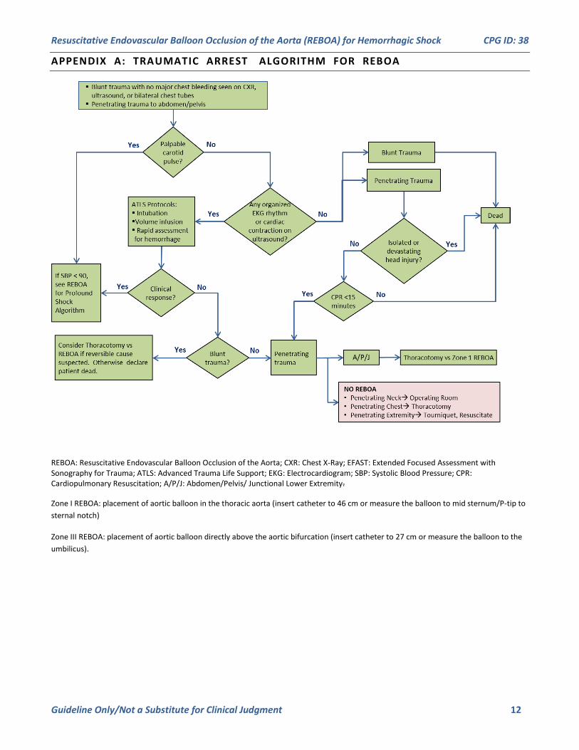
APPENDIX A: TRAUMATIC ARREST ALGORITHM FOR REBOA
REBOA: Resuscitative Endovascular Balloon Occlusion of the Aorta; CXR: Chest X-Ray; EFAST: Extended Focused Assessment with Sonography for Trauma; ATLS: Advanced Trauma Life Support; EKG: Electrocardiogram; SBP: Systolic Blood Pressure; CPR: Cardiopulmonary Resuscitation; A/P/J: Abdomen/Pelvis/ Junctional Lower Extremity.
Zone I REBOA: placement of aortic balloon in the thoracic aorta (insert catheter to 46 cm or measure the balloon to mid sternum/P-tip to
sternal notch)
Zone III REBOA: placement of aortic balloon directly above the aortic bifurcation (insert catheter to 27 cm or measure the balloon to the
umbilicus).
Resuscitative Endovascular Balloon Occlusion of the Aorta (REBOA) for Hemorrhagic Shock CPG ID: 38
Guideline Only/Not a Substitute for Clinical Judgment 13
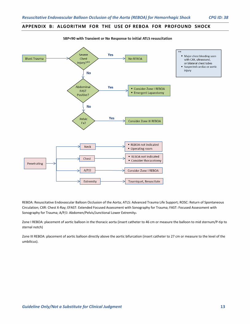
APPENDIX B: ALGORITHM FOR THE USE OF REBOA FOR PROFOUND SHOCK
REBOA: Resuscitative Endovascular Balloon Occlusion of the Aorta; ATLS: Advanced Trauma Life Support; ROSC: Return of Spontaneous
Circulation; CXR: Chest X-Ray; EFAST: Extended Focused Assessment with Sonography for Trauma; FAST: Focused Assessment with
Sonography for Trauma; A/P/J: Abdomen/Pelvis/Junctional Lower Extremity.
Zone I REBOA: placement of aortic balloon in the thoracic aorta (insert catheter to 46 cm or measure the balloon to mid sternum/P-tip to
sternal notch)
Zone III REBOA: placement of aortic balloon directly above the aortic bifurcation (insert catheter to 27 cm or measure to the level of the
umbilicus).
Resuscitative Endovascular Balloon Occlusion of the Aorta (REBOA) for Hemorrhagic Shock CPG ID: 38
Guideline Only/Not a Substitute for Clinical Judgment 14
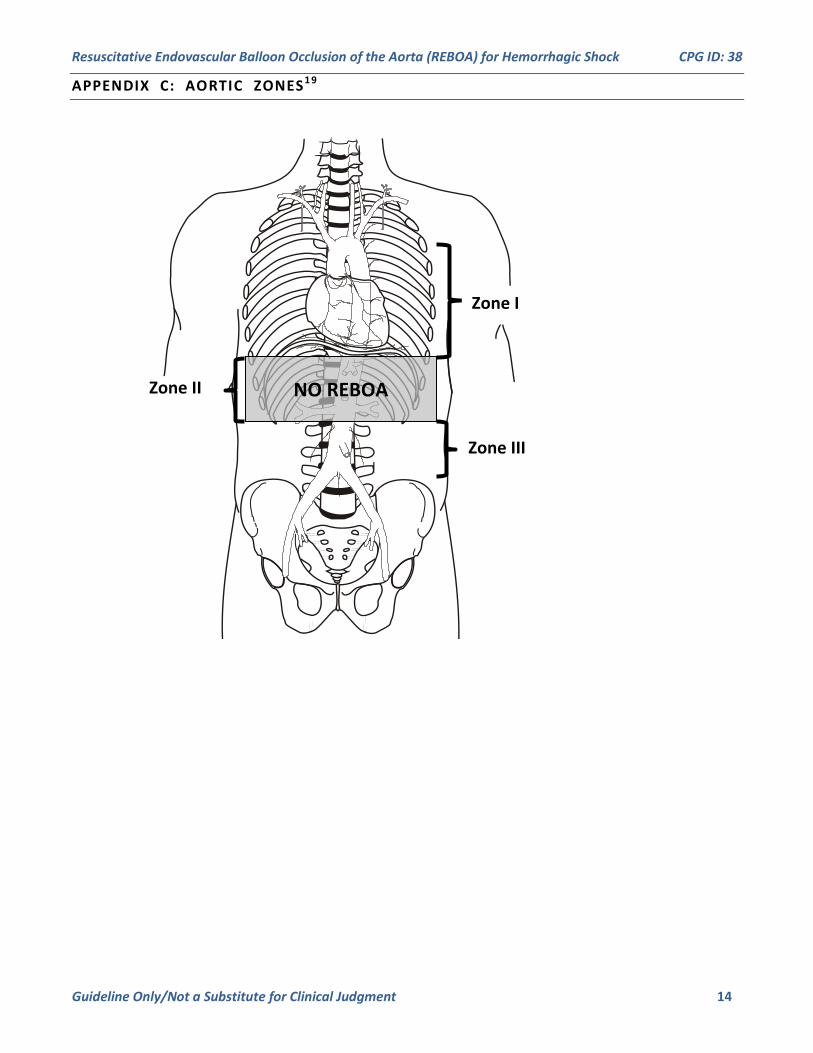
APPENDIX C: AORTIC ZONES1 9
Zone I
Zone II
Zone III
NO REBOA
Resuscitative Endovascular Balloon Occlusion of the Aorta (REBOA) for Hemorrhagic Shock CPG ID: 38
Guideline Only/Not a Substitute for Clinical Judgment 15
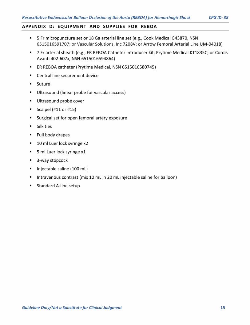
APPENDIX D: EQUIPMENT AND SUPPLIES FOR REBOA
5 Fr micropuncture set or 18 Ga arterial line set (e.g., Cook Medical G43870, NSN 6515016591707; or Vascular Solutions, Inc 7208V; or Arrow Femoral Arterial Line UM-04018)
7 Fr arterial sheath (e.g., ER REBOA Catheter Introducer kit, Prytime Medical KT1835C; or Cordis Avanti 402-607x, NSN 6515016594864)
ER REBOA catheter (Prytime Medical, NSN 6515016580745)
Central line securement device
Suture
Ultrasound (linear probe for vascular access)
Ultrasound probe cover
Scalpel (#11 or #15)
Surgical set for open femoral artery exposure
Silk ties
Full body drapes
10 ml Luer lock syringe x2
5 ml Luer lock syringe x1
3-way stopcock
Injectable saline (100 mL)
Intravenous contrast (mix 10 mL in 20 mL injectable saline for balloon)
Standard A-line setup

Resuscitative Endovascular Balloon Occlusion of the Aorta (REBOA) for Hemorrhagic Shock CPG ID: 38
Guideline Only/Not a Substitute for Clinical Judgment 16
APPENDIX E: REBOA STEPS USING THE 7 FRENCH ER-REBOA
The procedure may reviewed online using the following links: Part 1 : https://www.youtube.com/watch?v=-U7MkU3eA7E Part 2 : https://www.youtube.com/watch?v=DZ5LCEt7PBk
STEP 1: ARTERIAL ACCESS AND POSITIONING OF THE SHEATH
Establishing Arterial Access:
Access to the arterial circulation for REBOA for trauma should be obtained through the common femoral artery using one of three techniques: percutaneous, open exposure (e.g., cut down), or exchange over a guide wire from an existing common femoral arterial line. Percutaneous access can be obtained via landmark identification or ultrasound. Using landmarks, the location of the inguinal ligament is identified between the Anterior Superior Iliac Spine (ASIS) and pubic symphysis. The common femoral artery is then accessed 2 cm below the inguinal ligament. Alternatively, ultrasound is used to identify the common femoral artery above the branch of the profunda and the needle visualized passing into the common femoral artery (linear array transducer preferred). Ultrasound or direct surgical identification of the femoral artery is especially important in the hypotensive patient with no palpable pulse. Once identified, the artery should be entered at a 45-degree angle with the needle, using either a 5 Fr micropuncture kit or 18 gauge femoral arterial line kit. After the wire has been passed into the artery, the needle is removed and a small incision made at the interface of the wire and skin and the catheter is passed over the wire.
Selection and Positioning of Initial Sheath:
If REBOA is indicated, the arterial access catheter must be upsized to a 7 Fr sheath if an 18 gauge or 5 Fr arterial catheter was placed initially. This maneuver is accomplished by placing a 0.035 guide wire greater than twice the length of the existing arterial catheter through its inner lumen allowing the catheter to be removed over the wire while maintaining arterial access. After a larger opening is created at the wire/skin interface, the 7 Fr working sheath with its internal dilator in position can be inserted over the wire.
When urgently needed, a 7 Fr sheath may be placed as the initial step by placing the 7F sheath over the 0.035 guide wire.
It is important that any time a sheath is passed over a wire into the arterial system, the sheath’s internal dilator is firmly held in place to allow a smooth reverse taper from the wire to the diameter of the sheath to avoid arterial intimal injury. Once the dilator and sheath have been advanced over the wire through the skin into the artery, the dilator and wire are removed, leaving the sheath in place. To avoid bleeding from the side port of the sheath after the dilator is removed, it is important that the operator assure that the stopcock is in the “off” position to the patient.
STEP 2: SELECTION AND POSITIONING OF THE BALLOON
Selection of a Balloon:
The balloon which is best suited for austere environments is the ER REBOA (Prytime Medical) which is wire-free and smaller caliber than previously used balloons, allowing fewer steps for insertion and a smaller introducer sheath (7 Fr). It also has arterial pressure monitoring capability.
Alternative balloons should only be used by personnel with experience in the use of the specific catheter. Examples of compliant balloons suitable for aortic occlusion include: (1) Coda balloon (Cook Medical): 32 mm to
Resuscitative Endovascular Balloon Occlusion of the Aorta (REBOA) for Hemorrhagic Shock CPG ID: 38
Guideline Only/Not a Substitute for Clinical Judgment 17
40 mm, 14 Fr; (2) Reliant balloon (Medtronic): 10 mm to 46 mm, 12 Fr; and (3) Berenstein balloon (Boston Scientific): 11.5 mm, 6 Fr.
Balloon Preparation:
The ER REBOA balloon should be prepared by attaching a 25 ml syringe to the balloon port and ensuring that all air is evacuated from the balloon. The stop-cock can then be turned to the off position toward the balloon to ensure negative pressure within the balloon and allow the balloon to pass easily into the peel-away sheath.
If using the pressure monitoring capabilities, the pressure sensor and tubing should be attached to the catheter’s arterial stopcock and flushed with saline using standard arterial line setup and transducer connected to a monitor. Once the catheter is inserted, care must be taken to prevent inadvertent emboli (air, thrombus, etc.). This can be accomplished by always aspirating the catheter prior to flushing while the catheter is in place.
Balloon Positioning:
Aortic zones are designated Zone I, II, and III spanning from proximal to distal (Appendix C). Zone I is the descending thoracic aorta between the origin of the left subclavian to the celiac artery. Zone II is the paravisceral aorta between the celiac artery and the lowest renal artery and Zone III is the infrarenal aorta between the lowest renal artery and the aortic bifurcation. In most instances of shock and impending cardiovascular collapse, the goal is to position the balloon in Zone I, similar to an open thoracic aorta cross-clamp. Inflation of a balloon in Aortic Zone III is used for control of pelvic or junctional femoral hemorrhage.
For Zone I occlusion, the catheter should be inserted 46 cm (or measured with the balloon from the mid-sternal/P-tip from the sternal notch to the femoral access catheter). For Zone III occlusion, the P-tip should be inserted 27 cm (or measured from the umbilicus to the femoral access catheter). Distances are noted on the catheter shaft. The peel away sheath is advanced over the P-tip and balloon to protect these as they enter the 7F sheath. The peel away sheath is advanced into the end of the 7F sheath approximately 5mm or until it hits a “stop.” The REBOA catheter is then advanced 10-20 cm into the sheath. The peel away sheath can then be slid back onto the catheter hub or removed, if full advancement is necessary. The catheter should be advanced to the predetermined depth. Plain film x-ray, ultrasound, or fluoroscopy can confirm correct positioning of the catheter and adjustments can be made if necessary, prior to inflation. There are two radio-opaque markers on the catheter to designate the location of the balloon.
STEP 3: INFLATION OF THE BALLOON AND SECURING OF THE APPARATUS
Inflation of the Balloon:
A 10 ml syringe is filled with a 1:1 solution of sterile saline and iodinated contrast. The balloon should be inflated until the blood pressure is augmented and contralateral femoral pulse is stopped, approximately 8 ml for Zone I or 3 ml for Zone III. Do not over-inflate the balloon—balloon capacity is 24 ml—over-inflation will rupture the balloon or the blood vessel. If contrast or X-ray is not available, only sterile saline is used, however the use of contrast allows visualization of balloon inflation on X-ray and dilution allows rapid inflation and deflation times by reducing the viscosity of full-strength contrast. Balloon inflation should be guided by fluoroscopy, hemodynamic response, and/or loss of the contralateral pulse. When fluoroscopy is available, inflate the balloon until the outer edges of the balloon change from convex to parallel as the balloon takes on the contour of the aortic wall. When inflation appears adequate to gain aortic wall apposition and/or central blood pressure is augmented, the three-way stopcock on the shaft of the balloon should be turned off toward the balloon to maintain inflation and occlusion while other maneuvers are undertaken. If image guidance was not used for inflation, a confirmatory X-ray or ultrasound is recommended for radiographic confirmation of location. If no imaging is available in the austere environment, definitive confirmation of the balloon positioning should be accomplished directly with “hands-on” at the time of laparotomy. If the balloon is found to be mal-positioned (e.g., Zone II) the balloon should be deflated and catheter positioned to Zone I or III and the balloon re-inflated.
Resuscitative Endovascular Balloon Occlusion of the Aorta (REBOA) for Hemorrhagic Shock CPG ID: 38
Guideline Only/Not a Substitute for Clinical Judgment 18
Care should be made not to over-inflate the balloon as this may rupture the balloon and may damage or rupture the vessel.
Securing the Inflated Balloon and Sheath:
As the central aortic pressure improves, the catheter may be pushed caudally. To prevent catheter migration, a central line attachment device is sutured to the skin as close as possible to the sheath. For added monitoring and security, assign an assistant the task of holding the apparatus until balloon deflation is desired.
The assistant should monitor and communicate the “big three” factors imperative to maintenance of successful REBOA: mean arterial pressure, maintenance of position, and maintenance of occlusion (balloon inflation).
STEP 4: OPERATIVE/PROCEDURAL CONTROL OF BLEEDING
Control of bleeding below the diaphragm must occur very quickly when a Zone I REBOA is in place, with a goal to keep the total aortic occlusion time as short as possible, preferably less than 30 minutes and no more than 60 minutes. It is therefore important to start with temporizing maneuvers to control bleeding such as clamping of the splenic or renal hilum, Pringle maneuver, clamping of any injured blood vessel, packing, or obtaining proximal and distal control of an injured blood vessel. At times, definitive control of bleeding such as solid organ removal, ligation of clamped vessels, or vascular shunt placement, may be deferred until after the REBOA has been deflated.
Interventional radiology embolization may be considered when available, particularly with pelvic fractures after intra-abdominal hemorrhage has been ruled out or controlled and the REBOA has been positioned in Zone III.
STEP 5: DEFLATION OF THE BALLOON
Once specific vascular control or definitive hemorrhage control has been obtained, balloon deflation should be performed. Communication with the assistant holding the apparatus and the anesthesia team is critical before consideration of balloon deflation. Once a decision to attempt deflation is made, care must be taken to turn the three-way stopcock and deflate the balloon slowly as this step can be anticipated to result in a significant decrease in afterload and hypotension. Generally speaking, the main operator should be the person to deflate the balloon while the identified assistant continues to hold the balloon and sheath in the desired location. After deflation of the balloon, the team should anticipate hemodynamic changes related to reperfusion, washout of metabolic byproducts, and acidosis. As such, intermittent balloon inflation and deflation may be necessary with ongoing resuscitation until some hemodynamic stability is restored.
STEP 6: REMOVAL OF THE BALLOON AND SHEATH
After REBOA is no longer required, the deflated balloon may be removed from the 7 Fr sheath. The side port of this sheath can be used for arterial pressure monitoring. If the sheath is no longer necessary and coagulation has normalized, then the sheath can be removed. Ideally, the sheath should then be flushed with 100 mL of heparinized saline (1,000 units of heparin in 1 L of saline) as some thrombus may have formed during the process. If placed percutaneously, the sheath may be removed and direct pressure held at the arterial site, above the skin puncture site. Full occlusive direct pressure is held for 10 minutes, with gradual decrease in pressure every 5 minutes for 30 minutes total.
If the introducer sheath was placed by open arterial cutdown, then open surgical repair of the arterial access site is necessary. The femoral artery proximal and distal to the sheath entry site should be exposed to allow control. Proximally, this may require dissection for 2 cm to 3 cm underneath the inguinal ligament as an assistant uses a narrow handheld retractor (e.g., short Wylie renal vein retractor) to lift the inguinal ligament off of the femoral sheath. During this maneuver, the surgeon must be mindful of the circumflex iliac veins which course over the

Resuscitative Endovascular Balloon Occlusion of the Aorta (REBOA) for Hemorrhagic Shock CPG ID: 38
Guideline Only/Not a Substitute for Clinical Judgment 19
top of the distal external iliac and proximal common femoral artery. Exposure distal to the sheath entry site often requires identification and control of both the superficial and profunda femoris arteries. Once proximal and distal exposure and control with vessel loops or vascular clamps have been accomplished, the sheath may be removed. Consideration should be made for passage of embolectomy catheters distally to remove any potential clot and assure back bleeding. The resulting arteriotomy, especially the intima, should be closely examined and tailored with Potts scissors if necessary to allow primary transverse closure. Closure of the arteriotomy should be performed transversely using 5-0 or 6-0 permanent monofilament suture in either an interrupted or running fashion with care to capture all layers of the arterial wall with passage of the needle. Before closing the last suture, forward bleeding and back bleeding of the arterial segments should be allowed followed by flushing of the surface with heparinized saline. Restoration of flow through the arterial segment should be confirmed using manual palpation for pulses distally and use of continuous wave Doppler of both the artery and more distal extremity. If there is any question of flow, it is recommended to perform an angiogram and appropriate intervention if any abnormalities are noted. Closure of the soft tissues above the femoral artery is accomplished in layers using absorbable suture in the soft tissues.

Resuscitative Endovascular Balloon Occlusion of the Aorta (REBOA) for Hemorrhagic Shock CPG ID: 38
Guideline Only/Not a Substitute for Clinical Judgment 20
APPENDIX F: ER-REBOA PROCEDURE CHECKLIST
1. Resuscitate per ATLS
2. “Clear” chest (CXR/EFAST/Bilateral chest tubes)
3. FAST abdomen
4. Pelvic X-ray if blunt trauma
5. Check bilateral femoral pulses
6. Establish arterial access with 7 Fr Sheath
7. Measure balloon distance
8. Evacuate the balloon
9. Flush the arterial line
10. Insert the catheter
11. Inflate the balloon
12. Secure the catheter
13. Control major bleeding
14. Resuscitate and deflate the balloon
15. Complete damage control surgery
16. Remove sheath when coagulopathy corrected
17. Check distal pulses after sheath removal

Resuscitative Endovascular Balloon Occlusion of the Aorta (REBOA) for Hemorrhagic Shock CPG ID: 38
Guideline Only/Not a Substitute for Clinical Judgment 21
APPENDIX G: ADDITIONAL INFORMATION REGARDING OFF-LABEL USES IN CPGS
PURPOSE
The purpose of this Appendix is to ensure an understanding of DoD policy and practice regarding inclusion in CPGs of “off-label” uses of U.S. Food and Drug Administration (FDA)–approved products. This applies to off-label uses with patients who are armed forces members.
BACKGROUND
Unapproved (i.e., “off-label”) uses of FDA-approved products are extremely common in American medicine and are usually not subject to any special regulations. However, under Federal law, in some circumstances, unapproved uses of approved drugs are subject to FDA regulations governing “investigational new drugs.” These circumstances include such uses as part of clinical trials, and in the military context, command required, unapproved uses. Some command requested unapproved uses may also be subject to special regulations.
ADDITIONAL INFORMATION REGARDING OFF-LABEL USES IN CPGS
The inclusion in CPGs of off-label uses is not a clinical trial, nor is it a command request or requirement. Further, it does not imply that the Military Health System requires that use by DoD health care practitioners or considers it to be the “standard of care.” Rather, the inclusion in CPGs of off-label uses is to inform the clinical judgment of the responsible health care practitioner by providing information regarding potential risks and benefits of treatment alternatives. The decision is for the clinical judgment of the responsible health care practitioner within the practitioner-patient relationship.
ADDITIONAL PROCEDURES
1.0 Balanced Discussion
Consistent with this purpose, CPG discussions of off-label uses specifically state that they are uses not approved by the FDA. Further, such discussions are balanced in the presentation of appropriate clinical study data, including any such data that suggest caution in the use of the product and specifically including any FDA-issued warnings.
2.0 Quality Assurance Monitoring
With respect to such off-label uses, DoD procedure is to maintain a regular system of quality assurance monitoring of outcomes and known potential adverse events. For this reason, the importance of accurate clinical records is underscored.
3.0 Information to Patients
Good clinical practice includes the provision of appropriate information to patients. Each CPG discussing an unusual off-label use will address the issue of information to patients. When practicable, consideration will be given to including in an appendix an appropriate information sheet for distribution to patients, whether before or after use of the product. Information to patients should address in plain language: a) that the use is not approved by the FDA; b) the reasons why a DoD health care practitioner would decide to use the product for this purpose; and c) the potential risks associated with such use.