Embed Size (px)
Citation preview
Wig1 prevents cellular senescence by regulatingp21 mRNA decay through control of RISCrecruitment
Bong Cho Kim1,6, Hyung Chul Lee1,5,6,Je-Jung Lee1, Chang-Min Choi2,Dong-Kwan Kim3, Jae Cheol Lee4,Young-Gyu Ko5 and Jae-Seon Lee1,*1Division of Radiation Cancer Research, Korea Institute of Radiologicaland Medical Sciences, Seoul, Korea, 2Department of Pulmonary andCritical Care Medicine, Asan Medical Center, College of Medicine,University of Ulsan, Seoul, Korea, 3Department of Thoracic andCardiovascular Surgery, Asan Medical Center, College of Medicine,University of Ulsan, Seoul, Korea, 4Department of Oncology, AsanMedical Center, College of Medicine, University of Ulsan, Seoul,Korea and 5School of Life Sciences and Biotechnology, Korea University,Seoul, Korea
Premature senescence, a key strategy used to suppress
carcinogenesis, can be driven by p53/p21 proteins in
response to various stresses. Here, we demonstrate that
Wig1 plays a critical role in this process through regula-
tion of p21 mRNA stability. Wig1 controls the association
of Argonaute2 (Ago2), a central component of the
RNA-induced silencing complex (RISC), with target p21
mRNA via binding of the stem-loop structure near the
microRNA (miRNA) target site. Depletion of Wig1 prohib-
ited miRNA-mediated p21 mRNA decay and resulted in
premature senescence. Wig1 plays an essential role in
cell proliferation, as demonstrated in tumour xenografts
in mice, and Wig1 and p21 mRNA levels are inversely
correlated in human normal and cancer tissues. Together,
our data indicate a novel role of Wig1 in RISC target
accessibility, which is a key step in RNA-mediated gene
silencing. In addition, these findings indicate that fine-
tuning of p21 levels by Wig1 is essential for the prevention
of cellular senescence.
The EMBO Journal (2012) 31, 4289–4303. doi:10.1038/
emboj.2012.286; Published online 19 October 2012Subject Categories: RNA; cell cycleKeywords: cellular senescence; p21 mRNA stability; RISC
recruitment; RNA-binding protein; Wig1
Introduction
Cellular senescence was originally defined by the phenotype
of human fibroblasts undergoing replicative exhaustion
in vitro (Hayflick and Moorhead, 1961). Although this
phenotype represents a stable state of cell-cycle arrest,
cellular senescence is prematurely triggered in response to
diverse forms of cellular damage or stress. Because cellular
senescence limits the proliferative potential of premalignant
cells, this process is thought to be a key strategy for the
suppression of carcinogenesis (Schmitt et al, 2002; Chen et al,
2005; Lee et al, 2010). Premature senescence can be triggered
through two complementary pathways, which involve the
p53/p21 and p16/retinoblastoma (pRb) tumour suppressor
proteins. First, p21 (also known as Cip1/Waf1/CDKN1A),
which is a direct inhibitor of cyclin/cyclin-dependent kinase
(CDK) complexes, is an important player that induces cellular
senescence in response to DNA damage or oncogene
imbalance (Brugarolas et al, 1995; Deng et al, 1995; Wang
et al, 1999). The expression of p21 is strictly regulated
at the transcriptional level through p53-dependent and/or -
independent mechanisms and also at the post-transcriptional
and post-translational levels through mechanisms involving
mRNA stability, subcellular localization, and/or protein
stability (Sheikh et al, 1994; Gartel and Tyner, 1999; Abbas
and Dutta, 2009). Recently, post-transcriptional control
through a microRNA (miRNA)-mediated mRNA silencing
mechanism has been implicated as an important
mechanism of p21 regulation (Wu et al, 2010).
The miRNAs comprise a group of short (typically B22
nucleotides), non-coding RNAs that suppress the expression
of protein-coding genes by mRNA decay and/or suppress
translation through guiding the ribonucleoprotein RNA-
induced silencing complex (RISC), which contains the
Argonaute (Ago) proteins (Bartel, 2009). RISC-loaded
miRNAs recognize target sites in the 30-untranslated regions
(UTRs) of their target mRNAs (Kawamata and Tomari, 2010).
The most critical requirement for target recognition is
complementary base pairing between the target site and the
50 region of the miRNA, the so-called canonical ‘seed’ region
(Grimson et al, 2007; Bartel, 2009). For post-transcriptional
control, modulation of miRNA function through miRNA
biogenesis, localization, and degradation is a key process.
In addition, miRNA activity is enhanced or hindered by
RNA-binding proteins (RBPs; van Kouwenhove et al, 2011).
For example, HuR, an Elav-like protein, binds to the 30UTR of
cationic amino-acid transporter 1 (CAT1) mRNA and relieves
miR-122-mediated repression (Bhattacharyya et al, 2006).
On the other hand, HuR facilitates c-Myc repression by
recruiting let-7-loaded RICS via an association with the c-Myc
30UTR that neighbours a let-7-binding site (Kim et al, 2009a).
Thus, RBPs play fundamental roles in post-transcriptional
control, which is regulated by different processes of mRNA
metabolism and translation (Kim et al, 2009b). Since miRNA
target recognition is a key process for RISC functions, the RBPs
governing RISC-loaded miRNA recruitment to its target are
waiting to be uncovered (Wiemer, 2007; Kawamata and
Tomari, 2010; van Kouwenhove et al, 2011).
Wig1 (wild-type p53-induced gene 1; official gene symbol
is ZMAT3, zinc finger, matrin-type 3) encodes a Cys2His2-type
zinc finger protein that contains three zinc fingers (ZFs) and a
*Corresponding author. Division of Radiation Cancer Research, KoreaInstitute of Radiological and Medical Sciences, 75 Nowongil, Nowon Gu,Seoul 139-706, Korea. Tel.: þ82 2 970 1388; Fax: þ82 2 970 1388;E-mail: [email protected] authors contributed equally to this work.
Received: 13 March 2012; accepted: 27 September 2012; publishedonline: 19 October 2012
The EMBO Journal (2012) 31, 4289–4303 | & 2012 European Molecular Biology Organization |All Rights Reserved 0261-4189/12
www.embojournal.org
EMBO
THE
EMBOJOURNAL
THE
EMBOJOURNAL
4289&2012 European Molecular Biology Organization The EMBO Journal VOL 31 | NO 22 | 2012
nuclear localization signal (NLS) (Israeli et al, 1997; Varmeh-
Ziaie et al, 1997). Wig1 was identified as a p53-responsive
gene and is highly conserved from fish to humans, especially
within the ZFs, which are almost completely conserved
(Wilhelm et al, 2002; Hellborg and Wiman, 2004). Wig1
interacts with heterogeneous nuclear ribonucleoprotein
(hnRNP) A2/B1 and RNA helicase A (RHA) via dsRNA
binding and regulates p53 mRNA stability through the
protection of deadenylation (Mendez Vidal et al, 2006;
Prahl et al, 2008; Vilborg et al, 2009). Despite several
previous studies, the function and biological significance of
Wig1 remain nebulous.
In this study, we show that Wig1 depletion induces pre-
mature senescence due to p21 mRNA stabilization. Wig1
preferentially interacts with the stem-loop structure of the
30UTR that neighbours miRNA target sites to determine
the target accessibility of RISC. Our data describe a novel
role for Wig1 in modulating miRNA-mediated mRNA decay.
Results
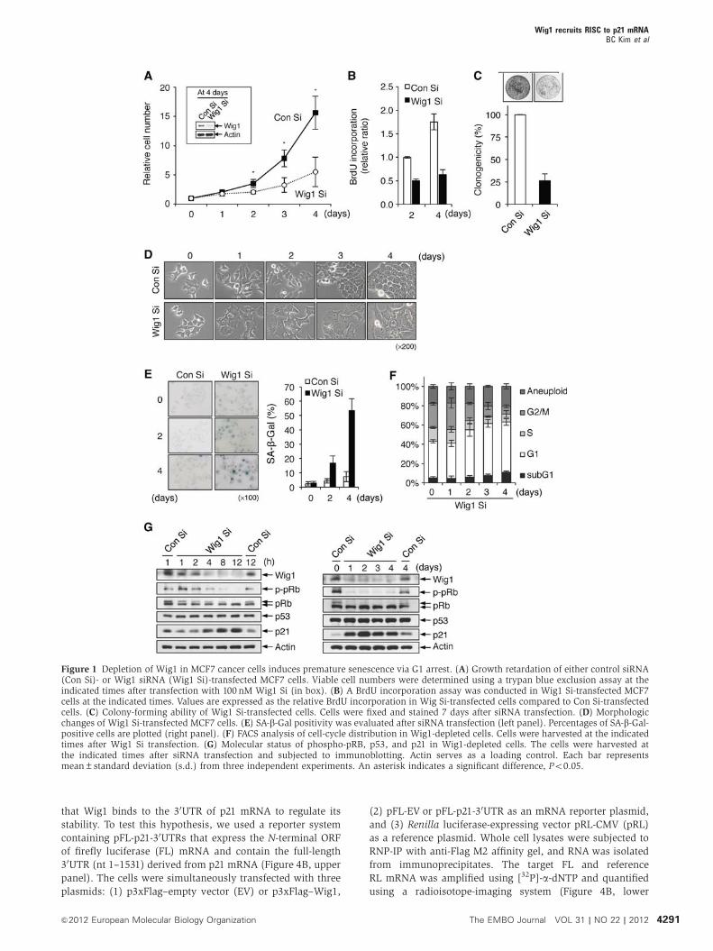
Wig1 depletion induces premature senescence
Wig1 has been identified as a p53-responsive gene and has
been suggested to play a role in the p53-dependent stress
response (Israeli et al, 1997; Hellborg et al, 2001); however,
its biological significance has yet to be elucidated. To investi-
gate the biological function of Wig1, we first examined the
influence of Wig1 depletion on cell growth, cellular morphol-
ogy, and apoptosis. MCF7 breast cancer cells, which were
treated with an siRNA against Wig1 (Wig Si), exhibited decreased
cell proliferation and clonogenic ability (Figure 1A–C).
Interestingly, Wig1 depletion induced typical senescence-
associated phenotypic changes: large and flattened morpho-
logy, positive senescence-associated b-galactosidase (SA-b-Gal) staining, and G1 cell-cycle arrest (Figure 1D–F). No
significant increase in the subG1 phase was observed
(Figure 1F). Wig1 depletion contributed to the gradual de-
crease in phospho-pRb and the increase in p21, and such
changes were sustained regardless of p53 activation (Figure 1G).
The premature senescent phenotypes were observed up to 10
days after the depletion of Wig1 (Supplementary Figure S1).
We confirmed a lack of off-target effects of Wig1 Si in p21
accumulation and induction of premature senescence
(Supplementary Figure S2). Wig1 depletion also resulted in
premature senescence phenotypes including p21 induction in
other cell types, such as H460 lung carcinoma cells, U2OS
osteosarcoma cells, MCF10A normal mammary epithelial
cells, and primary human diploid fibroblasts (Supplementary
Figures S3 and S4). Overexpression of siRNA-resistant Wig1
resulted in rescue of the abovementioned premature senes-
cent phenotypes observed following the depletion of Wig 1
(Supplementary Figure S5). These data demonstrate that
Wig1 depletion induces p21 accumulation and hypopho-
sphorylation of pRb and ultimately leads to premature senes-
cence in both normal and cancer cells.
p21 plays an essential role in Wig1 depletion-mediated
premature senescence
Next, we investigated the involvement of p53 and p21 in
premature senescence induced by Wig1 depletion. Wig1
depletion resulted in p21 accumulation and decreased cell
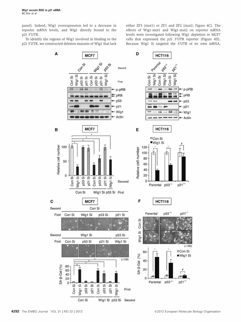
proliferation in p53-depleted MCF7 cells (Figure 2A and B).
Analysis of SA-b-Gal staining demonstrated that Wig1 deple-
tion induced premature senescence even in the absence of
p53 (Figure 2C). In contrast, p21 depletion abrogated Wig1
depletion-mediated premature senescence (Figure 2A–C). To
obtain additional evidence, we examined Wig1 depletion-
mediated premature senescence in HCT116parental,
HCT116p53� /� , and HCT116p21� /� isogenic cell lines. In
accordance with HCT116parental cells, HCT116p53� /� cells
exhibited increased p21 levels, limited cell proliferation, and
positivity of SA-b-Gal staining in the absence of Wig1
(Figure 2D–F), whereas Wig1 depletion did not induce pre-
mature senescence in HCT116p21� /� cells, which exhibited
no significant alterations in cell growth rate and SA-b-Galpositivity (Figure 2D–F). In the absence of p53, however,
Wig1 depletion-mediated premature senescence was lessened
due to a decreased level of p21 as compared to cells with
intact p53 (Figure 2). These results indicate that p21 accu-
mulation is indispensible for the Wig1 depletion-mediated
premature senescence.
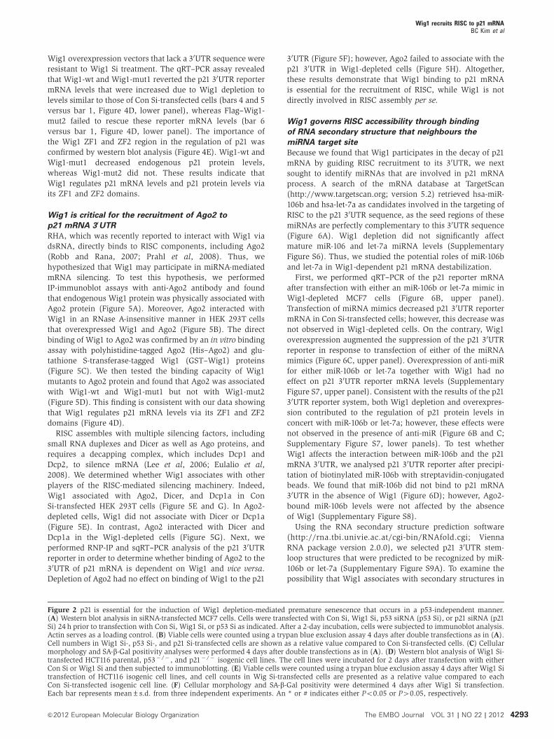
Wig1 modulates p21 levels via the regulation of
p21 mRNA stability
To define the regulatory mechanism of p21 accumulation in
Wig1-depleted cells, we examined the transcriptional activity,
mRNA stability, and protein stability of p21. No significant
difference was found in p21 promoter activity, which was
measured using a luciferase reporter containing the p21 pro-
moter (WWP-Luc), in Wig1 Si- and control siRNA (Con Si)-
transfected MCF7 cells (Figure 3A). To determine whether
Wig1 regulates p21 expression at the post-translational level,
we treated cells with cycloheximide (CHX), an inhibitor of
protein biosynthesis. Western blot analysis revealed that the
p21 protein degradation rate was not affected by Wig1 deple-
tion (Figure 3B). Next, to examine the change in p21 mRNA
levels following Wig1 depletion, we performed quantitative
real-time reverse transcription-PCR (qRT–PCR). The p21
mRNA levels were inversely correlated with Wig1 mRNA
levels (Figure 3C). To explore the decay rate of the p21
transcript, actinomycin D (ActD) chase and qRT–PCR assays
were performed. As shown in Figure 3D, the half-life of the
p21 transcript was significantly increased due to Wig1 deple-
tion (3.29 versus 5.34 h). To obtain additional evidence of p21
regulation by Wig1, the levels of p21 mRNA and protein were
measured in Flag-tagged Wig1 (Flag–Wig1)-overexpressed
and in Wig1-depleted cells. Overexpression and depletion of
Wig 1 downregulated and upregulated p21, respectively, at
both the mRNA and protein levels (Figure 3E). These results
demonstrate that Wig1 regulates the p21 protein level via
regulation of p21 mRNA decay.
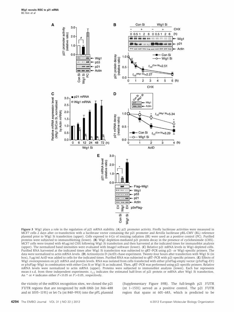
Wig1 binds to the 30UTR of p21 mRNA through ZF
domains 1 and 2
Because Wig1 is a ZF protein that contains an unusual
dsRNA-binding domain (Mendez Vidal et al, 2006), we
investigated whether Wig1 directly binds to p21 mRNA
using ribonucleoprotein immunoprecipitation (RNP-IP) and
a semiquantitative (sq) RT–PCR assay. As shown in
Figure 4A, Wig1 associated with the p21 mRNA and reduced
its level in Wig1-overexpressing MCF7 cells. In general,
cis-acting determinants located in the 30UTR regulate the
stability of mRNA by association with specific proteins
(Guhaniyogi and Brewer, 2001). Therefore, we hypothesized
Wig1 recruits RISC to p21 mRNABC Kim et al
4290 The EMBO Journal VOL 31 | NO 22 | 2012 &2012 European Molecular Biology Organization
that Wig1 binds to the 30UTR of p21 mRNA to regulate its
stability. To test this hypothesis, we used a reporter system
containing pFL-p21-30UTRs that express the N-terminal ORF
of firefly luciferase (FL) mRNA and contain the full-length
30UTR (nt 1–1531) derived from p21 mRNA (Figure 4B, upper
panel). The cells were simultaneously transfected with three
plasmids: (1) p3xFlag–empty vector (EV) or p3xFlag–Wig1,
(2) pFL-EV or pFL-p21-30UTR as an mRNA reporter plasmid,
and (3) Renilla luciferase-expressing vector pRL-CMV (pRL)
as a reference plasmid. Whole cell lysates were subjected to
RNP-IP with anti-Flag M2 affinity gel, and RNA was isolated
from immunoprecipitates. The target FL and reference
RL mRNA was amplified using [32P]-a-dNTP and quantified
using a radioisotope-imaging system (Figure 4B, lower
Figure 1 Depletion of Wig1 in MCF7 cancer cells induces premature senescence via G1 arrest. (A) Growth retardation of either control siRNA(Con Si)- or Wig1 siRNA (Wig1 Si)-transfected MCF7 cells. Viable cell numbers were determined using a trypan blue exclusion assay at theindicated times after transfection with 100 nM Wig1 Si (in box). (B) A BrdU incorporation assay was conducted in Wig1 Si-transfected MCF7cells at the indicated times. Values are expressed as the relative BrdU incorporation in Wig Si-transfected cells compared to Con Si-transfectedcells. (C) Colony-forming ability of Wig1 Si-transfected cells. Cells were fixed and stained 7 days after siRNA transfection. (D) Morphologicchanges of Wig1 Si-transfected MCF7 cells. (E) SA-b-Gal positivity was evaluated after siRNA transfection (left panel). Percentages of SA-b-Gal-positive cells are plotted (right panel). (F) FACS analysis of cell-cycle distribution in Wig1-depleted cells. Cells were harvested at the indicatedtimes after Wig1 Si transfection. (G) Molecular status of phospho-pRB, p53, and p21 in Wig1-depleted cells. The cells were harvested atthe indicated times after siRNA transfection and subjected to immunoblotting. Actin serves as a loading control. Each bar representsmean±standard deviation (s.d.) from three independent experiments. An asterisk indicates a significant difference, Po0.05.
Wig1 recruits RISC to p21 mRNABC Kim et al
4291&2012 European Molecular Biology Organization The EMBO Journal VOL 31 | NO 22 | 2012
panel). Indeed, Wig1 overexpression led to a decrease in
reporter mRNA levels, and Wig1 directly bound to the
p21 30UTR.
To identify the regions of Wig1 involved in binding to the
p21 30UTR, we constructed deletion mutants of Wig1 that lack
either ZF3 (mut1) or ZF1 and ZF2 (mut2; Figure 4C). The
effects of Wig1-mut1 and Wig1-mut2 on reporter mRNA
levels were investigated following Wig1 depletion in MCF7
cells that expressed the p21 30UTR reporter (Figure 4D).
Because Wig1 Si targeted the 30UTR of its own mRNA,
Wig1 recruits RISC to p21 mRNABC Kim et al
4292 The EMBO Journal VOL 31 | NO 22 | 2012 &2012 European Molecular Biology Organization
Wig1 overexpression vectors that lack a 30UTR sequence were
resistant to Wig1 Si treatment. The qRT–PCR assay revealed
that Wig1-wt and Wig1-mut1 reverted the p21 30UTR reporter
mRNA levels that were increased due to Wig1 depletion to
levels similar to those of Con Si-transfected cells (bars 4 and 5
versus bar 1, Figure 4D, lower panel), whereas Flag–Wig1-
mut2 failed to rescue these reporter mRNA levels (bar 6
versus bar 1, Figure 4D, lower panel). The importance of
the Wig1 ZF1 and ZF2 region in the regulation of p21 was
confirmed by western blot analysis (Figure 4E). Wig1-wt and
Wig1-mut1 decreased endogenous p21 protein levels,
whereas Wig1-mut2 did not. These results indicate that
Wig1 regulates p21 mRNA levels and p21 protein levels via
its ZF1 and ZF2 domains.
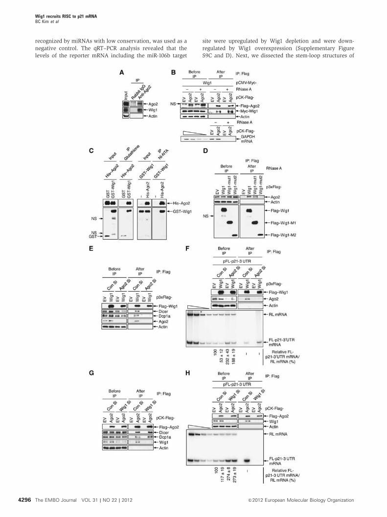
Wig1 is critical for the recruitment of Ago2 to
p21 mRNA 30UTR
RHA, which was recently reported to interact with Wig1 via
dsRNA, directly binds to RISC components, including Ago2
(Robb and Rana, 2007; Prahl et al, 2008). Thus, we
hypothesized that Wig1 may participate in miRNA-mediated
mRNA silencing. To test this hypothesis, we performed
IP-immunoblot assays with anti-Ago2 antibody and found
that endogenous Wig1 protein was physically associated with
Ago2 protein (Figure 5A). Moreover, Ago2 interacted with
Wig1 in an RNase A-insensitive manner in HEK 293T cells
that overexpressed Wig1 and Ago2 (Figure 5B). The direct
binding of Wig1 to Ago2 was confirmed by an in vitro binding
assay with polyhistidine-tagged Ago2 (His–Ago2) and glu-
tathione S-transferase-tagged Wig1 (GST–Wig1) proteins
(Figure 5C). We then tested the binding capacity of Wig1
mutants to Ago2 protein and found that Ago2 was associated
with Wig1-wt and Wig1-mut1 but not with Wig1-mut2
(Figure 5D). This finding is consistent with our data showing
that Wig1 regulates p21 mRNA levels via its ZF1 and ZF2
domains (Figure 4D).
RISC assembles with multiple silencing factors, including
small RNA duplexes and Dicer as well as Ago proteins, and
requires a decapping complex, which includes Dcp1 and
Dcp2, to silence mRNA (Lee et al, 2006; Eulalio et al,
2008). We determined whether Wig1 associates with other
players of the RISC-mediated silencing machinery. Indeed,
Wig1 associated with Ago2, Dicer, and Dcp1a in Con
Si-transfected HEK 293T cells (Figure 5E and G). In Ago2-
depleted cells, Wig1 did not associate with Dicer or Dcp1a
(Figure 5E). In contrast, Ago2 interacted with Dicer and
Dcp1a in the Wig1-depleted cells (Figure 5G). Next, we
performed RNP-IP and sqRT–PCR analysis of the p21 30UTR
reporter in order to determine whether binding of Ago2 to the
30UTR of p21 mRNA is dependent on Wig1 and vice versa.
Depletion of Ago2 had no effect on binding of Wig1 to the p21
30UTR (Figure 5F); however, Ago2 failed to associate with the
p21 30UTR in Wig1-depleted cells (Figure 5H). Altogether,
these results demonstrate that Wig1 binding to p21 mRNA
is essential for the recruitment of RISC, while Wig1 is not
directly involved in RISC assembly per se.
Wig1 governs RISC accessibility through binding
of RNA secondary structure that neighbours the
miRNA target site
Because we found that Wig1 participates in the decay of p21
mRNA by guiding RISC recruitment to its 30UTR, we next
sought to identify miRNAs that are involved in p21 mRNA
process. A search of the mRNA database at TargetScan
(http://www.targetscan.org; version 5.2) retrieved hsa-miR-
106b and hsa-let-7a as candidates involved in the targeting of
RISC to the p21 30UTR sequence, as the seed regions of these
miRNAs are perfectly complementary to this 30UTR sequence
(Figure 6A). Wig1 depletion did not significantly affect
mature miR-106 and let-7a miRNA levels (Supplementary
Figure S6). Thus, we studied the potential roles of miR-106b
and let-7a in Wig1-dependent p21 mRNA destabilization.
First, we performed qRT–PCR of the p21 reporter mRNA
after transfection with either an miR-106b or let-7a mimic in
Wig1-depleted MCF7 cells (Figure 6B, upper panel).
Transfection of miRNA mimics decreased p21 30UTR reporter
mRNA in Con Si-transfected cells; however, this decrease was
not observed in Wig1-depleted cells. On the contrary, Wig1
overexpression augmented the suppression of the p21 30UTR
reporter in response to transfection of either of the miRNA
mimics (Figure 6C, upper panel). Overexpression of anti-miR
for either miR-106b or let-7a together with Wig1 had no
effect on p21 30UTR reporter mRNA levels (Supplementary
Figure S7, upper panel). Consistent with the results of the p21
30UTR reporter system, both Wig1 depletion and overexpres-
sion contributed to the regulation of p21 protein levels in
concert with miR-106b or let-7a; however, these effects were
not observed in the presence of anti-miR (Figure 6B and C;
Supplementary Figure S7, lower panels). To test whether
Wig1 affects the interaction between miR-106b and the p21
mRNA 30UTR, we analysed p21 30UTR reporter after precipi-
tation of biotinylated miR-106b with streptavidin-conjugated
beads. We found that miR-106b did not bind to p21 mRNA
30UTR in the absence of Wig1 (Figure 6D); however, Ago2-
bound miR-106b levels were not affected by the absence
of Wig1 (Supplementary Figure S8).
Using the RNA secondary structure prediction software
(http://rna.tbi.univie.ac.at/cgi-bin/RNAfold.cgi; Vienna
RNA package version 2.0.0), we selected p21 30UTR stem-
loop structures that were predicted to be recognized by miR-
106b or let-7a (Supplementary Figure S9A). To examine the
possibility that Wig1 associates with secondary structures in
Figure 2 p21 is essential for the induction of Wig1 depletion-mediated premature senescence that occurs in a p53-independent manner.(A) Western blot analysis in siRNA-transfected MCF7 cells. Cells were transfected with Con Si, Wig1 Si, p53 siRNA (p53 Si), or p21 siRNA (p21Si) 24 h prior to transfection with Con Si, Wig1 Si, or p53 Si as indicated. After a 2-day incubation, cells were subjected to immunoblot analysis.Actin serves as a loading control. (B) Viable cells were counted using a trypan blue exclusion assay 4 days after double transfections as in (A).Cell numbers in Wig1 Si-, p53 Si-, and p21 Si-transfected cells are shown as a relative value compared to Con Si-transfected cells. (C) Cellularmorphology and SA-b-Gal positivity analyses were performed 4 days after double transfections as in (A). (D) Western blot analysis of Wig1 Si-transfected HCT116 parental, p53� /� , and p21� /� isogenic cell lines. The cell lines were incubated for 2 days after transfection with eitherCon Si or Wig1 Si and then subjected to immunoblotting. (E) Viable cells were counted using a trypan blue exclusion assay 4 days after Wig1 Sitransfection of HCT116 isogenic cell lines, and cell counts in Wig Si-transfected cells are presented as a relative value compared to eachCon Si-transfected isogenic cell line. (F) Cellular morphology and SA-b-Gal positivity were determined 4 days after Wig1 Si transfection.Each bar represents mean±s.d. from three independent experiments. An * or # indicates either Po0.05 or P40.05, respectively.
Wig1 recruits RISC to p21 mRNABC Kim et al
4293&2012 European Molecular Biology Organization The EMBO Journal VOL 31 | NO 22 | 2012
the vicinity of the miRNA recognition sites, we cloned the p21
30UTR regions that are recognized by miR-106b (nt 366–488
and nt 1035–1191) or let-7a (nt 840–993) into the pFL plasmid
(Supplementary Figure S9B). The full-length p21 30UTR
(nt 1–1531) served as a positive control. The p21 30UTR
region that spans nt 601–683, which is predicted to be
Figure 3 Wig1 plays a role in the regulation of p21 mRNA stability. (A) p21 promoter activity. Firefly luciferase activities were measured inMCF7 cells 2 days after co-transfection with a luciferase vector containing the p21 promoter and Renilla luciferase pRL-CMV (RL) referenceplasmid prior to Wig1 Si transfection (upper). Cells exposed to 6Gy of ionizing radiation (IR) were used as a positive control (PC). Purifiedproteins were subjected to immunoblotting (lower). (B) Wig1 depletion-mediated p21 protein decay in the presence of cycloheximide (CHX).MCF7 cells were treated with 40 mg/ml CHX following Wig1 Si transfection and then harvested at the indicated times for immunoblot analysis(upper). The normalized band intensities were evaluated with ImageJ software (lower). (C) Relative p21 mRNA levels in Wig1-depleted cells.Purified RNA harvested at the indicated times after Wig1 Si transfection was subjected to qRT–PCR using p21- or Wig1-specific primers. Thedata were normalized to actin mRNA levels. (D) Actinomycin D (ActD) chase experiment. Twenty-four hours after transfection with Wig1 Si (inbox), 5 mg/ml ActD was added to cells for the indicated times. Purified RNAwas subjected to qRT–PCR with p21-specific primers. (E) Effects ofWig1 overexpression on p21 mRNA and protein levels. RNAwas isolated from cells transfected with either p3xFlag-empty vector (p3xFlag–EV)or p3xFlag–Wig1 in combination with either Con Si or Wig1 Si as indicated. Then, qRT–PCR was performed using p21-specific primers. RelativemRNA levels were normalized to actin mRNA (upper). Proteins were subjected to immunoblot analysis (lower). Each bar representsmean±s.d. from three independent experiments. t1/2 indicates the estimated half-lives of p21 protein or mRNA after Wig1 Si transfection.An * or # indicates either Po0.05 or P40.05, respectively.
Wig1 recruits RISC to p21 mRNABC Kim et al
4294 The EMBO Journal VOL 31 | NO 22 | 2012 &2012 European Molecular Biology Organization
Figure 4 Wig1 binds to the 30UTR of the p21 mRNA through zinc finger domains 1 and 2. (A) Ribonucleoprotein immunoprecipitation(RNP-IP) assay. MCF7 cells were harvested 2 days after transfection with either p3xFlag–EVor p3xFlag–Wig1 (Flag–Wig1), and then an RNP-IPassay using anti-Flag M2 affinity gel was performed. Immunoprecipitated proteins were then subjected to immunoblotting using anti-Flagantibody (upper). Isolated total RNA was radiolabelled with RT–PCR using p21-specific primers (lower), and band intensities were quantifiedusing imaging software. The four leftmost lanes represent two-fold serial dilutions of RNA and demonstrate that the RT–PCR issemiquantitative. (B) Interaction of Wig1 and p21 30UTR reporter. Schematic representations of pFL-EV and pFL-p21-30UTR, which encodesthe N-terminal portion of firefly luciferase with the full-length 30UTR of p21 (upper). HEK 293T cells were transfected with either pFL-EV orpFL-p21-30UTR in combination with p3xFlag–EVor p3xFlag–Wig1. Cells were also co-transfected with pRL-CMV (pRL) as a reference plasmid.Cells were harvested 2 days after transfection and subjected to RNP-IP using anti-Flag M2 affinity gel. Purified RNA from IP was radiolabelledusing RT–PCR using RL- and FL-specific primers, and products were subjected to PAGE. The p21 30UTR reporter mRNA levels were quantifiedusing imaging software, and data are presented as mean±s.d. from three independent experiments (lower). (C) Schematic representations ofthe p3xFlag–Wig1-wild-type (wt), p3xFlag–Wig1-mutant 1 (mut1), and p3xFlag–Wig1-mutant 2 (mut2) overexpression plasmids. ZF, zincfinger; NLS, nuclear localization signal. (D) Immunoblotting and qRT–PCR. MCF7 cells were transfected with Wig1 Si prior to transfectionwith pFL-p21-30UTR in combination with p3xFlag–EV, p3xFlag–Wig1-wt, p3xFlag–Wig1-mut1, or p3xFlag–Wig1-mut2. The cells were alsoco-transfected with pRL-CMV as a reference plasmid. Whole cell lysates and RNA were isolated 2 days after transfection. Endogenous Wig1depletion and recombinant Wig1 overexpression were assessed using immunoblot analysis (upper). The qRT–PCR analysis shows the relativelevels of FL-p21-30UTR reporter mRNA (lower). Each bar represents mean±s.d. from three independent experiments. An * or # indicates eitherPo0.05 or P40.05, respectively. (E) Endogenous p21 protein levels in cells overexpressing recombinant Wig1. MCF7 cells were harvested 2days after transfection with p3xFlag–EV, p3xFlag–Wig1-wt, p3xFlag–Wig1-mut1, or p3xFlag–Wig1-mut2 and subjected to immunoblotting.
Wig1 recruits RISC to p21 mRNABC Kim et al
4295&2012 European Molecular Biology Organization The EMBO Journal VOL 31 | NO 22 | 2012
recognized by miRNAs with low conservation, was used as a
negative control. The qRT–PCR analysis revealed that the
levels of the reporter mRNA including the miR-106b target
site were upregulated by Wig1 depletion and were down-
regulated by Wig1 overexpression (Supplementary Figure
S9C and D). Next, we dissected the stem-loop structures of
Wig1 recruits RISC to p21 mRNABC Kim et al
4296 The EMBO Journal VOL 31 | NO 22 | 2012 &2012 European Molecular Biology Organization
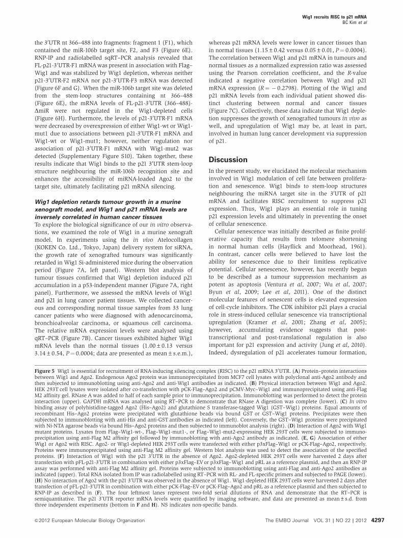
the 30UTR nt 366–488 into fragments: fragment 1 (F1), which
contained the miR-106b target site, F2, and F3 (Figure 6E).
RNP-IP and radiolabelled sqRT–PCR analysis revealed that
FL-p21-30UTR-F1 mRNAwas present in association with Flag–
Wig1 and was stabilized by Wig1 depletion, whereas neither
p21-30UTR-F2 mRNA nor p21-30UTR-F3 mRNA was detected
(Figure 6F and G). When the miR-106b target site was deleted
from the stem-loop structures containing nt 366–488
(Figure 6E), the mRNA levels of FL-p21-30UTR (366–488)-
DmiR were not regulated in the Wig1-depleted cells
(Figure 6H). Furthermore, the levels of p21-30UTR-F1 mRNA
were decreased by overexpression of either Wig1-wt or Wig1-
mut1 due to associations between p21-30UTR-F1 mRNA and
Wig1-wt or Wig1-mut1; however, neither regulation nor
association of p21-30UTR-F1 mRNA with Wig1-mut2 was
detected (Supplementary Figure S10). Taken together, these
results indicate that Wig1 binds to the p21 30UTR stem-loop
structure neighbouring the miR-106b recognition site and
enhances the accessibility of miRNA-loaded Ago2 to the
target site, ultimately facilitating p21 mRNA silencing.
Wig1 depletion retards tumour growth in a murine
xenograft model, and Wig1 and p21 mRNA levels are
inversely correlated in human cancer tissues
To explore the biological significance of our in vitro observa-
tions, we examined the role of Wig1 in a murine xenograft
model. In experiments using the in vivo Atelocollagen
(KOKEN Co. Ltd., Tokyo, Japan) delivery system for siRNA,
the growth rate of xenografted tumours was significantly
retarded in Wig1 Si-administered mice during the observation
period (Figure 7A, left panel). Western blot analysis of
tumour tissues confirmed that Wig1 depletion induced p21
accumulation in a p53-independent manner (Figure 7A, right
panel). Furthermore, we assessed the mRNA levels of Wig1
and p21 in lung cancer patient tissues. We collected cancer-
ous and corresponding normal tissue samples from 33 lung
cancer patients who were diagnosed with adenocarcinoma,
bronchioalveolar carcinoma, or squamous cell carcinoma.
The relative mRNA expression levels were analysed using
qRT–PCR (Figure 7B). Cancer tissues exhibited higher Wig1
mRNA levels than the normal tissues (1.00±0.13 versus
3.14±0.54, P¼ 0.0004; data are presented as mean±s.e.m.),
whereas p21 mRNA levels were lower in cancer tissues than
in normal tissues (1.15±0.42 versus 0.05±0.01, P¼ 0.0004).
The correlation between Wig1 and p21 mRNA in tumours and
normal tissues as a normalized expression ratio was assessed
using the Pearson correlation coefficient, and the R-value
indicated a negative correlation between Wig1 and p21
mRNA expression (R¼ � 0.2798). Plotting of the Wig1 and
p21 mRNA levels from each individual patient showed dis-
tinct clustering between normal and cancer tissues
(Figure 7C). Collectively, these data indicate that Wig1 deple-
tion suppresses the growth of xenografted tumours in vivo as
well, and upregulation of Wig1 may be, at least in part,
involved in human lung cancer development via suppression
of p21.
Discussion
In the present study, we elucidated the molecular mechanism
involved in Wig1 modulation of cell fate between prolifera-
tion and senescence. Wig1 binds to stem-loop structures
neighbouring the miRNA target site in the 30UTR of p21
mRNA and facilitates RISC recruitment to suppress p21
expression. Thus, Wig1 plays an essential role in tuning
p21 expression levels and ultimately in preventing the onset
of cellular senescence.
Cellular senescence was initially described as finite prolif-
erative capacity that results from telomere shortening
in normal human cells (Hayflick and Moorhead, 1961).
In contrast, cancer cells were believed to have lost the
ability for senescence due to their limitless replicative
potential. Cellular senescence, however, has recently begun
to be described as a tumour suppression mechanism as
potent as apoptosis (Ventura et al, 2007; Wu et al, 2007;
Byun et al, 2009; Lee et al, 2011). One of the distinct
molecular features of senescent cells is elevated expression
of cell-cycle inhibitors. The CDK inhibitor p21 plays a crucial
role in stress-induced cellular senescence via transcriptional
upregulation (Kramer et al, 2001; Zhang et al, 2005);
however, accumulating evidence suggests that post-
transcriptional and post-translational regulation is also
important for p21 expression and activity (Jung et al, 2010).
Indeed, dysregulation of p21 accelerates tumour formation,
Figure 5 Wig1 is essential for recruitment of RNA-inducing silencing complex (RISC) to the p21 mRNA 30UTR. (A) Protein–protein interactionsbetween Wig1 and Ago2. Endogenous Ago2 protein was immunoprecipitated from MCF7 cell lysates with polyclonal anti-Ago2 antibody andthen subjected to immunoblotting using anti-Ago2 and anti-Wig1 antibodies as indicated. (B) Physical interaction between Wig1 and Ago2.HEK 293T cell lysates were isolated after co-transfection with pCK-Flag–Ago2 and pCMV-Myc–Wig1 and immunoprecipitated using anti-FlagM2 affinity gel. RNase Awas added to half of each sample prior to immunoprecipitation. Immunoblotting was performed to detect the proteininteraction (upper). GAPDH mRNA was analysed using RT–PCR to demonstrate that RNase A digestion was complete (lower). (C) In vitrobinding assay of polyhistidine-tagged Ago2 (His–Ago2) and glutathione S transferase-tagged Wig1 (GST–Wig1) proteins. Equal amounts ofrecombinant His–Ago2 proteins were precipitated with glutathione beads via bound GST or GST–Wig1 proteins. Precipitates were thensubjected to immunoblotting with anti-His and anti-GST antibodies as indicated (left). Conversely, the GST–Wig1 proteins were precipitatedwith Ni-NTA agarose beads via bound His–Ago2 proteins and then subjected to immunoblot analysis (right). (D) Interaction of Ago2 with Wig1mutant proteins. Lysates from Flag–Wig1-wt-, Flag–Wig1-mut1-, or Flag–Wig1-mut2-expressing HEK 293T cells were subjected to immuno-precipitation using anti-Flag M2 affinity gel followed by immunoblotting with anti-Ago2 antibody as indicated. (E, G) Association of eitherWig1 or Ago2 with RISC. Ago2- or Wig1-depleted HEK 293T cells were transfected with either p3xFlag–Wig1 or pCK-Flag–Ago2, respectively.Proteins were immunoprecipitated using anti-Flag M2 affinity gel. Western blot analysis was used to detect the association of the specifiedproteins. (F) Interaction of Wig1 with the p21 30UTR in the absence of Ago2. Ago2-depleted HEK 293T cells were harvested 2 days aftertransfection with pFL-p21-30UTR in combination with either p3xFlag–EVor p3xFlag–Wig1 and pRL as a reference plasmid, and then an RNP-IPassay was performed with anti-Flag M2 affinity gel. Proteins were subjected to immunoblotting using anti-Flag and anti-Ago2 antibodies asindicated (upper). Total RNA isolated from IP was radiolabelled using RT–PCR with RL- and FL-specific primers and subjected to PAGE (lower).(H) No interaction of Ago2 with the p21 30UTR was observed in the absence of Wig1. Wig1-depleted HEK 293Tcells were harvested 2 days aftertransfection of pFL-p21-30UTR in combination with either pCK-Flag–EVor pCK-Flag–Ago2 and pRL as a reference plasmid and then subjected toRNP-IP as described in (F). The four leftmost lanes represent two-fold serial dilutions of RNA and demonstrate that the RT–PCR issemiquantitative. The p21 30UTR reporter mRNA levels were quantified by imaging software, and data are presented as mean±s.d. fromthree independent experiments (bottom in F and H). NS indicates non-specific bands.
Wig1 recruits RISC to p21 mRNABC Kim et al
4297&2012 European Molecular Biology Organization The EMBO Journal VOL 31 | NO 22 | 2012
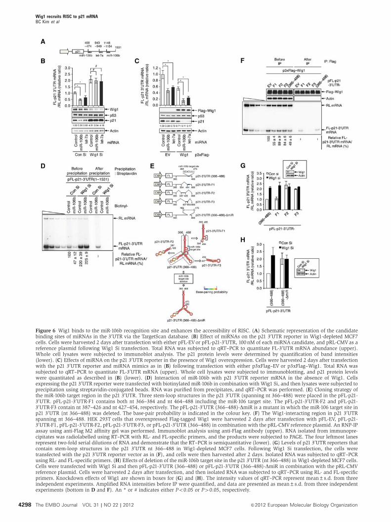
Figure 6 Wig1 binds to the miR-106b recognition site and enhances the accessibility of RISC. (A) Schematic representation of the candidatebinding sites of miRNAs in the 30UTR via the TargetScan database. (B) Effect of miRNAs on the p21 30UTR reporter in Wig1-depleted MCF7cells. Cells were harvested 2 days after transfection with either pFL-EVor pFL-p21-30UTR, 100 nM of each miRNA candidate, and pRL-CMVas areference plasmid following Wig1 Si transfection. Total RNA was subjected to qRT–PCR to quantitate FL-30UTR mRNA abundance (upper).Whole cell lysates were subjected to immunoblot analysis. The p21 protein levels were determined by quantification of band intensities(lower). (C) Effects of miRNA on the p21 30UTR reporter in the presence of Wig1 overexpression. Cells were harvested 2 days after transfectionwith the p21 30UTR reporter and miRNA mimics as in (B) following transfection with either p3xFlag–EV or p3xFlag–Wig1. Total RNA wassubjected to qRT–PCR to quantitate FL-30UTR mRNA (upper). Whole cell lysates were subjected to immunoblotting, and p21 protein levelswere quantitated as described in (B) (lower). (D) Interaction of miR-106b with p21 30UTR reporter mRNA in the absence of Wig1. Cellsexpressing the p21 30UTR reporter were transfected with biotinylated miR-106b in combination with Wig1 Si, and then lysates were subjected toprecipitation using streptavidin-conjugated beads. RNA was purified from precipitates, and qRT–PCR was performed. (E) Cloning strategy ofthe miR-106b target region in the p21 30UTR. Three stem-loop structures in the p21 30UTR (spanning nt 366–488) were placed in the pFL-p21-30UTR. pFL-p21-30UTR-F1 contains both nt 366–384 and nt 464–488 including the miR-106 target site. The pFL-p21-30UTR-F2 and pFL-p21-30UTR-F3 contain nt 387–426 and nt 427–454, respectively. The pFL-p21-30UTR (366–488)-DmiR is a mutant in which the miR-106 target site inp21 30UTR (nt 366–488) was deleted. The base-pair probability is indicated in the colour key. (F) The Wig1-interacting region in p21 30UTRspanning nt 366–488. HEK 293T cells that overexpressed Flag-tagged Wig1 were harvested 2 days after transfection with pFL-EV, pFL-p21-30UTR-F1, pFL-p21-30UTR-F2, pFL-p21-30UTR-F3, or pFL-p21-30UTR (366–488) in combination with the pRL-CMV reference plasmid. An RNP-IPassay using anti-Flag M2 affinity gel was performed. Immunoblot analysis using anti-Flag antibody (upper). RNA isolated from immunopre-cipitates was radiolabelled using RT–PCR with RL- and FL-specific primers, and the products were subjected to PAGE. The four leftmost lanesrepresent two-fold serial dilutions of RNA and demonstrate that the RT–PCR is semiquantitative (lower). (G) Levels of p21 30UTR reporters thatcontain stem-loop structures in the p21 30UTR nt 366–488 in Wig1-depleted MCF7 cells. Following Wig1 Si transfection, the cells weretransfected with the p21 30UTR reporter vector as in (F), and cells were then harvested after 2 days. Isolated RNA was subjected to qRT–PCRusing RL- and FL-specific primers. (H) Effects of deletion of the miR-106b target site in the p21 30UTR (nt 366–488) in Wig1-depleted MCF7 cells.Cells were transfected with Wig1 Si and then pFL-p21-30UTR (366–488) or pFL-p21-30UTR (366–488)-DmiR in combination with the pRL-CMVreference plasmid. Cells were harvested 2 days after transfection, and then isolated RNAwas subjected to qRT–PCR using RL- and FL-specificprimers. Knockdown effects of Wig1 are shown in boxes for (G) and (H). The intensity values of qRT–PCR represent mean±s.d. from threeindependent experiments. Amplified RNA intensities before IP were quantified, and data are presented as mean±s.d. from three independentexperiments (bottom in D and F). An * or # indicates either Po0.05 or P40.05, respectively.
Wig1 recruits RISC to p21 mRNABC Kim et al
4298 The EMBO Journal VOL 31 | NO 22 | 2012 &2012 European Molecular Biology Organization
Figure 7 Wig1 depletion in a murine xenograft model and Wig1 and p21 mRNA levels in human cancer tissues. (A) H460 cells (1�106) wereinjected subcutaneously into the femurs of nude mice. When tumours reached 50mm3, 200 ml AteloGene gel containing 20mM of Wig1 Si wasinjected to encompass the whole tumour mass. Tumour size was then measured at the indicated times. Error bars represent s.d. (nX3) (left).Xenograft tissue lysates at 5 days after siRNA injection were subjected to immunoblot analysis (right). An asterisk indicates a significantdifference, Po0.05. (B) Quantitative RT–PCR analysis of Wig1 and p21 mRNA in patient tissues. RNAwas purified from lung cancer tissues andcorresponding normal counterparts from 33 patients and subjected to qRT–PCR using Wig1- and p21-specific primers. Expression levels werenormalized to actin mRNA. The correlation between the relative levels of Wig1 and p21 mRNA was analysed by tumour versus normalcounterpart expression ratio of either Wig1 or p21 mRNA using the Pearson correlation coefficient. Error bars represent standard error of themean (s.e.m.). (C) In vivo correlation between Wig1 and p21 mRNA expression in each patient tissue. Expression levels in either normal ortumour tissues were clustered as either solid or dotted lines, respectively. Normalized Wig1 and p21 mRNA levels from each individual samplewere plotted on logarithmic scales. (D) Proposed model for Wig1-dependent target recognition of RISC. Wig1 is a critical determinant for thedecision between proliferation and cellular senescence via fine-tuning of p21 mRNA levels through facilitation of the accessibility of RISC to itstarget. In the presence of Wig1, RISC is effectively recruited to the target p21 mRNA, modulates its decay, and finally results in cell-cycleprogression. In the case of Wig1 depletion, p21 mRNA is stabilized, leading to the onset of cellular senescence.
Wig1 recruits RISC to p21 mRNABC Kim et al
4299&2012 European Molecular Biology Organization The EMBO Journal VOL 31 | NO 22 | 2012
suggesting that p21 expression and activity must be tightly
regulated by multiple mechanisms (Brugarolas et al, 1995;
Deng et al, 1995). In addition, induction of p21 expression or
p21 stabilization promotes cellular senescence (Jascur et al,
2005; Vigneron et al, 2005). Recently, regulation of p21
mRNA by various miRNAs and RBPs has begun to be
explored. Our results provide strong evidence to support a
molecular mechanism that involves tight regulation of p21 by
the RBP Wig1 and miRNAs at the post-transcriptional level to
allow cell proliferation. With respect to a clinical application
for these findings, we demonstrated that regulation of Wig1
levels is important for determining final cell fate, either
premature senescence or cell proliferation, in various
cancer cell lines and in cancer patient tissues. Vilborg et al
(2009) reported that Wig1 depletion resulted in a decrease in
p53 protein levels through the regulation of p53 mRNA
stability; however, we observed no correlation between
Wig1 and p53 in various cancer cells, including U2OS cells,
and in normal cells (Supplementary Figure S11; also
in Figure 1 and Supplementary Figures S3 and S5).
Our findings were consistent with the recent report by
Sedaghat et al (2012).
The miRNA-mediated mRNA decay plays a central role in
the regulation of gene expression in eukaryotic cells
(Hutvagner et al, 2001; Lee et al, 2003; Bartel, 2009). Here,
we showed that Wig1 overexpression facilitated miR-106b-
and let-7a-mediated degradation of p21 reporter mRNA
following transfection of miRNA mimics (Figure 6C).
In addition, Wig1 depletion suppressed reporter mRNA
decay in miRNA mimic-transfected cells (Figure 6B). In the
absence of let-7a mimic transfection, however, the effect of
Wig1 on FL-p21-30UTR (840–993) reporter mRNA levels was
barely detectable (Supplementary Figure S9C and D), perhaps
due to the low endogenous level of let-7a compared to that of
miR106b (Supplementary Figure S12). Since miR-106b and
let-7a target sites likely form a stem-loop structure due to
considerable base-pairing probabilities, we further defined
the exact role of this stem-loop structure of the p21 30UTR in
mRNA decay by Wig1. We found that Wig1 preferentially
associated with stem-loop fragments containing the miR-106b
target site in order to regulate the reporter mRNA levels
(Figure 6F and G).
Although several proteins modulate the efficacy of miRNA
biogenesis, target recognition of miRNAs is based on base-
pairing interactions via thermodynamic asymmetry and the
50-nucleotide identity between the miRNA ‘seed’ sequence
and the complementary nucleotides in the 30UTR of target
mRNAs (van Kouwenhove et al, 2011). Recently, several
reports have suggested that an alternative regulatory
mechanism is involved in influencing miRNA activity, since
most miRNAs pair imperfectly with their cognate mRNAs
(Jing et al, 2005; Kedde et al, 2007; Kedde et al, 2010; van
Kouwenhove et al, 2011). RBPs have been proposed to
be key components in the determination of miRNA
function. Indeed, alterations of RBP expression and activity
could affect many physiological and developmental processes
(van Kouwenhove et al, 2011). Although 4500 human RBPs
have been identified, the mechanisms of action of only a few
have been elucidated. Therefore, as RBPs play an essential
role in RNA metabolism, functional studies of various classes
of RBPs should be the focus of more attention (Hock et al,
2007; Landthaler et al, 2008).
If an miRNA target site is located in a stem-loop structure,
then the site may be concealed by the secondary structure to
prevent pairing with the miRNA. Thus, RBPs may affect
target site accessibility by facilitating an open structure via
association with the secondary structure of the mRNA and
may, thereby, modulate gene expression regulation by
miRNAs (Brodersen and Voinnet, 2009). Pumilio-1 (PUM1)
binds to the 30UTR of p27 and induces a local change in
mRNA structure to favour association with miR-221 and
miR-222 (Kedde et al, 2010). The AU-rich element (ARE)
binding protein, tristetraprolin (TTP), is also required in
miR16-mediated ARE-RNA decay (Jing et al, 2005).
Furthermore, competitive binding of RBP and miRNA to the
target sites may lead to modulation of miRNA-mediated
mRNA decay. Studies have demonstrated that hnRNP L,
Dnd1, and RBM38 relieve miRNA-mediated mRNA
repression because the target site overlaps with a binding
site for an RBP (Kedde et al, 2007; Jafarifar et al, 2011;
Leveille et al, 2011). In this study, we proposed possible
mechanisms for the regulation of p21 expression at the
post-transcriptional level by Wig1 (Figure 7D). Wig1 may
dictate miRNA-mediated mRNA decay through the regulation
of RISC accessibility to its target site via relaxation of the
mRNA secondary structure and/or recruitment of miRNA-
loaded RISC by direct association with the Ago2 protein. Our
results not only provide evidence for a novel contribution
to the prevention of premature senescence onset but also
suggest the molecular architecture to shed light on the
target recognition mechanism of miRNA-containing RISCs.
Furthermore, we demonstrated that Wig1 depletion inhibited
tumour progression in a murine xenograft model and pro-
vided evidence of an inverse correlation between Wig1 and
p21 mRNAs in human normal and cancer tissues, indicating
that Wig1 may play a critical role in tumour progression via
its regulation of p21 (Figure 7A–C). Since dysregulation of a
cell-cycle regulatory protein may contribute to malignant
transformation, we suggest that fine-tuning of cell-cycle
regulators via the coordination between miRNAs and the
RBP Wig1 may offer a therapeutic and prognostic strategy
for cancer treatment. We observed that levels of Wig1 and
p21 were not significantly altered during the replicative
senescence of human diploid fibroblasts (Supplementary
Figure S13), suggesting that these molecules may not be
critical executioners in the process of replicative senescence.
Recently, co-expression of miRNAs and their target genes
was documented, suggesting that these miRNAs may, indeed,
be involved in negative transcriptional co-regulation circuits,
which are termed as miRNA-mediated feed-forward loops
(Tsang et al, 2007). This circuit may have the potential to
provide fine-tuning and maintenance of protein steady state,
although the functional significance of the circuit has
not been fully characterized. If a temporal gap in the
activation of the target gene and the miRNA exists, then the
circuit may be utilized for either the delayed shutdown
of the regulated genes or buffering for noisy fluctuations
(Shalgi et al, 2007). Although both miR-17/92 and E2F are
transcriptionally activated by c-Myc, a tight regulatory
connection is involved as miR-17/92 represses E2F
(O’Donnell et al, 2005). Interestingly, we found that Wig1
induced by p53 negatively regulated another p53-responsive
gene, p21. Under normal conditions, the basal levels of
Wig1 negatively regulate p21 to prevent the onset of
Wig1 recruits RISC to p21 mRNABC Kim et al
4300 The EMBO Journal VOL 31 | NO 22 | 2012 &2012 European Molecular Biology Organization
cellular senescence (Figure 1; Supplementary Figure S3).
Under stressed conditions, activated p53 induces Wig1
expression as well as p21 expression, and this induced
Wig1 functions as a guardian to regulate the induced p21
level (Supplementary Figure S14A and B). Summarily, our
findings provide evidence that a TF controls the transcription
of both a target mRNA and the RBP involved in regulation of
the decay of the same target mRNA, providing more precise
regulation via a combined transcriptional/post-transcrip-
tional regulatory network (Supplementary Figure S14C).
Materials and methods
Cell cultureMCF7, HEK 293T, U2OS, and HDF cells were cultured in DMEM(PAA Laboratories GmbH). H460 cells were cultured in RPMI 1640(WelGENE). HCT116 parental, p53� /� , and p21� /� isogenic celllines were cultured in McCoy’s 5A medium (WelGENE). Cells weresupplemented with 10% FBS (Lonza Group Ltd.) and 1% penicillinand streptomycin solution (WelGENE) at 371C in a 5% CO2incubator.
RNA interference and plasmid transfectionCells were transfected with siRNA duplexes or miRNA mimicsusing Lipofectamine RNAiMAX (Invitrogen Corp.), and transfectionof plasmids was carried out using Lipofectamine 2000 reagent(Invitrogen Corp.) according to manufacturer’s instructions. ThesiRNAs used in this study are listed in Supplementary Table I.
Immunoblot analysisImmunoblotting was performed as described (Lee et al, 2011),and the antibodies used in this study are listed in SupplementaryTable II.
Analysis of p21 mRNA stability and protein half-lifeWig1 Si-transfected cells were treated with 5 mg/ml actinomycin D(Sigma-Aldrich) for various periods of time (0–4 h). Total RNA fromeach time point was purified from the cells, and quantitative real-time PCR was performed. To analyse protein half-life, Wig1 Si-transfected cells were treated with 40mg/ml CHX (Sigma-AldrichCo. LLC.) for the indicated periods of time (0–6h). Cellular extractsobtained at each time point were subjected to immunoblottingusing the appropriate antibodies. The intensities of the bandsnormalized with actin were determined using the ImageJ software(http://rsb.info.nih.gov/ij/) from the National Institutes of Health(USA).
sqRT–PCR and quantitative real-time PCRsqRT–PCR was performed by radioisotope-labelling PCR andpolyacrylamide gel electrophoresis (PAGE). qRT–PCR wasperformed in a DNAEngine Thermal Cycler with a Chromo4Real-Time PCR Detector (Bio-Rad Laboratories). Primers used inthis study are given in Supplementary Table III.
ImmunoprecipitationLysates from cells that overexpress Flag-tagged Wig1 wereimmunoprecipitated with anti-Flags M2 affinity gel (Sigma-Aldrich Co. LLC.). Total RNA isolated from these immunoprecipi-tates was subjected to sqRT–PCR.
In vitro binding assayIn vitro binding assays were performed using recombinantFlag-tagged Wig1 (Novus Biologicals, Inc.) and polyhistidine-taggedAgo2 (Sino Biological Inc.). After incubation of either GST or GST–Wig1 in combination with His–Ago2 in binding buffer (10mMTris–HCl (pH 8.0), 150mM NaCl, 10% glycerol, 0.1% BSA, 0.1%Triton X-100) at 41C for 2 h, samples were incubated with eitherglutathione Sepharose 4B (GE Healthcare Bio-Sciences AB) orNi-NTA agarose resin (Qiagen GmbH) for 1.5 h. After adsorption,the resin was washed five times with binding buffer, and thenbound proteins were resolved by 10% SDS–PAGE and subjectedto immunoblot using either His probe (Santa Cruz Biotechnology)
or HRP-conjugated anti-GST antibody (GE Healthcare Bio-Sciences AB).
Biotin precipitationWig1 Si-transfected HEK 293T cells were transfected with thepFL-p21-30UTR reporter plasmid and biotinyl-miR-106b (BioneerInc.). In addition, pRL-CMV was used as a reference plasmid. After2 days of incubation, the cells were rinsed and sonicated in NET-2buffer as described above for IP. Yeast tRNA-coated streptavidin beads(Sigma-Aldrich Co. LLC.) were added to cell lysates and incubated for3h at 41C. The beads were then washed 5–7 times with NET-2 buffer.RNA was isolated from the beads, and the mRNA associated with thebiotinylated miR-106b was amplified using RT–PCR with FL- andRL-specific primers as described above for sqRT–PCR.
Establishment of xenograft tumours and siRNA transfectionusing AteloGenes gelA single-cell suspension of H460 cells (1�106) was injected sub-cutaneously into lateral hind legs of 6-week-old BALB/c nude mice(n¼ 6). Average tumour length was determined as (L�W2)/2 withmeasurements of tumour length (L) and width (W) taken with acaliper. When the tumour reached an average volume of B50mm3
according to this formula, a Wig1 Si mixture with AteloGenes LocalUse (KOKEN Co. Ltd.) was injected so as to wrap up the wholetumour mass (Takei et al, 2004). Tumour volumes were determineddaily after the siRNA gel injection. The mice were sacrificed on days5 and 7 after xenograft injection, and tumours were removed forfurther analysis. All animal protocols and studies were conducted inaccordance with the guidelines of the Institutional Animal Care andUse Committee of the Korea Institute Radiological and MedicalSciences (IRB Approval No. KIRAMS 2012-2).
Human tissue collection and qRT–PCR analysisThis study involving human tissues was approved by theInstitutional Review Board of Korea Institute of Radiological andMedical Sciences (IRB Approval No. KIRAMS 2010-0526) and theAsan Medical Center Institutional Review Board (IRB ApprovalNo. K-1007-002-067). Snap-frozen tissues from lung tissues wereobtained from patients with lung cancer that had been diagnosed aslung adenocarcinoma, squamous cell carcinoma, or bronchioloal-veolar carcinoma. Samples were stored in liquid nitrogen. Tomeasure Wig1 and p21 mRNA expression in human specimens,total RNA was isolated and subjected to reverse transcription usingSuperScript III Reverse Transcriptase (Invitrogen Corp.) accordingto manufacturer’s recommendations. Then, qRT–PCR was per-formed using the Chromo4 Real-Time PCR Detection System(Bio-Rad Laboratories). The relative fold-change in RNA expressionwas calculated using the 2�DDCt method, where the average of DCtvalues for the amplicon of interest was normalized to that of anendogenous control gene (b-actin), and compared with normalcounterpart specimens.
Statistical analysisDifferences between the various experimental groups were calcu-lated using Student’s two-tailed t-test. P-values of o0.05 wereconsidered as significant. The correlation between Wig1 and p21mRNA levels in tumours versus normal tissues was assessed basedon the tumour/normal expression ratio of relative mRNA levels ofeach target using Pearson’s correlation coefficient analysis.
Supplementary dataSupplementary data are available at The EMBO Journal Online(http://www.embojournal.org).
Acknowledgements
Primary human diploid fibroblasts and Ago2 overexpressing vectorwere kindly provided by Professor Kyung A Cho at Chonnam NationalUniversity and Professor V Narry Kim at Seoul National University,respectively. We thank Professor Yoon Ki Kim and Professor Young SikLee at Korea University, Dr Kyung S Lee at National Cancer Institute,NIH, and Professor Edward KL Chan at University of Florida for criticalreview of the manuscript. This work was supported by the NuclearResearch and Development Program of the National ResearchFoundation funded by the Korean Government (MEST).
Wig1 recruits RISC to p21 mRNABC Kim et al
4301&2012 European Molecular Biology Organization The EMBO Journal VOL 31 | NO 22 | 2012

Author contributions: BCK, HCL, JJL, CMC, DKK, JCL, YGK, andJSL designed the experiments and analysed the data. BCK and HCLperformed the experiments. CMC and DKK collected the patienttissue samples. BCK and JSL wrote the manuscript.
Conflict of interest
The authors declare that they have no conflict of interest.
ReferencesAbbas T, Dutta A (2009) p21 in cancer: intricate networks and
multiple activities. Nat Rev Cancer 9: 400–414Bartel DP (2009) MicroRNAs: target recognition and regulatory
functions. Cell 136: 215–233Bhattacharyya SN, Habermacher R, Martine U, Closs EI, Filipowicz
W (2006) Relief of microRNA-mediated translational repression inhuman cells subjected to stress. Cell 125: 1111–1124
Brodersen P, Voinnet O (2009) Revisiting the principles ofmicroRNA target recognition and mode of action. Nat Rev MolCell Biol 10: 141–148
Brugarolas J, Chandrasekaran C, Gordon JI, Beach D, Jacks T,Hannon GJ (1995) Radiation-induced cell cycle arrest compro-mised by p21 deficiency. Nature 377: 552–557
Byun HO, Han NK, Lee HJ, Kim KB, Ko YG, Yoon G, Lee YS,Hong SI, Lee JS (2009) Cathepsin D and eukaryotic translationelongation factor 1 as promising markers of cellular senescence.Cancer Res 69: 4638–4647
Chen Z, Trotman LC, Shaffer D, Lin HK, Dotan ZA, Niki M, KoutcherJA, Scher HI, Ludwig T, Gerald W, Cordon-Cardo C, Pandolfi PP(2005) Crucial role of p53-dependent cellular senescencein suppression of Pten-deficient tumorigenesis. Nature 436:725–730
Deng C, Zhang P, Harper JW, Elledge SJ, Leder P (1995) Micelacking p21CIP1/WAF1 undergo normal development, but aredefective in G1 checkpoint control. Cell 82: 675–684
Eulalio A, Huntzinger E, Izaurralde E (2008) GW182 interactionwith Argonaute is essential for miRNA-mediated trans-lational repression and mRNA decay. Nat Struct Mol Biol 15:346–353
Gartel AL, Tyner AL (1999) Transcriptional regulation of thep21WAF1/CIP1 gene. Exp Cell Res 246: 280–289
Grimson A, Farh KK, Johnston WK, Garrett-Engele P, Lim LP,Bartel DP (2007) MicroRNA targeting specificity in mammals:determinants beyond seed pairing. Mol Cell 27: 91–105
Guhaniyogi J, Brewer G (2001) Regulation of mRNA stability inmammalian cells. Gene 265: 11–23
Hayflick L, Moorhead PS (1961) The serial cultivation of humandiploid cell strains. Exp Cell Res 25: 585–621
Hellborg F, Qian W, Mendez-Vidal C, Asker C, Kost-Alimova M,Wilhelm M, Imreh S, Wiman KG (2001) Human wig-1, a p53target gene that encodes a growth inhibitory zinc finger protein.Oncogene 20: 5466–5474
Hellborg F, Wiman KG (2004) The p53-induced Wig-1 zinc fingerprotein is highly conserved from fish to man. Int J Oncol 24:1559–1564
Hock J, Weinmann L, Ender C, Rudel S, Kremmer E, Raabe M,Urlaub H, Meister G (2007) Proteomic and functional analysis ofArgonaute-containing mRNA-protein complexes in human cells.EMBO Rep 8: 1052–1060
Hutvagner G, McLachlan J, Pasquinelli AE, Balint E, Tuschl T,Zamore PD (2001) A cellular function for the RNA-interferenceenzyme Dicer in the maturation of the let-7 small temporal RNA.Science 293: 834–838
Israeli D, Tessler E, Haupt Y, Elkeles A, Wilder S, Amson R,Telerman A, Oren M (1997) A novel p53-inducible gene,PAG608, encodes a nuclear zinc finger protein whose overexpres-sion promotes apoptosis. EMBO J 16: 4384–4392
Jafarifar F, Yao P, Eswarappa SM, Fox PL (2011) Repression ofVEGFA by CA-rich element-binding microRNAs is modulated byhnRNP L. EMBO J 30: 1324–1334
Jascur T, Brickner H, Salles-Passador I, Barbier V, El Khissiin A,Smith B, Fotedar R, Fotedar A (2005) Regulation of p21WAF1/CIP1
stability by WISp39, a Hsp90 binding TPR protein. Mol Cell 17:237–249
Jing Q, Huang S, Guth S, Zarubin T, Motoyama A, Chen J, Di PadovaF, Lin SC, Gram H, Han J (2005) Involvement of microRNA inAU-rich element-mediated mRNA instability. Cell 120: 623–634
Jung YS, Qian Y, Chen X (2010) Examination of the expandingpathways for the regulation of p21 expression and activity.Cell Signal 22: 1003–1012
Kawamata T, Tomari Y (2010) Making RISC. Trends Biochem Sci 35:368–376
Kedde M, Strasser MJ, Boldajipour B, Oude Vrielink JA, Slanchev K,le Sage C, Nagel R, Voorhoeve PM, van Duijse J, Ørom UA,Lund AH, Perrakis A, Raz E, Agami R (2007) RNA-bindingprotein Dnd1 inhibits microRNA access to target mRNA. Cell131: 1273–1286
Kedde M, van Kouwenhove M, Zwart W, Oude Vrielink JA, Elkon R,Agami R (2010) A Pumilio-induced RNA structure switch inp27-30 UTR controls miR-221 and miR-222 accessibility. Nat CellBiol 12: 1014–1020
Kim HH, Kuwano Y, Srikantan S, Lee EK, Martindale JL, Gorospe M(2009a) HuR recruits let-7/RISC to repress c-Myc expression.Genes Dev 23: 1743–1748
Kim MY, Hur J, Jeong S (2009b) Emerging roles of RNA and RNA-binding protein network in cancer cells. BMB Rep 42: 125–130
Kramer DL, Chang BD, Chen Y, Diegelman P, Alm K, Black AR,Roninson IB, Porter CW (2001) Polyamine depletion inhuman melanoma cells leads to G1 arrest associated with induc-tion of p21WAF1/CIP1/SDI1, changes in the expression of p21-regulated genes, and a senescence-like phenotype. Cancer Res 61:7754–7762
Landthaler M, Gaidatzis D, Rothballer A, Chen PY, Soll SJ, Dinic L,Ojo T, Hafner M, Zavolan M, Tuschl T (2008) Molecular char-acterization of human Argonaute-containing ribonucleoproteincomplexes and their bound target mRNAs. RNA 14: 2580–2596
Lee JJ, Kim BC, Park MJ, Lee YS, Kim YN, Lee BL, Lee JS (2011)PTEN status switches cell fate between premature senescenceand apoptosis in glioma exposed to ionizing radiation. Cell DeathDiffer 18: 666–677
Lee JJ, Lee JH, Ko YG, Hong SI, Lee JS (2010) Prevention ofpremature senescence requires JNK regulation of Bcl-2 andreactive oxygen species. Oncogene 29: 561–575
Lee Y, Ahn C, Han J, Choi H, Kim J, Yim J, Lee J, Provost P,Radmark O, Kim S, Kim VN (2003) The nuclear RNase III Droshainitiates microRNA processing. Nature 425: 415–419
Lee Y, Hur I, Park SY, Kim YK, Suh MR, Kim VN (2006) The role ofPACT in the RNA silencing pathway. EMBO J 25: 522–532
Leveille N, Elkon R, Davalos V, Manoharan V, Hollingworth D, OudeVrielink J, le Sage C, Melo CA, Horlings HM, Wesseling J, Ule J,Esteller M, Ramos A, Agami R (2011) Selective inhibition ofmicroRNA accessibility by RBM38 is required for p53 activity.Nat Commun 2: 513
Mendez Vidal C, Prahl M, Wiman KG (2006) The p53-inducedWig-1 protein binds double-stranded RNAs with structural char-acteristics of siRNAs and miRNAs. FEBS Lett 580: 4401–4408
O’Donnell KA, Wentzel EA, Zeller KI, Dang CV, Mendell JT (2005)c-Myc-regulated microRNAs modulate E2F1 expression. Nature435: 839–843
Prahl M, Vilborg A, Palmberg C, Jornvall H, Asker C, Wiman KG(2008) The p53 target protein Wig-1 binds hnRNP A2/B1 andRNA Helicase A via RNA. FEBS Lett 582: 2173–2177
Robb GB, Rana TM (2007) RNA helicase A interacts with RISC inhuman cells and functions in RISC loading. Mol Cell 26:523–537
Sedaghat Y, Mazur C, Sabripour M, Hung G, Monica BP (2012)Genomic analysis of wig-1 pathways. PLoS ONE 7: e29429
Schmitt CA, Fridman JS, Yang M, Lee S, Baranov E, Hoffman RM,Lowe SW (2002) A senescence program controlled by p53 andp16INK4a contributes to the outcome of cancer therapy. Cell 109:335–346
Shalgi R, Lieber D, Oren M, Pilpel Y (2007) Global and localarchitecture of the mammalian microRNA-transcription factorregulatory network. PLoS Comput Biol 3: e131
Wig1 recruits RISC to p21 mRNABC Kim et al
4302 The EMBO Journal VOL 31 | NO 22 | 2012 &2012 European Molecular Biology Organization

Sheikh MS, Li XS, Chen JC, Shao ZM, Ordonez JV, Fontana JA(1994) Mechanisms of regulation of WAF1/Cip1 geneexpression in human breast carcinoma: role of p53-dependentand independent signal transduction pathways. Oncogene 9:3407–3415
Takei Y, Kadomatsu K, Yuzawa Y, Matsuo S, Muramatsu T (2004)A small interfering RNA targeting vascular endothelialgrowth factor as cancer therapeutics. Cancer Res 64:3365–3370
Tsang J, Zhu J, van Oudenaarden A (2007) MicroRNA-mediatedfeedback and feedforward loops are recurrent network motifs inmammals. Mol Cell 26: 753–767
van Kouwenhove M, Kedde M, Agami R (2011) MicroRNA regula-tion by RNA-binding proteins and its implications for cancer.Nat Rev Cancer 11: 644–656
Varmeh-Ziaie S, Okan I, Wang Y, Magnusson KP, Warthoe P, StraussM, Wiman KG (1997) Wig-1, a new p53-induced gene encoding azinc finger protein. Oncogene 15: 2699–2704
Ventura A, Kirsch DG, McLaughlin ME, Tuveson DA, Grimm J,Lintault L, Newman J, Reczek EE, Weissleder R, Jacks T (2007)Restoration of p53 function leads to tumour regression in vivo.Nature 445: 661–665
Vigneron A, Roninson IB, Gamelin E, Coqueret O (2005) Srcinhibits adriamycin-induced senescence and G2 checkpointarrest by blocking the induction of p21waf1. Cancer Res 65:8927–8935
Vilborg A, Glahder JA, Wilhelm MT, Bersani C, Corcoran M,Mahmoudi S, Rosenstierne M, Grander D, Farnebo M, NorrildB, Wiman KG (2009) The p53 target Wig-1 regulates p53 mRNAstability through an AU-rich element. Proc Natl Acad Sci USA 106:15756–15761
Wang Y, Blandino G, Givol D (1999) Induced p21waf expression inH1299 cell line promotes cell senescence and protectsagainst cytotoxic effect of radiation and doxorubicin. Oncogene18: 2643–2649
Wiemer EA (2007) The role of microRNAs in cancer: no smallmatter. Eur J Cancer 43: 1529–1544
Wilhelm MT, Mendez-Vidal C, Wiman KG (2002) Identification offunctional p53-binding motifs in the mouse wig-1 promoter.FEBS Lett 524: 69–72
Wu CH, van Riggelen J, Yetil A, Fan AC, Bachireddy P, Felsher DW(2007) Cellular senescence is an important mechanism of tumorregression upon c-Myc inactivation. Proc Natl Acad Sci USA 104:13028–13033
Wu S, Huang S, Ding J, Zhao Y, Liang L, Liu T, Zhan R, He X (2010)Multiple microRNAs modulate p21Cip1/Waf1 expressionby directly targeting its 30 untranslated region. Oncogene 29:2302–2308
Zhang X, Li J, Sejas DP, Pang Q (2005) The ATM/p53/p21 pathwayinfluences cell fate decision between apoptosis and senescence inreoxygenated hematopoietic progenitor cells. J Biol Chem 280:19635–19640
Wig1 recruits RISC to p21 mRNABC Kim et al
4303&2012 European Molecular Biology Organization The EMBO Journal VOL 31 | NO 22 | 2012