Embed Size (px)
Citation preview
João Miguel Vicente Ventura
MECHANISTIC STUDIES ON THE ENZYMATIC
CONJUGATION OF BIOPOLYMERS WITH
BIOACTIVE COMPOUNDS
Dissertação no âmbito do Mestrado em Bioquímica orientada pela Doutora
Alexandra Teresa Pires Carvalho e pela Senhora Professora Paula Cristina
Veríssimo Pires e apresentada ao Departamento de Ciências da Vida.
Julho de 2021


Mechanistic studies on the enzymatic
conjugation of polyesters with
bioactive compounds
João Miguel Vicente Ventura
Master’s degree in Biochemistry
Department of Life Sciences
Faculty of Science and Technology
University of Coimbra
Supervisors: Alexandra Carvalho, PhD, Rational Protein Engineering ,Group – CNC Paula Veríssimo, PhD, Department of Life Sciences


i
Index
ABSTRACT .............................................................................................................. III
SUMÁRIO ................................................................................................................. IV
ABBREVIATIONS .................................................................................................... V
FIGURE INDEX ........................................................................................................ V
EQUATION INDEX .............................................................................................. VII
OBJECTIVES ........................................................................................................... VII
INTRODUCTION .................................................................................................... 1
Antimicrobial Peptides ..................................................................................................................... 1
Polymers as delivery mechanism .................................................................................................... 9
Enzymes as biocatalysts .................................................................................................................. 14
COMPUTATIONAL METHODS ........................................................................ 18
Molecular Mechanics ....................................................................................................................... 18
Quantum Mechanics ........................................................................................................................ 20
Quantum Mechanics/Molecular Mechanics .................................................................................. 21
Molecular Dynamics ........................................................................................................................ 22
Python programming language ..................................................................................................... 23
Micelle trajectory analysis ............................................................................................................... 24
RESULTS AND DISCUSSION ............................................................................ 26
Molecular modelling ........................................................................................................................ 26
Building base conjugate & Micelle first attempt ......................................................................... 27
Python Attempt File Editing .......................................................................................................... 30
Python attempt Micelle builder ..................................................................................................... 33

ii
Micelle stability ................................................................................................................................. 36
Membrane model ............................................................................................................................. 38
CONCLUSIONS AND FUTURE PERSPECTIVES .......................................... 42
REFERENCES ......................................................................................................... 43
ANEX II .................................................................................................................... 52
Part 1 ................................................................................................................................................. 52
Part 2 ................................................................................................................................................. 52
Part 3 ................................................................................................................................................. 53

iii
Abstract
Infection by multi-resistant bacteria is rising in likelihood and is often life threatening.
One of the proposed solutions is the introduction of antimicrobial peptides as
antibiotics. However, these compounds suffer from a short half-life and can display
cytotoxic effects, particularly severe haemolytic activity. Polymeric modification of
their moieties allows an increase in their effectiveness, altering their pharmacokinetics
and pharmacodynamics properties.
In this work we evaluated the conjugation effectiveness with polycaprolactone as a
modification to improve the AMPs, specifically polymyxin B and polyphemusin I.
Polymyxin B is one of the last line antibiotics and polyphemusin I, - while not in
medical use, has been shown to be effective against a broad spectrum of
microorganisms.
We intended to 1) verify the impact in their mechanism of action, particularly in the
interaction between the cell membranes and the conjugates and 2) the micelles remain
stable in water. We have performed classical Molecular Dynamics simulations and
analysed their structure and dynamics in water.
In this work, we succeeded in creating a stable micelle with PCL and polymyxin B and
prepared a membrane model to test the micelle.

iv
Sumário
Infeções por bactérias multirresistentes são cada vez mais prováveis e são
frequentemente um risco de vida. Uma das potenciais soluções para combater o
aparecimento de é a introdução de péptidos antimicrobianos como antibióticos.
Contudo, estes compostos têm limitações, como tempo de semivida demasiado curto
ou efeitos secundários demasiado perigosos,
Uma das soluções para os tornar mais eficazes é a sua modificação com polímeros,
para alterar a sua farmacocinética e farmacodinâmica. A polimixina B é um antibiótico
de última linha e a polifemusina I mesmo não tendo uso medico, demonstrou em
laboratório ser eficaz contra uma vasta gama de microrganismos
Este trabalho teve por objetivo avaliar a eficácia da conjugação dos péptidos
antimicrobianos polimixina B e polifemusina I com o polímero policaprolactona como
potencial modificação para melhorar os seus efeitos.
Procurou verificar que 1) não há impacto no mecanismo de ação, nomeadamente a
interação entre membranas e os conjugados e 2) verificar que as micelas se mantem
estáveis em água. Foram realizadas simulações de dinâmica molecular clássica e
analisada a estrutura e dinâmica dos compostos em água.
Neste trabalho, conseguimos com sucesso criar uma micela estável com PCL e
polimixina B e criar uma membrane modelo para testar a membrana.

v
Abbreviations
AMBER- Assisted Model Building with Energy Refinement
AMP- Antimicrobial Peptides
CalB- Candida antartica Lipase B
CHARMM-GUI- Chemical Harvard Molecular Mechanics graphical user interface
CMS- colistin methanesulfaonate
DFT- Density Functional Theory
DNA- Desoxiribonucleic Acid
LPS - Lipopolysaccharides
MD- Molecular Dynamics
MIC- Minimum Inhibitory Concentration
MM- Molecular Mechanics
NAMD- Nanoscale Molecular Dynamics
QM- Quantum Mechanics
QM/MM- Quantum Mechanics/Molecular Mechanics
PCL- Polycaprolactone
PDB- Protein Data Bank
PEG- Polyethylene glycol
PMB- Polymyxin B
RNA- Ribonucleic Acid
SASA- Solvent Accessible Surface Area
vdW- van der Waals
VMD- Visual Molecular Dynamics
Figure Index
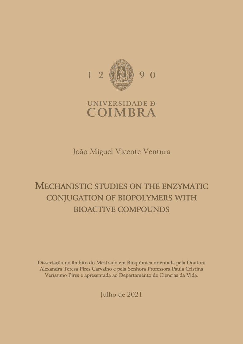
Figure 1- A representation of a Gram-negative bacteria cell, illustrating the
mechanisms of action of antimicrobial peptides (on the left of the line) and
mechanisms of resistance against antibiotics (right of the line). Adapted from Magana
et al., 2020.
Figure 2- Chemical representation of a Polymyxin B molecule. The structure is
composed of a cyclic portion (shaded in blue), a linear “panhandle” (in orange) and a

vi
fatty acyl tail (as yellow). In colistin, R6 D-phenylalanine is replaced by D-leucine.
Adapted from Vaara, 2019.
Figure 3- Schematic representation of the metabolization of PCL (Mandal &
Shunmugam, 2020).
Figure 4 - Visual representation of bond (r), angle (θ), and dihedral (ϕ) with atoms 1
to 4, the numbers of each represent the atoms involved. Adapted from Sharma,
Kumar, & Chandra, 2019.
Figure 5- Images of the conjugated starting structures before molecular dynamics,
polyphemusin I (A), and polymyxin B (B).
Figure 6- Image of the conjugated molecules after 20 nanoseconds of simulation. The
molecules have been separated by the repulsion of the positive charges.
Figure 7- The end point of a PCL chain with the AMP at the extremity, before (A) and
after (B) using the sorting python code and LeAP.
Figure 8- Comparison of both methods of point generation displayed using the module
matplotlib.pyplot. (A) is the first method (combinations) and (B) is the alternative
method
Figure 9- AMP (A) and control (B) micelles, after 20 ns simulation and the starting
position for the AMP micelle. Both micelles shape remained stable during the
simulation. The control micelle starting structure used the same base as the one with
AMP, but the peptide was removed.
Figure 10- Visualization of the construction of the membrane in CHARMM-GUI,
demonstrating the flaw of the micelle placement. LPS are shown as individual atoms;
other membrane molecules are shown as full molecules. The red molecules are water
and the large grey spheres are calcium ions.
Figure 11- Close-up of lipid A molecules in the membrane, highlighting the overlaps
of lipid tails. Each residue is shown in a different colour to add contrast.

vii
Figure 12- Membrane structure, where the atoms in red represent the unrestrained
lipid tails of LPS, with a beta value of 0, and in blue the remaining atoms, with a beta
value of 1. Omitted in this image, water and ions also have a beta value of 1.
Equation Index
Equation 1- Potential energy function used by amber software, as per the
AMBER manual 2020
Equation 2- Time dependent Schrödinger’s equation
Equation 3- Formula to calculate the eccentricity (𝑒) of a micelle
Equation 4- Equation to calculate the distance between points in three
dimensions, using their Cartesian coordinates.
Objectives
Our aim was to perform a in silico study about the conjugation of antimicrobial
peptides (AMPs), specifically Polymyxin B and Polyphemusin I, with a
polycaprolactone (PCL, a biopolymer), to improve their therapeutic potential.
To achieve this, we proposed to perform: (i) Establish molecular models for
Polymyxin B, Polyphemusin I, and Polycaprolactone; (ii) Simulate (with
Molecular Mechanics) the behaviour of the conjugated AMP with PCL in water
to guaranty the formation of stable micelles; (iii) Simulate (with Molecular
Mechanics) the interaction with a membrane-model to verify if the mechanism
of action is not compromised by modification; and last (iv) Characterize the
formation of the conjugate though Quantum mechanics/Molecular mechanics
simulation.

viii

1
Introduction
Antimicrobial Peptides
Antimicrobial peptides (AMPs) are part of the innate defence systems of many
organisms (Lei et al., 2019). In microorganisms they evolved as a strategy to
overcome competition, while in multicellular organisms they are part of the
non specific defence system against infections.
The most common mechanism is the disruption of the membrane stability with
the formation of pores that lead to cell lysis (Martin-Serrano, Gómez, Ortega,
& Mata, 2019), although some have immunomodulating functions or regulate
inflammation (Magana et al., 2020) and some have intracellular targets like
DNA, RNA or proteins (Zhu, Liu, & Niu, 2017).
They are classified into two groups: non-ribosomal synthesis, which are
produced by enzymes; and ribosomal synthesis which are encoded in genes.
The former is most common in bacteria whereas the later is common in all
species (Hancock, 2000). Natural AMPs are small peptide sequences - 12 to
50 amino acids - with a broad spectrum of target pathogens (Zhu et al., 2017),
and have good water solubility and thermal stability (Lei et al., 2019). About
50% are hydrophobic and mainly composed of basic amino acids (lysine,
arginine and histidine), which gives them a positive charge, from +2 to +9
(Hancock, 2000).
AMPs are characterized by their secondary structure as α-helix, β-sheet
extended and loop (Sandreschi, Piras, Batoni, & Chiellini, 2016; Zhu et al.,
2017). The most common are α-helix and β-sheet, with α-helix being the most
studied. There are two main types of tertiary structure: an amphipathic two-
faced shape where one is apolar and the other positively charged; and a
hydrophobic core with two wings of hydrophilic pockets (Hancock, 2000).
Often they only obtain the functional structure in contact with the membranes,

2
having a random structure in free solution (Hancock, 2000). AMPs, rather than
acting on specific intracellular targets like conventional antibiotics, disrupt the
membrane with electrostatic interactions (Sun et al., 2018). However they have
poor stability, salt sensitivity, and toxicity to mammalian cells (Sun et al.,
2018).
In this age of antibiotic resistance where all classes of antibiotics have at least
one known mechanism of resistance (Sandreschi et al., 2016), there are
approximately 25000 yearly deaths in the EU caused by bacterial resistance to
antibiotic (Sandreschi et al., 2016).
AMPs are part of the initiative to replace the failing antibiotics. Since these
peptides can act both on membrane lipids and internal components, their non-
specific mechanism is leaves fewer opportunities for the development of
resistance compared to other antibiotics (Javia et al., 2018; Sun et al., 2018).
In figure 1 is represented both the mechanisms of antibacterial action of AMPs
and various mechanisms of antibiotic resistance of bacteria
3
Figure 1- A representation of a Gram-negative bacteria cell, illustrating the
mechanisms of action of antimicrobial peptides (on the left of the line) and
mechanisms of resistance against antibiotics (right of the line). Adapted from
Magana et al., 2020.
However, their use is limited due to cytotoxic side effects. There is a growing
interest in designing new AMPs to surpass the limitations of natural AMPs
(Martin-Serrano et al., 2019).
Among these are the polymyxins, a group with two antibiotics currently in use
(polymyxin B and colistin), cyclic lipodecapeptides which are effective against
Gram-negative bacteria. They are non-ribosomal peptides synthesized by a
enzyme complex, containing nonstandard amino acids in their composition,
with a total charge of +5 (Hancock, 2000).
In 2013, the International Conference on Polymyxins established the “Prato
Polymyxin Consensus”- the framework for optimizing the clinical use of

4
colistin and polymyxin B, which was refined in the following conferences
(Lenhard, Bulman, Tsuji, & Kaye, 2019).
The population pharmacokinetics of colistin and polymyxin B were elucidated
in 2011 and 2013 respectively, allowing the improvement of the dosage
required for the intended plasma concentration. Colistin is administered in an
inactive form (prodrug colistin methanesulfaonate, CMS) for it to be slowly
converted into its active form, taking into account the patient’s renal function
for dosage determination, while polymyxin B (PMB) is not affected by renal
function and is administrated in an active form, allowing its pharmacokinetics
to be more predictable (Lenhard et al., 2019).
Of the two molecules, polymyxin B was chosen as the one to be studied in this
work, as it is conventionally applied in an active form and, since it is shown to
be less toxic, it has a broader therapeutic spectrum.
Figure 2- Chemical representation of a Polymyxin B molecule. The structure is
composed of a cyclic portion (shaded in blue), a linear “panhandle” (in orange)
and a fatty acyl tail (as yellow). In colistin, R6 D-phenylalanine is replaced by
D-leucine. Adapted from Vaara, 2019.
The mechanism of action of these molecules (figure 2) starts with the binding
to anionic groups like phosphate and pyrophosphate in lipopolysaccharides

5
(LPS) and lipids exclusive to outer membranes of Gram-negative bacteria,
releasing them from the membrane by creating pores and compromising the
integrity of the membrane. This allows periplasmatic components and external
molecules to flow through (Vaara, 2019). This is followed by the damage of
the cytoplasmatic membrane, leading to the leakage of components such as
adenosine 5'-triphosphate (ATP) (Vaara, 2019). Possible additional effects
include inhibiting the NADH-quinone oxidoreductase, thus producing
hydroxyl radicals and reactive oxygen species (Vaara, 2019). Strains exposed
to polymyxins, even if they are resistant against them, are sensitized towards
other classes of antibiotics, allowing a synergistic combination of drugs
(Lenhard et al., 2019; Vaara, 2019).
These molecules were considered too toxic for systemic use, due to nephrotoxic
side effects (Hancock, 2000; Lenhard et al., 2019) thus making their research
very limited since then (Lenhard et al., 2019). This toxicity is the result of
reabsorption by the proximal tubular kidney cells, inside which the polymyxins
inhibit the mitochondrial electron transport chain, that increase super oxide
production and induce apoptosis by caspases activation (Lenhard et al., 2019;
Vaara, 2019). There is an upregulation of cholesterol production that is
thought to be an attempt to protect the cell. Several attempts to administrate
antioxidants simultaneously to reduce nephrotoxicity have not produced data
supporting the routine co-administration (Vaara, 2019).
Polymyxins re-entered clinical use as last-resort antibiotics, due to increasing
instances of extremely multiresistant bacteria, specially Enterobactericeae,
Pseudomonas aeruginosa and Acinetobacter baumannii (Lenhard et al., 2019; Vaara,
2019). Furthermore, their toxicity limits the dosage to suboptimal efficacy
(Vaara, 2019). While most antibiotics from the “golden age of antibiotic
discovery” have been replaced by new and improved versions, polymyxins
remain part of the exceptions that do not have newer developed versions in
clinical use (Vaara, 2019).

6
Their usage resurged against Enterobactericeae due to the spread of mobile
plasmids encoding Carbapenemase (β-lactam degrading enzyme), that when
acquired by strains with resistance mechanisms to other classes of antibiotics,
leaving very few classes of drugs with active effects (Lenhard et al., 2019). This
β-lactamase is not affected by conventional inhibitors, being addressed by next-
generation β-lactamase inhibitors (Lenhard et al., 2019). Although they have
been shown to improve the clinical outcome of patients, mortality remains
high, so the clinical use may include polymyxins for the synergy, though
preliminary studies have not shown results yet (Lenhard et al., 2019).
In combat of multidrug-resistant Pseudomonas aeruginosa, polymyxins are being
replaced by safer anti-pseudomonal antibiotics. Nevertheless preliminary in
vitro studies did show that there is synergy effects between some of them and
polymyxins, but further research is needed (Lenhard et al., 2019).
Currently, the treatment of Acinetobacter baumannii is reliant on polymyxins
unless drugs able to affect the β-lactam resistant strains, which by 2010, was
the case in approximately half the clinical isolates of A.baumannii in the USA
(Lenhard et al., 2019). Attempts are being made to improve treatment of A.
baumannii infections, for example combination regimes of polymyxins with
traditional antibiotics. Nonetheless there has not yet been a trial demonstrating
effectiveness of such methods (Lenhard et al., 2019). The alternative is the
development of novel antibacterials since the recently developed β-lactamase
inhibitors are not effective against the enzyme oxacillinase that grants
A.baumannii resistance, oxacillinase (Lenhard et al., 2019).
Polymyxin B nonapeptide is a derivative of polymyxin B that does not have the
fatty acyl tail (Figure 2) and the N-terminal diaminobutyryl (DAB) (French et
al., 2020; Lenhard et al., 2019). Instead of having bacterial activity, can make
the outer membrane permeable to antibiotics that normally would not be able
to enter the periplasm (Lenhard et al., 2019; Rose et al., 2000). In animal

7
testing, it showed less nephrotoxicity, but it is not going active clinical
development (Lenhard et al., 2019).
SPR741 is a low toxicity analogue that also does not have bactericide activity,
but instead is able to improve the effect of other antibiotics (French et al.,
2020; Lenhard et al., 2019). It lacks a fatty acyl tail and has a lower positive
charge (+3 compared to +5 of PMB) which change its activity but also improve
its safety profile (French et al., 2020; Lenhard et al., 2019). It is being
developed by Spero Therapeutic Inc, having completed clinical trials of Phase
I (Lenhard et al., 2019).
MRX-8 is a polymyxin analogue whose fatty acyl is bonded with a ester bond,
which can be hydrolyzed in the blood (Lenhard et al., 2019; Lepak, Wang, &
Andes, 2020). It is the product of “soft drug design”(drug design which aims
to develop safer drugs by designing the metabolism and detoxification of the
compound), intended to create an analogue that is metabolized for less
nephrotoxic metabolites after activity (Lepak et al., 2020), which has been
shown to be less toxic to kidneys than polymyxin B in rats studies while
maintaining the same effect (Lenhard et al., 2019). It is developed by MicuRx
Pharmaceuticals, which has received 5.2 million dollars (Lenhard et al., 2019;
Vaara, 2019).
Analogues have the potential to become therapeutic agents but few have
reached clinical studies (Lenhard et al., 2019). The possibility of an analogue
having less nephrotoxicity or more antibacterial potency would be an
improvement for combating infections (Lenhard et al., 2019).
While safer alternatives to polymyxins are being developed, bacterial resistance
to those drugs is also being developed (Lenhard et al., 2019). The polymyxin
class of antibiotics may eventually be relegated to therapy in combination,
whether as the current ones or as analogues, but presently they are still
necessary as last line antibiotics (Lenhard et al., 2019).

8
Another antimicrobial peptide family, polyphemusins, which albeit not having
the same medical history, have shown results in suppressing growth in vitro of
both Gram-negative and Gram-positive bacteria, fungi and mammalian cancer
cells (Marggraf et al., 2018).
The polyphemusin family are β-hairpin antimicrobial peptides that were
discovered in hemocytes of Limulus polyphemus, the American horseshoe crab,
that have a chain of 18 amino acids with 2 disulfide bonds and several lysine
and arginine residues, which produce a high net positive charge, while being
amphiphilic (Marggraf et al., 2018; Powers, Martin, Goosney, & Hancock,
2006).
Besides their broad spectrum of activity, they also have an affinity for LPS and
may degrade biofilms of Staphylococcus aureus (Marggraf et al., 2018).
Polyphemusin have preferential interaction with negatively charged
membranes, being able to disrupt both outer and inner Gram-negative
membranes and their cationic and amphilipathic properties are considered
essential for their activity (Marggraf et al., 2018).
The polyphemusin family has 3 isoforms. Of them, polyphemusin III has the
least bactericidal activity and the highest cytotoxicity (Marggraf et al., 2018).
This toxicity is likely due to a higher hydrophobicity, which correlates to
increase affinity towards all membranes (Marggraf et al., 2018). From the
remaining, polyphemusin I was chosen for this work, as it is the most studied.
Polyphemusin I has a rapid killing effect (5 minute of contact) and a low
minimum inhibitory concentration (sometimes lower than 1 µg per millilitre)
and great affinity for lipopolysaccharides but affects greatly both Gram-
negative and Gram-positive (Powers et al., 2006).
Although polyphemusin I is among the most effective antimicrobial peptides,
there is not a consensus on the mechanism of action (Yurkova, Zenin,

9
Sadykhov, & Fedorov, 2020). Polyphemusin I induces flip-flop movements of
membranes lipids, with a preference for negatively charged membranes (Powers
et al., 2006). Studies have shown that Polyphemusin I bactericide effect comes
in part from translocating the membrane and reaching the interior vesicles
(Powers et al., 2006). Peptides from similar families, tachypesins, were shown
to bind to DNA (Powers et al., 2006).
It has been established that it does enter the cell with minimal damage to the
membrane and mainly targets intracellular components, rather than creating
pores for molecule to enter or exit the cell (Powers et al., 2006; Yurkova et al.,
2020). Tests of dimerization, which should decrease the number of molecules
required to make a pore, did not change the Minimum Inhibitory
Concentration (MIC), suggesting pore formation to not be the killing
mechanism (Yurkova et al., 2020).
Polymers as delivery mechanism
Applications of AMPs in vivo faces a hurdle in achieving an effective
concentration, as they can: i) be inactivated by binding to proteins, ii) cause
toxicity or immune response by interacting with human cells, iii) be excreted
by the liver and kidneys, and iv) undergo degradation in the highly proteolytic
environment, such as infected tissues (Martin-Serrano et al., 2019; Nordström
& Malmsten, 2017).
The goals of designing a delivery system is the reduction of toxicity, protection
from biodegradation, and improving environmental stability and
physiochemical properties (Dash & Konkimalla, 2012).
The conjugation of AMPs to polymers can preserve the function of both
components, diminish undesired properties like cytotoxicity and/or improve
the ones that lack stability (Martin-Serrano et al., 2019; Sun et al., 2018)

10
Polymers are molecules formed by the repetition of small units called
monomers, whether natural like DNA or cellulose or synthetic like
Polyethylene glycol (PEG) or PCL. In the last decades, the biomedicine and
food industry have increasingly used biocompatible or biodegradable polymers
(Martin-Serrano et al., 2019). Biodegradable polymers have the advantageous
property that their metabolites can be eliminated from the body by innate
metabolic processes (Dash & Konkimalla, 2012). There are several methods to
apply polymers in biomedicine, such as polymer coated surfaces, nanofibers,
and polymer conjugates (Martin-Serrano et al., 2019).
Functional polymers can be conjugated with AMPs to improve their properties
(Sun et al., 2018). Conjugating AMPs with functional polymers enhances
antibacterial activity, stability and selectivity (Sun et al., 2018). PEGylaction
is able to increase circulation time by improving water solubility, reducing renal
filtration and help AMPs avoid immune system cells (Sun et al., 2018).
Similar to traditional amphiphilic copolymers, AMP-polymer conjugates can
be designed to self-assemble into various nanostructures such as nanosheets,
micelles, nanoparticles or vesicles (Sun et al., 2018). There are many potential
applications of nanostructures of AMP-polymer conjugates, such as
antibiofilm, implant coating, wound dressing, drug delivery and more (Sun et
al., 2018). Nanostructures have shown advantages in many fields, and AMPs
can further improve these advantages (Sun et al., 2018). AMPs conjugates in
nanoparticles demonstrated to have a better performance than their
conventional counterparts (Sun et al., 2018).
The therapeutic treatment of infection would benefit from the synergy between
AMPs-polymer conjugates and traditional antibiotics, as it may be an effective
way to prevent resistance from developing, and result in better antimicrobial
effects than individual components (Sun et al., 2018).

11
The properties of both the polymer and the bond to the AMP can influence
the antibacterial activity of the conjugate, and can be engineered to respond to
pH or temperature (Sun et al., 2018) .
The plurality of production methods allow a big arsenal of polymer variants
with different properties in assembly behaviour, drug loading, and cellular
uptake, which allows fine-tuning according to the therapeutic need (Grossen,
Witzigmann, Sieber, & Huwyler, 2017). Nanoparticles can be modified to
specifically interact with a target by including a ligand that has selective
interactions such as antibodies (Grossen et al., 2017). However over-
engineering structures can be detrimental as excessive chemical modification
may decrease biocompatibility or effectiveness (Sun et al., 2018).
Amphiphilic AMP-polymer conjugates can organize themselves into micelles
where the AMP is either the core or corona (Sun et al., 2018). Polymeric
micelles are nano-sized particles that are formed by amphiphilic polymer self-
assembling into a usually spherical structure in monophasic or biphasic liquid
(Dash & Konkimalla, 2012). There have been extensive studies of micelle
properties such as shape (Debye, Anacker, & Anacker’, 1950; Tanford, 1972)
and formation (Phillips, 1954; Tanford, 1974).
Micelles that use AMPs as building blocks have potential as therapeutic agents
because they are “armed”, which can be combined with drugs loaded in the
micelle for multiple effects (Sun et al., 2018).
An example of environmental response is a micelle with an AMP block that is
only positively charged in a lower pH than the blood, such as a tumour site
(Sun et al., 2018).
It is possible to improve upon conventional drug formulations using polymers,
in multiple form such as crosslinked polymers, polymeric micelles or multiple
component polyplexes (Liechty, Kryscio, Slaughter, & Peppas, 2010). Modern

12
research is supplementing traditional drug development with methods to
improve both new and existing. One of these methods is the application of
polymers as delivery systems, which can be designed to improve drug
formulations in various ways, such as increasing effectiveness and targeting
specific tissues (Dash & Konkimalla, 2012).
Micelles are the product of the amphiphilic behaviour of compounds in water,
as they self-assemble into a structure, usually spherical, where the hydrophobic
components form the core and the hydrophilic surface (Kedar, Phutane,
Shidhaye, & Kadam, 2010). These structures can be used to transport
substances and designed to target specific cells or having a controlled substance
release (Kedar et al., 2010; Seidi, Jenjob, & Crespy, 2018).
Micellar delivery can be achieved with either the inclusion (loading) of
apolar/hydrophobic drugs in the micelle core (Shuai, Ai, Nasongkla, Kim, &
Gao, 2004), or bonding the drug is to the micelle components, with both
methods being able to improve drug solubility (Owen, Chan, & Shoichet,
2012). Further modifications can improve the formulation of the micelle
molecules, such as adding compounds to the core such as benzyl groups to
increase hydrophobicity, or crosslinking to decrease the number of molecules
needed to create a stable micelle (Lu, Zhang, Yang, & Cao, 2018)
In this work the micelles were made with the AMP as hydrophilic components,
as they are charged and water-soluble, and as PCL is not charged, it will not
interfere with the bonding process, so that the conjugate AMP-PCL will be able
to exhibit its membrane disrupting mechanism.
In this work the polymer that will be used is PCL, because it is biocompatible
and bioresorbable, i.e., can be metabolized and eliminated by the biological
processes of an organism (Lam, Hutmacher, Schantz, Woodruff, & Teoh,
2009), and has been used in many different biomedical applications with
13
success. For example, as a scaffold for tissue repair (Martinez-Diaz et al., 2010)
and transporting of medication in the form of micelles (Shuai et al., 2004).
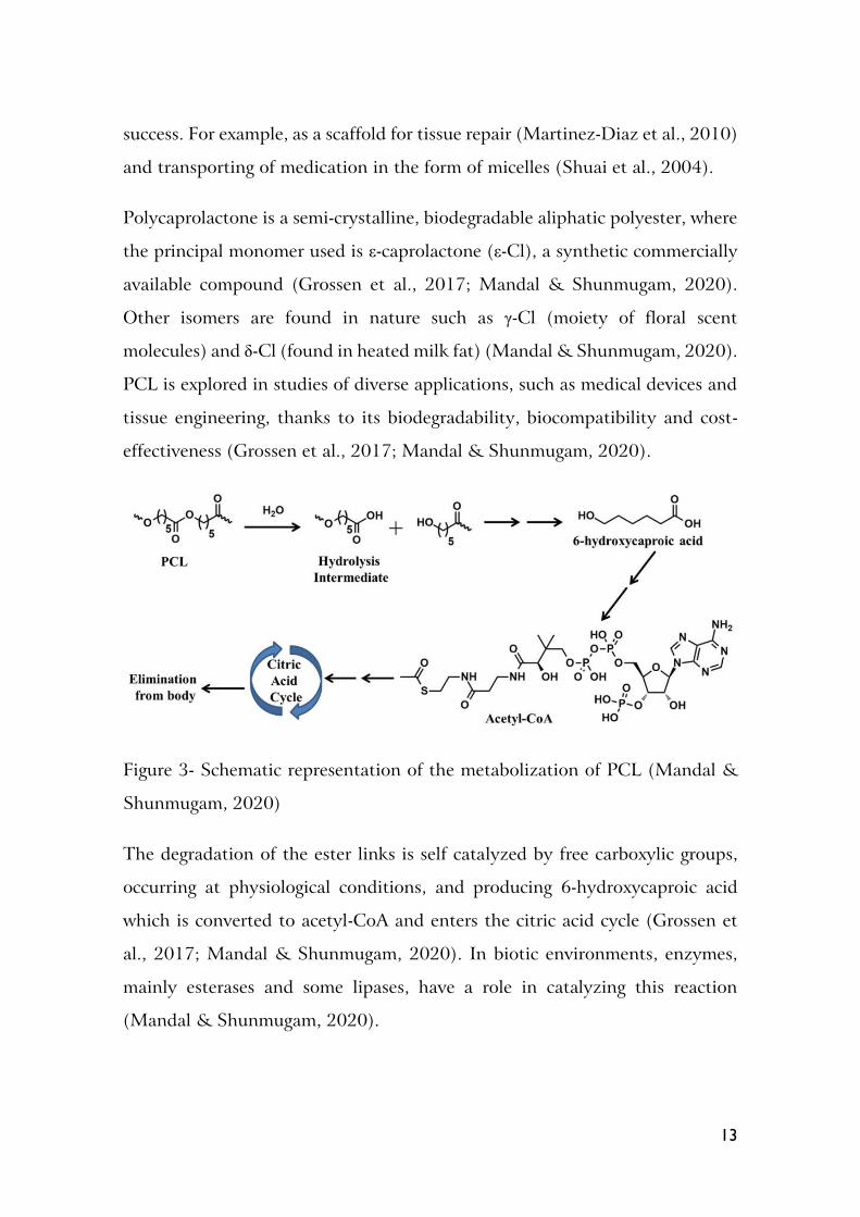
Polycaprolactone is a semi-crystalline, biodegradable aliphatic polyester, where
the principal monomer used is ε-caprolactone (ε-Cl), a synthetic commercially
available compound (Grossen et al., 2017; Mandal & Shunmugam, 2020).
Other isomers are found in nature such as γ-Cl (moiety of floral scent
molecules) and δ-Cl (found in heated milk fat) (Mandal & Shunmugam, 2020).
PCL is explored in studies of diverse applications, such as medical devices and
tissue engineering, thanks to its biodegradability, biocompatibility and cost-
effectiveness (Grossen et al., 2017; Mandal & Shunmugam, 2020).
Figure 3- Schematic representation of the metabolization of PCL (Mandal &
Shunmugam, 2020)
The degradation of the ester links is self catalyzed by free carboxylic groups,
occurring at physiological conditions, and producing 6-hydroxycaproic acid
which is converted to acetyl-CoA and enters the citric acid cycle (Grossen et
al., 2017; Mandal & Shunmugam, 2020). In biotic environments, enzymes,
mainly esterases and some lipases, have a role in catalyzing this reaction
(Mandal & Shunmugam, 2020).

14
PEG-PCL micelles remain stable for months in phosphate buffered serum, but
in the biological medium they are affected by protein opsonisation (Grossen et
al., 2017). In animal studies, PEG-PCL unimers were removed from plasma
with a half-life of 10.2h, where micelles lasted triple of the time, and no
accumulation of metabolites was observed as they were excreted from the body
(Grossen et al., 2017).
Enzymes as biocatalysts
Enzymes are biological catalysts that are advantageous over conventional
metallic catalysts because of their exceptional product selectivity, mitigation of
waste generation, ability to function at mild conditions, lower energy
requirements, simplified production routes and lower toxicity (Chapman,
Ismail, & Dinu, 2018).
Enzymes are applied in many industries such as food and beverage, detergent,
pharmaceutical, and bio fuel (Chapman et al., 2018). Their application is
projected to grow with the improvement of the economics of their use, with
the global enzyme market projected to reach 6.32 billion dollars in 2021
(Chapman et al., 2018). Most enzymes in use are hydrolytic, mainly proteases
followed by carbohydrases (Kirk, Borchert, & Fuglsang, 2002).
Modern biotechnology is the origin of the contemporaneous application of
enzymes, where natural enzymes from spontaneously growing microorganism
were applied in the production of food like cheese, bread, beer, wine, vinegar,
and commodities like leather, indigo and linen (Kirk et al., 2002).
During the last century, the production of enzymes as select strains, purified
and well-characterized, even in large amounts, allowed them to become viable
products and catalysts in the industrial context, for example in detergent,
textile and starch industries (Kirk et al., 2002).
The production of recombinant gene cells, allowed for further improvements

15
in the production of established enzymes and the manufacture of new enzymes
from microorganism that cannot be cultivated yet (Kirk et al., 2002).
Protein engineering allowed enzymes to be tailor made for new reactions and
specific conditions (Kirk et al., 2002). The improvements in protein design
directed evolutionary approaches also allowed the production of better
enzymes with improved activity, stability, and substrate affinity and cheaper
isolation (Chapman et al., 2018). Directed evolution utilizes random
modifications to the amino acid sequence, then the mutants are screened for
improvements, and the beneficial alteration is further improved on, which has
produced variants able to function in extreme pH or high temperatures and
some have reached several orders of higher catalytic activity (Chapman et al.,
2018).
Molecular dynamics (MD) simulations can provide understanding of the
phenomenon that determines the enzymes physical and catalytic
characteristics, at an atomic level (Chapman et al., 2018). These simulations
apply Newton’s laws of motion at the atomic scale, providing insight into
interactions such as the binding mechanism, which led to the optimization of
the studied enzymes (Chapman et al., 2018).
The main application of enzymes is the detergent industry, which is constantly
improving the current engineered version of the detergent enzymes to fit ever-
updating performance requirements (Kirk et al., 2002). This includes the
ability to fulfil their role at lower temperatures and alkaline pH (Kirk et al.,
2002). The development of detergents that are both cost-effective and
environmentally benign relies on enzymes, not as a catalyst for a step in
production but as a product (Chapman et al., 2018). The specificity of enzymes
also avoids the damage to fabrics and surfaces that harsh detergent agents can
cause, and the ratio of enzymes in a detergent mixture can be fine-tuned for a
specific application (Chapman et al., 2018)

16
In the pharmaceutical industry product specificity is a high priority, so
enzymes’ high selectivity allows production with less effort to purify the desired
stereoisomer and lessen the use of high temperatures or chemically harsh
substances (Chapman et al., 2018).
Traditional chemical synthesis, with metallic catalysts, is not viable in food
production due to toxicity, but enzymes are an alternative that allows for
simple, safer, and efficient production (Chapman et al., 2018).
In the production of animal feed, there are enzymes turn components
otherwise indigestible for monogastric, such as cellulose into digestible sugars,
decreasing the amount of feed needed, or increase the uptake of phosphorus to
prevent environmental release (Kirk et al., 2002).
The industries where excess waste is punished monetarily by aregulatory
agency, such as paper, textiles, leather and biofuel, are expected to embrace
more the use of enzymes along with progress (Chapman et al., 2018). For
example, in textile industry, enzymes allow for cotton scouring to be performed
at lower temperatures with less water consumption (Kirk et al., 2002). From
an ecological perspective, enzymes like lipases, esterases, and proteases, which
have the ability to hydrolyse ester bonds, are interesting means of
bioremediation in cases of industrial waste contamination (Melani, Tambourgi,
& Silveira, 2020).
However the application of enzymes is limited by their lack of stability at high
temperatures, turbulent reaction systems and some industrial solvents
(Chapman et al., 2018).
Enzymes can be improved for a task by immobilization, the attachment to the
desired material, which can improve activity and stability in conditions outside
the range of the free enzyme and imparts new functionality according to both
the method of immobilization and the physical, chemical, electrical or

17
mechanical properties of the material (Chapman et al., 2018). Immobilized
enzymes also reduce the number of steps required for processing as it is simpler
to remove the catalyst from the reaction mixture, they maintain their catalytic
ability and can be reused, even if the immobilization is time-consuming and
costly (Chapman et al., 2018).
The industrial application of enzymes is associated with decreased
consumption of energy, reduced chemical input and lower waste production,
indicating the potential for enzymes to make industry both more eco-friendly
and lucrative (Chapman et al., 2018).
For the reactions in this work, the enzyme class chosen was lipases, more
specifically Candida antartica lipase B (CalB).
Lipases are serine hydrolases whose catalytic activity includes the hydrolysis of
triaglycerols, synthesis of esters and transesterification and aminolysis
(Navvabi, Razzaghi, Fernandes, Karami, & Homaei, 2018), but can be used to
catalyse reactions of esterification, transesterification, interesterification,
amidation, transamidation, aminolysis, aldol condensation, and Michael
addition (Jiang & Loos, 2016). Lipases are most active on long-chain fatty
acids, namely ten or more carbon atoms (Navvabi et al., 2018).
The active site is composed of the classic catalytic triad composed by a serine,
histidine and aspartate residues (Melani et al., 2020). The aspartate residue is
sometimes substituted by a glutamic acid, another catalytic acidic residue
(Jiang & Loos, 2016). Lipases are stable even in non-aqueous solution, and do
not require a cofactor (Melani et al., 2020). They have high activity and most
have a alkaline optimum pH, at 8 to 9 (Melani et al., 2020).
Lipases have a characteristic α/β hydrolase fold that grants the distinguishing
property of interfacial activation, as the enzyme conformation is obtained in
contact with an oil-water interface so the lipase operates in the interface of

18
biphasic systems (Goswami & Van Lanen, 2015; Melani et al., 2020). In
aqueous media the hydrophobic active site is blocked by a lid with an internal
hydrophobic face and external hydrophilic face (Melani et al., 2020). However
not all lipases have this lid, so they do not have interfacial activation (Melani
et al., 2020).
Commercial lipases were obtained from the fungi genera Rhizopus, Candida and
Rhizomucor and the bacteria genera Pseudomonas and Choromobacterium (Navvabi
et al., 2018), with the most used being CalB, thanks to its versatility (Goswami
& Van Lanen, 2015).
Computational Methods
Molecular Mechanics
Molecular Mechanics (MM) calculations provides a way to simulate large and
complex molecular systems without the massive resource consumption of
Quantum Mechanics calculations that overwhelm even supercomputers
(Durrant & Mccammon, 2011). The calculation of the forces in the system is
achieved with a simplified potential function (equation 1, Case et al., 2020)
using bonded and non-bonded terms. When solving the potential function,
the variables are replaced with parameters obtained with theoretical
calculations or determined experimentally according to atom types, element
and chemical environment (Y. Wang, Fass, & Chodera, 2020).
𝐸𝑡𝑜𝑡𝑎𝑙 = ∑ 𝐾𝑏(𝑟 − 𝑟𝑒𝑞)2 +
𝑏𝑜𝑛𝑑𝑠
∑ 𝐾𝜃(𝜃 − 𝜃𝑒𝑞)2 +
𝑎𝑛𝑔𝑙𝑒𝑠
∑ 𝑉𝜙[1 + cos(𝑛𝜙 − 𝛾)] +∑ ∑ [𝐴𝑖𝑗
𝑅𝑖𝑗12 −
𝐵𝑖𝑗
𝑅𝑖𝑗6 +
𝑞𝑖𝑞𝑗
𝜀𝑅𝑖𝑗]
𝑁
𝑗=𝑖+1
𝑁−1
𝑖=1𝑑𝑖ℎ𝑒𝑑𝑟𝑎𝑙𝑠
(1)
In this work, the force fields used are AMBER’s ff14SB, AMBER lipid17
(Dickson et al., 2014) and the General Amber Force Field (GAFF) (Sprenger,
Jaeger, & Pfaendtner, 2015; J. Wang, Wolf, Caldwell, Kollman, & Case, 2004)
which were obtained for proteins and lipids respectively, with the later offering
support to non-included residues -by using AMBER antechamber tools to

19
adjust atom types and add missing parameters (Javanainen & Martinez-seara,
2016; Maier et al., 2015; Salomon-ferrer, Case, & Walker, 2012). For the
membrane simulation, the CHARMM force field was used, as it was the one
employed by the software that constructed the membrane. CHARMM force
fields are similar to amber force fields but they are not compatible because non-
bonded parameter development follows different strategies (Vanommeslaeghe
et al., 2009).
The terms labelled bonds, angles, and dihedral are the bonded terms, which are
related to the covalent bonds between atoms. The first is the distance between
atoms, the second is the angle between atoms and the third is the dihedrals (ϕ)
the angle between the planes defined by two sets of three atoms, as illustrated
in figure 4. These variables in the simulation are compared with “eq” are the
ideal values for the atoms, i.e., most stable configuration; and a constant K for
stiffness (the larger, the more energy is required to move away from the ideal)
(Harrison et al., 2018; Langham & Kaznessis, 2010). These variables are
defined as the balance of attracting and repelling forces between atoms, and
the more stable (least energetic the point of equilibrium), the more energy is
required to move away from that point.
Figure 4 - Visual representation of bond (r), angle (θ), and dihedral (ϕ) with atoms 1 to
4, the number of each represent the atoms involved. Adapted from Sharma, Kumar, &
Chandra, 2019.

20
Non-bonded terms account for van der Waals and electrostatic
interactions, which are calculated using the Lennard-Jones potential and
Coulomb’s law respectively (Durrant & Mccammon, 2011). They cover
the distant interactions by atoms either not connected or that are at a
distance greater than three covalent bonds. Although for atoms distant
enough that the vdW interaction can be ignored, there is a cut-off to
reduce the number of calculations (Langham & Kaznessis, 2010). This is
not precise in electrostatic interactions, so after the cut-off, the Ewald
summation is applied and, distant interactions are modelled with Fourier
transform to reduce workload (Stenberg & Stenqvist, 2020).
Quantum Mechanics
Any simulation that requires a change in the quantum electronic structure
of the material, to break or create chemical bonds, requires Quantum
Mechanics (QM) calculations (Mendieta-moreno & Marcos-alcalde,
2015). QM, as a methodology, describes the behaviour of individual
particles, including electrons, with time dependent Schrödinger’s
equation (equation 2, Levine, 2000) as a means of calculating the
position of the electron in a wave function as a probability (Engel &
Dreizler, n.d.).
−ħ
𝑖
𝜕Ψ(𝑥, 𝑡)
𝜕𝑡= −
ħ2
2𝑚
𝜕2Ψ(𝑥, 𝑡)
𝜕𝑥2+ V(𝑥, 𝑡)Ψ(𝑥, 𝑡) (2)
In equation 2, ħ (called “h bar”) is the Plank constant (h) divided by 2π
(two times pi), Ψ is the wave function with coordinates of particle 𝑥 at
time 𝑡 and i stands for √−1.
This equation cannot be directly used in calculations for a system as it is
for a single particle, but some formulations can be applied to a complete
system, such as Born– Oppenheimer approximation, where the motion of

21
electrons and nuclei is partially depopulated due to the difference in
timescale (Engel & Dreizler, n.d.; Levine, 2000).
While all quantum calculations are more computationally demanding
than any force field to the point of impracticality on any large system (Y.
Wang et al., 2020), ab initio methods are so resource demanding that they
are reserved to the most complex or accuracy-demanding cases, and
Density Functional Theory (DFT) is used in most works (Thiel, 2009;
Mardirossian & Head-gordon, 2017).
With DFT, electronic energy is calculated with a functional of the density
of electrons instead of a many-body electronic wavefunction (Neese,
2009). The basis of modern DFT are two theorems by Hohenberg and
Kohn: 1)all ground-state properties of an electronic system is determined
by the ground-state electron density uniquely and 2) a functional of the
electron density is a minimum for the ground-state density can describe
the energy of an electron distribution (Neese, 2009).Rather than calculate
a many body equation, in DFT, the calculations rely on minimizing the
density functional (Neese, 2009).
Quantum Mechanics/Molecular Mechanics
The hybrid Quantum Mechanics/Molecular Mechanics (QM/MM) is a
method that allows a less resource intensive simulation while maintaining
accuracy, by limiting QM to a section of the system where the reaction
happens, while MM is applied to the remaining system (Gerrit Groenhof,
2013).
QM/MM coupling the methods can be subtractive, where the interaction
is modulated entirely in molecular mechanics, as MM is applied to the
whole system and also to just the subsection for the quantum calculations
and is then subtracted to avoid double counting of the area (Groenhof,

22
2013; Thiel, 2009); or additive, where molecular mechanics is not applied
inside the quantum section but requires correction for the interactions
and bonds that cross the borders between sections (Groenhof, 2013;
Thiel, 2009). While subtractive coupling is simpler to implement, the QM
section may not have a compatible force field, or it cannot support
chemical changes from the reaction (Groenhof, 2013; Thiel, 2009). On
the other hand, additive methods requires explicit terms for the
interactions between subsystems : mechanic embedding in which the force
field is adjusted to not allow the QM section to be polarized by the
environment of the MM section, which is not the best method for
biomolecules, as they can be highly polar (Groenhof, 2013; Thiel, 2009);
or electrostatic embedding in which the electrostatic interaction between
subsystems is calculated alongside the electronic wave function treating
charged atoms of the MM section as one electron term, allowing the QM
subsystem to react to the environment and mimic reality better
(Groenhof, 2013; Thiel, 2009).
Molecular Dynamics
Molecular Dynamics (MD) simulations calculate the movement of atoms
according to classical laws of motion. By calculating the sum of forces
applied on an atom, it is possible to determine acceleration and by
applying the laws of motion, it determines the position as a function of
time (Langham & Kaznessis, 2010). Each atom is moved to the new
position, updating the model to have a position corresponding to after a
small interval of time in the order of femtoseconds (fs) (Durrant &
Mccammon, 2011).
MD simulation’s starting models can be provided by many different
techniques, such as X-ray crystallography or nuclear magnetic resonance
(Hollingsworth & Dror, 2018), or be custom built in software.

23
The molecular dynamics first step is the preparation of the system that is
going to be simulated. In this work, that was accomplished with LEaP
(link, edit and parm), a tool built in AMBER software package, which can
set the force fields that are used, bond atoms (for example sulphite bridges
have to be bonded manually) and solvates and adds ions to the system.
The simulations’ environment in this work were run with water as the
solvent, because it is the environment in which the conjugate will be
applied and the standard solvent for enzymatic reaction. It was also
applied periodic box conditions, in which the system is simulated as
infinite copies of the primary simulation box, side by side. Only the atoms
of primary box are tracked but interactions are simulated across boxes to
include atoms within cut-off distance. The atoms leaving the primary box
are replaced by the equivalent atoms entering from the opposite side,
keeping the pressure constant while better mimicking real-world
conditions. The starting kinetic energy of each atom was randomly
assigned from a Boltzmann distribution.
Python programming language
Python (Python Software Foundation, www.python.org) is a free, open-source,
cross-compatible and beginner-friendly programming language (Millman &
Aivazis, 2011). Python is an interpreted language, rather than a compiled one,
so it runs on a virtual machine to be independent of operating systems; it is
dynamically typed, where using variables does not require declaring their type
for the compiler, making it easier for beginners. Python error messages can be
easily searched in the internet to understand what went wrong.
Instead of using parenthesis { } to delimit sections of code, python uses
indentations, which are more readable for beginners. By using indentations
with conditionals (the “while” and “for” conditions), it is possible to set a loop

24
to run a block of code multiple times until the condition is reached. Using
if/else conditions, it is possible to run a block of code when appropriate.
The many available modules allow the user to extend the capabilities of python
without needing to code them. Of the available modules the ones used were:
Numpy to extend the numerical operations (Harris et al., 2020), Pandas to
process and analyse data (McKinney, 2010), Matplotlib, a library of utilities
for visualization, more specifically Pyplot, a sub-module for graphical
representation(Hunter, 2007).
Using Pandas it is possible to load data into columns to be edited and
processed. In this manner it is possible to maintain the connection between
values while applying functions such as sorting or mathematical operations.
The Numpy module was used in this work mostly for the trigonometry
functions is provides. Said functions were used to make the spherical custom
micelle model.
The Pyplot sub-module allowed the better awareness of the coordinate
processing, by enabling the visualization using a three dimensional graph.
Micelle trajectory analysis
The polymer of this work was chosen, among other characteristics for its
ability to self-assemble into micelles when in water (Raman, Pajak, &
Chiew, 2018). There is still necessary to characterise the properties of this
specific micelle. Several mathematical means of evaluating the structure
of micelles such as eccentricity, the radius of gyration, solvent accessible
surface area (SASA), and internal hydration (Faramarzi et al., 2017;
Lebecque, Crowet, Nasir, Deleu, & Lins, 2016).
The shape of a micelle is evaluated by how much it deviates from an ideal
spherical form. Eccentricity (𝑒) is a unitless measurement of that

25
deviation, the lower it is the closer to the ideal shape, and is calculated
with equation 3 , where 𝑙𝑚𝑖𝑛 and 𝑙𝑎𝑣𝑔 are respectively the minimum (min)
and average (avg) moments of inertia along the principal axis (Krüger &
Kamerlin, 2017; Lebecque et al., 2016), determined by diagonalization of
the inertial matrix from the specified atoms coordinates with CPPTRAJ
tool command “principal”.
𝑒 = 1 −𝑙𝑚𝑖𝑛
𝑙𝑎𝑣𝑔 (3)
The radius of gyration is a measurement of the distribution of atoms
relative to the center of mass. The effective radius of a micelle can be
obtained by multiplying the radius of gyration by √5
3 (Krüger & Kamerlin,
2017). Experimental measurements of the radius of gyration tend to yield
larger measurements when compared to simulations due to including
water molecules in the hydration shell (Faramarzi et al., 2017). It is
calculated with the CPPTRAJ command “radgyr”.
The solvent accessible surface area represents the area of vdW that is
exposed to the environment and therefore, able to be interacted with.
There are two ways of calculating SASA: the Connolly method calculates
the surface area by simulating a probe moving across the surface to
determine what is the shape of the molecule surface (Connolly, 1983);
and the LCPO (linear combination of pairwise overlaps) method that
calculates an atoms overlap of vdW radii to subtract from the total, with
correction terms to avoid errors from multiple overlaps (Weiser, Shenkin,
& Still, 1998). These methods are calculated with the CPPTRAJ tool, the
commands “molsurf” and “surf” respectively.
Internal hydration or hydrophobic tails hydration is a debated subject as
molecular simulations do not show noticeable amounts of water in the

26
micelle core but experimentally it appears to be noticeably hydrated
(Faramarzi et al., 2017). It is calculated as the average number of water
molecules in proximity to hydrophobic components of the micelle with
the CPPTRAJ tool command “watershell”.
Results and discussion
Molecular modelling
The AMPs molecular models were obtained from the RCSB Protein Data Bank
archive, a database that contains information, including structure models, for
biological macromolecules (Burley et al., 2019; Goodsell et al., 2020). The
structural files were prepared for simulation by the AMBER tool LEaP, which
is the principal tool to create and edit systems for simulation. LEaP allows
choosing what force fields to use, adds missing atoms according to reference
files, and enables editing structures such as adding bonds.
The pdb code for the structure of Polyphemusin is 1RKK, obtained with NMR
and deposited by Powers, Rozek, & Hancock, 2004. The file was edited for the
cysteine’s disulfide bridges to be CYX (bonded) rather than CYS (free form),
and manually bonded the residues 4 to 17 and 8 to 13, and the C-terminal
amination was renamed to match the AMBER force field terminology.
Polymyxin B was not available by itself, but included in complex with a
different protein, LSD1-CoREST1, in pdb code 5L3F (resolution 3.50 Å)
obtained with X-ray diffraction and deposited by Speranzini et al., 2016. Since
it is not a standard protein but rather a non-ribosomal peptide with modified
amino acids, the AMBER tool antechamber was used to generate topology
information which was saved as a file in the mol2 format that includes bond
and charge information.

27
The structure for polycaprolactone was previously custom made in molecular
design software for another project (Almeida, Figueiredo, & Carvalho,
2019)and was used in strands of 5 monomers.
Building base conjugate & Micelle first attempt
The conjugates were made by connecting the N-terminal amino acid of
polyphemusin I to polycaprolactone’s distal carboxyl group as shown in figure
5 (A). In studies with AMP-chitosan conjugates, the orientation of the peptide
relative to the backbone influences the activity of the conjugates, with N-
terminal linkage showing more antibacterial activity (Sun et al., 2018).
Due to polymyxins lack of n-terminal, the bond was made using a side chain
nitrogen atom of polymyxin B that doesn’t interact with the membrane
(Velkov, Thompson, Nation, & Li, 2010) to the same atom of PCL, as shown
in figure 5 (B).
(A)

28
(B)
Figure 5- Images of the conjugated starting structures before molecular dynamics,
polyphemusin I (A) and polymyxin B (B).
They were simulated in water to closer match the environment in which they
are intended to be applied. To spare computational resources we started the
simulation near the theoretical micellar optimum position, the choice of start
the simulation near the optimum position, hoping to reduce the required time
to reach equilibrium.
The conjugates were distributed in a starting position that approximates the
final micelle structure by using CHARM-GUI input generator micelle builder
(Cheng, Jo, Lee, Klauda, & Im, 2013; Jo, Kim, Iyer, & Im, 2008), which
produced a structure file whose atoms positions were used as a basis. The
molecule for the base structure, an Alkyl-PEG referred to as C13EG8, was
chosen because it had enough atoms for the entire PCL chain without
branching or ring atoms and still had room for three atoms of the AMP, so
LEaP could use as basis to add the remaining atoms.
The micelle was composed of thirty molecules, and required the order of some
atoms in the file to be switched as the input generator ordered atoms starting
in the middle of the molecule and counting towards the extremities, first the
alkyl part then the PEG. This led to a residue per molecule being split into two

29
parts that appeared separated in the file. As a consequence LEaP interpreted it
as being two different residues and misplaced atoms in excess.
The molecule was edited manually using Microsoft® Excel to alter the main
chain atom types and residue names to the intended names, repositioning lines,
and using the option to save as space separated values to maintain the columns
in the pdb format. However manual editing was very error-prone and many
corrections were required to create the final working file.
The first AMP to be tested was polymyxin B because it is the most medically
relevant of the two AMPs.
LEaP produced a micelle system that would crash during the heating step, and
multiple variations, including systems with fewer conjugates, were used for
troubleshooting. The issue was the double bond oxygen atoms of the carboxyl
group placed by leap being too close to the main chain oxygen, which yielded
a very high energy that couldn’t be computed. The solution was to perform
more minimization steps using a mask (atom selection) where only the oxygen
atoms are allowed to move.
However, the micelle was not stable and the molecules drift apart, as seen in
figure 6, most likely due to the high positive charge of Polymyxin B. The
solution was to have PCL molecules without AMP acting as stabilizer to the
micelle by increasing the interaction inside the micelle core. The ratio chosen
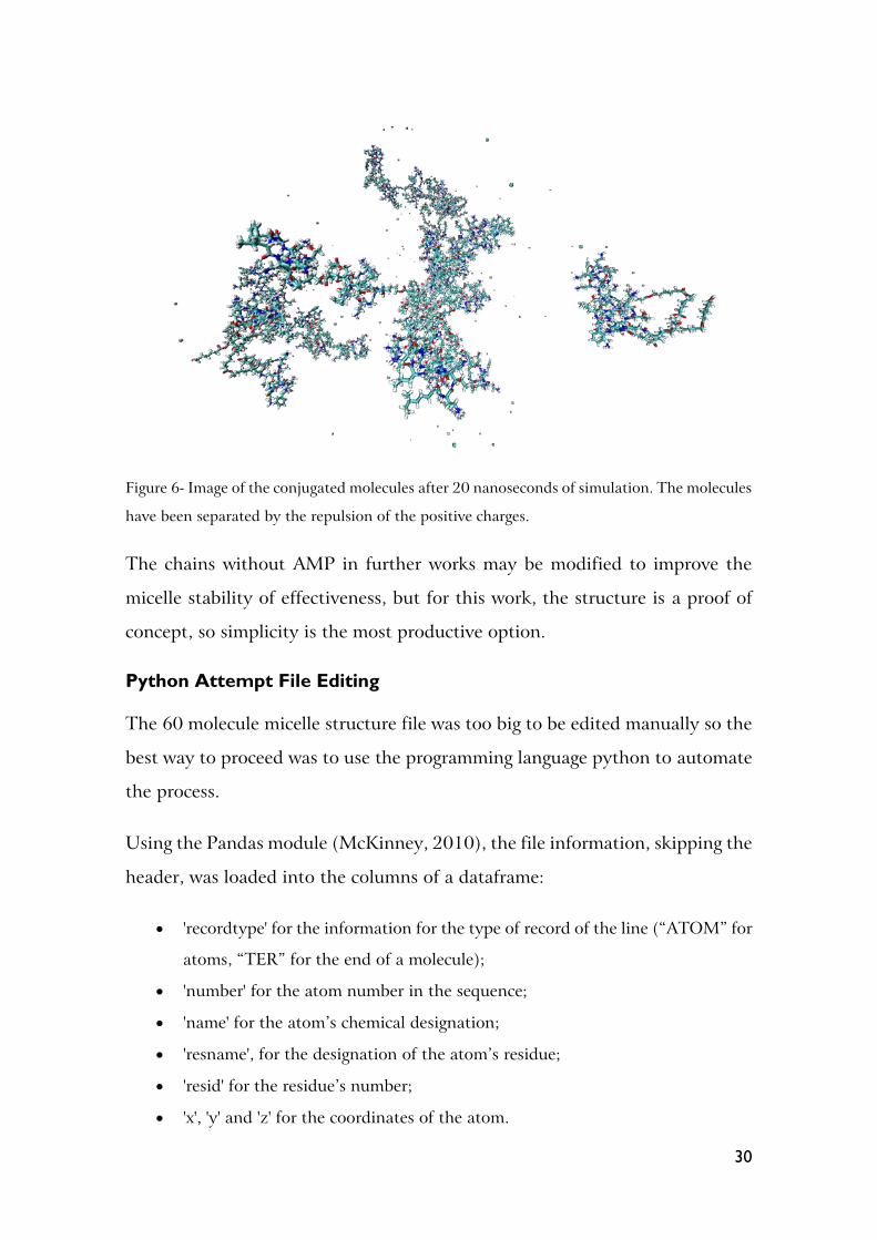
to attempt this was 1:1, making the micelle composition thirty chains with
AMPs and thirty chains without.
30
Figure 6- Image of the conjugated molecules after 20 nanoseconds of simulation. The molecules
have been separated by the repulsion of the positive charges.
The chains without AMP in further works may be modified to improve the
micelle stability of effectiveness, but for this work, the structure is a proof of
concept, so simplicity is the most productive option.
Python Attempt File Editing
The 60 molecule micelle structure file was too big to be edited manually so the
best way to proceed was to use the programming language python to automate
the process.
Using the Pandas module (McKinney, 2010), the file information, skipping the
header, was loaded into the columns of a dataframe:
• 'recordtype' for the information for the type of record of the line (“ATOM” for
atoms, “TER” for the end of a molecule);
• 'number' for the atom number in the sequence;
• 'name' for the atom’s chemical designation;
• 'resname', for the designation of the atom’s residue;
• 'resid' for the residue’s number;
• 'x', 'y' and 'z' for the coordinates of the atom.

31
The coordinates of x, y, and z were used to create a column (referred to by the
designation of ‘distance’) of values calculated as the addiction of the
coordinates squared, which is an adaptation of the equation to determine the
distance between points in Cartesian coordinates (equation 4), when one of
the points is the origin (0, 0, 0). The square root was omitted because the values
maintain the same sequence. Therefore the calculation is simplified to shorten
calculations.
𝑑 = √(𝑥1 − 𝑥2)2 + (𝑦1 − 𝑦2)
2 + (𝑧1 − 𝑧2)2 (4)
The lines in the dataframe were ordered with the sort_values() function by resid
to prevent mixing molecules and distance to place the atoms in order of
proximity by how close they are to the origin. Then the number of the atoms
and their name were corrected to match the intended molecule.
This did not work because of curves in the molecule that made atoms to be
placed closer than the preceding atoms when they should have been further
away, leading to a zigzag placement of atom, shown in figure 7 that resulted in
errors when attempting to calculate the energy of those bonds.

32
(A)
(B)
Figure 7- The end point of a PCL chain with the AMP at the extremity, before
(A) and after (B) using the sorting python code and LeAP.

33
Python attempt Micelle builder
An alternative to the previous procedure was to use a custom micelle builder
that could place the already edited molecules in a spherical arrangement. To
achieve this goal, the coordinates were transformed into spherical coordinates,
because they can be rotated around the origin whilst maintaining relative
positions of atoms.
To make the micelle two functions to convert the coordinates to Spherical and
to turn them back to Cartesian were made based on online sources, shown in
ANEX II- Part 1. Spherical coordinates use a value for distance to origin
(radius) and two angles, azimuth (angle with the x-axis in the xy plane) and
inclination (angle of radius with the xy plane).
The original molecule distribution was based on the combination of possible
azimuths and inclination angles with more than 60 results by using the angles
π/6, π/3, π/2, 2π/3, 5π/6, 7π/6, 4π/3, 3π/2, 5π/3, 11π/6, 2π. The function
itertools.combinations_with_replacement() was used to make a list of points to
place the molecule. However the placement was flawed and resulted in the
superposition of molecules. An alternative method, shown in ANEX II-Part 2,
was used that places the points evenly and can be scaled up to more easily. The
method is based on an algorithm that places the atoms by in small increments
to achieve a spherical shape while keeping the points apart. This metod allows
for an even spacing of the points across the sphere, were the original placed
them closer near the top and bottom, as seen in figure 8.

34
(A)
(B)
Figure 8- Comparison of both methods of point generation displayed using the module
matplotlib.pyplot. (A) is the first method (combinations) and (B) is the alternative method.
The molecule used to rotate was aligned to the x-axis for simplicity, then the
coordinates converted to Spherical and for each molecule until the desired
amount, the Azimuth and Inclination of a point are added to the atom’s
coordinates. The output is rewritten with the string formatting option to space
and align the columns to make sure the decimal cases are according to pdb
format.
This method did not scale well to the amount of molecules required. The
micelles produced have overlaps, which yield incalculable energy in repulsive

35
forces. An attempt to produce linear molecules to be adjusted by minimization
also did not work because the bonds were too stretched and the energy
calculation became equally incalculable. So for the sake of time the 60 molecule
micelle was redefined to use the functional but unstable micelle file and reduce
the number of antimicrobial peptides to obtain the same AMP.
So the micelle with thirty AMP was edited to have only fifteen AMP. A short
python code was written to copy the pdb file line by line and count the number
of atoms with the residue name of the AMP, until the desired amount was
reached to then skip the remaining AMP atoms. The micelle was simulated for
20 nanoseconds in segments of two and a half nanoseconds, during which it
remained stable. This method was repeated to create a micelle without AMP as
a control and simulated during the same amount of time (20 ns) in the same
conditions to be used in structural comparisons. Both can be seen in figure 9,
with the starting position for comparisson
(A)

36
(B)
(C)
Figure 9- AMP (A) and control (B) micelles, after 20 ns simulation and the starting position (C)
for the AMP micelle. Both micelles shape remained stable during the simulation. The control
micelle starting structure used the same base as the one with AMP, but the peptide was
removed.
Micelle stability
To understand the properties of the AMP bonded micelle, they were compared
to the control micelle without AMP. Due to the lack of more similar micelle
information for comparison, the structural information of software-made
detergent micelles from Krüger & Kamerlin, 2017 (supplemental table S1) was
used as a reference of normal micelle parameters.

37
The eccentricity of the micelles was evaluated from 17.5 ns to 20 ns. While the
eccentricity fluctuates, the micelle with polymyxin B averages at 0.172 with a
standard deviation of 0.026. When compared to simulations of the control
micelle with an average of 0.169 with a standard deviation of 0.029 the bonded
AMP appears to not drastically alter the eccentricity. However, the reference
micelles have their eccentricity between 0.083±0.031 (α-DDM) and
0.153±0.045 (β-NG) which is lower than either PCL micelle, but the micelles
appear to be stable even if they are not completely spherical.
The average effective micelle radius in the same interval is 32.14 Å for the
micelle with polymyxin B, while the control micelle was a radius of 18.783 Å.
The reference micelles have a minimum size of 24.73±0.39 Å (β-OM) and a
maximum size of 34.71±0.41 Å (β-DDM), a range that includes the created
micelles.
In the micelle with polymyxin B, the solvent accessible surface area (SASA) of
the micelle hydrophobic (PHO) components is on average 6258.486 Å2 and
the hydrophilic (PHI) components is 19253.975 Å2, which has a PHI/PHO
ratio of 3.083. When the ratio is higher, it corresponds to a more
thermodynamically favourable disposition of the micelle domains (Lebecque et
al., 2016). However in this specific case some of the hydrophilic AMPs are not
fully in contact with the core so it may not be wise to evaluate this micelle on
this parameter the way the others are since the ratio doesn’t correlate entirely
to how much the hydrophilic domains shields the core. The control micelle has
a SASA of 9270.661 Å2 so the micelles AMPs cover 67.5085% of the surface
area.
The hydrophobic residues appear to have contact with water but there is not
hydration at the core. Despite the contact of PCL with water, there is no water
inside the micelle.

38
The micelles appear to have the PCL molecules move within the core in such a
manner that some chains appear to be entirely on the surface. This is most
likely due to the extremely simplified conditions chosen for the micelle
formation. In future works, the micelle stability would be taken into account,
using PEG as a hydrophilic block for the chains without AMP or adding a more
hydrophobic component to PCL to maximize the hydrophobic potential. It
could be possible to attempt the simulations with alternative formulations of
the peptides, such as an analogue with a lower charge to avoid repulsion. The
choice of position used to bond the AMP with the PCL chain can be reviewed,
to minimize the impact on the functionality.
Membrane model
As it is most frequent, this work was based on E. coli as a model-organism for
the study of AMP membrane interactions. The membrane was created using
CHARMM-GUI membrane builder to create a pdb file with the molecules
distributes as intended in a bilayer.
For simplicity, the outer membrane external layer, was constituted of only LPS,
since other lipids have low composition enough to omit (Rice &
Wereszczynski, 2018; Wu et al., 2014). The inner leaflet of the outer
membrane was composed of phosphatidylethanolamine (PE) and
phosphatidylglycerol (PG), which are the two main classes of lipids of the inner
membrane leaflet (Wu et al., 2014). The ratio between them is 3: 1, since PE
constitutes 75% of the lipids in the inner leaflet and the other components are
far less present (Wu et al., 2014). The charges of lipid A phosphate groups were
neutralized with calcium and the remaining negative charges with sodium
(Patel, Qi, & Im, 2017; Rice & Wereszczynski, 2018).
Due to the variety and size of LPS, lipid A without core or O-antigen was used
as the simplest form rough LPS. The polymyxin B targets the phosphate groups
in the lipid A, therefore both the simulation time required for their interaction

39
and the number of atoms in the system is reduced. Unfortunately, the
software’s limitation causes the LPS to not be placed correctly, likely due to
the modularity of LPS construction being incompatible with the adjustments
applied to the other membrane molecules, as the software’s membrane
visualization option, seen in figure 10, does not recognise the LPS atoms as a
single molecule even though it recognizes the remaining molecules. The end
result is overlapping lipid tails as the LPS are placed as rigid structures, instead
of being adjusted like PE and PG were, as seen in figure 11.
Figure 10- Visualization of the construction of the membrane in CHARMM-
GUI, demonstrating the flaw of the micelle placement. LPS are shown as
individual atoms; other membrane molecules are shown as full molecules. The
red molecules are water and the large grey spheres are calcium ions.

40
Figure 11- Close-up of lipid A molecules in the membrane, highlighting the
overlaps of lipid tails. Each residue is shown in a different colour to add
contrast.
However the overlap of LPS lipid tails during the structure creation led to the
need for the membrane minimization to be more complex, using NAMD
(Phillips et al., 2020) to restrict the structure so only the LPS lipid tails move,
so the system can be properly equilibrated. This is done using the VMD
(Humphrey, Dalke, & Schulten, 1996) scripting tool to attribute a beta value
of 0 to the atoms that are intended to equilibrate, and to attribute a beta value
of 1 to the atoms that are restrained, which can be seen in figure 12.
Figure 12- Membrane structure, where the atoms in red represent the
unrestrained lipid tails of LPS, with a beta value of 0, and in blue the remaining

41
atoms, with a beta value of 1. Omitted in this image, water and ions also have
a beta value of 1
In this step the CHARMM force field was used, as the force field that the input
generator creates the parameters for by default. The equilibration of the
membrane first started by restricting every atom except those in LPS lipid tails
and lasted 1 nanosecond.
Then minimization continued for 20 ns without the restrictions on atoms
followed by 10 ns more of simulation without the minimization.
For compatibility, to simulate the membrane with the AMP, both must be
under the same force field.
Using AMBER’s charmm2amber.py script, it is possible to substitute the atom
and residue designations to AMBER’s for both inner leaflet lipids. The main
difference between CHARMM and AMBER lipid force field is the residue
labelling, as in CHARMM every residue is an entire lipid molecule and in
AMBER the molecule is split in smaller residues, as a form of modularity.
However, lipid A is not supported in this script so the antechamber tool was
used to create a mol2 file that includes the information for parameters. Calcium
ions were also not covered in this script, but the search and replace function
solved that oversight.
Other required modification of the file were the renumbering of lipid A
residues, as all were set as residue 1, and the separation of molecules in the file
with the indicator “TER”, in accordance with PDB format. This was done with
python code.
First, the file was opened with python to load the data in lists. Using the split()
function on each line, the values of each column can be assigned to variables,
which are then appended to a list for each. The residue numbers of lipid A were
replaced, by using a counter that increases when the first atom of the molecule

42
is encountered. Using string formatting, the columns were rewritten in the pdb
spacing and alignment format.
Before any of the first atoms either LPS or inner leaflet molecules, a “TER”
indicator was written on the file. The atoms were specifically: HB6S of LPS
and C12 of PA a residue common to both PG and PE, as part of AMBER lipids
modularity.
Conclusions and Future Perspectives
This project, while not having fulfilled the proposed objectives within the
available time, still accomplished useful results:
-A stable micelle with PCL and polymyxin B that is ready for testing;
-An AMBER-compatible equilibrated E.coli membrane model;
-Several snippets of python code for pdb editing, specifically reformatting files
and renumbering and separating residues.
The results in this work are preliminary, setting a precedent for a new way to
utilize antimicrobial peptides for medical purposes. Although this work is very
conservative, to minimize the number of variables, further research would
establish a better understanding that will allow designing an optimized micelle
to be tested. This would allow in vitro testing to not have unproductive
experiments.
The works elaborating on this one can take this base approach and incorporate
the above referred stabilization mechanisms.

43
References Almeida, B. C., Figueiredo, P., & Carvalho, A. T. P. (2019). Polycaprolactone Enzymatic
Hydrolysis: A Mechanistic Study. ACS Omega, 4(4), 6769–6774. https://doi.org/10.1021/acsomega.9b00345
Burley, S. K., Berman, H. M., Bhikadiya, C., Bi, C., Chen, L., Di Costanzo, L., … Ioannidis, Y. E. (2019). Protein Data Bank: The single global archive for 3D macromolecular structure data. Nucleic Acids Research, 47(D1), D520–D528. https://doi.org/10.1093/nar/gky949
Case, D. A., Belfon, K., Ben-Shalom, I. Y., Brozell, S. R., Cerutti, D. S., T.E. Cheatham, I., … Kollman, P. A. (2020). Amber 2020. University of California, San Francisco.
Chapman, J., Ismail, A. E., & Dinu, C. Z. (2018). Industrial applications of enzymes: Recent advances, techniques, and outlooks. Catalysts, 8(6), 20–29. https://doi.org/10.3390/catal8060238
Cheng, X., Jo, S., Lee, H. S., Klauda, J. B., & Im, W. (2013). CHARMM-GUI Micelle Builder for Pure/Mixed Micelle and Protein/Micelle Complex Systems. Journal of Chemical Information and Modeling, 53(8), 2171–2180. https://doi.org/10.1021/ci4002684
Connolly, M. L. (1983). Analytical molecular surface calculation. Journal of Applied Crystallography, 16(5), 548–558. https://doi.org/10.1107/s0021889883010985
Dash, T. K., & Konkimalla, V. B. (2012). Poly-ε-caprolactone based formulations for drug delivery and tissue engineering: A review. Journal of Controlled Release, 158(1), 15–33. https://doi.org/10.1016/j.jconrel.2011.09.064
Debye, P., Anacker, E., & Anacker’, E. W. (1950). Micelle shape from dissymetry measurements. J. Phys. Chem., 644–655.
Dickson, C. J., Madej, B. D., Skjevik, Å. a., Betz, R. M., Teigen, K., Gould, I. R., & Walker, R. C. (2014). Lipid14: The Amber Lipid Force Field. Journal of Chemical Theory and Computation, 10(2), 865–879. https://doi.org/dx.doi.org/10.1021/ct4010307
Durrant, J. D., & Mccammon, J. A. (2011). Molecular dynamics simulations and drug discovery. BMC Biology. Retrieved from http://www.biomedcentral.com/1741-7007/9/71
Engel, E., & Dreizler, R. M. (n.d.). Density Functional Theory.
Faramarzi, S., Bonnett, B., Scaggs, C. A., Ho, A., Grodi, D., Harvey, E., … Virginia, W. (2017). Molecular Dynamics Simulations as a Tool for Accurate Determination of Surfactant Micelle Properties. https://doi.org/10.1021/acs.langmuir.7b02666
French, S., Farha, M., Ellis, M. J., Sameer, Z., Côté, J. P., Cotroneo, N., … Brown, E. D. (2020). Potentiation of Antibiotics against Gram-Negative Bacteria by Polymyxin B Analogue SPR741 from Unique Perturbation of the Outer Membrane. ACS Infectious Diseases, 6(6), 1405–1412. https://doi.org/10.1021/acsinfecdis.9b00159
Gerrit Groenhof. (2013). Introduction to QM/MM Simulations. Methods Mol Biol (Vol. 924). https://doi.org/10.1007/978-1-62703-017-5
Goodsell, D. S., Zardecki, C., Di Costanzo, L., Duarte, J. M., Hudson, B. P., Persikova, I., … Burley, S. K. (2020). RCSB Protein Data Bank: Enabling biomedical research and drug discovery. Protein Science, 29(1), 52–65. https://doi.org/10.1002/pro.3730

44
Goswami, A., & Van Lanen, S. G. (2015). Enzymatic strategies and biocatalysts for amide bond formation: Tricks of the trade outside of the ribosome. Molecular BioSystems, 11(2), 338–353. https://doi.org/10.1039/c4mb00627e
Groenhof, G. (2013). Introduction to QM/MM Simulations. In Biomolecular Simulations: Methods and Protocols, Methods in Molecular Biology (Vol. 924, pp. 43–66). https://doi.org/10.1007/978-1-62703-017-5
Grossen, P., Witzigmann, D., Sieber, S., & Huwyler, J. (2017). PEG-PCL-based nanomedicines : A biodegradable drug delivery system and its application. Journal of Controlled Release, 260(May), 46–60. https://doi.org/10.1016/j.jconrel.2017.05.028
Hancock, R. E. W. (2000). Expert Opinion on Investigational Drugs- Cationic antimicrobial peptides : towards clinical applications, 1723–1729.
Harris, C. R., Millman, K. J., van der Walt, S. J., Gommers, R., Virtanen, P., Cournapeau, D., … Oliphant, T. E. (2020). Array programming with NumPy. Nature, 585(7825), 357–362. https://doi.org/10.1038/s41586-020-2649-2
Harrison, J. A., Schall, J. D., Maskey, S., Mikulski, P. T., Knippenberg, M. T., Morrow, B. H., … Morrow, B. H. (2018). Review of force fields and intermolecular potentials used in atomistic computational materials research, 031104. https://doi.org/10.1063/1.5020808
Hollingsworth, S. A., & Dror, R. O. (2018). Molecular Dynamics Simulation for All. Neuron, 99(6), 1129–1143. https://doi.org/10.1016/j.neuron.2018.08.011
Humphrey, W., Dalke, A., & Schulten, K. (1996). VMD - Visual Molecular Dynamics. Journal of Molecular Graphics, 14, 33–38.
Hunter, J. D. (2007). Matplotlib: A 2D graphics environment. Computing in Science and Engineering, 9(3), 90–95. https://doi.org/10.1109/MCSE.2007.55
Javanainen, M., & Martinez-seara, H. (2016). Efficient preparation and analysis of membrane and membrane protein systems. Biochimica et Biophysica Acta- Biomembranes, 1858(10), 2468–2482. https://doi.org/10.1016/j.bbamem.2016.02.036
Javia, A., Amrutiya, J., Lalani, R., Patel, V., Bhatt, P., & Misra, A. (2018). Antimicrobial peptide delivery: An emerging therapeutic for the treatment of burn and wounds. Therapeutic Delivery, 9(5), 375–386. https://doi.org/10.4155/tde-2017-0061
Jiang, Y., & Loos, K. (2016). Enzymatic synthesis of biobased polyesters and polyamides. Polymers, 8(7). https://doi.org/10.3390/polym8070243
Jo, S., Kim, T., Iyer, V. G., & Im, W. (2008). CHARMM-GUI: A web-based graphical user interface for CHARMM. Journal of Computational Chemistry, 29(11), 1859–1865. https://doi.org/https://doi.org/10.1002/jcc.20945
Kedar, U., Phutane, P., Shidhaye, S., & Kadam, V. (2010). Advances in polymeric micelles for drug delivery and tumor targeting. Nanomedicine: Nanotechnology, Biology, and Medicine, 6(6), 714–729. https://doi.org/10.1016/j.nano.2010.05.005
Kirk, O., Borchert, T. V., & Fuglsang, C. C. (2002). Industrial enzyme applications. Current Opinion in Biotechnology, 13(4), 345–351. https://doi.org/10.1016/S0958-1669(02)00328-2

45
Krüger, D. M., & Kamerlin, S. C. L. (2017). Micelle Maker: An Online Tool for Generating Equilibrated Micelles as Direct Input for Molecular Dynamics Simulations. ACS Omega, 2(8), 4524–4530. https://doi.org/10.1021/acsomega.7b00820
Lam, C. X. F., Hutmacher, D. W., Schantz, J. T., Woodruff, M. A., & Teoh, S. H. (2009). Evaluation of polycaprolactone scaffold degradation for 6 months in vitro and in vivo. Journal of Biomedical Materials Research. Part A, 90(3), 906–919. https://doi.org/10.1002/jbm.a.32052
Langham, A., & Kaznessis, Y. N. (2010). Molecular Simulations of Antimicrobial Peptides. Methods Mol Biol, (618), 267–285. https://doi.org/10.1007/978-1-60761-594-1_17
Lebecque, S., Crowet, J. M., Nasir, M. N., Deleu, M., & Lins, L. (2016). Molecular Dynamics Study of Micelles Properties According to their Size. Journal of Molecular Graphics and Modelling. https://doi.org/10.1016/j.jmgm.2016.12.007
Lei, J., Sun, L. C., Huang, S., Zhu, C., Li, P., He, J., … He, Q. (2019). The antimicrobial peptides and their potential clinical applications. American Journal of Translational Research, 11(7), 3919–3931.
Lenhard, J. R., Bulman, Z. P., Tsuji, B. T., & Kaye, K. S. (2019). Shifting gears: The future of polymyxin antibiotics. Antibiotics, 8(2), 1–13. https://doi.org/10.3390/antibiotics8020042
Lepak, A. J., Wang, W., & Andes, D. R. (2020). Pharmacodynamic Evaluation of MRX-8, a Novel Polymyxin, in the Neutropenic Mouse Thigh and Lung Infection Models against Gram-Negative Pathogens. Antimicrobial Agents and Chemotherapy, 64(11), 1–11. https://doi.org/10.1128/AAC.01517-20
Levine, I. N. (2000). Quantum Chemistry - Ira Levine - 5th edition (5th ed.).
Liechty, W. B., Kryscio, D. R., Slaughter, B. V., & Peppas, N. A. (2010). Polymers for drug delivery systems. Annual Review of Chemical and Biomolecular Engineering, 1, 149–173. https://doi.org/10.1146/annurev-chembioeng-073009-100847
Lu, Y., Zhang, E., Yang, J., & Cao, Z. (2018). Strategies to improve micelle stability for drug delivery. Nano Res, (11), 4985–4998. https://doi.org/doi:10.1007/s12274-018-2152-3
Magana, M., Pushpanathan, M., Santos, A. L., Leanse, L., Fernandez, M., Ioannidis, A., … Tegos, G. P. (2020). The value of antimicrobial peptides in the age of resistance. The Lancet Infectious Diseases, 20(9), e216–e230. https://doi.org/10.1016/S1473-3099(20)30327-3
Maier, J. A., Martinez, C., Kasavajhala, K., Wickstrom, L., Hauser, K., & Simmerling, C. (2015). ff14SB : Improving the accuracy of protein side chain and backbone parameters from ff99SB.
Mandal, P., & Shunmugam, R. (2020). Polycaprolactone: a biodegradable polymer with its application in the field of self-assembly study. Journal of Macromolecular Science, Part A: Pure and Applied Chemistry, 58(2), 111–129. https://doi.org/10.1080/10601325.2020.1831392
Mardirossian, N., & Head-gordon, M. (2017). Thirty years of density functional theory in computational chemistry : an overview and extensive assessment of 200 density functionals, 8976. https://doi.org/10.1080/00268976.2017.1333644

46
Marggraf, M. B., Panteleev, P. V., Emelianova, A. A., Sorokin, M. I., Bolosov, I. A., Buzdin, A. A., … Ovchinnikova, T. V. (2018). Cytotoxic potential of the novel horseshoe crab peptide polyphemusin III. Marine Drugs, 16(12), 1–19. https://doi.org/10.3390/md16120466
Martin-Serrano, Á., Gómez, R., Ortega, P., & Mata, F. J. D. La. (2019). Nanosystems as vehicles for the delivery of antimicrobial peptides (Amps). Pharmaceutics, 11(9), 1–24. https://doi.org/10.3390/pharmaceutics11090448
Martinez-Diaz, S., Garcia-Giralt, N., Lebourg, M., Gómez-Tejedor, J. A., Vila, G., Caceres, E., … Monllau, J. C. (2010). In Vivo Evaluation of 3-Dimensional Polycaprolactone Scaffolds for Cartilage Repair in Rabbits. American Journal of Sports Medicine, 38(3), 509–519. https://doi.org/10.1177/0363546509352448
McKinney, W. (2010). Data Structures for Statistical Computing in Python. Proceedings of the 9th Python in Science Conference, 1(Scipy), 56–61. https://doi.org/10.25080/majora-92bf1922-00a
Melani, N. B., Tambourgi, E. B., & Silveira, E. (2020). Lipases: From Production to Applications. Separation and Purification Reviews, 49(2), 143–158. https://doi.org/10.1080/15422119.2018.1564328
Mendieta-moreno, J. I., & Marcos-alcalde, I. (2015). Molecular Mechanics Method for the Dynamical Study of Reactions in Biomolecules. Combined Quantum Mechanical and Molecular Mechanical Modelling of Biomolecular Interactions (1st ed., Vol. 100). Elsevier Inc. https://doi.org/10.1016/bs.apcsb.2015.06.003
Millman, K. J., & Aivazis, M. (2011). Python for scientists and engineers. Computing in Science and Engineering, 13(2), 9–12. https://doi.org/10.1109/MCSE.2011.36
Navvabi, A., Razzaghi, M., Fernandes, P., Karami, L., & Homaei, A. (2018). Novel lipases discovery specifically from marine organisms for industrial production and practical applications. Process Biochemistry, 70(March), 61–70. https://doi.org/10.1016/j.procbio.2018.04.018
Neese, F. (2009). Density functional theory, 443–453. https://doi.org/10.1007/s11120-009-9404-8
Nordström, R., & Malmsten, M. (2017). Delivery systems for antimicrobial peptides. Advances in Colloid and Interface Science (Vol. 242). Elsevier B.V. https://doi.org/10.1016/j.cis.2017.01.005
Owen, S. C., Chan, D. P. Y., & Shoichet, M. S. (2012). Polymeric micelle stability. Nano Today, 7(1), 53–65. https://doi.org/10.1016/j.nantod.2012.01.002
Patel, D. S., Qi, Y., & Im, W. (2017). Modeling and simulation of bacterial outer membranes and interactions with membrane proteins. Current Opinion in Structural Biology. Elsevier Ltd. https://doi.org/10.1016/j.sbi.2017.01.003
Phillips, J. C., Hardy, D. J., Maia, J. D. C., Stone, J. E., Ribeiro, J. V., Bernardi, R. C., … Tajkhorshid, E. (2020). Scalable molecular dynamics on CPU and GPU architectures with NAMD. Journal of Chemical Physics, 153(4). https://doi.org/10.1063/5.0014475
Phillips, J. N. (1954). The energetics of micelle formation. Transactions of the Faraday Society, 561–569.

47
Powers, J. S., Martin, M. M., Goosney, D. L., & Hancock, R. E. W. (2006). The Antimicrobial Peptide Polyphemusin Localizes to the Cytoplasm of Escherichia coli following Treatment. ANTIMICROBIAL AGENTS AND CHEMOTHERAPY, 50(4), 1522–1524. https://doi.org/10.1128/AAC.50.4.1522
Powers, J. S., Rozek, A., & Hancock, R. E. W. (2004). Structure-activity relationships for the β-hairpin cationic antimicrobial peptide polyphemusin I. Biochimica et Biophysica Acta - Proteins and Proteomics, 1698(2), 239–250. https://doi.org/10.1016/j.bbapap.2003.12.009
Raman, A. S., Pajak, J., & Chiew, Y. C. (2018). Interaction of PCL based self-assembled nano-polymeric micelles with model lipid bilayers using coarse-grained molecular dynamics simulations. Chemical Physics Letters, 712(August), 1–6. https://doi.org/10.1016/j.cplett.2018.09.049
Rice, A., & Wereszczynski, J. (2018). Atomistic Scale Effects of Lipopolysaccharide Modifications on Bacterial Outer Membrane Defenses. Biophysj, 114(6), 1389–1399. https://doi.org/10.1016/j.bpj.2018.02.006
Rose, F., Heuer, K. U., Sibelius, U., Hombach-Klonisch, S., Kiss, L., Seeger, W., & Grimminger, F. (2000). Targeting lipopolysaccharides by the nontoxic polymyxin B nonapeptide sensitizes resistant Escherichia coli to the bactericidal effect of human neutrophils. Journal of Infectious Diseases, 182(1), 191–199. https://doi.org/10.1086/315669
Salomon-ferrer, R., Case, D. A., & Walker, R. C. (2012). An overview of the Amber biomolecular simulation package, 00(February), 1–13. https://doi.org/10.1002/wcms.1121
Sandreschi, S., Piras, A. M., Batoni, G., & Chiellini, F. (2016). Perspectives on polymeric nanostructures for the therapeutic application of antimicrobial peptides. Nanomedicine, 11(13), 1729–1744. https://doi.org/10.2217/nnm-2016-0057
Seidi, F., Jenjob, R., & Crespy, D. (2018). Designing Smart Polymer Conjugates for Controlled Release of Payloads. Chemical Reviews, 118(7), 3965–4036. https://doi.org/10.1021/acs.chemrev.8b00006
Sharma, S., Kumar, P., & Chandra, R. (2019). Introduction to molecular dynamics. Molecular Dynamics Simulation of Nanocomposites Using BIOVIA Materials Studio, Lammps and Gromacs, 23, 1–38. https://doi.org/10.1016/B978-0-12-816954-4.00001-2
Shuai, X., Ai, H., Nasongkla, N., Kim, S., & Gao, J. (2004). Micellar carriers based on block copolymers of poly(ε-caprolactone) and poly(ethylene glycol) for doxorubicin delivery. Journal of Controlled Release, 98(3), 415–426. https://doi.org/10.1016/j.jconrel.2004.06.003
Speranzini, V., Rotili, D., Ciossani, G., Pilotto, S., Marrocco, B., Forgione, M., … Mattevi, A. (2016). Polymyxins and quinazolines are LSD1/KDM1A inhibitors with unusual structural features. Science Advances, 2(9), 2–10. https://doi.org/10.1126/sciadv.1601017
Sprenger, K. G., Jaeger, V. W., & Pfaendtner, J. (2015). The general AMBER force field (GAFF) can accurately predict thermodynamic and transport properties of many ionic liquids. Journal of Physical Chemistry B, 119(18), 5882–5895. https://doi.org/10.1021/acs.jpcb.5b00689
Stenberg, S., & Stenqvist, B. (2020). An Exact Ewald Summation Method in Theory and

48
Practice. Journal of Physical Chemistry A, 124(19), 3943–3946. https://doi.org/10.1021/acs.jpca.0c01684
Sun, H., Hong, Y., Xi, Y., Zou, Y., Gao, J., & Du, J. (2018). Synthesis, Self-Assembly, and Biomedical Applications of Antimicrobial Peptide-Polymer Conjugates. Biomacromolecules, 19(6), 1701–1720. review-article. https://doi.org/10.1021/acs.biomac.8b00208
Tanford, C. (1972). Micelle Shape and Size’. J. Phys. Chem, 76(21), 3020–3024.
Tanford, C. (1974). Theory of Micelle Formation in Aqueous Solutions. J. Phys. Chem., 2469–2479.
Thiel, W. (2009). QM / MM Methodology : Fundamentals , Scope , and Limitations. Multiscale Simulation Methods in Molecular Sciences, 42, 203–214.
Vaara, M. (2019). Polymyxins and their potential next generation as therapeutic antibiotics. Frontiers in Microbiology, 10(JULY), 1–6. https://doi.org/10.3389/fmicb.2019.01689
Vanommeslaeghe, K., Hatcher, E., Acharya, C., Kundu, S., Zhong, S., Shim, J., … Mackerell jr, A. D. (2009). CHARMM General Force Field: A Force Field for Drug-Like Molecules Compatible with the CHARMM All-Atom Additive Biological Force Fields. Journal of Computational Chemistry, 3(Jornal of Computional Chemistry), 174–182. https://doi.org/10.1002/jcc.21367
Velkov, T., Thompson, P. E., Nation, R. L., & Li, J. (2010). Structure-activity relationships of polymyxin antibiotics. Journal of Medicinal Chemistry, 53(5), 1898–1916. https://doi.org/10.1021/jm900999h
Wang, J., Wolf, R. M., Caldwell, J. W., Kollman, P. A., & Case, D. A. (2004). Development and testing of a general Amber force field. Journal of Computational Chemistry, 25(9), 1157–1174. https://doi.org/10.1002/jcc.20035
Wang, Y., Fass, J., & Chodera, J. D. (2020). End-to-End Differentiable Molecular Mechanics Force Field Construction.
Weiser, J., Shenkin, P. S., & Still, W. C. (1998). Approximate Atomic Surfaces from Linear Combinations of Pairwise, 20(2), 217–230.
Wu, E. L., Fleming, P. J., Yeom, M. S., Widmalm, G., Klauda, J. B., Fleming, K. G., & Im, W. (2014). E. coli outer membrane and interactions with OmpLA. Biophysical Journal, 106(11), 2493–2502. https://doi.org/10.1016/j.bpj.2014.04.024
Yurkova, M. S., Zenin, V. A., Sadykhov, E. G., & Fedorov, A. N. (2020). Dimerization of Antimicrobial Peptide Polyphemusin I into One Polypeptide Chain: Theoretical and Practical Consequences. Applied Biochemistry and Microbiology, 56(9), 893–897. https://doi.org/10.1134/S0003683820090100
Zhu, M., Liu, P., & Niu, Z. W. (2017). A perspective on general direction and challenges facing antimicrobial peptides. Chinese Chemical Letters, 28(4), 703–708. https://doi.org/10.1016/j.cclet.2016.10.001

49

50
ANEX I
List of software
AMBER
A compilation of programs for molecular dynamics focused in biomolecules.
The name also applies to the empirical force fields implemented for said
programs.( Case et al, 2020)
Leap- program to create and modify systems for molecular dynamics, using
molecules in input files such pdb or mol2, and building the parameter and
topology files. ( Case et al, 2020)
Antechamber - program that allows calculate force field of small molecules
that aren’t included in the force field, such as drugs and modified amino
acids. ( Case et al, 2020)
Sander- a tool that can equilibrate the system and run molecular dynamics.
( Case et al, 2020)
Pmemd- an improvement of sander, allowing faster simulations and the
support for GPU (graphic processing unit) acceleration. ( Case et al, 2020)
Cpptraj- main program for analyzing dynamic output files, such as
calculating values of bonds or angles, measure fluctuation and analyze
hydrogen bonds. ( Case et al, 2020)
CHARMM-GUI online input generator
Web-based plataform to generate complex systems for molecular dynamics,
that supports multiple force fields and with several application such as the
construction of large structures including membranes and micelles (S. Jo, T.
Kim, V.G. Iyer, and W. Im, 2008).
NAMD
51
A cross-platform molecular simulation software that uses scalable parallel
processing for high performance simulations (Phillips et al., 2020). NAMD
supports multiple force fields, including AMBER and CHARMM(Phillips et
al., 2020).
VMD(Visual Molecular Dynamics)
Visualization software for displaying, animating and analysing large bio
molecular systems. Supports various file types, such as pdb, although Windows
version requires AMBER output to be converted with cpptraj(Humphrey, W.,
Dalke, A. and Schulten, K, 1996)

52
ANEX II
Python Code
Part 1
Coordinate convertion code, adapted from
https://stackoverflow.com/questions/57794357 (visited on 15th of January of
2021) 1
2
3
4
5
6
7
8
9
10
11
12
13
14
15
16
17
18
19
20
def Cartesian2Spherical(p):
x,y,z = p
r = np.sqrt(x*x+y*y+z*z)
phi = np.arctan2(y,x) # Inclination
theta = np.arccos(z/r) # Azimuth
q = np.array([r,theta,phi])
return q
def Spherical2Cartesian(q):
r,theta,phi = q
SinTheta = np.sin(theta)
CosTheta = np.cos(theta)
SinPhi = np.sin(phi)
CosPhi = np.cos(phi)
rSinTheta = r*SinTheta
x = rSinTheta*CosPhi
y = rSinTheta*SinPhi
z = r*CosTheta
p = np.array([x,y,z])
return p
Part 2
Spherical point generator, adapted from
https://stackoverflow.com/questions/9600801 (visited on 24th of February
2021) 1
2
3
4
5
6
7
8
9
def fibonacci_sphere(samples):
points = []
phi = math.pi * (3. - math.sqrt(5.)) # golden angle in
radians
for i in range(samples):
y = 1 - (i / float(samples - 1)) * 2 # y goes
from 1 to -1
radius = math.sqrt(1 - y * y) # radius at y
theta = phi * i # golden angle increment

53
10
11
x = math.cos(theta) * radius
z = math.sin(theta) * radius
points.append((x, y, z))
return points
Part 3
Code to Renumber the residues, write them to file with the pdb format and
add a “TER” indicator between residues. Original.
1
2
3
4
5
6
7
8
9
10
11
12
13
14
15
16
17
18
19
20
21
22
23
24
25
26
27
28
29
30
31
32
33
34
35
36
output = open ("Membrane_renumbered.pdb", 'w')
space = ' '
newline= '\n'
recordtype_list = []
name_list = []
number_list = []
resname_list = []
resid_list = []
X_list = []
Y_list = []
Z_list = []
t_factor_list = []
occupancy_list = []
chain_list = []
guide_list = []
n=0
LPS = 'LPSA'
firstatom_LPS = 'HB6S'
firstatom_PA = 'C12 PA'
with open('Membrane_reference.pdb', 'r') as f:
for line in f:
line_data = line.split()
if "ATOM" in line_data:
recordtype, number, name, resname, resid,
X, Y, Z, occupancy, t_factor, chain = line_data
guide_list.append(recordtype)
recordtype_list.append(recordtype)
number_list.append(number)
name_list.append(name)
resname_list.append(resname)
X_list.append(float(X))
Y_list.append(float(Y))
Z_list.append(float(Z))
t_factor_list.append(float(t_factor))
occupancy_list.append(float(occupancy))
chain_list.append(chain)

54
37
38
39
40
41
42
43
44
45
46
47
48
49
50
51
52
53
54
55
56
57
58
59
60
61
62
63
64
65
66
67
if LPS not in line_data:
resid_list.append(resid)
elif LPS in line_data:
if firstatom_LPS in line_data:
guide_list.append('TER')
n+=1
resid_list.append(n)
elif "TER" in line_data:
guide_list.append('TER')
number_list = [str(num) for num in number_list]
resid_list = [str(res) for res in resid_list]
for i in range(len(recordtype_list)):
if guide_list == 'TER':
output.writelines('TER\n')
else:
output.writelines(f"{recordtype_list[i] :
<4}{number_list[i] : >7s}{space : >1}{name_list[i] : <4}{space :
>1}{resname_list[i] : <4}{resid_list[i] : >5s}{X_list[i] :
>12.3f}{Y_list[i] : >8.3f}{Z_list[i] : >8.3f}{t_factor_list[i] :
>6.2f}{occupancy_list[i] : >6.2f}{chain_list[i] : >10}{newline :
>1}")
output.close()
output = open ("Membrane_AMBER_TER_resname_test.pdb", 'w')
with open('Membrane_resnumbered.pdb', 'r') as f:
for line in f:
if "ATOM" in line:
if firstatom_LPS in line or firstatom_PA
in line:
output.writelines('TER\n')
output.writelines(line)
else:
output.writelines(line)
output.close()


![STUDOS DA GUERRA João Nunes Vicente[1]database.jornaldefesa.pt/assuntos_diversos_defesa/Estudos sobre o...2011/05/09 ESTUDOS SOBRE O FUTURO DO FENÓMENO DA GUERRA João Nunes Vicente[1]](https://img.pdfslide.net/doc/110x75/5c62993109d3f2ea708b5ca3/studos-da-guerra-joao-nunes-vicente1-sobre-o20110509-estudos-sobre-o-futuro.jpg)