Embed Size (px)
Citation preview

1
14-3-3γ meditated transport of plakoglobin to the cell border is required for the initiation
of desmosome assembly in vitro and in vivo.
Lalit [Sehgal]1,2, Amitabha [Mukhopadhyay]1, Anandi [Rajan]1#,, Nileema [Khapare]1#,
Mugdha [Sawant]1#,, Sonali S. [Vishal]1, Khyati [Bhatt]1, Srikant [Ambatipudi]1, Noelle
[Antao]1, Hunain [Alam]1, Mansa [Gurjar]1, Srikanta [Basu]1, Rohit [Mathur]2, Lalit
[Borde]3, Amol S. [Hosing]1, Milind M. [Vaidya]1, Rahul [Thorat]1, Felipe [Samaniego]2,
Ullas [Kolthur-Seetharam]3 and Sorab N. [Dalal]1*.
*Corresponding author
1KS215, ACTREC, Tata Memorial Centre Kharghar Node, Navi Mumbai 410210 India, 2
Department of Lymphoma/Myeloma, The University of Texas MD Anderson Cancer
Center, 1515 Holcombe Blvd., Houston, Texas 77030 and 3Department of Biological
Sciences, Tata Institute of Fundamental Research, Homi Bhabha Road, Mumbai 400005
India.
Phone number: 91-22-27405007
Email address: [email protected]
Key words 14-3-3γ, desmosome, plakoglobin, KIF5B, spermatogenesis
Running title. 14-3-3γ desmosome formation
#These authors contributed equally to the work
© 2014. Published by The Company of Biologists Ltd.Jo
urna
l of C
ell S
cien
ceA
ccep
ted
man
uscr
ipt
JCS Advance Online Article. Posted on 7 March 2014
2
SUMMARY
The regulation of cell-cell adhesion is important for the processes of tissue formation and
morphogenesis. Here we report that loss of 14-3-3γ leads to a decrease in cell-cell
adhesion and a defect in the transport of plakoglobin (PG) and other desmosomal proteins
to the cell border in HCT116 cells and in the mouse testis. 14-3-3γ binds to PG in a
PKCµ dependent fashion resulting in microtubule dependent transport of PG to the
border. Transport of PG to the border is dependent on the KIF5B/KLC1 complex.
Knockdown of KIF5B in HCT116 cells or in the mouse testis, results in a phenotype
similar to that observed with 14-3-3γ knockdown. Our results suggest that loss of 14-3-3γ
leads to decreased desmosome formation and a decrease in cell-cell adhesion in vitro and
in vivo in the mouse testis leading to defects in testis organization and spermatogenesis.
Jour
nal o
f Cel
l Sci
ence
Acc
epte
d m
anus
crip
t

3
INTRODUCTION.
Desmosomes are adherens like junctions that anchor intermediate filaments leading to the
generation of a tissue wide intermediate filament network. Three different protein
families contribute to desmosome structure and function: the desmosomal cadherins -
desmocollins (DSC) and desmogleins (DSG), the armadillo (ARM) proteins, and the
plakins (Green and Gaudry, 2000). Desmosome composition varies with respect to tissue
type and differentiation status as the cadherins and associated ARM family members
show tissue and cell type specific expression (Bass-Zubek et al., 2009; Dusek et al.,
2007) leading to changes in the organization and function of desmosomes in different
tissues.
The ARM proteins participate in the regulation of desmosome assembly and cell-cell
adhesion (Marcozzi et al., 1998; Palka and Green, 1997). Plakoglobin (PG) localizes to
both desmosomes and adherens junctions and is required for the initiation of desmosome
formation by adherens junctions (Acehan et al., 2008; Knudsen and Wheelock, 1992;
Lewis et al., 1997). Decreases in DSG3 and in the density of the plaque and the levels of
plakophilin (PKP) 1 at the cell border have been observed in PG -/- keratinocytes
(Acehan et al., 2008; Caldelari et al., 2001), suggesting that PG is required for
desmosome formation and function in cultured keratinocytes. PG knockout mice die
during embryogenesis due to defects in desmosome formation in cardiac tissue (Ruiz et
al., 1996). Some discrepancies are observed in the literature on the effects of PG loss on
desmosome formation in the epidermis. Ruiz et. al. have reported that the epidermis of
11.5 dpc embryos are normal upon PG loss (Ruiz et al., 1996), whereas Bierkamp et. al
have reported that defects in epidermal organization and desmosome function are
Jour
nal o
f Cel
l Sci
ence
Acc
epte
d m
anus
crip
t

4
observed in mice lacking PG at 17.5 days dpc (Bierkamp et al., 1996). PG has been
reported to form a complex with both P-cadherin and E-cadherin and the total levels of
the classical cadherins mediate desmosome formation and organization (Lewis et al.,
1997; Michels et al., 2009; Tinkle et al., 2008). These results suggest that PG and other
ARM proteins might serve as a link between adherens junction formation and
desmosome formation. Consistent with this hypothesis, PG and E-cadherin are
independently required for the recruitment of plakophilin3 (PKP3) to the cell border to
initiate desmosome formation in HCT116 cells (Gosavi et al., 2011), while PG and the
plakophilin (PKP) family members collaborate with the desmoplakin (DP) N-terminus to
regulate the clustering of the desmosomal cadherins at the cell surface (Chen et al.,
2002). These results suggest that PG is required for the initiation of desmosome
formation and maintenance. However, the mechanisms by which the ARM proteins are
transported to the cell border to initiate the process of desmosome formation remain
unclear.
Spermatogenesis occurs in seminiferous tubules in the testis and is intrinsically
dependent upon cell-cell adhesion between spermatocytes and Sertoli cells, and the
formation of the Blood Testes Barrier (BTB) between two Sertoli cells (Russell et al.,
1990). These adhesive interactions are critical for the progression of spermatogenesis.
The BTB is composed of tight junctions, basal ectoplasmic specializations, gap junctions
and desmosome like junctions (Cheng and Mruk, 2002; Lie et al., 2011). Disruption of
cell-cell adhesion, disrupts the normal progression of spermatogenesis (Wong et al.,
2004). Importantly, decreasing the expression of PKP2, DSG2 and DSC2 affected cell-
Jour
nal o
f Cel
l Sci
ence
Acc
epte
d m
anus
crip
t

5
cell adhesion, indicating that the development of spermatozoa is regulated by the
formation of desmosome-like junctions in the testis (Li et al., 2009; Lie et al., 2010).
The 14-3-3 protein family is a family of small, acidic proteins (Yaffe, 2002) that bind to
proteins containing a phosphorylated Serine residue in a consensus motif (Muslin et al.,
1996; Yaffe et al., 1998). Loss of 14-3-3ε and 14-3-3γ led to an override of checkpoint
function and premature entry into mitosis (Hosing et al., 2008; Telles et al., 2009).
Therefore, we wished to determine whether loss of 14-3-3γ in the mouse led to defects in
checkpoint function. When we attempted to generate 14-3-3γ knockdown mice using a
novel transgenic protocol developed in our laboratory (Sehgal et al., 2011), we observed
that loss of 14-3-3γ led to sterility in male mice due to a decrease in cell-cell adhesion
and a defect in the transport of PG and other desmosomal proteins to the cell border in
the seminiferous tubules of mice. Similar results were obtained in HCT116 cells in
culture. Further, our results demonstrate that the 14-3-3γ might load PG onto the
KIF5B/KLC1 complex to transport PG to the cell border to initiate desmosome formation
both in HCT116 cells in culture and in the mouse testis thus demonstrating that 14-3-3γ is
required for desmosome formation.
Jour
nal o
f Cel
l Sci
ence
Acc
epte
d m
anus
crip
t

6
RESULTS
To determine if loss of 14-3-3γ leads to a loss of checkpoint regulation in vivo, we
attempted to generate knockdown mice for 14-3-3γ using a Sperm Mediated Gene
Transfer (SMGT) protocol developed in our laboratory (Sehgal et al., 2011). However,
when mice injected with viruses expressing the sh-14-3-3γ construct were mated with
female mice, no pups were obtained. 14-3-3γ levels were substantially decreased in the
testis of mice injected with viruses expressing the sh14-3-3γ construct as compared to the
mice injected with the vector control (Vec) (figure1A). Loss of 14-3-3γ led to an almost
complete absence of mature spermatozoa in the epididymis as compared with the Vec
mice (figure1B-C). In addition, the organization of the seminiferous tubule was severely
disrupted upon 14-3-3γ knockdown as individual sections of the seminiferous tubule
were dissociated from one another in comparison to Vec mice (figure1B). It was also
observed that primary germ cells and Sertoli cells are detached from the basal lamina.
This did not lead to a huge increase in transferase dUTP nick end labeling (TUNEL)
positive cells (SF1A). Further, histological examination revealed an abrogation of cell-
cell adhesion between Sertoli cells and Sertoli cells and germ cells in the testis with a
knockdown of 14-3-3γ (figure1D), which was confirmed by electron microscopy
(figure1E). These results suggested that loss of 14-3-3γ leads to a decrease in cell-cell
adhesion in vivo.
To identify the mechanisms leading to a decrease in cell-cell adhesion, we used a
previously described cell line model for 14-3-3γ knockdown (sh14-3-3γ) derived from
HCT116 cells (Hosing et al., 2008). 14-3-3γ mRNA and protein levels are lower in the
sh14-3-3γ cells as compared to the Vec cells (figure2A-B). The protein levels of 14-3-3ε
Jour
nal o
f Cel
l Sci
ence
Acc
epte
d m
anus
crip
t
7
and 14-3-3σ or the mRNA levels of 14-3-3ε, 14-3-3β, 14-3-3τ and 14-3-3ζ (figure2A-B)
were not altered in the sh14-3-3γ cells as compared with Vec cells (figure2A). Actin and
glyceraldehyde 3-phosphate dehydrogenase (GAPDH) served as loading controls (figure
2A-B). In comparison with the Vec cells, the sh14-3-3γ cells showed a decrease in cell-
cell adhesion in hanging drop assays (figure 2C-D) and cell adhesion to fibronectin and
Collagen IV was also diminished in these cells (SF1B), which is consistent with the
detachment of cells in the testis from the basal lamina.
To determine whether the defect in cell-cell adhesion was specifically induced by the loss
of 14-3-3γ, knockdown cells for 14-3-3ε were generated (sh14-3-3ε). A Western blot
analysis demonstrated that although14-3-3ε protein levels were decreased in sh14-3-3ε
cells, the levels of 14-3-3γ were unaltered. Actin served as a loading control (figure2E).
No significant difference was observed in cell-cell adhesion in the sh14-3-3ε knockdown
cells as compared with the Vec cells (figure2F-G). These results suggest that the
differences in cell-cell adhesion observed upon 14-3-3γ knockdown are specific to the
14-3-3γ isoform.
To determine the causes of the decrease in cell-cell adhesion, the levels of adhesion
proteins in the Vec and sh14-3-3γ were determined by Western blot analysis or reverse
transcriptase coupled PCR (RT-PCR) (figure3A-C). Western blots for actin and a RT-
PCR for GAPDH were performed as loading controls. The levels of these proteins or
mRNAs were not decreased upon 14-3-3γ knockdown. However, in the case of DSG2
and DSC2/3, increased protein levels were observed in the 14-3-3γ knockdown cells
(figure3B) although no great increase was observed in mRNA levels (figure3C). PKP2
mRNA levels were not altered significantly in the sh-14-3-3γ cells as compared with the
Jour
nal o
f Cel
l Sci
ence
Acc
epte
d m
anus
crip
t

8
Vec cells (figure3C) and our previous reports have suggested that HCT116 cells do not
express PKP1 (Kundu et al., 2008). Importantly, the levels of the desmosomal proteins
PG, PKP3, DP, DSC2/3, DSG2 and PKP2 were significantly lower at the border in the
sh14-3-3γ cells than in the Vec cells (figure3E-F), despite their expression not being
altered in the sh-14-3-3γ cells. Intensity profiles for the staining are shown in SF2.
Importantly, no change in detergent solubility for the desmosomal proteins was observed
in the 14-3-3γ knockdown cells as compared with the Vec cells (SF4D).
In contrast, the levels of adherens junction components (E-cadherin, P-cadherin, β-
catenin, p120-catenin and α-E-catenin), tight junction components (ZO-1) and polarity
proteins (Par-3) were not diminished at the cell border in the sh14-3-3γ cells (SF1D-E).
Loss of 14-3-3ε did not result in a decrease in the levels of PG at the cell border
(figure3D). HCT116 cells lacking both copies of 14-3-3σ (Chan et al., 1999) showed a
decrease in PG levels (SF4A) and showed a decrease in cell-cell adhesion (SF4B).
However, there was no defect in PG localization to the border in these cells (SF4C)
suggesting that 14-3-3σ might be required to maintain PG protein levels but not PG
localization to the border.
Expression of a Green Fluorescent Protein (GFP)-tagged shRNA resistant 14-3-3γ
cDNA, GFP-14-3-3γR, resulted in recruitment of PG to the cell border in sh14-3-3γ cells
in comparison with cells transfected with GFP alone (figure4A). To determine if the
desmosomal proteins formed a complex with 14-3-3γ, protein extracts from HCT116
cells were incubated with either Glutathione S-Transferase (GST) alone or GST 14-3-3γ.
14-3-3γ formed a complex with PG, PKP3 and DP, but not with DSG2 or adherens
junctions proteins such as E-cadherin (figure4B). Our previous results suggested that PG
Jour
nal o
f Cel
l Sci
ence
Acc
epte
d m
anus
crip
t

9
is present at the cell border in low calcium medium and is required for the recruitment of
other desmosomal proteins to the cell border in HCT116 cells and for the initiation of
desmosome formation upon calcium addition (Gosavi et al., 2011). To determine whether
14-3-3γ is required for the initiation of desmosome formation, calcium switch assays
were performed. PG was present at the cell border in low calcium medium and the levels
at the border increased upon calcium addition in the Vec cells. DSC2/3 was not
detectable at the border in the Vec cells in low calcium medium. However, DSC2/3
localized to the border after 60 minutes of calcium addition in the Vec cells. In contrast,
in the sh14-3-3γ cells, neither PG nor DSC2/3 were present at the border in low calcium
medium and accumulated to significantly lower levels at the border upon the addition of
calcium as compared to the Vec cells (figure4C). E-cadherin levels at the border were not
affected in the sh14-3-3γ cells as compared with the Vec cells (figure4C). These results
suggest that 14-3-3γ specifically is required for PG border localization and is required for
the initiation of desmosome formation.
We observed that while the levels of DSC2/3 at the border in the 14-3-3γ knockdown
cells were lower than in the Vec cells, a substantial fraction of the DSC2/3 localized to
the border. These results suggested that to some extent, DSC2/3 localization to the border
might be independent of the presence of 14-3-3γ and the presence of PG at the cell
border. We had previously shown in this cell system that PG is required to recruit PKP3
and DP to the cell border (Gosavi et al., 2011). To determine whether PG is required for
the recruitment of desmosomal cadherins to the border, their localization was studied in
PG knockdown cells (sh-PG) (Gosavi et al., 2011). The levels of DSC2/3 and DSG2 were
not reduced at the border in the sh-PG cells (SF1C), suggesting that 14-3-3γ in addition to
Jour
nal o
f Cel
l Sci
ence
Acc
epte
d m
anus
crip
t

10
being required for PG localization to the border might have other functions in
desmosome formation and is consistent with the fact that 14-3-3γ forms a complex with
PKP3 and DP (figure4B).
Analysis of the PG sequence led to the identification of a potential 14-3-3 binding site at
Serine 236 (S236) (SF3A) (Obenauer et al., 2003). To determine if S236 is required for
PG-14-3-3γ interaction and mediate PG targeting to the surface, S236 was altered to
Alanine (S236A) and the ability of this mutant to bind 14-3-3γ and localize to the border
determined. GST-14-3-3γ formed a complex with WT PG but not with the S236A
mutant (figure5A). In contrast to WT PG which localized to the cell border, the S236A
protein showed lowered levels at the border and an increased pan-cellular localization
(figure5B). Although some of the S236A mutant protein still localized to the border,
border localization was attenuated in comparison with the WT PG suggesting that 14-3-
3γ association is required for the efficient localization of PG to the border.
The S236 site is a potential site for phosphorylation by PKCµ (SF3A) suggesting that
phosphorylation of PG by PKCµ is required for PG localization to the border. HCT116
cells were treated with either the vehicle control dimethyl dulfoxide (DMSO), a pan PKC
inhibitor that doesn’t inhibit PKCµ, Bisindolylmaleimide1 (Bis I), or an inhibitor specific
for PKCµ and PKCα (Go6976) followed by a GST pull down assay using 14-3-3γ.
Importantly, GST 14-3-3γ was unable to pull down PG from protein extracts prepared
from cells treated with G06976 in comparison with cells treated with DMSO (figure5C)
suggesting that the activity of either PKCα or PKCµ is required for complex formation
with 14-3-3γ and localization of PG to the cell border. However, 14-3-3γ formed a
complex with DP and PKP3 in extracts formed from cells treated with the inhibitor
Jour
nal o
f Cel
l Sci
ence
Acc
epte
d m
anus
crip
t
11
(figure5C) suggesting that the activity of PKCµ or PKCα is not required for the
association of DP or PKP3 with 14-3-3γ. In agreement with these results, an
immunofluorescence analysis with antibodies to PG demonstrated that treatment of cells
with Go6976 decreased PG localization at the cell border as compared with cells treated
with either DMSO or BisI (figure5D).
Because the motif scan software identified S236 as a potential site for PKCµ, we
inhibited the expression of PKCµ using vector driven RNAi in HCT116 cells using a
previously described sequence that inhibits PKCµ expression but not PKCα expression
(Park et al., 2009). HCT116 cells were transfected with either the vector plasmid or a
plasmid expressing shRNA sequences targeting PKCµ (shPKCµ). Forty-eight hours post
transfection cells were transferred to media containing puromycin to enrich for
transfected cells. A Western blot analysis demonstrated that PKCµ levels were
diminished as expected, however, PG and DP protein levels were reduced in cells
transfected with the PKCµ shRNA suggesting that PKCµ might also regulate the stability
of these proteins (figure5E). No change in PKP3 levels was observed and Western blots
for actin were performed as loading controls. The levels of PG, DP and PKP3 at the cell
border were decreased in cells transfected with the PKCµ shRNA as compared with cells
transfected with the vector control and the remaining protein in these cells was not
localized to the border (figure5F). These results suggest that in addition to the decrease in
protein levels, there is a decrease in localization of the demsomal components to the
border in the absence of PKCµ. This is in contrast to the results obtained for cells lacking
14-3-3σ in which a decrease in PG levels was observed but there was no defect in PG
localization to the border (SF4C). To determine if PKCµ phosphorylated PG directly the
Jour
nal o
f Cel
l Sci
ence
Acc
epte
d m
anus
crip
t

12
first 300 amino acids of PG containing the putative 14-3-3 binding site were purified
from bacteria as a GST fusion protein and used as a substrate in an in vitro kinase assay
using purified PKCµ (Signal Chem). A peptide derived from CREB (Signal Chem) was
used as a positive control in these assays. As shown in figure 5G, the GSTPG 1-300
fusion protein was phosphorylated in vitro by PKCµ and the level of phosphorylation
increased markedly with an increase in substrate concentration in contrast to the results
observed with GST alone. These results suggest that PKCµ might regulate localization of
PG to the cell border and PG expression or stability.
A proteomic screen conducted in our laboratory had identified the kinesin 1 family
member, KIF5B (Cross and Carter, 2000), as a potential ligand for 14-3-3γ (data not
shown). As kinesin motor proteins are required for the transport of proteins to the cell
border (Cross and Carter, 2000) and have been shown to be required for the transport of
the desmosomal cadherins to the cell border (Nekrasova et al., 2011), we hypothesized
that PG was transported to the border by KIF5B. To determine whether 14-3-3γ forms a
complex with KIF5B, HCT116 cells were transfected with either the vector control
(pCDNA3) or HA-14-3-3γ and immunoprecipitation reactions were performed with
antibodies to the HA epitope. 14-3-3γ formed a complex with KIF5B suggesting that 14-
3-3γ may load PG onto KIF5B and lead to the transport of PG to the cell border
(figure6A). To test this hypothesis, KIF5B expression was stably down-regulated using
vector driven RNA interference (RNAi). Five clones (K1-K5) were isolated that showed
a decrease in KIF5B protein levels as compared with the Vec cells (figure6B). A Western
blot for actin was performed as a loading control. Loss of KIF5B led to a perinuclear
accumulation of mitochondria as previously reported (Tanaka et al., 1998) (figure6C).
Jour
nal o
f Cel
l Sci
ence
Acc
epte
d m
anus
crip
t

13
Further, hanging drop assays demonstrated that loss of KIF5B led to a decrease in cell-
cell adhesion as compared with the Vec cells (SF3B and figure6D).
To determine if the decrease in cell-cell adhesion observed upon KIF5B knockdown was
accompanied by a decrease in the localization of the desmosomal proteins to the cell
border, the Vec cells or the KIF5B knockdown clones K3 and K5 were stained with
antibodies to components of desmosomes or adherens junctions. In a phenotype similar to
that observed in the sh14-3-3γ cells, the levels of PG, PKP3, DP, DSC2/3 and DSG2
were substantially decreased at the border in K3 and K5 KIF5B knockdown cells as
compared with the Vec cells (figure6E-F). Intensity profiles for the staining are shown in
SF 2. In contrast, KIF5B knockdown did not lead to a decrease in the levels of E-cadherin
p120 catenin, α-E-catenin and β-catenin at the cell border (SF3C and SF3F-G). The
decrease in desmosomal protein levels at the border is not due to a decrease in protein
levels as indicated by a Western blot analysis. In contrast to results observed for PG,
PKP3 and DP a large increase in the levels of DSC2/3 and DSG2 were observed in the
kinesin knockdown cells (figure6F), though this was not observed at the mRNA level
(SF3E). In cells fixed with methanol, we observed low levels of DSC2/3 and DSG2 in the
cytoplasma (figure6E), however, fixation with paraformaldehyde revealed high levels of
DSC2/3 and DSG2 in the cytoplasm (SF4E) suggesting that these proteins do accumulate
in the cytoplasm upon loss of KIF5B. PKP2 mRNA levels were not altered upon kinesin
knockdown (SF3E).
Because kinesin motor proteins transport their cargo on microtubules, we investigated
whether a disruption of the microtubule network would lead to a decrease in the
concentration of PG at the cell border. Treatment with nocodazole, but not the vehicle
Jour
nal o
f Cel
l Sci
ence
Acc
epte
d m
anus
crip
t

14
control, led to a decrease in the levels of PG at the border (SF3D). To determine if
complex formation between KIF5B and PG is dependent upon PKCµ, protein extracts
from Go6976 or DMSO treated HCT116 cells treated were incubated with GST14-3-3γ.
GST14-3-3γ formed a complex with KIF5B under both conditions but did not interact
with PG in protein extracts from cells treated with Go6976 (figure5C).
To further confirm the role of KIF5B in transporting PG to the border, we determined
whether a dominant negative KIF5B (KIF5B DN) construct would inhibit transport of PG
to the cell border. The dominant negative kinesin heavy chain constructs used here have a
point mutation in the ATPase domain (Materials and Methods) resulting in an inability of
the proteins to move along the microtubules while preserving their ability to bind to cargo
(Cross and Carter, 2000). YFP-tagged versions of WT KIF5B or the KIF5BDN or GFP-
tagged kinesin 2 family member, KIF3A (Cross and Carter, 2000), or a KIF3A dominant
negative construct (KIF3A DN) constructs were transfected into HCT116 cells. Post
transfection the cells were stained with antibodies to PG and visualized by confocal
microscopy. Cells expressing YFP KIF5B showed border staining for PG. In contrast, PG
was not localized at the cell border in cells expressing YFP KIF5B DN (figure7A). Cells
transfected with both KIF3A constructs showed border staining for PG. Cells transfected
with either DN construct had round edges and showed a morphology that was different
from cells transfected with either WT construct, presumably because over-expression of
the DN constructs affects other cellular processes and might affect microtubule
organization (Silver and Harrison, 2011). However, despite the change in morphology,
only cells transfected with the KIF5BDN construct, and not those transfected with the
Jour
nal o
f Cel
l Sci
ence
Acc
epte
d m
anus
crip
t

15
KIF3ADN construct, showed a disruption of PG localization (figure7A). These results
suggest that KIF5B is specifically required for PG transport to the border.
Kinesin motor proteins are heterodimers consisting of two heavy chains and two light
chains (Cross and Carter, 2000). KIF5B associates with two different kinesin light chains
(KLC), KLC1 and KLC2 (Verhey and Hammond, 2009). To determine which of these is
required for PG transport to the cell border, bacterially produced GST-tagged KLC1 and
KLC2 were incubated with protein extracts prepared from HCT116 cells and the
reactions were resolved on SDS-PAGE gels followed by Western blotting with antibodies
to PG, KIF5B and 14-3-3γ. Both KLC1 and KLC2 formed a complex with KIF5B,
however, only KLC1 could form a complex with PG and 14-3-3γ (figure7B). KLC’s
contain a coiled-coiled (CC) domain that is required for complex formation with the
kinesin heavy chain and a cargo binding domain (TPR) (Verhey and Hammond, 2009)
(figure7C). To determine whether the CC domain from KLC1 could form a complex with
PG, protein extracts from HCT116 cells were incubated with either GSTKLC1 or
GSTKLC1CC. GSTKLC1 could form a complex with KIF5B, PG and 14-3-3γ, whereas
GSTKLC1CC bound to KIF5B but not to PG and 14-3-3γ (figure7D). Therefore, the CC
domain from KLC1 could serve as a dominant negative mutant as it should bind to the
heavy chain, but fail to form a complex with PG and 14-3-3γ. To test this hypothesis,
HCT116 cells were transfected with either GFP-KLC1 or GFP-KLC1CC (containing
only the CC domain) and stained with antibodies to PG. Over-expression of KLC1CC
disrupted the transport of PG to the cell border, while cells expressing the WT protein did
not show any alteration in PG localization (figure7E). These results suggest that the
KIF5B KLC1 complex is required for the transport of PG to the cell border.
Jour
nal o
f Cel
l Sci
ence
Acc
epte
d m
anus
crip
t

16
The results described above suggest that 14-3-3γ and KIF5B are required for desmosome
formation in epithelial cells. To determine if loss of KIF5B led to a decrease in cell-cell
adhesion in the seminiferous epithelium, KIF5B expression was inhibited in the testis as
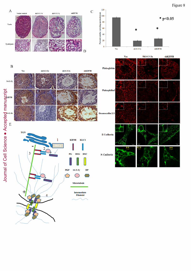
described. Loss of KIF5B in the testis led to a phenotype similar to that observed for the
loss of 14-3-3γ (figure8A-C) and suggested that both KIF5B and 14-3-3γ were
independently required to regulate cell-cell adhesion in the testis. Importantly, a
immunohistochemical analysis demonstrated that loss of either KIF5B or 14-3-3γ did not
lead to a decrease in the levels of the other protein (figure8B). Loss of 14-3-3ε did not
lead to a decrease in cell-cell adhesion and spermatogenesis suggesting that the effects
observed upon 14-3-3γ knockdown are specific to 14-3-3γ (figure8A-B), a result
consistent with those observed in HCT116 cells (figure2). In addition, a loss of either 14-
3-3γ or KIF5B lead to a detachment of the cells from the basal lamina. To determine if
the detachment of the primary germ cells and the Sertoli cells from the basal lamina lead
to an increase in cell death, the testis sections were stained using a TUNEL staining kit.
As shown in SF1A, treatment of the Vec sections with DNAse resulted in a strong
positive signal in the TUNEL assay. Testis sections from mice injected with the 14-3-3γ
knockdown construct showed low levels of TUNEL positivity in comparison with testis
sections from the Vec mice or the KIF5B knockdown mice. Given that the testis
morphology of the KIF5B knockdown mice and the 14-3-3γ knockdown mice are
virtually identical, it is likely that the loss of cell-matrix adhesion does not lead to cell
death which is consistent with our results in the cell line model. These results suggest that
cell-cell adhesion in the testis requires both 14-3-3γ and KIF5B and that loss of either
protein leads to defects in cell-cell adhesion.
Jour
nal o
f Cel
l Sci
ence
Acc
epte
d m
anus
crip
t

17
To determine if desmosome organization was altered in the 14-3-3γ and KIF5B
knockdown testis, testis sections were stained with antibodies to PG, PKP3, DSC2/3, N-
cadherin and E-cadherin. PG, PKP3 and DSC2/3 localized to the border in testis injected
with the control virus, however the levels of these proteins at the cell border were greatly
diminished in the 14-3-3γ and KIF5B knockdown testis (figure8D). In contrast, there was
no change in E-cadherin localization in the 14-3-3γ and KIF5B knockdown testis as
compared with testis sections injected with the vector control, a phenotype similar to that
observed in HCT116 cells in culture. In contrast to the results obtained for E-cadherin, it
was observed that N-cadherin localized to the border in the vector control and 14-3-3γ
knockdown testis but not in the KIF5B knockdown testis (figure8D). These results are
consistent with our observations that the KIF5B knockdown testis show a more severe
adhesion phenotype than the 14-3-3γ knockdown testis and suggest that KIF5B might be
required for the transport of other cell-cell adhesion molecules to the border in addition to
desmosomal proteins. These results suggest that 14-3-3γ and KIF5B are required for
desmosome formation in vivo.
Jour
nal o
f Cel
l Sci
ence
Acc
epte
d m
anus
crip
t

18
DISCUSSION
Our results suggest that 14-3-3γ and the KIF5B/KLC1 complex are required for the
transport of PG to the border in human cell lines and in the mouse testis leading to a
defect in the localization of PG to the cell border. In addition, loss of 14-3-3γ leads to a
disruption of cell-matrix adhesion, which might affect cell-cell adhesion in vitro and in
vivo. The association of PG with 14-3-3γ and transport of PG to the border is dependent
on PKCµ activity. Loss of either 14-3-3γ or KIF5B inhibits PG transport to the border
resulting in a decrease in desmosome formation and cell-cell adhesion in HCT116 cells
and in the testis leading to male sterility. The decrease in the levels of N-cadherin at the
border in KIF5B knockdown testis resulted in a more drastic phenotype in these animals
than in those with a knockdown of 14-3-3γ suggesting that N-cadherin is required for
cell-cell adhesion in the testis as previously reported (Andersson et al., 1994; Lee et al.,
2003).
Previous results have shown that loss of PG leads to a depletion of desmosomal proteins
from the cell border and defects in desmosome formation (Acehan et al., 2008; Gosavi et
al., 2011; Knudsen and Wheelock, 1992; Lewis et al., 1997). Although the presence of a
classical cadherin seems to be required for PG recruitment to the cell border (Michels et
al., 2009; Tinkle et al., 2008), the mechanisms by which PG is transported to the cell
border have not been identified. The results reported presented here suggest that
phosphorylation of PG by PKCµ at S236 leads to the generation of a binding site for 14-
3-3γ and that 14-3-3γ is required for transport of PG to the cell border to initiate
desmosome formation, presumably in complex with a classical cadherin (Michels et al.,
2009; Tinkle et al., 2008). 14-3-3γ might be required to load PG on the KIF5B/KLC1
Jour
nal o
f Cel
l Sci
ence
Acc
epte
d m
anus
crip
t

19
complex followed by transport of PG to the cell border on microtubules (figure8E). This
is consistent with observations that PKCµ localizes to the Golgi complex (Hausser et al.,
2002; Prestle et al., 1996) and is required for fission of vesicles carrying cargo to the cell
border (Liljedahl et al., 2001). A 14-3-3γ dimer is required for carrier formation at the
Golgi complex along with PKCµ (Valente et al., 2012) suggesting that 14-3-3γ loss could
disrupt desmosome formation due to defects in PG transport to the cell border.
Treating cells with an inhibitor that inactivates both PKCα and PKCµ leads to a decrease
in PG border localization and abolishes complex formation between PG and 14-3-3γ.
However, treatment with the inhibitor does not inhibit the interaction between 14-3-3γ
and other desmosomal proteins or KIF5B. These results suggest that phosphorylation of
PG by PKCµ might be required for complex formation between PG and 14-3-3γ and
loading of PG onto KIF5B, which is essential for transport of PG to the cell border.
Alternatively, it is possible that PG, DP and PKP3 are transported independently to the
border and the absence of PG at the border prevents the formation of a functional
desmosome. Further, it was observed that loss of PG did not affect the border localization
of DSC2/3 and DSG2 unlike the loss of 14-3-3γ or KIF5B. This might be because 14-3-
3γ is required for the localization of PKP3, DP and PG to the cell border and therefore the
defect in the localization of the desmosomal cadherins observed upon 14-3-3γ loss are
not observed upon PG loss. PG is probably required to stabilize and maintain the
organization of the desmosome, which is why loss of PG leads to a decrease in cell-cell
adhesion in these cells as previously reported (Gosavi et al., 2011). PKCµ was also
shown to directly phosphorylate the first 300 amino acids in PG which contain the S236
residue, suggesting that S236 might serve as site for phosphorylation by PG. This does
Jour
nal o
f Cel
l Sci
ence
Acc
epte
d m
anus
crip
t

20
not exclude the possibility that PKCµ might phosphorylate other residues in PG and
might provide an explanation for the observation that PG protein levels decrease when
PKCµ expression is inhibited. It is possible that PKCµ regulates other aspects of PG
function that are not limited to the transport of PG to the cell border such as the retention
of PG at the border or the stability of PG protein.
In the absence of KIF5B or upon expression of a dominant negative KIF5B construct, PG
does not localize to the cell border, leading to a decrease in the recruitment of other
components of the desmosome to the border as previously reported (Gosavi et al., 2011;
Lewis et al., 1997). Previous experiments have suggested that inhibiting KIF5B
expression does not affect PG localization to the border in SCC9 cells (Nekrasova et al.,
2011). Our results suggest that loss of KIF5B in HCT116 cells and in the testis results in
an inhibition of PG transport to the border. Consistent with the results reported by
Nekrasova et. al. (Nekrasova et al., 2011), we did not observe any defects in PG
localization upon expression of a KIF3A DN mutant. The discrepancy in these two
reports might be due to the use of different cell types in the two reports. Other kinesin
family members could regulate PG transport in SCC9 cells in the absence of KIF5B.
Pasdar et. al. have reported that a microtubule network is not essential for desmosome
formation (Pasdar et al., 1991). However, the results from this report and others suggest
that the transport of desmosomal components to the border is dependent on an intact
microtubule network (Gloushankova et al., 2003; Nekrasova et al., 2011). This is
consistent with reports suggesting that desmosome organization, function and
composition might vary in different cell types (reviewed in (Cross and Carter, 2000;
Garrod and Chidgey, 2007; Getsios et al., 2004; Green and Gaudry, 2000; Hatzfeld,
Jour
nal o
f Cel
l Sci
ence
Acc
epte
d m
anus
crip
t

21
2007)) and is consistent with the observation that loss of the different desmosomal
components in the mouse leads to a vast variety of phenotypes (Chidgey et al., 2001;
Gallicano et al., 1998; Grossmann et al., 2004; Koch et al., 1997; Lechler and Fuchs,
2007; Ruiz et al., 1996; Sklyarova et al., 2008; Vasioukhin et al., 2001).
Although 14-3-3γ seems to be required for transporting PG to the cell border, it might
also be required for transporting PKP3 and DP to the cell border in a manner that is
independent of PG transport. This is consistent with our observation that inhibiting both
PKCα and PKCµ does not lead to a disruption of the interaction between 14-3-3γ and DP
or PKP3. The transport of DSG2 to the border is dependent on KIF5B while DSC2
transport requires KIF3A and PKP2 (Nekrasova et al., 2011). Our results suggest that PG
transport to the border is dependent on KIF5B but not KIF3A suggesting that the
transport of desmosomal cadherins and the plaque proteins to the border might occur via
independent microtubule dependent pathways. Alternatively, since 14-3-3 proteins bind
to their ligands as dimers with each member of the dimer forming a complex with a
phospho-peptide (Brunet et al., 2002; Yaffe et al., 1998), it is possible that 14-3-3γ
bridges interactions between the different desmosomal plaque proteins allowing
formation of an intact desmosome as previously postulated by Bonne et. al. (Bonne et al.,
2003). The only argument against this hypothesis is that as far as we can tell 14-3-3γ does
not localize to the desmosome (figure4A). In fact, the lack of any co-localization between
14-3-3γ and PG suggests that any functions that 14-3-3γ performs with respect to PG
localization are transient in nature, which is why we favor the hypothesis that 14-3-3γ is
required for loading PG onto the KIF5B/KLC1 complex.
Jour
nal o
f Cel
l Sci
ence
Acc
epte
d m
anus
crip
t

22
In contrast to the results reported in this report, previous work has suggested that 14-3-3γ
knockout mice are viable and no adhesion or sterility defects have been reported in these
mice (Steinacker et al., 2005). PG knockout mice die during embryogenesis due to
cardiac defects as a result of decreased desmosome formation and as these mice die
before the testis is formed, no information is available on the effects of PG loss in the
testis (Ruiz et al., 1996). Taken together these previously published reports suggest that
PG functions required for desmosome formation are not altered in the 14-3-3γ-/- mice.
Our results suggest that 14-3-3γ might regulate desmosome formation in multiple cell
types, as loss of 14-3-3γ in HCT116 cells derived from the colon and in the seminiferous
epithelium leads to a decrease in cell-cell adhesion and in desmosome formation and is
consistent with the reported role of 14-3-3γ in the transport of proteins from the Golgi
complex to the cell border (Valente et al., 2012). One reason for the differences in these
results and those reported by Steinacker et al (Steinacker et al., 2005) could be that
another 14-3-3 family member binds to PG and stimulates desmosome formation in the
14-3-3γ-/- mice and that this compensation is not observed upon shRNA mediated
knockdown in the testis. Another possibility is that these are strain specific differences
that are due to differences in the genetic background of the mice used in the two studies.
Generation of an inducible knockdown of 14-3-3γ in either the Swiss mice used in this
study or in other mouse strains could help determine whether loss of 14-3-3γ leads to
defects in desmosome formation and cell-cell adhesion in other tissues and not just in the
testis.
The results described above lead to the generation of the following model. 14-3-3γ binds
to PG phosphorylated at S236 by PKCµ and loads PG onto the KIF5B/KLC1 complex
Jour
nal o
f Cel
l Sci
ence
Acc
epte
d m
anus
crip
t

23
for transport to the cell border (figure8E). Loss of either 14-3-3γ or KIF5B or DN
versions of KIF5B and KLC1 inhibit PG transport to the border. As 14-3-3γ forms a
complex with PKP3 and DP, loss of 14-3-3γ might also lead to defects in transport of
these proteins to the border. As PG loss does not significantly affect the localization of
the cadherins to the border, it is possible that loss of PG leads to a defect in cadherin
retention or the formation of an intact desmosome at the border in the absence of 14-3-3γ.
Further, loss of KIF5B and KLC1 in the testis led to sterility and a decrease in
desmosome formation. This is consistent with previously reported results, which showed
that disruption of cell-cell adhesion (Cheng and Mruk, 2002) and desmosome like
junction formation in the testis leads to an increase in sterility (Li et al., 2009; Lie et al.,
2010).
To conclude, 14-3-3γ and the KIF5B/KLC1 complex are required for regulating the
transport of PG to the cell border. A decrease in 14-3-3γ levels leads to a decrease in
desmosome formation and the recruitment of other desmosomal proteins to the border.
14-3-3γ might be required for loss of cell-cell adhesion in multiple tissues; however this
can only be confirmed by additional experiments in vivo.
Jour
nal o
f Cel
l Sci
ence
Acc
epte
d m
anus
crip
t

24
MATERIALS AND METHODS
Animals. Swiss mice Crl:CFW(SW) were bred and maintained in the laboratory animal
facility of the Advanced Centre for Treatment Research and Education in Cancer
(ACTREC). Protocols for the experiments were approved by the Institutional Animal
Ethics Committee (IAEC) of ACTREC. The animal study proposal number is 11/2008
dated August 19, 2008. Testis injections were performed as previously described (Sehgal
et al., 2011).
Plasmids. Details of the oligonucleotides used in this study are in Supplementary Table
1. The GST-14-3-3γ and HA-epitope tagged 14-3-3γ constructs and the shRNA resistant
GFP-14-3-3γ construct were previously described (Hosing et al., 2008). Site directed
mutagenesis (Stratagene) was used to generate the MYCPGS236A construct. The full-
length KLC-1 and KLC-2 cDNA (Rahman et al., 1998), deletion constructs of KLC-1
(GST-KLC1 WT, GST-KLC1-CC and GST-KLC1-TPR) (Aoyama et al., 2009),
constructs expressing KIF-3A- GFP (Haraguchi et al., 2006), KIF-5B - YFP wild type
(Gu et al., 2006), the dominant negative (DN) kinesin constructs, KIF3A T107N-GFP
and KIF5B T92N-YFP (Wiesner et al., 2010), GFP-KLC1-WT and GFP-KLC1-CC
(Araki et al., 2007) have been described previously. To generate the KIF5B and PKCµ
shRNA constructs, oligonucleotide pairs (Supplementary Table 1) were ligated into
pLKO.1 Puro or pLKO.1 EGFP-f Puro as described previously (Sehgal et al., 2011). GST
PG 1-300 was generated by amplifying the first 300 amino acids of PG (Table 1) and
cloning the fragment into pGEX4T1 (Amersham).
Cell lines and transfections. The HCT116 (ATCC) and the HCT116-derived stable cell
lines were cultured as described previously (Hosing et al., 2008). To generate the KIF5B
Jour
nal o
f Cel
l Sci
ence
Acc
epte
d m
anus
crip
t

25
knockdown clones in the HCT116 cell line, cells were transfected with 1 μg of the
shRNA constructs targeting human KIF5B. Sixty hours post transfection, the cells were
transferred to medium containing 0.5 µg/ml of puromycin (Sigma) to generate single-cell
clones. The clones K3 and K5 were further used for all experiments.
Antibodies and Western blotting. The antibodies for PKP3, DSC2/3, DSG2, PG, DP,
K8, actin, E-cadherin, β-catenin and α-Ecatenin were used in Western blots as previously
described (Gosavi et al., 2011; Khapare et al., 2012; Kundu et al., 2008). Tissue culture
supernatants of the anti-HA (12CA5), anti-14-3-3γ antibody (CG31) antibodies and anti-
14-3-3σ (CS112) were used at a dilution of 1:50. The anti-14-3-3ε antibody (T-16,
Santacruz) were used at a dilution of 1:2000, p120 catenin(Mouse monoclonal from BD
transductions. Cat. No# 610134 dilution 1:1000), PKCµ (rabbit monoclonal Ab from
Abcam. Cat. No. 3146-1, 1:500), were used for Western blot analysis. Respective
secondary antibodies were used at a dilution of 1:1000 (Invitrogen) or 1:5000 (Pierce).
Immunofluorescence and calcium switch assays. The cells were cultured on chromic
acid treated, poly-l-Lysine coated glass coverslips at a confluence of 70- 80%. Prior to
fixation, the cells were washed carefully twice with 1X phosphate buffered saline (PBS).
HCT116 derived clones were fixed in absolute methanol for 10min at - 20ᵒC to detect α-
tubulin, KIF5B, PKP2, Par3, ZO1, P-cadherin, DP, PG, DSC2/3, DSG2, E-Cadherin and
PKP3. In some experiments, cells were fixed in 4% paraformaldehyde and permiabilized
with Triton X-100 as described (Gosavi et al., 2011). The antibodies for PKP3, DSC2/3,
DSG2, PG, DP, K8, actin, E-cadherin, β-catenin, ZO-1 and α-Ecatenin were used in
immunofluoresecence analysis as previously described (Gosavi et al., 2011; Khapare et
al., 2012; Kundu et al., 2008). Antibodies to PKP2 (BD clontech, dilution 1:25), KIF5B
Jour
nal o
f Cel
l Sci
ence
Acc
epte
d m
anus
crip
t

26
(Abcam, dilution 1:100), α-tubulin (Abcam, dilution 1:150), Par3 (Millipore, dilution
1:50), ZO-1 (Abcam, dilution 1:100), P-cadherin (BD Transduction laboratories, dilution
1:100), HA (12CA5, supernatant), p120 catenin (BD transductions dilution 1:100), N-
cadherin (Life technologies. Cat. No# 33-3900 1:50) and α-E-catenin (Santa Cruz
Biotechnology 1:25) E-cadherin (clone 36/E-Cadherin, mouse monoclonal, BD
Transduction laboratories, dilution 1:100) were incubated with the cells for 1 hour at
room temperature at the indicated dilutions as described (Gosavi et al., 2011). To stain
mitochondria, Mitotraker GreenFM (Invitrogen) was used at 100nM to stain live cells.
Confocal images were obtained by using a LSM 510 Meta Carl Zeiss confocal system
with an Argon 488 nm and Helium/Neon 543 nm lasers. All images were obtained using
an Axio Observer Z.1 microscope (numerical aperture [NA] 1.4) at a magnification of
630X with 2X or 4X optical zoom. The surface intensity of staining was measured for the
different proteins in a minimum of twenty four cells using the Axiovision software and
the mean and standard deviation plotted.
Hanging drop assays. Hanging drop assays were used to measure cell adhesion as
previously described (Kundu et al., 2008).
GST pulldown and immunoprecipitation assays. These assays were performed as
previously described (Dalal et al., 1999).
GST PG production and kinase assays. GST alone or GST PG1-300 was purified from
bacteria as described (Dalal et al., 2004). Purified proteins were used in kinase assays
using recombinant PKCµ and a peptide derived from CREB as a positive control (Signal
Chem) and kinase activity determined using the ADP glow assay kit (Promega) according
to the manufacturer’s instructions.
Jour
nal o
f Cel
l Sci
ence
Acc
epte
d m
anus
crip
t

27
Histology and Immunohistochemistry. Mouse testes were fixed in 10% formaldehyde
overnight and processed for histology as described (Kundu et al., 2008). TUNEL assays
were performed as per the manufacturer’s instructions (Promega).
Electron microscopy. WT and 14-3-3γ KNOCKDOWN testis were fixed with 3%
glutaraldehyde and postfixed with 1% osmium tetraoxide (Tedpella). Grids were
contrasted with alcoholic uranyl acetate for 1 minute and lead citrate for half a minute.
The grids were observed under a Carl Zeiss LIBRA120 EFTEM transmission electron
microscope at an accelerating voltage of 120 kV and at 325,000 magnification. Images
were captured using a Slow Scan CCD camera (TRS, Germany).
Jour
nal o
f Cel
l Sci
ence
Acc
epte
d m
anus
crip
t

28
Acknowledgements. We thank Dr.’s Young Lee, Stefan Linder, Tetsu Akiyoma, Chen
Gu and Lawrence Goldstein for supplying us with constructs used during the course of
this study. We would also like to thank the ACTREC imaging facility for help with
confocal microscopy, the ACTREC animal facility and Ms. Sharda. Sawant for helping
with the preparation of grids for electron microscopy.
This work was supported by grants from the Department of Biotechnology
(http://dbtindia.nic.in/index.asp) (grants BT/PR6521/Med/14/828/2005 and
BT/PR12578/MED/31/75/2009) and ACTREC (www.actrec.gov.in) (LS and SND) and a
fellowship from the University Grants Commission (http://www.ugc.ac.in/) (AM).
Jour
nal o
f Cel
l Sci
ence
Acc
epte
d m
anus
crip
t

29
References
Acehan, D., Petzold, C., Gumper, I., Sabatini, D. D., Muller, E. J., Cowin, P. and Stokes, D. L. (2008). Plakoglobin is required for effective intermediate filament anchorage to desmosomes. J Invest Dermatol 128, 2665-75. Andersson, A. M., Edvardsen, K. and Skakkebaek, N. E. (1994). Expression and localization of N- and E-cadherin in the human testis and epididymis. Int J Androl 17, 174-80. Aoyama, T., Hata, S., Nakao, T., Tanigawa, Y., Oka, C. and Kawaichi, M. (2009). Cayman ataxia protein caytaxin is transported by kinesin along neurites through binding to kinesin light chains. J Cell Sci 122, 4177-85. Araki, Y., Kawano, T., Taru, H., Saito, Y., Wada, S., Miyamoto, K., Kobayashi, H., Ishikawa, H. O., Ohsugi, Y., Yamamoto, T. et al. (2007). The novel cargo Alcadein induces vesicle association of kinesin-1 motor components and activates axonal transport. Embo J 26, 1475-86. Bass-Zubek, A. E., Godsel, L. M., Delmar, M. and Green, K. J. (2009). Plakophilins: multifunctional scaffolds for adhesion and signaling. Curr Opin Cell Biol. Bierkamp, C., McLaughlin, K. J., Schwarz, H., Huber, O. and Kemler, R. (1996). Embryonic heart and skin defects in mice lacking plakoglobin. Dev Biol 180, 780-5. Bonne, S., Gilbert, B., Hatzfeld, M., Chen, X., Green, K. J. and Van Roy, F. (2003). Defining desmosomal plakophilin-3 interactions. J Cell Biol 161, 403-16. Brunet, A., Kanai, F., Stehn, J., Xu, J., Sarbassova, D., Frangioni, J. V., Dalal, S. N., DeCaprio, J. A., Greenberg, M. E. and Yaffe, M. B. (2002). 14-3-3 transits to the nucleus and participates in dynamic nucleo-cytoplasmic transport. J. Cell Biol. 156, 817-828. Caldelari, R., de Bruin, A., Baumann, D., Suter, M. M., Bierkamp, C., Balmer, V. and Muller, E. (2001). A central role for the armadillo protein plakoglobin in the autoimmune disease pemphigus vulgaris. J Cell Biol 153, 823-34. Chan, T. A., Hermeking, H., Lengauer, C., Kinzler, K. W. and Vogelstein, B. (1999). 14-3-3 is required to prevent mitotic catastrophe after DNA damage. Nature 401, 616-620. Chen, X., Bonne, S., Hatzfeld, M., van Roy, F. and Green, K. J. (2002). Protein binding and functional characterization of plakophilin2: evidence for its diverse roles in desmosomes and -catenin signaling. J. Biol. Chem. 277, 10512-10522. Cheng, C. Y. and Mruk, D. D. (2002). Cell junction dynamics in the testis: Sertoli-germ cell interactions and male contraceptive development. Physiol Rev 82, 825-74. Chidgey, M., Brakebusch, C., Gustafsson, E., Cruchley, A., Hail, C., Kirk, S., Merritt, A., North, A., Tselepis, C., Hewitt, J. et al. (2001). Mice lacking desmocollin 1 show epidermal fragility accompanied by barrier defects and abnormal differentiation. J Cell Biol 155, 821-32. Cross, R. A. and Carter, N. J. (2000). Molecular motors. Curr Biol 10, R177-9. Dalal, S. N., Schweitzer, C. M., Gan, J. and DeCaprio, J. A. (1999). Cytoplasmic localization of human cdc25C during interphase requires an intact 14-3-3 binding site. Mol. Cell. Biol. 19, 4465-4479.
Jour
nal o
f Cel
l Sci
ence
Acc
epte
d m
anus
crip
t

30
Dalal, S. N., Yaffe, M. B. and DeCaprio, J. A. (2004). 14-3-3 family members act coordinately to regulate mitotic progression. Cell Cycle 3, 672-677. Dusek, R. L., Godsel, L. M. and Green, K. J. (2007). Discriminating roles of desmosomal cadherins: beyond desmosomal adhesion. J Dermatol Sci 45, 7-21. Gallicano, G. I., Kouklis, P., Bauer, C., Yin, M., Vasioukhin, V., Degenstein, L. and Fuchs, E. (1998). Desmoplakin is required early in development for assembly of desmosomes and cytoskeletal linkage. J Cell Biol 143, 2009-22. Garrod, D. and Chidgey, M. (2007). Desmosome structure, composition and function. Biochim Biophys Acta. Getsios, S., Huen, A. C. and Green, K. J. (2004). Working out the strength and flexibility of desmosomes. Nat Rev Mol Cell Biol 5, 271-81. Gloushankova, N. A., Wakatsuki, T., Troyanovsky, R. B., Elson, E. and Troyanovsky, S. M. (2003). Continual assembly of desmosomes within stable intercellular contacts of epithelial A-431 cells. Cell Tissue Res 314, 399-410. Gosavi, P., Kundu, S. T., Khapare, N., Sehgal, L., Karkhanis, M. S. and Dalal, S. N. (2011). E-cadherin and plakoglobin recruit plakophilin3 to the cell border to initiate desmosome assembly. Cell Mol Life Sci 68, 1439-54. Green, K. J. and Gaudry, C. A. (2000). Are desmosomes more than tethers for intermediate filaments? Nat Rev Mol Cell Biol 1, 208-16. Grossmann, K. S., Grund, C., Huelsken, J., Behrend, M., Erdmann, B., Franke, W. W. and Birchmeier, W. (2004). Requirement of plakophilin 2 for heart morphogenesis and cardiac junction formation. J Cell Biol 167, 149-60. Gu, C., Zhou, W., Puthenveedu, M. A., Xu, M., Jan, Y. N. and Jan, L. Y. (2006). The microtubule plus-end tracking protein EB1 is required for Kv1 voltage-gated K+ channel axonal targeting. Neuron 52, 803-16. Haraguchi, K., Hayashi, T., Jimbo, T., Yamamoto, T. and Akiyama, T. (2006). Role of the kinesin-2 family protein, KIF3, during mitosis. J Biol Chem 281, 4094-9. Hatzfeld, M. (2007). Plakophilins: Multifunctional proteins or just regulators of desmosomal adhesion? Biochim Biophys Acta 1773, 69-77. Hausser, A., Link, G., Bamberg, L., Burzlaff, A., Lutz, S., Pfizenmaier, K. and Johannes, F. J. (2002). Structural requirements for localization and activation of protein kinase C mu (PKC mu) at the Golgi compartment. J Cell Biol 156, 65-74. Hosing, A. S., Kundu, S. T. and Dalal, S. N. (2008). 14-3-3 Gamma is required to enforce both the incomplete S phase and G2 DNA damage checkpoints. Cell Cycle 7, 3171-9. Khapare, N., Kundu, S. T., Sehgal, L., Sawant, M., Priya, R., Gosavi, P., Gupta, N., Alam, H., Karkhanis, M., Naik, N. et al. (2012). Plakophilin3 Loss Leads to an Increase in PRL3 Levels Promoting K8 Dephosphorylation, Which Is Required for Transformation and Metastasis. PLoS One 7, e38561. Knudsen, K. A. and Wheelock, M. J. (1992). Plakoglobin, or an 83-kD homologue distinct from beta-catenin, interacts with E-cadherin and N-cadherin. J Cell Biol 118, 671-9. Koch, P. J., Mahoney, M. G., Ishikawa, H., Pulkkinen, L., Uitto, J., Shultz, L., Murphy, G. F., Whitaker-Menezes, D. and Stanley, J. R. (1997). Targeted disruption of the pemphigus vulgaris antigen (desmoglein 3) gene in mice causes loss of
Jour
nal o
f Cel
l Sci
ence
Acc
epte
d m
anus
crip
t
31
keratinocyte cell adhesion with a phenotype similar to pemphigus vulgaris. J Cell Biol 137, 1091-102. Kundu, S. T., Gosavi, P., Khapare, N., Patel, R., Hosing, A. S., Maru, G. B., Ingle, A., Decaprio, J. A. and Dalal, S. N. (2008). Plakophilin3 downregulation leads to a decrease in cell adhesion and promotes metastasis. Int J Cancer 123, 2303-2314. Lechler, T. and Fuchs, E. (2007). Desmoplakin: an unexpected regulator of microtubule organization in the epidermis. J Cell Biol 176, 147-54. Lee, N. P., Mruk, D., Lee, W. M. and Cheng, C. Y. (2003). Is the cadherin/catenin complex a functional unit of cell-cell actin-based adherens junctions in the rat testis? Biol Reprod 68, 489-508. Lewis, J. E., Wahl, J. K., 3rd, Sass, K. M., Jensen, P. J., Johnson, K. R. and Wheelock, M. J. (1997). Cross-talk between adherens junctions and desmosomes depends on plakoglobin. J Cell Biol 136, 919-34. Li, M. W., Mruk, D. D., Lee, W. M. and Cheng, C. Y. (2009). Connexin 43 and plakophilin-2 as a protein complex that regulates blood-testis barrier dynamics. Proc Natl Acad Sci U S A 106, 10213-8. Lie, P. P., Cheng, C. Y. and Mruk, D. D. (2010). Crosstalk between desmoglein-2/desmocollin-2/Src kinase and coxsackie and adenovirus receptor/ZO-1 protein complexes, regulates blood-testis barrier dynamics. Int J Biochem Cell Biol 42, 975-86. Lie, P. P., Cheng, C. Y. and Mruk, D. D. (2011). The biology of the desmosome-like junction a versatile anchoring junction and signal transducer in the seminiferous epithelium. Int Rev Cell Mol Biol 286, 223-69. Liljedahl, M., Maeda, Y., Colanzi, A., Ayala, I., Van Lint, J. and Malhotra, V. (2001). Protein kinase D regulates the fission of cell surface destined transport carriers from the trans-Golgi network. Cell 104, 409-20. Marcozzi, C., Burdett, I. D., Buxton, R. S. and Magee, A. I. (1998). Coexpression of both types of desmosomal cadherin and plakoglobin confers strong intercellular adhesion. J Cell Sci 111 ( Pt 4), 495-509. Michels, C., Buchta, T., Bloch, W., Krieg, T. and Niessen, C. M. (2009). Classical cadherins regulate desmosome formation. J Invest Dermatol 129, 2072-5. Muslin, A. J., Tanner, J. W., Allen, P. M. and Shaw, A. S. (1996). Interaction of 14-3-3 with signaling proteins is mediated by recognition of phosphoserine. Cell 84, 889-897. Nekrasova, O. E., Amargo, E. V., Smith, W. O., Chen, J., Kreitzer, G. E. and Green, K. J. (2011). Desmosomal cadherins utilize distinct kinesins for assembly into desmosomes. J Cell Biol 195, 1185-203. Obenauer, J. C., Cantley, L. C. and Yaffe, M. B. (2003). Scansite 2.0: Proteome-wide prediction of cell signaling interactions using short sequence motifs. Nucleic Acids Res 31, 3635-41. Palka, H. L. and Green, K. J. (1997). Roles of plakoglobin end domains in desmosome assembly. J Cell Sci 110 ( Pt 19), 2359-71. Park, J. E., Kim, Y. I. and Yi, A. K. (2009). Protein kinase D1 is essential for MyD88-dependent TLR signaling pathway. J Immunol 182, 6316-27.
Jour
nal o
f Cel
l Sci
ence
Acc
epte
d m
anus
crip
t

32
Pasdar, M., Krzeminski, K. A. and Nelson, W. J. (1991). Regulation of desmosome assembly in MDCK epithelial cells: coordination of membrane core and cytoplasmic plaque domain assembly at the plasma membrane. J Cell Biol 113, 645-55. Prestle, J., Pfizenmaier, K., Brenner, J. and Johannes, F. J. (1996). Protein kinase C mu is located at the Golgi compartment. J Cell Biol 134, 1401-10. Rahman, A., Friedman, D. S. and Goldstein, L. S. (1998). Two kinesin light chain genes in mice. Identification and characterization of the encoded proteins. J Biol Chem 273, 15395-403. Ruiz, P., Brinkmann, V., Ledermann, B., Behrend, M., Grund, C., Thalhammer, C., Vogel, F., Birchmeier, C., Gunthert, U., Franke, W. W. et al. (1996). Targeted mutation of plakoglobin in mice reveals essential functions of desmosomes in the embryonic heart. J Cell Biol 135, 215-25. Russell, L. D., Ettlin, R., Sinha Hikim, A. P. and Clegg, E. D. (1990). Histological and Histopathological Evaluation of the Testis: Clearwater Fl. Cache River. Sehgal, L., Thorat, R., Khapare, N., Mukhopadhaya, A., Sawant, M. and Dalal, S. N. (2011). Lentiviral mediated transgenesis by in vivo manipulation of spermatogonial stem cells. PLoS One 6, e21975. Silver, K. E. and Harrison, R. E. (2011). Kinesin 5B is necessary for delivery of membrane and receptors during FcgammaR-mediated phagocytosis. J Immunol 186, 816-25. Sklyarova, T., Bonne, S., D'Hooge, P., Denecker, G., Goossens, S., De Rycke, R., Borgonie, G., Bosl, M., van Roy, F. and van Hengel, J. (2008). Plakophilin-3-deficient mice develop hair coat abnormalities and are prone to cutaneous inflammation. J Invest Dermatol 128, 1375-85. Steinacker, P., Schwarz, P., Reim, K., Brechlin, P., Jahn, O., Kratzin, H., Aitken, A., Wiltfang, J., Aguzzi, A., Bahn, E. et al. (2005). Unchanged survival rates of 14-3-3gamma knockout mice after inoculation with pathological prion protein. Mol Cell Biol 25, 1339-46. Tanaka, Y., Kanai, Y., Okada, Y., Nonaka, S., Takeda, S., Harada, A. and Hirokawa, N. (1998). Targeted disruption of mouse conventional kinesin heavy chain, kif5B, results in abnormal perinuclear clustering of mitochondria. Cell 93, 1147-58. Telles, E., Hosing, A. S., Kundu, S. T., Venkatraman, P. and Dalal, S. N. (2009). A novel pocket in 14-3-3e is required to mediate specific complex formation with cdc25C and to inhibit cell cycle progression upon activation of checkpoint pathways. Exp Cell Res 315, 1448-1457. Tinkle, C. L., Pasolli, H. A., Stokes, N. and Fuchs, E. (2008). New insights into cadherin function in epidermal sheet formation and maintenance of tissue integrity. Proc Natl Acad Sci U S A 105, 15405-10. Valente, C., Turacchio, G., Mariggio, S., Pagliuso, A., Gaibisso, R., Di Tullio, G., Santoro, M., Formiggini, F., Spano, S., Piccini, D. et al. (2012). A 14-3-3gamma dimer-based scaffold bridges CtBP1-S/BARS to PI(4)KIIIbeta to regulate post-Golgi carrier formation. Nat Cell Biol 14, 343-54. Vasioukhin, V., Bowers, E., Bauer, C., Degenstein, L. and Fuchs, E. (2001). Desmoplakin is essential in epidermal sheet formation. Nat Cell Biol 3, 1076-85. Verhey, K. J. and Hammond, J. W. (2009). Traffic control: regulation of kinesin motors. Nat Rev Mol Cell Biol 10, 765-77.
Jour
nal o
f Cel
l Sci
ence
Acc
epte
d m
anus
crip
t
33
Wiesner, C., Faix, J., Himmel, M., Bentzien, F. and Linder, S. (2010). KIF5B and KIF3A/KIF3B kinesins drive MT1-MMP surface exposure, CD44 shedding, and extracellular matrix degradation in primary macrophages. Blood 116, 1559-69. Wong, C. H., Mruk, D. D., Lui, W. Y. and Cheng, C. Y. (2004). Regulation of blood-testis barrier dynamics: an in vivo study. J Cell Sci 117, 783-98. Yaffe, M. B. (2002). How do 14-3-3 proteins work? - Gatekeeper phosphorylation and the molecular anvil hypothesis. FEBS Letters 513, 53-57. Yaffe, M. B., Rittinger, K., Volinia, S., Caron, P. R., Aitken, A., Leffers, H., Gamblin, S. J., Smerdon, S. J. and Cantley, L. C. (1998). The structural basis for 14-3-3 phosphopeptide binding specificity. Cell 91, 961-971.
Jour
nal o
f Cel
l Sci
ence
Acc
epte
d m
anus
crip
t

34
Figure Legends.
Figure 1. Loss of 14-3-3γ leads to disruption of cell-cell adhesion. A-D. Tissue
sections from mouse testis injected with either the 14-3-3γ knockdown construct (sh14-3-
3γ) or the vector control (Vec) were stained with antibodies to 14-3-3γ and visualized by
light microscopy (A and D) or stained with hematoxylin and eosin to visualize either the
seminiferous tubules (top panels) or the epididymis (bottom panels) (B). The percentage
of vesicles containing spermatozoa in the epididymis is graphed on the y-axis and the bar
represents the mean and standard deviation for three different animals (C). Bars in A
correspond to 5µM, B to 5µM and D to 5µM. E. Electron micrographs of the testis
injected with either the 14-3-3γ KNOCKDOWN viruses (sh14-3-3γ) or the vector control
(Vec). Sertoli cells (SC) and germ cells (GC) are indicated. The panels on the extreme
right are higher magnification images of the boxed areas as indicated. Bars correspond to
2µM.
Figure 2. Loss of 14-3-3γ leads to a decrease in cell-cell adhesion. A. Protein extracts
from the Vec and sh14-3-3γ cells were resolved on SDS-PAGE gels followed by Western
blotting with the indicated antibodies. B. mRNA was prepared from the Vec and sh14-3-
3γ cells and RT-PCR reactions performed with oligonucleotide pairs specific for the
indicated genes. GAPDH served as a loading control. C-D. Hanging drop assays were
performed on the Vec and sh14-3-3γ cells. The images of the clumps (C) and the
quantitation (D) of cluster number and size are shown. Bars in C correspond to 200µM.
E. Protein extracts from the Vec and sh14-3-3ε cells were resolved on SDS-PAGE gels
followed by Western blotting with the indicated antibodies. F-G. Hanging drop assays
Jour
nal o
f Cel
l Sci
ence
Acc
epte
d m
anus
crip
t

35
were performed on the Vec and sh14-3-3γ cells. The images of the clumps (F) and the
quantitation (G) of cluster number and size are shown. Bars in F correspond to 200µM.
Figure 3. Localization of PG is altered in the sh-14-3-3γ cells. A-B. Protein extracts
from the Vec and sh14-3-3γ cells were resolved on SDS-PAGE gels followed by Western
blotting with the indicated antibodies. C. mRNA was prepared from the Vec and sh14-3-
3γ cells and RT-PCR reactions performed with oligonucleotide pairs specific for the
indicated genes. GAPDH served as a loading control. D. PG levels at the border were
determined in the vector control cells (Vec) and the 14-3-3ε knockdown cells (sh14-3-3ε)
Bars correspond to 5µM. Note that PG levels did not change at the border upon
knockdown of 14-3-3ε. E-F. Vec and sh14-3-3γ cells were stained with the indicated
antibodies followed by confocal microscopy The bars in E correspond to 5µM and the
bars in F correspond to 10µM. Representative images are shown. The border intensity
was measured for at least twenty cells in three different experiments. The mean and
standard deviation for three independent experiments is shown.
Figure 4. 14-3-3γ is required to initiate desmosome formation. A. sh14-3-3γ cells
were transfected with either GFP alone or GFP 14-3-3γR. The cells were stained with
antibodies to PG (red), followed by confocal microscopy. Bars correspond to 5µM. B.
Protein extracts from HCT116 cells were incubated with either GST or GST14-3-3γ. The
reactions were resolved on SDS-PAGE gels followed by Western blotting with the
indicated antibodies. C. Vec and sh14-3-3γ cells were incubated in low calcium medium
for 24 hours (0 minute). After calcium addition for 60 min, cells were fixed and stained
with the indicated antibodies followed by confocal microscopy. Note that the levels of the
desmosomal proteins do not increase at the border in the sh-14-3-3γ cells as compared to
Jour
nal o
f Cel
l Sci
ence
Acc
epte
d m
anus
crip
t

36
the Vec cells. No change in E-cadherin staining is observed. Bars correspond to 10 µM.
The border intensity was measured for at least 20 cells in three different experiments. The
mean and standard deviation for three independent experiments is shown.
Figure 5. 14-3-3γ association with PG requires PKCµ activity. A and B. HCT116
cells were transfected with either MYC-epitope tagged WT PG or the S236A mutant of
PG. 48 hours post transfection protein extracts were incubated with GST or GST14-3-3γ
followed by Western blotting with antibodies to MYC (A) or the cells were stained with
antibodies to the MYC-epitope (B). DIC images for the fields are shown in the lower
panels. Bars correspond to 10µM. C and D. HCT116 cells were treated with either the
vehicle control (DMSO), the pan PKC inhibitor (BisI) and the PKCα and PKCµ specific
inhibitor (Go6976). Protein extracts from these cells were incubated with either GST or
GST14-3-3γ, the reactions resolved on SDS-PAGE gels and Western blots performed
with the indicated antibodies (C) or stained with antibodies to PG (D). Bars correspond to
5µM. E-F. Protein extracts from vector control or PKCµ knockdown cells (sh PKCµ)
were resolved on SDS-PAGE gels and Western blots performed with the indicated
antibodies. Note that there is a decrease in PG and DP levels upon PKCµ knockdown but
no difference in PKP3 levels is observed. F. Vec cells and sh PKCµ cells were fixed and
stained with the indicated antibodies. Bars correspond to 5µM. G. PG 1-300 was
produced in bacteria as a GST fusion and kinase assays performed using recombinant
PKCµ according to the manufacturer’s instructions. GST alone was used as a negative
control in this assay. Different concentrations of the substrate are shown on the x-axis
and enzyme activity is graphed on the y-axis and bars represent standard deviation. Note
Jour
nal o
f Cel
l Sci
ence
Acc
epte
d m
anus
crip
t

37
that an increase in kinase activity is observed upon an increase in the concentration of
PG1-300 but not with an increase in the concentration of GST alone.
Figure 6. KIF5B is required for transport of PG to the cell border. A. HCT116 cells
were transfected with the vector control (pCDNA3) or HA-14-3-3γ and
immunoprecipitations were performed with antibodies to the HA epitope. The reactions
were resolved on SDS-PAGE gels and Western blots performed with the indicated
antibodies. B. HCT116 cells were transfected with constructs expressing an shRNA
targeting KIF5B. Individual cell clones were expanded and protein extracts from these
clones were resolved on SDS-PAGE gels followed by Western blotting with antibodies to
KIF5B. Note that the knockdown clones have a lower level of KIF5B (K1-K5) than the
vector control (Vec). A Western blot for actin served as a loading control. C. Vector
control (Vec) or KIF5B knockdown clones (K3 and K5) were incubated with Mitotraker
GreenFM (Invitrogen). Bars correspond to 10µM. D. Hanging drop assays were
performed to determine cell-cell adhesion. K3 and K5 formed fewer and smaller clumps
as compared to the Vec cells. E. Vec, K3 and K5 cells were stained with antibodies to
PG, DSC2/3, DSG2, DP, and PKP3 and surface intensity quantitated using confocal
microscopy. Total magnification was 630X with 2X optical zoom. Bars correspond to
5µM. F. Protein extracts prepared from the Vec, K3 and K5 cells were resolved on SDS-
PAGE gels followed by Western blotting with the indicated antibodies. G. Vec, K3 and
K5 cells were stained with antibodies to p120 catenin and α-Ecatenin and surface
intensity quantitated using confocal microscopy. Total magnification is 630X with 2X
optical zoom. Bars correspond to 5µM.
Jour
nal o
f Cel
l Sci
ence
Acc
epte
d m
anus
crip
t

38
Figure 7. Dominant negative mutants of KIF5B and KLC1 inhibit PG transport to
the border. A. HCT116 cells were transfected with YFP-tagged WT or DN KIF5B or
GFP-tagged WT or DN KIF3A. Post transfection the cells were fixed and stained with
antibodies to PG. B. Protein extracts from HCT116 cells were incubated with GST alone
or GST-KLC1 or GST-KLC2. The reactions were resolved on SDS-PAGE gels followed
by Western blotting with the indicated antibodies. C. Cartoon of KLC1 mutants. D.
Protein extracts from HCT116 cells were incubated with GST alone or GST-KLC1WT or
GST-KLC1 TPR and GST-KLC1 CC. The reactions were resolved on SDS-PAGE gels
followed by Western blotting with the indicated antibodies. E. GFP-KLC1 or GFP-KLC1
CC were transfected into HCT116 cells. Forty-eight hours post transfection the cells were
fixed and stained with antibodies to PG. DNA was stained with DAPI. Total
magnification is 630X with 2X optical zoom. All bars correspond to 5µM.
Figure 8. Loss of KIF5B and 14-3-3γ in the testis leads to a disruption of desmosome
formation and sterility. A-D. Lentiviruses encoding either the vector control or
shRNA’s targeting either 14-3-3ε, 14-3-3γ or KIF5B were injected into the testes of
Swiss mice. Thirty-five days post-injection the mice were sacrificed, sections of the
epididymis and testes were stained with hematoxylin and eosin and examined
microscopically (A). A immunohistochemical analysis with antibodies to the different
proteins that were knocked down demonstrated that the expression of 14-3-3ε, 14-3-3γ
and KIF5B were inhibited in the testis injected with the appropriate lentivirus (B). Bar
corresponds to 20µM. The percentage of epididymal vesicles showing the presence of
mature spermatozoa in three different animals was determined and the mean and standard
deviation plotted (C). Testis sections were stained with antibodies to PG, PKP3, DSC2/3,
Jour
nal o
f Cel
l Sci
ence
Acc
epte
d m
anus
crip
t

39
N-cadherin and E-cadherin Bar corresponds to 10µM. (D). Total magnification was
630X with 2X optical zoom. (E) 1. PKCµ phosphorylates PG thereby allowing
association with 14-3-3γ resulting in loading of PG on the KIF5B/KLC1 complex (2).
The motor protein complex containing PG moves along microtubules (3) resulting in
desmosome assembly at the border (4).
Jour
nal o
f Cel
l Sci
ence
Acc
epte
d m
anus
crip
t

Jour
nal o
f Cel
l Sci
ence
Acc
epte
d m
anus
crip
t

Jour
nal o
f Cel
l Sci
ence
Acc
epte
d m
anus
crip
t

Jour
nal o
f Cel
l Sci
ence
Acc
epte
d m
anus
crip
t

Jour
nal o
f Cel
l Sci
ence
Acc
epte
d m
anus
crip
t

Jour
nal o
f Cel
l Sci
ence
Acc
epte
d m
anus
crip
t

Jour
nal o
f Cel
l Sci
ence
Acc
epte
d m
anus
crip
t

Jour
nal o
f Cel
l Sci
ence
Acc
epte
d m
anus
crip
t
Jour
nal o
f Cel
l Sci
ence
Acc
epte
d m
anus
crip
t


![LIGHT UNFLAVORED MESONS S C B - 2020 Reviewpdg.lbl.gov/2019/tables/rpp2019-sum-mesons.pdf · 3γ C < 1.6 × 10⁻⁵ CL=90% 274 4π0 P,CP < 6.9 × 10⁻⁷ CL=90% 40 π0e+e− C [f]](https://img.pdfslide.net/doc/110x75/5ff2a557e84a5178d42e40aa/light-unflavored-mesons-s-c-b-2020-3-c-16-10a5-cl90-274-40-pcp.jpg)