Embed Size (px)
Citation preview

JANUARY 2013 Vol. 4 No. 1 Your partner in paediatric and O&G practice
JOURNAL OF PAEDIATRICS, OBSTETRICS & GYNAECOLOGY
cimsasia.com get connected get addictedTHE MOST POWERFUL DRUG SEARCH
JOURNAL WATCH
GYNAECOLOGYGYNAECOLOGY
Bacterial Vaginosis
CASE STUDYCASE STUDY
A Rare Case of Buschke Lowenstein Tumour in Pregnancy CME ARTICLE
Intrauterine Foetal Growth Restriction
PAEDIATRICSPAEDIATRICS
Management of Eating Disorders in Children and Adolescents
OBSTETRICS
IN PRACTICE
GYNAECOLOGYGYNAECOLOGY
OBSTETRICS
Methods of Induction of Labour−CriticalReview of Literature (Part II)

get connected
get addicted
cimsasia.com
make CIMS your home page at the point of care

JANUARY 2013
Vol. 4 No. 1
JOURNAL OF PAEDIATRICS, OBSTETRICS & GYNAECOLOGY
JPOG JANUARY 2013 • i
1
2
Editorial Board Board Director, PaediatricsProfessor Pik-To CheungAssociate ProfessorDepartment of Paediatrics and Adolescent MedicineThe University of Hong Kong
Board Director, Obstetrics and Gynaecology Professor Pak-Chung HoHead, Department of Obstetrics and GynaecologyThe University of Hong Kong
Professor Biran AffandiUniversity of IndonesiaDr Karen Kar-Loen ChanThe University of Hong KongAssociate Professor Oh Moh ChayKK Women’s and Children’s Hospital, SingaporeAssociate Professor Anette JacobsenKK Women’s and Children’s Hospital, SingaporeProfessor Rahman JamalUniversiti Kebagsaan MalaysiaDato’ Dr Ravindran JegasothyHospital Kuala Lumpur, MalaysiaAssociate Professor Kenneth KwekKK Women’s and Children’s Hospital, SingaporeDr Siu-Keung LamKwong Wah Hospital, Hong KongProfessor Terence LaoChinese University of Hong KongDr Kwok-Yin LeungThe University of Hong KongDr Tak-Yeung LeungChinese University of Hong KongProfessor Tzou-Yien LinChang Gung University, TaiwanProfessor Somsak LolekhaRamathibodi Hospital, ThailandProfessor Lucy Chai-See LumUniversity of Malaya, MalaysiaProfessor SC NgNational University of Singapore
Professor Hextan Yuen-Sheung NganThe University of Hong KongProfessor Carmencita D PadillaUniversity of the Philippines ManilaProfessor Seng-Hock QuakNational University of SingaporeDr Tatang Kustiman SamsiUniversity of Tarumanagara, IndonesiaProfessor Perla D Santos OcampoUniversity of the PhilippinesAssociate Professor Alex SiaKK Women’s and Children’s Hospital, SingaporeDr Raman SubramaniamFetal Medicine and Gynaecology Centre, MalaysiaProfessor Walfrido W Sumpaico MCU-DFT Medical Foundation, PhilippinesProfessor Cheng Lim TanKK Women’s and Children’s Hospital, Singapore
Associate Professor Kok Hian TanKK Women’s and Children’s Hospital, SingaporeDr Surasak TaneepanichskulChulalongkorn University, ThailandProfessor Eng-Hseon TayThomson Women’s Cancer Centre, SingaporeProfessor Gulardi H WiknjosastroUniversity of IndonesiaDr PC WongNational University of SingaporeDr George SH YeoKK Women’s and Children’s Hospital, SingaporeProfessor Hui-Kim YapNational University of SingaporeProfessor Tsu-Fuh YehChina Medical University, Taiwan
Indian Editorial BoardObstetrics & Gynaecology
Editor
Dr. JB SharmaAssociate Professor, All India Institute of Medical Sciences, New DelhiDr. Ashok KumarProfessor, MAMC, New DelhiDr. P. Reddi RaniProfessor, JIPMER, Pondicherry
PaediatricsBharat J Parmar, Associate ProfessorBJ Medical College & Civil Hospital, AhmedabadDeepak Chawla, Assistant ProfessorGovernment Medical College & Hospital, ChandigarhDr. Sangeeta Sharma, HOD, Dept. of PaediatricsLRS Institute of Tuberculosis & Respiratory Disease, New DelhiDr. Asha Pherwani, ConsultantPD Hinduja Hospital & Research Centre, Mumbai
Journal Watch
1 • Hormonal contraception and cardiovascular risk
• Midurethral sling during vaginal prolapse surgery to reduce post-operative incontinence
• Effect of contraception on maternal mortality rates
Review ArticlePaediatrics
2 Management of Eating Disorders in Children and Adolescents The article summarizes current recommendations for the psychological and pharmacological management ofearly-onset eating disorders in childhood and adolescence, and highlights the important principles of treatmentin both inpatient and outpatient settings.Reenee Barton, Dasha Nicholls

JANUARY 2013
Vol. 4 No. 1
JOURNAL OF PAEDIATRICS, OBSTETRICS & GYNAECOLOGY
8
JPOG JANUARY 2013 • ii
In Practice
7 Case of the Month: Large Fistula in Supratrigonal Region after Lower Segment Caesarean Delivery Mallazzar Masaarapa Vijaykumar, Shivappa Prasanth, Madhusudan, Shobana Bhojaraj
Review ArticleObstetrics
8 Methods of Induction of Labour−Critical Review of Literature (Part II)In this review authors have analysed different methods of induction of labour and tried to give defi nitive guidance regarding their use. Numerous clinical studies, Cochrane database reviews, Royal College of Obstetricians and Gynaecologists (RCOG) 2008 and American Congress of Obstetricians and Gynecologists (ACOG) 2009 guidelines on induction of labour are taken into account.Varsha Deshmukh, Kanan Yelikar, Sonali Deshpande, Pradeep Ingale
Review ArticleGynaecology
21 Bacterial Vaginosis Bacterial vaginosis is the commonest cause of abnormal vaginal discharge in women of childbearing age, with a prevalence as high as 50% in some communities. Bacterial vaginosis is a risk factor for acquisition of sexually transmitted infections including HIV, and for post-abortion endometritis and adverse pregnancy outcomes such as late miscarriage and preterm birth. Studies of antibiotics in pregnancy have not consistently shown reduced adverse outcomes, so better strategies need to be studied to improve pregnancy outcome.Phillip Hay
29 In Practice (Answer)
7
21
JANUARY 2013
Vol. 4 No. 1
JOURNAL OF PAEDIATRICS, OBSTETRICS & GYNAECOLOGY
JPOG JANUARY 2013 • iii
Case Study
30 A Rare Case of Buschke Lowenstein Tumour in PregnancyManika Agarwal, Rajlaxmi Mundhra, Santa Singh, Kalkambe A Sangma
Continuing Medical Education
32 Intrauterine Foetal Growth Restriction Intrauterine foetal growth restriction is a placental function disorder in which Doppler ultrasound has a major role in foetal surveillance. Sequential changes in Doppler velocimetry of the umbilical artery, middle cerebral artery and ductus venosus can be seen in progressively worsening placental dysfunction. Management requires balancing the risk of prematurity and the risk of intrauterine asphyxia.May Li Lim, Kenneth Kwek, Kok Hian Tan, George SH Yeo
The Journal of Paediatrics, Obstetrics and Gynaecology contains articles under license from UBM Medica India Pvt. Ltd.
30
32

JOURNAL OF PAEDIATRICS, OBSTETRICS & GYNAECOLOGY
JANUARY 2013
Vol. 4 No. 1
INTERNATIONAL PUBLISHING TEAM PublisherBen YeoPublication Manager Marisa LamDeputy Managing EditorPaul Pimentel Associate Editor Audrey WongDesign Manager Rowena Sim DesignerCandice NgProductionEdwin Yu, Ho Wai HungCirculationLily OngAccounting Manager Minty KwanAdvertising CoordinatorRebecca Leung
INDIAN PUBLISHING TEAMManaging Director & PublisherDr. Monica BhatiaScientifi c ContentSukanya GhildiyalDesignersArun KharkwalAshim Sarkar Himani Kukreti
Published by: UBM Medica India Pvt LtdEmpire Towers, No. 53, Railway Parallel Road, Kumara Park West, Bengaluru-560020Email: [email protected]
ChinaTeo Wai ChooTel: (86 21) 5213 6622Email: [email protected]
Hong KongKristina Lo-Kurtz, Pagon LoTel: (852) 2559 5888Email: [email protected]
IndiaDr. Monica BhatiaTel: (91 022) 6612 2678Email: [email protected]
IndonesiaHafta Hasibuan, Sri Damayanti Tel: (62 21) 5365 2977Email: [email protected]
JapanMamoru TakagiTel: (81 3) 5562 6961Email: [email protected]
KoreaYoung Taek LeeTel: (82 2) 2007 5400Email: [email protected]
MalaysiaMeera Jassal, Lee Pek Lian, Irene Lee, Vincent LioTel: (60 3) 7954 2910Email: [email protected]
PUBLISHER: Journal of Paediatrics, Obstetric & Gynaecology (JPOG) is published 12 times a year by UBM Medica, a division of United Business Media.CIRCULATION: JPOG is for medical practitioners in Asia. It is available on subscription to members of allied professions.
SUBSCRIPTION: The price per copy is Rs. 200/-. The price per annum is Rs. 2040/-. EDITORIAL MATTER published herein has been prepared by professional editorial staff. Views expressed are not necessarily those of UBM Medica. Although great care has been taken in compiling and checking the information given in this publication to ensure that it is accurate, the authors, the publisher and their servants or agents shall not be responsible or in any way liable for the continued currency of the information or for any errors, omissions or inaccuracies in this publication whether arising from negligence or otherwise howsoever, or for any consequences arising therefrom. The inclusion or exclusion of any product does not mean that the publisher advocates or rejects its use either generally or in any particular fi eld or fi elds. COPYRIGHT: © 2013 UBM Medica. All rights reserved. No part of this publication may be reproduced, stored in a retrieval system or transmitted in any form or by any means, electronic, mechanical, photocopying, recording or otherwise, in any language, without written consent of copyright owner. Permission to reprint must be obtained from the publisher. ADVERTISEMENTS are subject to editorial acceptance and have no infl uence on editorial content or presentation. UBM Medica does not guarantee, directly or indirectly, the quality or effi cacy of any product or service described in the advertisements or other material which is commercial in nature.
Review ArticlesComprehensive reviews providing the latest clinical information on all aspects of the management of medical conditions affecting children and women.
Case StudiesInteresting cases seen in general practice and their management.
Pictorial MedicineVignettes of illustrated cases with clinical photographs.
For more information, please contact:The EditorUBM Medica India Pvt. Ltd., 404, 4th Floor, DLF City, Phase-IV, Gurgaon, Haryana-122 009, Indiaemail: [email protected]
PhilippinesDenise Javier, Arlene ToribioTel: (63 2) 886 0333Email: [email protected]
SingaporeKim Teo, Carrie Ong, Petrine Ong, Kenric KohTel: (65) 6223 3788Email: [email protected]
TaiwanCarol KuoTel: (886 2) 2577 6096Email: [email protected]
ThailandWipa SriwijitchokTel: (66 2) 741 5354Email: [email protected]
VietnamNguyen Thi Lan Huong, Bui Thi Cam TrucTel: (84 8) 3829 7923Email: [email protected]
Europe/USAKristina Lo-Kurtz, Maria KaiserTel: (852) 2116 4352Email: [email protected], [email protected]
Enquiries and Correspondence
JPOG JANUARY 2013 • iv
Publisher of CIMS/IDR

Journal Watch
JPOG JANUARY 2013 • 1
GYNAECOLOGY
Hormonal contraception and
cardiovascular risk
A Danish registry study has provided more data
about cardiovascular risks associated with hor-
monal contraception.
Data were obtained from four national reg-
istries over a 15-year period about non-pregnant
women aged 15–49 with no history of cardiovascu-
lar disease or cancer. The data included 1,626,158
women with 14,251,063 person-years of observa-
tion, during which there were 3,311 thrombotic
strokes and 1,725 myocardial infarctions. The rate
of thrombotic stroke was 21.4 per 100,000 per-
son-years and of myocardial infarction, 10.1 per
100,000 person-years. Among women using oral
contraceptives including ethinyl oestradiol at a
dose of 30–40 µg, the risk of thrombotic stroke was
increased 1.5- to 2.2-fold according to progestin
type, compared with non-users. The risk of myocar-
dial infarction was increased 1.3- to 2.3-fold. At an
ethinyl oestradiol dose of 20 µg, the increase in risk
was less in general, and there was no increased
risk with drospirenone as the progestin. Transder-
mal patches were not associated with significantly
increased risk for either thrombotic stroke or myo-
cardial infarction. Vaginal ring was associated with
a significant 2.5-fold increase in risk of thrombotic
stroke but a non-significant increase in risk of myo-
cardial infarction.
Although hormonal contraception may in-
crease the risks of thrombotic stroke and myocar-
dial infarction, the absolute risks are low. An edito-
rialist concludes that they are ‘safe enough’.
Lidegaard Ø et al. Thrombotic stroke and myocardial infarction with hormonal contraception. NEJM 2012; 366: 2257–2266; Petitti DB. Hormonal contraceptives and arterial thrombosis – not risk-free but safe enough. Ibid: 2316–2318 (editorial).
Midurethral sling during vaginal prolapse surgery to reduce post-operative incontinence
About a quarter of women undergoing surgery for
pelvic organ prolapse who had no urinary incon-
tinence before surgery will develop incontinence
after surgery. The prophylactic insertion of a mi-
durethral sling during prolapse surgery has become
popular without good evidence of its effectiveness.
A multicentre US trial has been reported.
A total of 337 women undergoing prolapse
surgery but without a history of stress incontinence
were randomized to insertion of a midurethral sling
or a control group (sham incisions) and 327 women
were followed up for 1 year. At 3 months, there
was a significant reduction in urinary incontinence
in the urethral sling group (23.6% vs 49.4%). At 12
months, the rates of incontinence were 27.3% vs
43.0%. The number needed to treat to prevent one
case of urinary incontinence at 12 months was 6.3.
Bladder perforation occurred in 6.7% of the ure-
thral sling group but in none of the control group.
There were significant increases in the sling group
in urinary tract infection (31.0% vs 18.3%), major
bleeding (3.1% vs 0%), and incomplete bladder
emptying 6 weeks after surgery (3.7% vs 0%).
The insertion of a midurethral sling was ef-
fective in reducing the risk of postoperative urinary
incontinence but at the expense of increased risk
of complications.
Wei JT et al. A midurethral sling to reduce incontinence after vaginal prolapse repair. NEJM 2012; 366: 2358–2367; Iglesia CB. Vaginal prolapse repair – place midurethral sling now or later: Ibid: 2422–2424 (editorial).
Effect of contraception on maternal mortality rates
The Safe Motherhood Initiative begun in 1987 has
four strategies to reduce maternal mortality: family
planning, antenatal care, safe delivery, and postna-
tal care. Now, the effects of contraceptive use on
maternal mortality worldwide have been estimated
from three international databases.
Data were analysed from 172 countries for
2008. The number of deaths from maternal causes
in 2008 was estimated at 342,203 (data from 172
countries). The estimated number of maternal
deaths averted by contraception was 272,040, a
44% reduction of the potential total. It was also
estimated that expansion of contraceptive use
could avert another 104,000 maternal deaths each
year. The number of deaths averted increased with
increased contraceptive use. In countries with high
(> 65%) contraceptive use, almost 60% of potential
maternal deaths were averted, whereas in sub-Sa-
haran Africa (22% contraceptive use), only 32% of
potential maternal deaths were averted.
Increased use of contraception could prevent
many maternal deaths in developing countries.
Ahmed S et al. Maternal deaths averted by contraceptive use: an analysis of 172 countries. Lancet 2012; 380: 111–125; Gilmore K, Gebreyesus TA. What will it take to eliminate preventable maternal deaths? Ibid: 87–88 (comment).

JPOG JANUARY 2013 • 2
PAEDIATRICS
The treatment of eating disorders in childhood and adolescence presents dif-
ferent challenges from the treatment of adults with eating disorders. These
include the medical concerns specific to periods of increased growth; the fact
that children and adolescents are usually brought for treatment, which will influence moti-
vation; statutory responsibilities in relation to the protection of children and adolescents;
and the importance of family context as the primary provider of care and the need, there-
fore, to engage families rather than individuals in treatment.
The majority of the literature pertaining to this age group relates to anorexia nervosa,
reflecting both the frequency and severity with which eating disorders present at this age.
In addition, bulimia nervosa is often perceived as the less severe condition. In fact, it can
often be even more distressing both to sufferers and to their carers, clouded as it is in
secrecy, and associated with a number of multi-impulse and maladaptive behaviours, in-
cluding self-harm, substance misuse and depression. There is often a gap of several years
between the onset of symptoms and diagnosis. Hence, bulimia nervosa generally presents
to clinicians in the older adolescent population, giving a false reflection of its prevalence.
In addition, adolescents with bulimia nervosa may be reluctant to engage with services
that expect involvement of family members.1
ASSESSMENT AS INTERVENTION
A comprehensive assessment not only forms the basis for treatment planning for a child with
an eating disorder, but the experience of having concerns recognized and the sharing of anxi-
ety can be a powerful first step in regaining strength for the battle ahead. The assessment
process initiates the therapeutic alliance between the treating team, and the child and fam-
ily, ensuring a shared understanding about the nature of the eating difficulties and factors
influencing them. Assessment will inform diagnostic formulation, taking into consideration
predisposing, precipitating and perpetuating factors. Emphasis is placed on assessing the
Management of Eating Disorders in Children
and Adolescents Reenee Barton, MRCPsych; Dasha Nicholls, MRCPsych
PAEDIATRICS I PEER REVIEWEDPAEDIATRICS

JPOG JANUARY 2013 • 3
PAEDIATRICS
child in the context of both their family system and
their developmental stage at onset. An open discus-
sion about the history and development of the eating
problems ensures transparency, as well an opportunity
for young people to hear the reflections of others. For
the therapist, it is an opportunity to begin recognition
of patterns of anxiety and communication that have be-
come established around the problem.
A detailed family tree helps establish the systemic
context and sources of support. Individual psychopa-
thology is best assessed through a structured interview
such as the Eating Disorders Examination (EDE)2 or child
EDE for those under 13 years of age,3 and an individual
mental state examination is needed to assess co-mor-
bidity and to contribute to the assessment of risk. It is
important to clarify, with the child, issues around confi-
dentiality in relation to individual components of the as-
sessment, both in terms of the need to share aspects of
risk, but also the benefit of conveying some of the young
person’s hopes and anxieties to the rest of the family.
The use of timelines and Likert scales, for example, to
rate mood, can help to establish a collaborative rela-
tionship with the young person and his or her family.
Through this process, the family has an op-
portunity to review the context in which the eating
disorder has arisen, its severity and impact, and can
begin to take on its role as the primary source of
support. Motivations and expectations of each fam-
ily member are laid out from the start of treatment
and lines of communication established. This is es-
pecially important when parents are no longer living
together. Barriers to intervention, both practical (eg,
access to treatment facilities) and psychological (eg,
parental guilt about causing the problem), need to be
addressed directly.
The provision of information is a key component
of collaborative working and forms the core of inter-
vention at this stage. Families need to be given ver-
bal and written information about diagnosis, physi-
cal and psychological risks, course, potential com-
plications and treatment options. It is important to
acknowledge that although the majority of patients
will make a good recovery, up to one-third of young
people do not, and a significant proportion (around
40%) will have a second co-morbid Axis 1 diagnosis
on follow-up.4 A resource list, books and helplines
for parents and young people are very helpful; some
examples are given below.
TREATMENT
The aims of treatment are threefold:
To address eating behaviour and related cognitions •
directly, including nutritional deficiencies.
To facilitate emotional communication and problem-•
solving skills.
To address developmental issues and promote indi-•
viduation.
Addressing issues of control and responsibility is
central to successful intervention, as is the develop-
ment of good collaborative relationships with the young
person and his or her parents. A collaborative stance
is facilitated if the therapist is able to adopt a direc-
tive, client-centred style, which aims to help explore
and resolve ambivalence about behaviour change, and
empowers decision making by the young person and his
or her family. This may be difficult in the face of consid-
erable anxiety about a young person’s nutritional status,
but can be facilitated by good risk management pro-
tocols. Like most safety nets, these protocols are less
likely to be needed if everyone knows they are there.
A number of recommendations have been made in
the UK National Institute for Clinical Excellence (NICE)
2004 guidelines5 specifically regarding the manage-
ment of children and adolescents with anorexia nervosa,
based on a combination of research evidence and expert
opinion. The importance of clarifying areas of responsi-
bility and lines of communication between professionals
in writing is highlighted throughout the guidelines, with
an emphasis on ongoing risk assessment.

JPOG JANUARY 2013 • 4
PAEDIATRICS
Medical and Nutritional Management
The medical management of anorexia nervosa is dis-
cussed in greater detail elsewhere in this issue. How-
ever, it is important to highlight differences between
children and adults. Firstly, the increased medical
risks associated with food restriction in young people
are well documented,6 as is the increased risk associ-
ated with the tendency for younger patients to restrict
fluid as well as food intake. The implications in terms
of the need for close partnership with paediatric ser-
vices are clear, with ongoing medical assessment as
a core component of treatment. Weekly weighing and
physical review are standard practice in outpatient
treatment, with 3-monthly measurement of height
by a clinician skilled in growth assessment for those
who have not completed pubertal development.
Secondly, calorie requirements are age de-
pendent, but may be much higher (up to 3,000 kcal
per day) during the weight-gain phase, especially in
adolescents who are in puberty or very active. Views
differ on the benefits of structured meal plans, be-
cause they imply a ‘correct’ way to eat when the aim
ultimately is to empower parents to re-establish au-
thority over eating. Nevertheless, involvement of a di-
etitian in giving nutritional information and adjusting
meal plans can be helpful and should involve parents
as well as the young person. Weight gain of between
0.5 and 1 kg per week in an inpatient setting, or 0.5 kg
in an outpatient setting, is considered optimal.
Finally, the use of body mass index (BMI) in chil-
dren and adolescents is not appropriate as a measure
of nutritional status. BMI centiles, which adjust for age
and sex, are more helpful, although they do not take
delays in growth and development into consideration.
However, BMI centile charts facilitate discussion of
healthy weight ranges, and determinants of healthy
weight status can be clarified.7 For post-menarcheal
adolescents, this will usually be the resumption of
menses, but for pre-menarcheal girls, and for boys, re-
sumption of growth, advances in pubertal development
and maturation of pelvic ultrasonographic appear-
ances are more appropriate. Bone density is measured
annually in patients with chronic anorexia nervosa,
with adjustment for bone size being important to take
developmental delay into account with interpretation.
Although vitamin D and mineral supplements may be
helpful, oestrogen preparations or dehydroepiandros-
terone (DHEA) should not be used because of the risk
of premature fusion at the epiphyses.
Special considerations in younger people with
binge–purge behaviours include the risk of aspiration
pneumonia when they vomit, and an increased risk
of primary pneumomediastinum, pneumothorax, sub-
cutaneous emphysema and rib fractures. Menstrual
dysfunction including oligomenorrhoea or amenorrhoea
may also occur, but can be hard to differentiate from the
natural inconsistency of menses following menarche.
Psychological Treatments
Family Therapy
The evidence in young people supports a family-based
approach (NICE Category B recommendation), with a
model in which parents help their child fight the eating
disorder, reinforcing their role as experts on their child
and the primary source of support.8–10 This can be a
challenge in the inpatient setting, where the uninten-
tional effect of skilled nursing care can be to reduce
parental confidence and skills, but can be overcome if
acknowledged and addressed. The model is specific in
exonerating parents of blame for the eating disorder.
Externalization helps the family to see the illness as a
distinct entity, rather than purposeful misbehaviour.
Separated family therapy, where the parents and
child are seen separately, is most helpful when signifi-
cant criticism and negative expressed emotion are ap-
parent.11,12 Multi-family group interventions have also
been shown to be helpful and may provide an alter-
native to inpatient admission in some cases.13 An ap-
proach emphasizing that the responsibility for change
lies with young people, for example, cognitive–be-

JPOG JANUARY 2013 • 5
PAEDIATRICS
havioural therapy (CBT), is appropriate for some older
adolescents or where, for whatever reason, family
factors make a family-based approach difficult.
Cognitive–behavioural therapy for young peo-
ple with bulimia nervosa is more effective than a
family-based approach in reducing symptoms.14
Individual Work
In the treatment of anorexia nervosa, there is modest
evidence in adults for the use of a range of individual
therapies: cognitive analytical therapy, CBT and psy-
chodynamic psychotherapy (NICE Category C recom-
mendations). For young people, it can be more helpful
to tailor individual work to the motivation, develop-
mental stage and cognitive style of the young person.
For example, a child who is very rigid and anxious may
respond well to a more structured approach, whereas
a child who is struggling with verbal communication
may respond well to an explorative play-based ap-
proach. Those with more entrenched difficulties may
benefit from psychodynamic approaches.
The evidence base for CBT approaches in young
people with anorexia nervosa and bulimia nervosa
is limited, although randomized controlled trials are
under way. In adults with bulimia nervosa, there is
excellent evidence (NICE Category A) to support the
use of self-help manuals and CBT. Adolescents with
bulimia nervosa may be treated with CBT-BN, adapted
as needed to suit their age, circumstances and level of
development, and including the family as appropriate
(NICE Category C recommendations).
Inpatient vs Outpatient Treatment
Most adolescents with anorexia nervosa and bu-
limia nervosa should be managed as outpatients.
Nevertheless, the likelihood of children and young
people with anorexia nervosa needing an inpatient
admission to either a paediatric or a psychiatric in-
patient ward are around 50% or higher, depending
on the healthcare system. Successful admission to
paediatric wards is facilitated by the use of docu-
mented treatment protocols, agreed between pae-
diatric and mental health teams.
Admission to a psychiatric or specialist eating-dis-
orders inpatient unit is a serious decision, given a likely
length of stay on average of 6 months, and often longer.
It should be considered when there has been a failure
to progress psychologically as an outpatient or when
there are indications based on a paediatric admission
that outpatient work carries too high a risk. This may be
in terms of physical or psychological risk presented by
the young people themselves or because of the degree
of support available on an outpatient basis, due to ei-
ther family factors or health service resources. In other
words, many young people are treated in inpatient set-
tings for reasons of context rather than the severity of
their illness.15 This should be reflected in the aims of
admission. In an inpatient setting, the therapeutic goals
should be clearly agreed and documented. Role identi-
fication for staff and regular reviews to avoid splitting
is essential. Consistency between therapists, between
parents and over time should be maintained; if this is
not possible, recovery may be hampered.
Consent to Treatment
Consent in young people is a complex issue and beyond
the scope of this article to explore in full. Differences
between consent, assent and refusal vary with age and
competence, and it is important to remember that con-
sent is specific to each treatment decision. The current
UK NICE guidelines state that when essential treatment
is refused, clinicians may treat the child or adolescent
under the Children Act 1989 up to the age of 17 years,
with the responsible parent’s permission, or under the
Mental Health Act 1983, which has no lower-age limit.
However, the Mental Health Act 2007, to be fully imple-
mented in October 2008, now states that it is no longer
appropriate to admit informally a 16- or 17-year-old re-
fusing admission for treatment of a mental health dis-
order, even if his or her responsible parent consents to

JPOG JANUARY 2013 • 6
PAEDIATRICS
admission. In these cases, it is now recommended that
the Mental Health Act should be used. This recommen-
dation is made to ensure that these young people do not
end up detained in hospital against their will, but with-
out the protections offered to those formally detained
under the Mental Health Act. Which of these Acts is
used is a matter for individual judgement, with pros and
cons for each. The status of a young person in terms of
his or her consent to treatment should be documented,
and opportunities for appeal or advocacy provided to
young people being treated against their consent. Re-
lying indefinitely on parental consent to treatment of
the child who continues to refuse should be avoided.
Clinicians have statutory responsibilities in relation to
children’s well-being that override consent, but which
can impact on the therapeutic alliance.
Psychopharmacology
No medication is used in the first-line treatment of
anorexia nervosa or bulimia nervosa, but appropriate
drugs can be effective in the treatment of co-morbid
diagnoses and symptoms, especially anxiety. In an-
orexia, symptoms of depression and obsessive com-
pulsive disorder often resolve with weight restoration.
Fluoxetine may be prescribed to weight-restored ado-
lescents with anorexia nervosa, and supplementary
vitamins, folate, zinc and calcium can be used. In adult
patients, there have been increasing case reports and
retrospective studies on the use of atypical antipsy-
chotics, such as olanzapine. Given the risks of meta-
bolic syndrome in children, a short trial of risperidone
may be more appropriate in treatment-resistant pa-
tients where there is significant psychological rigidity
and anxiety. As drugs may prolong the QTc interval,
some authors suggest that medication in young peo-
ple should be used only in a healthy weight range. If
medication is used, this needs to be alongside careful
electrocardiogram monitoring.
Antidepressant drugs have been used for the
treatment of bulimia nervosa in young people, but
there is no evidence to support this approach. In
adults, they have been shown to reduce the frequen-
cy of binge eating and purging.
CONCLUSION
The management of eating disorders in children and
adolescents is a fascinating and challenging area that
requires further research. Working collaboratively
with the child within the context of their family and
the professional system, in the right therapeutic set-
ting, is crucial to recovery. The organization of servic-
es aimed at young people with eating disorders also is
an area that merits further work, given the challenges
of working on the interface between paediatrics and
mental health, inpatient and outpatient care, and for
transition to adult services.
© 2008 Elsevier Ltd. Initially published in Psychiatry 2008;7:167–170.
About the AuthorsDr Barton is Consultant in Child Psychiatry at St Ann’s Hospital, London, UK. Dr Nicholls is Consultant Child and Adolescent Psychiatrist at Great Ormond Street Hospital for Children NHS Trust, London, UK.
Further ReadingBeat™. Understanding eating disorders and how you can help. Avaliable
at: http://www.b-eat.co.uk/Home. Accessed 14 January 2008. (Information and help on all aspects of eating disorders, including anorexia nervosa, bulimia nervosa, binge-eating disorder and related eating-disorders. Beat is the United Kingdom’s leading eating-disorder charity.)
Bryant-Waugh R, Lask B. Eating Disorders: A Parent’s Guide. London, UK: Penguin; 1999. (A very helpful book to be read by professionals as well as recommended to parents.)
Jaffa T, McDermott B. Eating disorders in children and adolescents. Cambridge Child and Adolescent Psychiatry series. Cambridge, UK: Cambridge University Press; 2007. (International experts in the field have contributed to this well-written, balanced account of the assessment and treatment of young people with eating disorders.)
Lask B, Bryant-Waugh R. Anorexia Nervosa and Related Eating Disorders in Childhood and Adolescence. 3rd ed. Hove, UK: Brunner–Routledge; 2007. (Clearly written, concise chapters with a wide range of experts in the field contributing to the book, offering practical advice on assessment and management based on the authors’ experience of outpatient and inpatient work at Great Ormond Street Hospital.)
A list of references can be obtained upon request from the editorial office.
IN PRACTICE
JPOG JANUARY 2013 • 7
IN PRACTICE
Case ReportA female patient about 22 year old, para 1
admitted with history of passing menstrual
blood through urethra cyclically (menouria/
cyclical haematuria) for past 2 years. She
used to pass maximum menses through
urethra, and only few ml of through cervix.
She had no complains of pain abdomen and
incontinence. She had undergone caesarean
section 2 1/2 years back at rural hospital
during second stage labour. Post operative
period was uneventful. Following 6 months
of caesarean, she developed passing blood
through urethra once she got her periods.
During work up of the patient, when she
became pregnant of 8 weeks gestation, she
was given tablets for medical termination
of pregnancy. She expelled most products
of conception through urethra only.
On examination of the abdomen,
pfannenstiel incision was noticed, and
speculum examination revealed normal
cervix and vagina, but obliterated anterior
fornix. Examination per vagina showed
uterus normal, anteverted and fornices free.
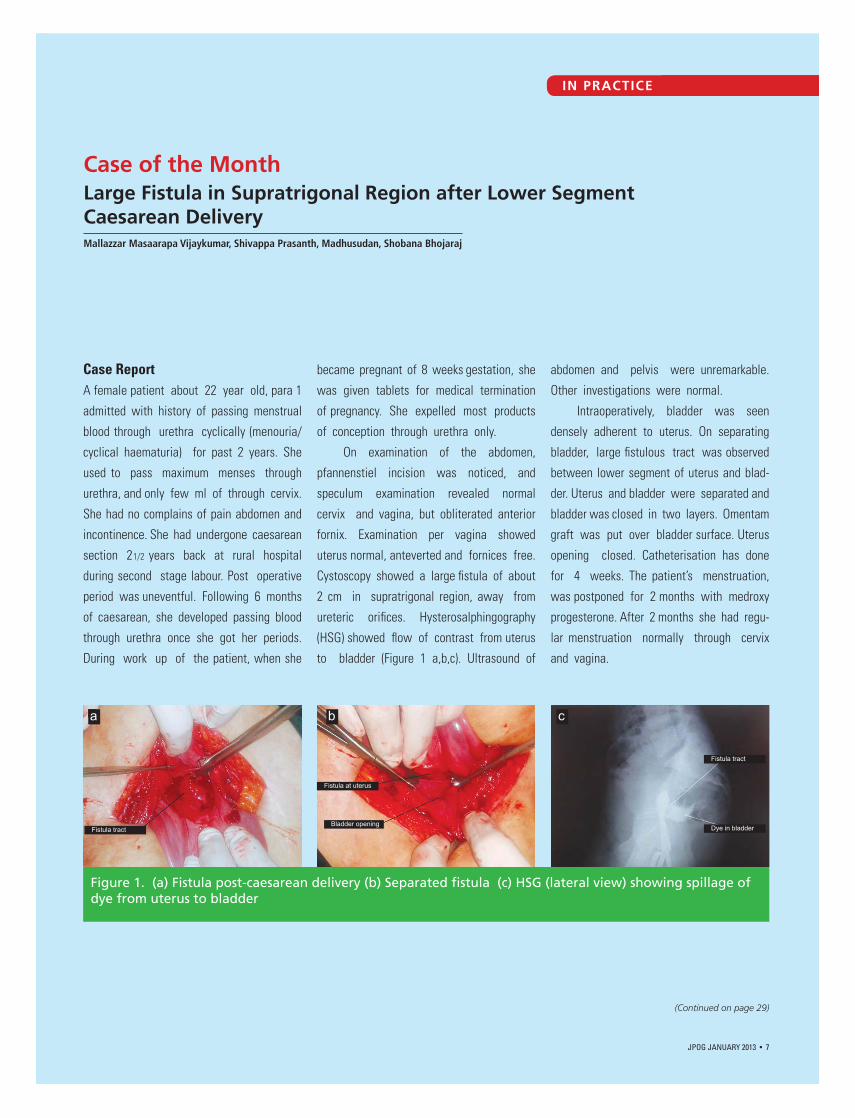
Cystoscopy showed a large fi stula of about
2 cm in supratrigonal region, away from
ureteric orifi ces. Hysterosalphingography
(HSG) showed fl ow of contrast from uterus
to bladder (Figure 1 a,b,c). Ultrasound of
abdomen and pelvis were unremarkable.
Other investigations were normal.
Intraoperatively, bladder was seen
densely adherent to uterus. On separating
bladder, large fi stulous tract was observed
between lower segment of uterus and blad-
der. Uterus and bladder were separated and
bladder was closed in two layers. Omentam
graft was put over bladder surface. Uterus
opening closed. Catheterisation has done
for 4 weeks. The patient’s menstruation,
was postponed for 2 months with medroxy
progesterone. After 2 months she had regu-
lar menstruation normally through cervix
and vagina.
Case of the MonthLarge Fistula in Supratrigonal Region after Lower Segment Caesarean DeliveryMallazzar Masaarapa Vijaykumar, Shivappa Prasanth, Madhusudan, Shobana Bhojaraj
Figure 1. (a) Fistula post-caesarean delivery (b) Separated fistula (c) HSG (lateral view) showing spillage of dye from uterus to bladder
(Continued on page 29)
Fistula tract
Fistula at uterus
Bladder opening
Fistula tract
Dye in bladder
aa b c

OBSTETRICS
JPOG JANUARY 2013 • 8
Methods of Induction of Labour–Critical Review of Literature (Part II)
Varsha Deshmukh, Kanan Yelikar, Sonali Deshpande, Pradeep Ingale
Misoprostol (PGE1)
Recent interest in inducing agents has been focused on misoprostol, a synthetic PGE1
analogue which was first used as treatment of gastric and duodenal ulcers. Misoprostol
is about 50 times cheaper than dinoprostone gel and as opposed to dinoprostone gel, is
stable at room temperature.
Although misoprostol can be administered vaginally/rectally/orally/sublingually,
vaginal route at present offers most benefits in terms of efficacy and minimising side-
effect profile.82
Different Routes of Administration
Vaginal
In 1987, Rabe T et al 81 first reported the use of misoprostol on pregnant uterus in the
first trimester of pregnancy. In a randomised controlled by Margulies et al 82 in 1992,
64 women beyond 28 weeks gestation undergoing indicated induction in the third
trimester of pregnancy, were given 50 µg doses of misoprostol vaginally or oxytocin
intravenously. Intravaginal misoprostol was found to be as effective as oxytocin and
without differences in neonatal outcomes.
Sanchez-Ramos et al 82 described their experience with intravaginal misopros-
tol 50 µg given every 4 hours compared to intravenous oxytocin in 129 subjects with
undilated and uneffaced cervices. There was reduction in induction delivery interval in
those with misoprostol (11 vs. 18 hours). However the frequency of uterine tachysystole
in the misoprostol treatment arm was 3 times that in oxytocin treatment arm (34.4%
JPOG JANUARY 2013 • 9
OBSTETRICS
vs. 13.8%). No differences were found in mode
of delivery or neonatal or maternal morbidity. The
authors concluded that misoprostol was safe and
effective for labour induction, and recommended
further investigation to detail the optimal route,
dose and dosing regimen.
Hofmeyr et al 83 in a Cochrane database
systematic review concluded that although vaginal
misoprostol is more effective than other induc-
ing agents, the apparent rise in uterine hyper
stimulation is a cause of concern. However, dosing
regimen less than 25 µg, 4 hourly is comparable
to traditional methods of induction in terms of
hyperstimulation. In dosages of 25 micrograms
three hourly or more, vaginal misoprostol is more
effective than conventional methods of cervical
ripening and labour induction. However, uterine
hyperstimulation with foetal heart rate changes
is increased. Although no differences in perinatal
outcome were shown, it states that the studies are
not sufficiently large to exclude the possibility of
uncommon serious adverse effects. The trend to an
increase in meconium-stained liquor also requires
further investigation. Anecdotal reports of uterine
rupture following labour induction with misoprostol
are cause for concern.
Misoprostol currently is approved by the US
Food and Drug Administration (FDA) for the preven-
tion of peptic ulcers. The manufacturer of misopro-
stol had issued a cautionary letter to health care
providers against the use of misoprostol in preg-
nant women. This stand was maintained despite
a large body of evidence supporting efficacy and
safety of misoprostol for induction of labour in term
pregnancy. This prompted a response by the Ameri-
can Congress of Obstetricians and Gynecologists
(ACOG) which endorsed its previous conclusions
regarding the efficacy of intravaginal misoprostol
tablets for labour induction in women with unfavor-
able cervices. Debate continues as to the optimal
dosing regimen of vaginally administered miso-
prostol. ACOG committee opinion stated that if
misoprostol is used for cervical ripening and labour
induction, 25 mg should be considered for the initial
dose to be repeated 3−6 hourly.
FDA has now approved a new label on the use of
misoprostol during pregnancy for cervical ripening
and for the induction of labour. This labeling does
not contain claims regarding the efficacy or safety
of misoprostol, nor does it stipulate doses or dose
intervals. It is now on the WHO essential drug list
for labour induction. Recently generic forms of
misoprostol have become available. Misoprostol
has been approved by Drug Controller General of
India (DCGI) in December for 25 µg, 100 mcg and
200 µg for cervical ripening, prevention of post-
partum haemorrhage and first trimester of abortion
with mifepristone with additional strength approval
for 50 µg for same approved indications in August
2008.
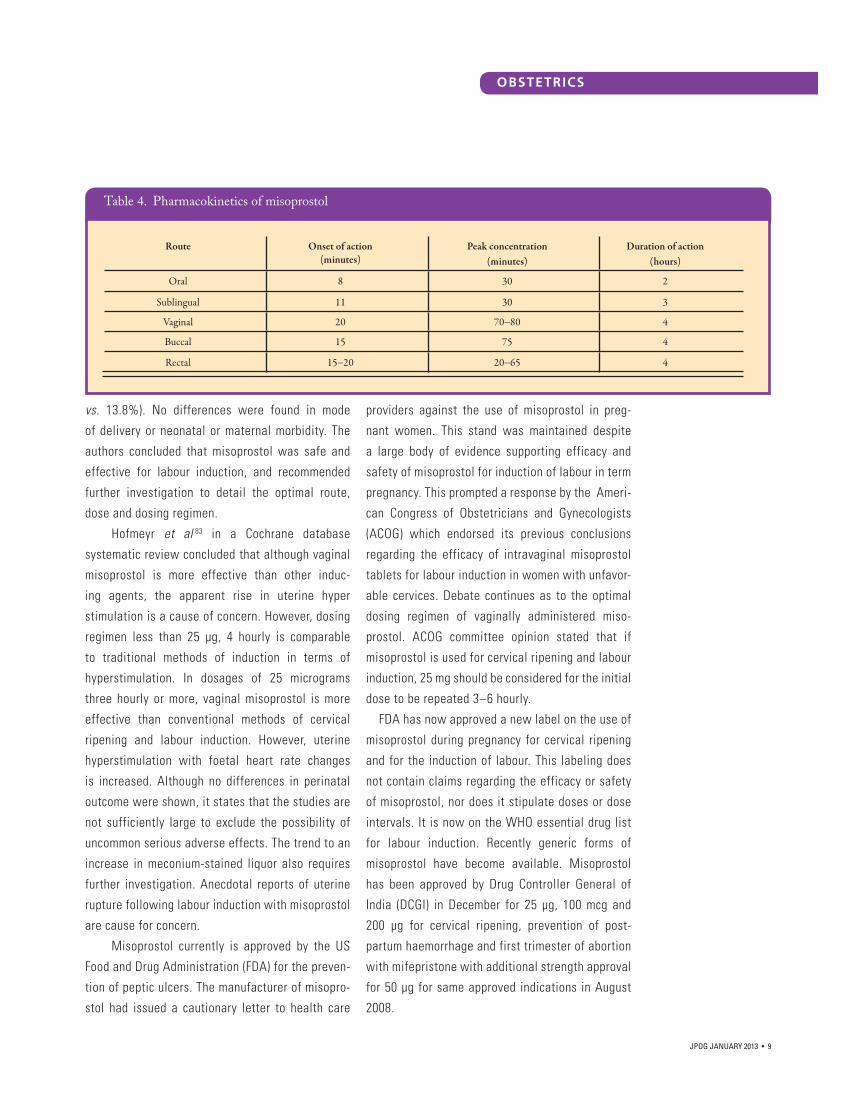
Table 4. Pharmacokinetics of misoprostol
Route Onset of action (minutes)
Peak concentration(minutes)
Duration of action (hours)
Oral 8 30 2
Sublingual 11 30 3
Vaginal 20 70−80 4
Buccal 15 75 4
Rectal 15−20 20−65 4

JPOG JANUARY 2013 • 10
OBSTETRICS
Thus, use of misoprostol for induction of labour
is associated with decrease in induction to delivery
interval, decreased need for oxytocin augmenta-
tion, increased chances of successful induction,
decrease in incidence of caesarean section; but
with increased rates of uterine hyperstimulation,
both with and without associated foetal heart rate
changes and no differences in perinatal or maternal
outcome. Though misoprostol shows promise as a
highly effective, inexpensive and convenient agent
for labour induction, the lack of global registration
for this purpose, and thus of well-established regi-
mens, is problematic.
Comparative Studies of Vaginal
Misoprostol
Vaginal Misoprostol vs. Vaginal
Dinoprostone Tablets
Fletcher et al 84 compared 32 cases of intravaginal
misoprostol (100 µg) with 31 cases of intravaginal
dinoprostone tablets (3 mg). The mean change
in Bishop’s score was significantly higher in the
misoprostol arm (5 vs. 3.3) and there were no
significant differences in labour outcome. The
author concluded that misoprostol was as effective
as dinoprostone for inducing labour at term.
Chang et al 85 in 1997 compared vaginal dino-
prostone tablets (3 mg) with vaginal misoprostol
tablets (200 µg) in a group of 60 women with term
singleton pregnancy. After 12 hours of induc-
tion, the mean Bishop score was 9.7 in Group II
compared with only 7.3 in Group I. The mean time
from insertion of the drug to delivery was shorter
in group II (16.5 hours) than in group I (25.7 hours).
There were no significant differences in the sponta-
neous labour rate, need for oxytocin augmentation,
type of delivery, and Doppler flow velocity wave-
forms of the umbilical artery. The average number
of required doses per patient was 1.8 in group II
and 2.7 in group I. The spontaneous vaginal delivery
rate was 88% in group I and 80% in group II; 6%
and 10%, respectively, were delivered by caesar-
ean section.
Thus it was concluded that inexpensive drug
misoprostol, is associated with higher Bishop
scores and a shorter interval to vaginal delivery
than dinoprostone tablets.
Vaginal Misoprostol vs. Vaginal
Dinoprostone Gel
Danielian et al 86 studied the efficacy of vaginal
misoprostol (50 µg 4 hourly) in comparison with
vaginal dinoprostone gel (1 mg 6 hourly) for
induction of labour. The misoprostol group had a
highly significant reduction in median induction-
delivery interval compared with the dinoprostone
group. There were no adverse neonatal outcomes
associated with the use of misoprostol. Women
in the misoprostol group experienced more pain
in the interval between induction and being
given analgesia in labour, but this did not reach
statistical significance. The authors concluded that
misoprostol 50 µg vaginally is a more effective
induction agent than 1 mg dinoprostone vaginal
gel, with no apparent adverse effects on mode of
delivery, or on the foetus. The higher pain scores
in the misoprostol group must be balanced against
the reduction in time spent having labour induced,
and the reduction in need for intravenous oxytocin
augmentation.
Van Gemund87 compared the efficacy of
vaginal misoprostol (25 µg 4 hourly) with vaginal
dinoprostone gel (1 mg 4 hourly). The median induc-
tion-delivery interval was longer in the misopros-
tol group compared with the dinoprostone group
(25 vs. 19 hours). The caesarean section rate was
lower in the misoprostol group: 16.1% vs. 21%, but
this difference was not statistically significant RR =
0.8 (95% CI 0.6−1.04). 'Adverse neonatal outcome'

JPOG JANUARY 2013 • 11
OBSTETRICS
was found to be similar in both groups: 21% in the
misoprostol and 23% in the dinoprostone groups.
Significantly fewer neonates were admitted to
neonatal ICU in the misoprostol group compared
with dinoprostone 19% vs. 26% (RR = 0.7, 95% CI
0.5-0.98). The authors concluded that misoprostol
in this dosing regimen is a safe method of labour
induction.
Gregson et al 88 conducted a single-blind
randomised controlled trial comparing the efficacy
of vaginal misoprostol tablet (25 µg 4 hourly) with
vaginal dinoprostone gel (1−2 mg 6 hourly). There
were no significant differences between the two
groups in induction-to-vaginal delivery interval,
mode of delivery, number of women delivering
within 24 hours and neonatal outcomes. The inci-
dence of uterine contraction abnormalities (tachy-
systole and hyperstimulation) and the incidence of
abnormal CTG recordings were also similar for both
groups. Thus both the drugs are equally effective,
misoprostol being much less costly.
Thus, misoprostol in doses 25−50 µg 4 hourly
is having comparable safety and efficacy when
compared to 1−2 mg dinoprostone gel 6 hourly.
Vaginal Misoprostol vs. Vaginal
Dinoprostone Insert
Garry et al 89 in 2003 compared the safety and
efficacy of vaginal misoprostol tablets (50 µg 3
hourly) with vaginal dinoprostone insert (10 mg
12 hourly) for induction of labour. The authors
concluded that intravaginal misoprostol and
dinoprostone were safe and effective medications
for use in cervical ripening before labour induction.
Misoprostol resulted in a shorter interval from
induction to delivery. However, Caesarean delivery
for a non-reassuring foetal heart rate tracing was
more common with misoprostol.
Wing et al 90 in October 2008 compared the
efficacy of dinoprostone vaginal insert to misopro-
stol vaginal insert for induction of labour. A total of
1,308 women requiring cervical ripening (modified
Bishop score less than or equal to 4) before induc-
tion of labour were randomly assigned to receive
misoprostol vaginal insert 100 µg (n=428), miso-
prostol vaginal insert 50 µg (n=443) or 10 mg dino-
prostone vaginal insert (n=436). The misoprostol
vaginal insert 100 µg and the dinoprostone vaginal
insert had similar median time intervals to vaginal
delivery, whereas the misoprostol vaginal insert 50
had a significantly longer time to vaginal delivery.
The three products had similar cesarean rates and
safety profiles.
Ozkan et al 91 in 2008 compared the efficacy
and safety of vaginal misoprostol with dinopro-
stone vaginal insert for labour induction in term
pregnancies. The subjects were randomised to
receive either [i] 50 µg misoprostol intravaginally
every 4 hour to a maximum of five doses or [ii] 10
mg dinoprostone vaginal insert kept for a maximum
of 12 hours. Time interval from induction to vagi-
nal delivery was found to be significantly shorter
in misoprostol group when compared to dinopro-
stone subjects (680 min vs. 1070). More subjects
required oxytocin augmentation in dinoprostone
group (62.5% vs. 35.7%). Cardiotocography trac-
ings revealed early decelerations occurring more
frequently with misoprostol induction (10.7 vs. 0%).
Tachysystole and uterine hyperstimulation, mode of
delivery, rate of caesarean sections due to foetal
distress and adverse neonatal outcome were not
demonstrated to be significantly different between
groups. The authors concluded that using vaginal
misoprostol is an effective way of labour induction,
misoprostol and dinoprostone being equally safe.
Vaginal Misoprostol vs. Endocervical
Dinoprostone Gel
Varaklis et al 92 compared 25 µg misoprostol every
2 hours to commercially available endocervical

JPOG JANUARY 2013 • 12
OBSTETRICS
preparation containing 0.5 mg of PGE2 gel
administered at 6 hour intervals for a maximum
of two doses. Women who received misoprostol
experienced a significantly reduced mean time from
drug administration to onset of three contractions
in 10 minutes (6.7 vs. 12.4 hours). Mean time to
rupture of membranes was also shorter in the
misoprostol group (9.7 vs. 13.6 hours), as was the
mean time to delivery (16 vs. 22.4 hours). Three
patients in the misoprostol group experienced
uterine hypertonus but not related foetal morbidity.
Thus, they concluded that misoprostol is more
effective than endocervical PGE2 in bringing about
labour and delivery.
Krithika et al 93 conducted a prospective
randomized controlled trial to compare the efficacy
and safety of intravaginal misoprostol with endo-
cervical dinoprostone gel for induction of labour in
cases of unfavourable cervix. One hundred women
with an unfavourable cervix requiring induction of
labour were randomised to receive either 25 µg
vaginal misoprostol 4 hourly or 0.5 mg of endocer-
vical dinoprostone 12 hourly. The improvement in
Bishop's score at 12 hours was significantly better
in the misoprostol group. Induction to delivery
interval was shorter in the misoprostol group, 16.59
+/− 5.13 hours vs. 27.77 +/−12.71 hours. The rate
of complications was comparable. Thus, it was
concluded that vaginal misoprostol 25 µg 4 hourly
is safe and effective for induction of labour with
shorter induction to delivery interval.
Shivarudraiah and Palaksha94 conducted a
prospective randomised controlled trial to compare
the efficacy and safety of intravaginal misoprostol
with endocervical dinoprostone gel for induction of
labour in cases of unfavorable cervix. Three hundred
and twenty women with an unfavourable cervix
requiring induction of labour were randomised to
receive either 25 µg vaginal misoprostol 4 hourly
or 0.5 mg of endocervical dinoprostone 6 hourly.
Median induction to active phase interval was simi-
lar in both groups (465 vs. 457), but there was no
significant difference in median induction to deliv-
ery interval in misoprostol and dinoprostone groups
(684 vs. 690). The proportion of women delivering
in 24 hours was similar, requirement of oxytocin
augmentation was similar. No difference was noted
in overall incidence of caesarean section, vaginal/
instrumental delivery rate.
Similar study was carried out in Government
Medical College and Hospital (GMCH), Aurangabad,
Maharashtra by Sonali D, Kanan Y and Pradeep I in
2012 to compare safety and efficacy of intravaginal
misoprostol tablet as compared to endocervical
dinoprostone gel for induction of labour in women
with unfavorable cervix. Primary outcome measure
was induction to delivery interval white secondary
outcome measures were labour characteristics,
maternal and neonatal outcomes. Of 100 women
randomised, 50 received tab misoprostol 25 µg
every 4 hourly (maximum 6 doses) and 50 received
dinoprostone gel 0.5 mg every 6 hourly (maximum 4
doses). Both groups were found to be comparable
with respect to primary and secondary outcome
measures except the cost of misoprostol was 50
times cheaper than dinoprostone gel.
Thus, misoprostol tablet is having compa-
rable safety, efficacy and side effect profile when
compared with the established agent dinoprosone
gel thus may be used as a cost effective alternative
to dinoprostone gel for induction of labour.
Vaginal Misoprostol vs. Mechanical Methods
Perry et al 95 studied the efficacy of vaginal
misoprostol (25 µg 4 hourly) to intracervical Foley’s
catheter and intravaginal dinoprostone. They
found Foley’s catheter/dinoprostone to have lesser
induction ripening interval (7.5 vs. 12.0 hours) as
well as induction delivery interval (17.4 vs. 21.2
hours).

JPOG JANUARY 2013 • 13
OBSTETRICS
Chung et al 96 studied efficacy of combination
intravaginal misoprostol and intracervical Foley
catheter for prelabour cervical ripening. Women
were assigned to one of three groups:
• Intravaginal misoprostol 25 µg every 3 hours,
• Intracervical 16F Foley catheter, or
• Combination misoprostol-Foley catheter
They concluded that intravaginal misoprostol
and intracervical Foley catheter are comparable for
preinduction cervical ripening. The combination of
the two methods did not provide additional efficacy.
Prager et al 97, included 588 subjects and
randomised them for induction of labour using
intravaginal dinoprostone (2 mg once every 6 hours)
or misoprostol (25 µg once every 4 hours) or a
transcervical balloon catheter. The shortest mean
induction-to-delivery interval was obtained with
the catheter (12.9 hours) vs. 16.8 and 17.3 hours
for dinoprostone and misoprostol, respectively. The
efficacies of the two prostaglandins were similar.
The maternal and neonatal outcomes associated
with each of the three procedures were similar.
The researchers concluded that induction of labour
with a transcervical balloon catheter is effective
and therefore safe and can be recommended as the
first choice.
Thus, transcervical balloon catheter can be
used as a safe and cost effective alternative to
prostaglandins. Though combination of catheter
with dinoprostone gel is found to be more effective,
similar efficacy could not obtained using catheter
with misoprostol tablet. The two prostaglandins,
dinoprostone and misoprostol, were shown to be
equally effective and safe, while misoprostol costs
significantly less and is easier to store.
Oral
Ngai et al 98 investigated the effectiveness of oral
misoprostol as a cervical priming agent for patients
presenting with pre-labour rupture of membranes
at term. Eighty patients were randomised to receive
either 200 µg of misoprostol or 50 mg of vitamin
B6 orally 1 hour after admission. The cervical score
was significantly improved and the induction rate
was also reduced in the misoprostol group when
compared with the control group. The interval from
recruitment to onset of labour, duration of labour,
and the interval from recruitment to delivery were
significantly shorter in the misoprostol group. The
mode of delivery and the perinatal outcome were
similar for the two groups. The authors concluded
that oral misoprostol is an effective agent for
cervical priming and labour induction in patients
with pre-labour rupture of membranes at term.
Thus, oral misoprostol is simple and nonin-
vasive. Onset of action is most rapid by this route
and there is almost complete absorption, peak
levels reached by 30 minutes. It might reduce the
incidence of chorioamnionitis (associated with
repeated vaginal examinations) particularly in the
presence of prelabour rupture of membranes.
Comparative Studies of Oral
Misoprostol
Oral Misoprostol vs. Vaginal Misoprostol
Wing et al 99 compared 50 µg of oral misoprostol
every 4 hourly with 25 µg vaginal misoprostol every
4 hourly for maximum 6 doses. Overall mean time
to delivery (hours ± SD) was 29 ± 14.1 in oral group
and 23.2 ± 12.8 in vaginal group. Mean number of
doses required were 3.3 ± 1.7 in oral group and 2.3
± 1.3 in vaginal group. There was no significant
difference in maternal and perinatal morbidity.
Thus, they concluded that oral administration of 50
µg dose of misoprostol appears less effective than
vaginal administration of 25 µg dose of misoprostol
for cervical ripening and labour induction.
Nopdonrattakoon L100, studied 106 preg-
nant women at term with unfavorable cervix with

JPOG JANUARY 2013 • 14
OBSTETRICS
Bishop’s score less than 4 to receive intravaginal
misoprostol 50 µg every 4 hourly or oral misoprostol
50 µg every 4 hourly. It was found that time interval
from induction to delivery in oral group was signifi-
cantly longer than intravaginal group (881 ± 443
min vs. 637 ± 373 min respectively). The number of
dosage required was higher in oral group but there
was no difference between both groups with regard
to failure of induction and maternal and neonatal
complications.
In cross sectional comparative study by Bano
et al 101, 200 term women were recruited and allot-
ted to receive 50 µg of misoprostol every 4 hourly
either vaginally or orally. Mean induction to deliv-
ery interval was similar in both groups (9.09 vs. 9.08
hours). This study concluded that oral route was
as effective as vaginal route in terms of induction
to delivery interval, number of dosage, caesarean
section rate, maternal complications and neonatal
outcome.
RCOG committee2 opinion states that in
women with an unfavorable cervix, oral misopros-
tol 50 µg is less likely than vaginal misoprostol 25
µg to achieve vaginal birth within 24 hours. Oral
misoprostol has similar efficacy to vaginal PGE2 gel
in terms of vaginal birth within 24 hours.
Thus oral misoprostol is having comparable
efficacy when compared with vaginal misoprostol
but rise in the incidence of uterine contraction
abnormalities is the cause of concern.
Oral Misoprostol vs. Vaginal Mechanical
Methods
Abramovici et al 102 compared oral misoprostol
(50 µg 4 hourly) with cervical Foley catheter and
intravenous oxytocin infusion. In multiparous
patients the percentage delivered of their neonates
within 24 hours and the median induction-to-
delivery times were similar in the 2 groups. In
nulliparous patients, however, delivery within
24 hours was significantly less likely in the
misoprostol group (53.4% vs. 82. 5%; p<0.001), and
the median induction-to-delivery time was longer
(23.3 hours vs. 17.2 hours; p<0.01). The incidence of
hyperstimulation was higher in the oxytocin-Foley
group (4.1% vs. 13.1%; p=0.02). They concluded
that oral misoprostol is as effective as oxytocin-
Foley’s catheter for inducing labour in multiparous
women, but appears less efficacious in nulliparous
patients.
Oral Misoprostol vs. Endocervical
Dinoprostone Gel
Bartha et al 103 compared the efficacy, safety, and
tolerance of oral misoprostol with endocervical
dinoprostone for cervical ripening and labour
induction. They concluded that a single dose of
200 µg oral misoprostol was more effective for
cervical ripening and labour induction than 0.5 mg
of dinoprostone endocervically every 6 hours.
Nagpal et al 104 from New Delhi conducted
a study in April 2009 comparing the efficacy of
oral misoprostol (50 µg 4 hourly) with endocervi-
cal dinoprostone gel (0.25 mg 6 hourly). Twenty
women in the misoprostol group (n=31) delivered
within 12 hours compared with 5 in the PGE2 group
(n=30). The induction-to-delivery interval in the
misoprostol group was shorter than in the PGE2 gel
group (615 vs. 1070 min). The mode of delivery was
comparable between the 2 groups. Abnormalities in
uterine contractions and neonatal outcomes were
also comparable. The requirement for oxytocin was
lower and patient satisfaction was better in the
misoprostol group.
Oral misoprostol is more effective than endo-
cervical dinoprostone gel for induction of labour.
Titrated oral misoprostol
This is the latest method of administration being
studied for induction of labour with misoprostol.

JPOG JANUARY 2013 • 15
OBSTETRICS
The rationale behind this is the short half-life of
misoprostol when administered by oral route (peak
concentration at 34 minutes and half-life of 20–40
minutes. This method is designed to reduce the risk
of hyperstimulation which is seen with misoprostol,
especially by the oral route. The principle used is
200 µg of misoprostol tablet with dissolved in 200
ml of water (1µg/ml), initial administration of 20 µg/
hour till adequate contractions are achieved. Dose
is increased to 40 µg/hour if sufficient contractions
are not reached in 4 doses. When adequate contrac-
tions are achieved, no further misoprostol is given
if contractions decrease; hourly doses of misopro-
stol are restarted at 10 µg/hour and increased to
20 or 40 µg/ hour if necessary. Induction failure
is defined as inability to reach active phase by 36
hours.
Hofmeyr et al 105 compared titrated oral misoprostol
solution with 2 mg vaginal dinoprostone for
induction of labour. They found similar efficacy and
hyperstimulation rates.
Cheng and Chen106 evaluated the efficacy of
titrated oral misoprostol in a study group of 77
women. The mean interval from induction to vaginal
delivery for all the women was 9.7 hours, with a 2.3
hour active phase. The mean misoprostol dosage
was 206 µg, with eight women (10.4%) requir-
ing oxytocin augmentation. There was no uterine
hyperstimulation or induction failure, except for
seven cases of uterine tachysystole (9.1%). They
concluded that titrated oral misoprostol is a safe
and effective method of labour induction because
the dosage can be adjusted according to individual
response.
Comparison of Titrated Oral
Misoprostol with Other Methods of
Induction of Labour
Cheng, Ming and Lee107 compared titrated oral
misoprostol vs. vaginal misoprostol (25 µg 4 hourly)
for induction of labour in women between 34 and
42 weeks with unfavorable cervices (Bishop’s score
≤ 6). Completed vaginal delivery occurred within
12 hours in 75 (74.3%) women in the titrated oral
group and 27 (25.5%) women in the vaginal group
(relative risk [RR] 8.44, 95% confidence interval
[CI] 4.52−15.76). The incidence of hyperstimulation
was 0.0% in the titrated oral group compared
with 11.3% in the vaginal group (RR 0.08, 95% CI
0.01−0.61). Although more women experienced
nausea (10.9%) in the titrated oral group (RR 27.07,
95% CI 1.57−465.70), fewer infants had APGAR
scores of less than 7 at 1 minute in the titrated oral
group than in the vaginal group (RR 0.10, 95% CI
0.01−0.76).
Kundodyiwa and colleagues108 conducted a
review of the use of low dose oral misoprostol for
induction of labour. They concluded that low-dose
oral misoprostol solution (20 micrograms) adminis-
tered every 2 hours appeared to be as effective as
both vaginal dinoprostone and vaginal misoprostol,
with lower rates of caesarean delivery and uterine
hyperstimulation, respectively.
Though, titrated oral misoprostol has proven
effective in few studies, it is considered that
future studies this area may give us definitive word
regarding its safety and efficacy.
Oral Misoprostol on Outpatient Basis
Kipikasa et al 109 evaluated the use of oral
misoprostol outpatient basis for pre-induction
cervical ripening and induction of labour. Forty nine
subjects followed in an outpatient obstetrical clinic
with pregnancies of at least 40 weeks' gestation,
and an unfavorable Bishop's score were assigned
randomly to receive oral misoprostol 50 or 25 µg
every 3 days for a maximum of three doses.
Twenty three subjects received misoprostol
25 µg and 26 received 50 µg. The mean interval
(+/-standard deviation) from start of cervical ripen-
JPOG JANUARY 2013 • 16
OBSTETRICS
ing to delivery was 2.4 days +/-0.3 vs. 3.9 days
+/-0.7 for the 50 and 25 µg. No adverse events were
noted.
Gaffney et al 110 studied the use of oral miso-
prostol 100 µg given orally on outpatient basis.
Subjects received either oral misoprostol 100 µg or
placebo daily for 3 days unless the subject devel-
oped significant cervical change or began labour
spontaneously. Study drug was repeated every 24
hours for a maximum of three doses if subjects
did not develop significant cervical change or
enter labour. A significant difference was noted in
reduction of time from study entry to both active
phase and delivery in the misoprostol group. Fewer
women remained undelivered after the 72 hours
study period in the misoprostol group. There were
no differences in route of delivery or neonatal
outcomes between groups.
RCOG committee2 opinion states that induc-
tion of labour in the outpatient setting should only
be carried out if safety and support procedures are
in place and the practice of induction of labour in an
outpatient setting should be audited continuously.
Endocervical
Srisomboon et al 111 compared the efficacy of
intracervical misoprostol with vaginal misoprostol.
No significant differences were noted between
intracervical and intravaginal misoprostol in terms
of Bishop's score change, (score 7.2 vs. 7.5),
interval from gel insertion to vaginal delivery (17.0
hours vs. 16.4 hours), route of delivery and perinatal
outcome. Uterine tachysystole occurred in 24% and
32% in the intracervical and intravaginal groups
respectively which did not significantly differ. No
evidence of foetal distress was noted in these
events. The authors concluded that the two routes
of misoprostol application appeared to be safe and
equally effective in ripening cervix and inducing
labour, however, the intravaginal application
was more convenient to administer practically as
compared with the intracervical.
Liu et al from Taiwan112 also evaluated the
safety and efficacy of intracervical misoprostol (50
µg 4 hourly). They concluded that misoprostol as a
single 50 µg intracervical dose is safe and effective.
However, they advised caution in repeated doses as
might be required in an unfavorable cervix.
Chang et al 113 compared the safety and effi-
cacy of intracervical misoprostol (50 µg 4 hourly)
with intracervical dinoprostone (0.5 mg 4 hourly).
They concluded that as compared to dinopros-
tone, intracervical misoprostol was more effective
in cervical ripening and labour induction at term.
The higher frequency of uterine hypercontractil-
ity associated with the use of misoprostol did not
increase the risk of adverse intrapartum and neona-
tal outcomes.
Although intracervical misoprostol has been
found to be effective some studies, the intravaginal
application is more convenient to administer practi-
cally as compared to intracervical.
Buccal and sublingual
With these routes there is rapid increase in
concentration and higher Cmax than oral route, as
first pass metabolism is avoided. RCOG committee2
opinion states that there is insufficient data to
determine the effectiveness of buccal/sublingual
misoprostol as compared with oral and vaginal
misoprostol.
Misoprostol for Labour Induction
with Prior Caesarean Births
The only randomised controlled trial by Wing et
al 114 comparing vaginal misoprostol with oxytocin
in women with prior caesareans was terminated
prematurely because of 2 uterine scar disruptions
in subjects who had received multiple doses of
vaginal misoprostol 25 µg.

JPOG JANUARY 2013 • 17
OBSTETRICS
Aslan et al 115 in a retrospective report
compared labour induction with vaginal miso-
prostol in previous caesarean delivery vs. that in
unscarred uteri. Uterine rupture occurred in 4 out of
41 patients in the prior caesarean group compared
to none in 50 patients without scarring.
Thus, use of misoprostol in prior caesarean deliver-
ies, is not recommended.
Oxytocin
Use of oxytocin drip was described by Theobald
and Helman in 1948. Oxytocin, an octapeptide
neurohormone that originates in the hypothalamus
is secreted by the posterior lobe of the pituitary
gland. This is secreted in a pulsatile manner.
The uterus becomes increasingly responsive to
oxytocin as pregnancy progresses; there is a
gradual increase in response from 20−30 weeks of
gestation, followed by a plateau from 34 weeks of
gestation until term, when sensitivity increases.
It seems that oxytocin has direct stimulatory
effects on the myometrium in addition to stimulat-
ing decidual prostaglandin production. An increased
level of prostaglandin F2α metabolite was demon-
strated in women who underwent successful oxyto-
cin induction of labour, whereas this increase was
not present in failed inductions.
The intravenous route is used almost exclu-
sively to stimulate the pregnant uterus because
it allows precise measurement of the amount of
medication being administered and a relatively
rapid discontinuation of the drug when side effects
occur. Oxytocin is administered as a dilute solu-
tion, with the flow rate into the intravenous line
precisely regulated by an infusion pump.
ACOG states that each hospital’s obstet-
rics and gynaecology department should develop
guidelines for the preparation and administration
of oxytocin. One United States Pharmacopoeia
unit being equivalent to 2 mg of oxytocin. Uterine
response ensues after 3–5 minutes of infusion, and
a steady level of oxytocin in plasma is achieved by
40 minutes.
Several trials (Blackmore et al 116, Mercer
et al 117 , Chua et al 118, Satin et al 119, Muller et
al 120) have compared options in oxytocin dosage
increases and time intervals between dose
increases. Starting doses have ranged from 0.5
to 2 mU/min, with some as much as 6 mU/min.
Increments of dose increase have ranged from as
low as 1−2 mU/min to as much as 6 mU/min, with
adjustments for increased uterine activity. Time
intervals between increases have ranged from 15
to 40 minutes. Although low-dose regimens (initial
dose 0.5–2 mU/min, with incremental increases of
1–2 mU/min every 15–40 min) are commonly used
in the United States, high-dose regimens (initial
dose 6–8 mU/min, with incremental increases of 6
mU/min every 15–40 min) have been shown to be
safe and effective for labour induction in patients
with viable pregnancies (Merrill and Zlatnic121). A
meta-analysis of 11 trials by Crane and Young122
compared low-dose with high-dose oxytocin for
labour induction. They found that greater increases
and shorter intervals were associated with shorter
labour and lower rates of intra-amniotic infections
and cesarean delivery for dystocia, but more hyper-
stimulation was noted. Based on recent pharmaco-
kinetic data, many obstetricians have moved to a
regimen whereby the dose of oxytocin is increased
by 1−2 mU/min every 40 minutes. Advantages of
this regimen derive from not increasing the oxyto-
cin dose before steady-state levels of oxytocin have
been reached. This approach should lead to a lower
total dosage of oxytocin required in addition to a
lower incidence of hyperstimulation that can result
from increasing the oxytocin dose before steady-
state is reached. A potential disadvantage is that
women who are relatively insensitive to oxytocin
may have a prolonged course before adequate

JPOG JANUARY 2013 • 18
OBSTETRICS
labour is achieved. Nearly 90% of patients respond
to 16 mU/min or less, whereas it is most unusual
for a patient to require more than 20−40 mU/min.
In 1978, Pavlou et al 123 were the first to
describe a protocol of pulsatile infusion. More
recently, several randomised trials compared the
safety and efficacy of pulsatile oxytocin administra-
tion with continuous infusion. Most authors (Sala-
malekis et al 124, Reid and Helewa125 , Wellcourt et
al 126) concluded that though there does not seem to
be a shortening of the intervals to delivery, pulsa-
tile administration of oxytocin reduces the amount
of oxytocin required for successful labour induc-
tion. The concentration of oxytocin administered,
the rate of infusion, and the interval between dose
increments are subjects of study and debate.
Mifepristone
Mifepristone, also known as RU−486, is an anti-
progestin and has been developed to antagonise
the action of progesterone. Mifepristone now has
an established role in the termination of pregnancy
and has been approved by FDA, in combination with
prostaglandins, for the first trimester abortion.
Some studies have used in post term pregnancies
and is probably a new field of research for labour
induction.
Cochrane database of systematic review by
Neilson JP127 (7 RCTs, 594 women, mixed parity
and Bishop score < 6) that evaluated the effects of
mifepristone vs. placebo/no treatment in women
at term, found insufficient information to support
the use of mifepristone to induce labour. However,
there is recent evidence of serious neonatal side
effects involving renal function in the form of isch-
emic hypoxic changes in the foetal kidney ultra-
structure when labour was induced by mifepristone
between 16 and 28 weeks of gestation. The smaller
is the foetus, more obvious the changes.
RCOG committee2 opinion states that at pres-
ent there is insufficient information to support the
use of mifepristone to induce labour in a women
with live foetus.
Hyaluronidase
The level of hyaluronic acid increases markedly
after the onset of labour. Cervical injection of
hyaluronidase was postulated to increase cervical
ripening.
Kavanagh et al 128 in Cochrane database of
systematic reviews assessed the effects of intra-
cervical hyaluronidase in women undergoing induc-
tion of labour. Women given hyaluronidase were
reported to achieve significant improvement in
cervical status (RR 0.62, 95% CI 0.52 to 0.74) and
there were significantly fewer caesarean births (RR
0.37, 95% CI 0.22 to 0.61) when compared with
placebo. No side effects for mother or baby were
reported. Thus, intracervical hyaluronidase is likely
to improve cervical ripening and reduce caesarean
rates when compared with placebo. Although intra-
cervical hyaluronidase may be effective in improv-
ing cervical ripening and reducing caesarean birth
rates, it is an invasive procedure that women may
find unacceptable when alternative available meth-
ods, such as vaginal PGE2, are less invasive.
RCOG committee opinion states that hyaluron-
idase should not be used for induction of labour.2
Corticosteroids
Corticosteroids are postulated to have a promoting
effect in induction of labour but their role in the
process of labour is not well understood. Kavanagh
et al in Cochrane database of systematic reviews
(one RCT involving 66 women, and unfavorable
cervix) assessed the effects of corticosteroids
versus intravenous oxytocin in cervical priming and
induction of labour.129 Vaginal birth within 24 hours
was not reported. There were no reports of uterine
hyperstimulation, APGAR score < 7 or maternal

JPOG JANUARY 2013 • 19
OBSTETRICS
fever in either group and caesarean birth rates were
not significantly different (RR 0.40, 95% CI 0.08 to
1.92).
The available evidence relating to the effects of
corticosteroids for cervical priming and induction of
labour is limited thus RCOG committee (2008)2 opin-
ion states that corticosteroids should not be used for
induction of labour.2
Oestrogen
The increase in the serum oestrogen-to-progesterone
ratio that occurs before the onset of labour is
believed to activate prostaglandin production, which
in turn stimulates cervical ripening. One systematic
review by Thomas et al 130 (6 RCTs involving 341
women, Bishop's score < 3) assessed the effects
of oestrogen in women undergoing induction of
labour. The included studies compared oestrogen
(intravenous, oral, vaginal or extra-amniotic) versus
placebo (four RCTs); versus vaginal PGE2 (one RCT);
versus intracervical PGE2 (one RCT); versus oxytocin
(one RCT); and versus extra-amniotic PGF2α (one
RCT). It reported no significant differences between
the oestrogens and the placebo groups in the rates
of caesarean births, instrumental vaginal births or
uterine hyperstimulation with or without foetal heart
rate (FHR) changes. There were insufficient data
for the remaining comparisons. Overall, there were
insufficient data to make any meaningful conclusion.
Limited evidence suggested that oestrogen and
placebo achieve similar maternal and foetal
outcomes. There was insufficient data available for
the comparisons between oestrogen and vaginal
PGE2, oxytocin alone or extra-amniotic PGF2α.
RCOG committee, 2008 opinion stated that
oestrogen should not be used for induction of labour.2
Vaginal Nitric Oxide Donors
Nitric oxide is considered a fundamental mediator
of cervical ripening without causing uterine
contractions or adverse effects on the mother and
foetus.
Studies by Chanrachakul and Bullabo state
that vaginal glyceryl trinitrate and nitric oxide donors
have not been shown to be of any particular benefit
when compared with vaginal PGE2 as induction
agents, although they seem to be associated with
less uterine hyperstimulation. However, there are
significant side effects associated with its use.131,132
RCOG committee 2008 opinion states that vaginal
nitric oxide donors should not be used for induction
of labour.2
Relaxin
Relaxin is a protein formed by two amino acid chain.
Its role in pregnancy and parturition is not clear.
Because of difficulty in using human, relaxin bovine
or porcine relaxin is used with controversial results.
Controlled studies indicate improvement in cervi-
cal status with no case of uterine hyperstimulation,
however further studies are required to assess its
safety and efficacy.
Homoepathy and Herbal Supplements
Homoepathy involves the administration in dilution
of substances aimed at the alleviation of symptoms,
that the same substances generally cause in their
undiluted form. It has been suggested that the herbs
belonging to the Caulophyllum genus are useful in
establishing labour, when uterine contractions are
short and/or irregular or when they stop.
RCOG committee 2008 opinion states that in
the absence of sufficient evidence to prove either
effectiveness or harm, homoepathy as a method of
induction is not recommended to be offered.2
The use of herbal supplements to promote
health has become popular. It is believed by some
that drinking herbal beverage teas while pregnant
nourishes and tones the uterus, supporting optimal
health in pregnancy.

JPOG JANUARY 2013 • 20
OBSTETRICS
RCOG committee 2008 opinion states that no
evidence was identified relating to the effects of
herbal supplements in cervical priming or induction
of labour.2
CONCLUSION
Amongst the numerous methods of induction,
prostaglandins are the preferred for induction of
labour. Dinoprostone gel used endocervically is
found to be both, safe and effective method, but
its cost and need of refrigeration limit its universal
use. Misoprostol vaginal tablet is rapidly becoming
a cost effective alternative to dinoprostone gel, but
lack of global consensus regarding its safety and
thus absence of global registration for this purpose
is a problem.
Oxytocin induction is not found to be as effec-
tive as prostaglandins. Though mifepristone was
found to be effective, there is inadequate data on
its safety profile. Other methods like use of hyal-
uronidase, steroids, oestrogen, herbal supplements,
homoepathy are not found to confer any advantage
over the established methods of labour induction.
Even with the availability of the armamen-
tarium of inducing agents, simple methods like
membrane sweeping should be offered whenever
opportunity exists, so as to decrease the rate of
formal induction.
About the AuthorsDr Varsha Deshmukh and Dr Sonali Deshpande are Associate Profes-sor, Dr Kanan Yelikar is Professor and Head and Dr Pradeep Ingale is Assistant Professor, Department of Obstetrics and Gynaecology, GMCH, Aurangabad, Maharashtra.
The complete list of references is available on request to the editorial office.

JPOG JANUARY 2013 • 21
Bacterial vaginosis is the most common cause of abnormal vaginal discharge in
women of childbearing age. It is a syndrome of unknown cause characterized by
depletion of the normal Lactobacillus population and an overgrowth of vaginal
anaerobes, accompanied by loss of the usual vaginal acidity. In 1983, the term ‘bacterial
vaginosis’ replaced the older term ‘Gardnerella vaginitis’. This recognized the fact that
many anaerobic or facultative anaerobic bacteria are present and that classical signs of
inflammation are absent.1
Women with symptomatic bacterial vaginosis report an offensive, fishy-smelling
discharge that is most noticeable after unprotected intercourse or at the time of men-
struation. The diagnosis can be confirmed by microscopy ± additional tests. About 50%
of cases are asymptomatic. Bacterial vaginosis is associated with infective complica-
tions in pregnancy and following gynaecological surgery, and is a risk factor for the
acquisition of sexually transmitted infections (STIs) including human immunodeficiency
virus (HIV).
EPIDEMIOLOGY
In unselected populations in the UK, the prevalence of bacterial vaginosis is 10–20%,
but it may be as high as 36% in women attending STI clinics and 28% in those seek-
ing elective termination of pregnancy.2 A prevalence of more than 50% was reported
in rural Uganda.3 The debate about whether bacterial vaginosis is an STI or merely
sexually associated continues. A meta-analysis has concluded that bacterial vagino-
sis has the characteristics of an STI: being associated with partner change and other
STIs.4 The strongest evidence against it being an STI has come from studies report-
ing similar rates in self-reported virgin and non-virgin women.5–7 This has been chal-
GYNAECOLOGY

JPOG JANUARY 2013 • 22
GYNAECOLOGY
lenged by a detailed study that reported no bacte-
rial vaginosis in women denying any oral or digital
genital contact.8 In many studies, it is associated
with black race and intrauterine device use. The
condition often arises spontaneously around
the time of menstruation and may resolve spon-
taneously in mid-cycle. It is not known how often
bacterial vaginosis occurs in post-menopausal
women.
AETIOLOGY AND PATHOGENESIS
Lactobacilli dominate the normal vaginal flora, al-
though other organisms may be present in small
numbers. When bacterial vaginosis develops, the
lactobacilli reduce in concentration and may dis-
appear whilst there is an increased concentration
of anaerobic and facultative anaerobic organisms.
Lactobacilli produce inhibitory mediators including
lactic acid, H2O2, defensins, and bacteriocins. The
triggers for bacterial vaginosis are probably mul-
tiple. An increase in vaginal pH from the normal
3.5–4.5 to as high as 7.0 is observed, which reduces
the inhibitory effect of H2O2 on anaerobic growth.
Hormonal changes and inoculation with organisms
from a partner might also be important.
The organisms classically associated with
bacterial vaginosis using culture and those more
recently identified using molecular techniques9,10
are shown in Table 1. The description of the biofilm
that develops in bacterial vaginosis by Swidsinski
and colleagues places Gardnerella vaginalis once
again at the centre of pathogenesis of bacterial
vaginosis.11 In some women, the biofilm covered
the entire biopsy; in others, it was more patchy.
Gardnerella accounted for 90% of bacteria seen in
the biofilm, with Atopobium vaginae the only other
numerically important organism. Lactobacilli pre-
dominated in women with normal flora but did not
form a biofilm.
Table 1. The organisms classically associated with bacterial vaginosis using culture are shown in the first column and those more recently identified through molecular techniques in the second
Gardnerella vaginalis Atopobium vaginae
Bacteroides (Prevotella) BVAB1-3 (Clostridiales)
Mycoplasma hominis Megasphaera
Mobiluncus species Sneathia
Leptotrichia
Table 2. Composite (Amsel) criteria for the diagnosis of bacterial vaginosis
• Vaginal pH > 4.5• Release of a fishy smell on addition of alkali (10% potassium hydroxide)• Characteristic discharge on examination• Presence of ‘clue cells’ on microscopy of vaginal fluid mixed with normal salineAt least three of the four criteria must be fulfilled to make a diagnosis of bacterial vaginosis.
DIAGNOSIS
Bacterial vaginosis should be suspected in any
woman presenting with an offensive, typically
fishy-smelling vaginal discharge. Speculum exami-
nation shows a thin, homogeneous, white or yellow
discharge adherent to the walls of the vagina. Am-
sel criteria (Table 2) have been the mainstay of di-
agnosis in settings such as genitourinary medicine
clinics where microscopy can be performed. Epithe-
lial cells covered with so many small bacteria that
the border is fuzzy are termed ‘clue cells’ because
their presence is a clue to the diagnosis.12
Any of the Amsel criteria can, however, be mis-
leading:
• The appearance of vaginal secretions may be

JPOG JANUARY 2013 • 23
GYNAECOLOGY
Figure 1. Gram-stained vaginal smear from a woman with normal flora. Epithelial cells and their nuclei can be seen clearly. Gram-positive rods are typical of lactobacilli.
Figure 2 Gram-stained vaginal smear from a woman with bacterial vaginosis. There are many small bacteria present, some Gram-positive and some Gram-negative. Large, curved rods, typical of Mobiluncus mulieris, are present. Clue cells are not part of most scoring systems for bacterial vaginosis, and none are seen in this field.

JPOG JANUARY 2013 • 24
GYNAECOLOGY
altered by factors such as recent intercourse
and douching.
• Both Candida and trichomoniasis can give a
similar clinical appearance.
• A positive potassium hydroxide test may be
found in the presence of semen.
• Vaginal pH may be elevated during menstrua-
tion or by the presence of semen.
• Detection of clue cells is the single most sensi-
tive and specific criterion, but the interpreta-
tion of microscopy is subjective. Debris or de-
generate cells can be mistaken for clue cells,
and lactobacilli sometimes adhere to epithelial
cells in low numbers.
Recent studies have concluded that there is
a continuum from normal Lactobacillus-dominated
flora to ‘severe bacterial vaginosis’. This is recog-
nized in Gram-stain scoring systems but not with
Amsel criteria. When the history is highly sugges-
tive of the condition but the tests are negative, of-
fer further testing if symptoms return.
Gram StainingExamination of a Gram-stained vaginal smear is
a quick and relatively simple means of diagnosis.
It enables recognition of intermediate flora, and
stored slides can be subsequently evaluated inde-
pendently in research studies.
Typical lactobacilli are large, Gram-positive
rods with blunt ends. Gardnerella is usually a Gram-
negative coccus. The normal flora includes plentiful
lactobacilli (Figure 1), whereas in bacterial vagino-
sis there are large numbers of Gram-negative cocci
and small rods (Figure 2). Curved rods (Mobiluncus
species) may be present. Recognition of interme-
diate categories can be more difficult and entails
subjective assessment of the morphotypes. Scoring
systems (eg, the Nugent) have attempted to reduce
interobserver variability.13 A simplified scoring sys-
tem (Hay–Ison criteria) has been recommended for
use in genitourinary medicine clinics in preference
to the Amsel criteria.14
Other TestsCommercially available tests detect biochemical
changes in vaginal fluid associated with bacterial
vaginosis. However, the relatively high cost of the
currently available tests compared with use of the
Gram stain or Amsel criteria has limited their up-
take. In routine practice, vaginal pH can be meas-
ured using pH-sensitive paper. A pH of less than
4.5 almost excludes bacterial vaginosis. If the pH is
high, a high vaginal swab can be sent to the micro-
biology laboratory for examination by wet mount or
Gram staining.
DIFFERENTIAL DIAGNOSIS (TABLE 3)
Other common causes of vaginal discharge are
cervicitis caused by Chlamydia or gonorrhoea, can-
didiasis and trichomoniasis, all of which can also
coexist with bacterial vaginosis. In cervicitis, there
may be contact bleeding, and purulent discharge
may be visible in the external os. Candida typi-
cally causes a curd-like discharge and is associated
Recent studies have
concluded that there is a
continuum from normal
Lactobacillus-dominated
flora to ‘severe bacterial
vaginosis’

JPOG JANUARY 2013 • 25
GYNAECOLOGY
with itching. Trichomonas causes a more purulent
discharge and is associated with soreness and ery-
thema. These organisms can be sought using spe-
cific diagnostic tests.
MANAGEMENT
Bacterial vaginosis is sometimes distressing and
must be managed with sensitivity. Because it has
a relapsing–remitting course in many women, the
value of treating asymptomatic bacterial vaginosis
has not been established. There is also no evidence
that treatment reduces the prevalence in the com-
munity. Treatment should therefore be prescribed
for control of symptoms and in situations in which
it might prevent complications of a procedure (eg,
termination of pregnancy) or in pregnancy.
AntibioticsAntibiotics targeting anaerobic organisms should
be effective in bacterial vaginosis. Metronidazole
and clindamycin are obvious choices. The standard
treatment for bacterial vaginosis is metronida-
zole, 400 mg orally 12-hourly for 5–7 days.15,16 An
alternative is a 2-g single dose, or tinidazole 2 g
which is more expensive. The cure rate immediately
after treatment with metronidazole is up to 95%,
but after 4 weeks this declines to 80% in open-
label studies and less than 70% in blinded
studies.17
Topical treatments with intravaginal 2% clin-
damycin cream or 0.75% metronidazole gels are
licensed for the treatment of bacterial vaginosis.
They are more expensive than oral metronidazole
but have similar efficacy and can be useful when
systemic treatment is not desirable.
Adverse Effects of TreatmentOral metronidazole is associated with nausea, a
metallic taste, and alcohol intolerance. Allergic
rashes occur occasionally. Initial concerns about
potential teratogenicity have not been substanti-
ated, and metronidazole can be used in pregnan-
cy.18 Oral clindamycin can induce rashes and occa-
sionally pseudomembranous colitis. About 10% of
women develop symptomatic candidiasis following
Table 3. Differential diagnosis of vaginal discharge
Symptoms and signs Candidiasis Bacterial
vaginosis Trichomoniasis Cervicitis
Itching or soreness ++ – +++ –Smell May be ‘yeasty’ Offensive, fishy May be offensive –Colour White White or yellow Yellow or green Clear or colouredConsistency Curdy Thin, homogeneous Thin, homogeneous Mucoid
Other signsPurulent mucus at cervical os
Potassium hydroxide test
– ++ ± –
pH < 4.5 4.5–7.0 4.5–7.0 < 4.5
ConfirmationMicroscopy and culture
MicroscopyMicroscopy and culture
Microscopy, tests for Chlamydia and gonorrhoea

JPOG JANUARY 2013 • 26
GYNAECOLOGY
treatment of bacterial vaginosis.
Male PartnersFour double-blind, placebo-controlled trials have
failed to show any difference in bacterial vagino-
sis relapse rates following treatment of male part-
ners with metronidazole, tinidazole or clindamycin.
Many physicians advocate screening for STIs in the
partners of women with recurrent bacterial vagino-
sis, but this is not based on prospective studies.
Alternative TreatmentsProbiotics and prebiotics have been studied as a
treatment for gastrointestinal conditions. Vaginal
lactobacilli differ from those considered optimal for
the gut, but several vaginal strains are now avail-
able. Another approach is to use lactic acid gel
to acidify the vagina. Both approaches have been
evaluated in small studies of variable quality, so
there is insufficient evidence to support their rou-
tine use in current guidelines.19,20
RelapsesIn some women, bacterial vaginosis recurs fre-
quently following treatment. Management of such
cases is difficult. It is reasonable to screen the
sex partner for infections. If available, the probiot-
ics or lactic acid gel may help to prevent relapse,
otherwise regular antibiotic treatment is the only
option. One study showed that use of metronida-
zole gel twice weekly reduced the rate of relapse,
although it was associated with increased rates of
candidiasis.21 The author usually prescribes metro-
nidazole in the dosage and preparation preferred by
the woman to self-treat at the first sign of relapse,
accompanied by an antifungal agent if there is a
history of candidiasis.
Patient Advice and Self-helpVaginal douching and the use of shower gel and
Bacterial vaginosis is sometimes distressing and must be managed with sensitivity.
bubble bath should be avoided. If the woman wash-
es her hair in the shower, she should avoid con-
tact between the shampoo and the vulval area. It is
sensible to use condoms with new sex partners to
protect against infections, possibly including bacte-
rial vaginosis.
COMPLICATIONS
PregnancyBacterial vaginosis is associated with second-tri-
mester miscarriage and preterm birth. The reported
odds ratio is 1.4–7.0. It is thought that women with
bacterial vaginosis are at increased risk of cho-
rioamnionitis, which can stimulate preterm birth
through the release of proinflammatory cytokines.22
Several studies have assessed the value of screen-
ing for and treatment of bacterial vaginosis in pre-
venting adverse outcomes in pregnancy. The results
have been variable; some studies showed a benefit
with treatment in terms of reducing preterm birth

JPOG JANUARY 2013 • 27
GYNAECOLOGY
rates, but the largest study to date showed no ben-
efit from treatment with short courses of metro-
nidazole.23 On the basis of these studies, it cannot
be concluded that antibiotic treatment of bacterial
vaginosis in pregnancy will universally reduce the
incidence of preterm birth. This was confirmed by
the most recent Cochrane review.24
Termination of PregnancyWomen infected with Chlamydia trachomatis who
undergo elective termination of pregnancy are
at high risk of endometritis and pelvic inflamma-
tory disease. Bacterial vaginosis also confers an
increased risk and may be present in almost 30%
of such women. A double-blind, placebo-controlled
trial in Sweden showed that the risk of endometri-
What’s new?
• Molecular techniques have identified several new organisms in bacterial vaginosis, including Atopobium vaginae
• The description of a vaginal biofilm containing predominantly Gardnerella vaginalis places the organism as central in pathogen-esis again
• The biofilm also offers the opportunity to study potential new treatments for bacterial vaginosis
• Probiotics and lactic acid gels need further study as alternative treatments to antibiotics
Practice points
• Metronidazole 400 mg twice daily for 5–7 days remains the first-line treatment for bacterial vaginosis
• The frequency of recurrence can be reduced by regular application of 0.75% metronidazole gel
• Routine screening and treatment in pregnancy to prevent preterm birth are not recommended, but symptomatic women should be treated tis in women without Chlamydia was 12.2% in a
placebo-treated group and 3.8% in those prescribed
oral metronidazole before termination.25 A more re-
cent randomized controlled trial in Sweden found
a fourfold reduction in infective complications with
clindamycin cream compared with placebo.26
Other Gynaecological SurgeryBacterial vaginosis has been associated with vagi-
nal cuff cellulitis, wound infection and abscess
formation after hysterectomy. No randomized trials
have been performed to investigate the value of
screening and treatment before such surgery. The
potential role of bacterial vaginosis in infections
following intrauterine device insertion, hysteros-
copy, and dilatation and curettage has not been
systematically studied.
HIV and STIsHIV has spread rapidly through sub-Saharan Africa
Because the aetiology
of bacterial vaginosis
is not fully understood,
it is not known
how to prevent it

JPOG JANUARY 2013 • 28
GYNAECOLOGY
REFERENCES
1. Easmon CS, Hay PE, Ison CA. Bacterial vagi-nosis: a diagnostic approach. Genitourin Med 1992;68:134–138.2. Blackwell AL, Thomas PD, Wareham K, Emery SJ. Health gains from screening for infection of the lower genital tract in women attending for termination of pregnancy. Lancet 1993;342:206–210.3. Wawer MJ, Gray RH, Sewankambo NK, et al. A randomized, community trial of intensive sexually transmitted disease control for AIDS prevention, Rakai, Uganda. AIDS 1998;12:1211–1225.4. Fethers KA, Fairley CK, Hocking JS, Gurrin LC, Bradshaw CS. Sexual risk factors and bacterial vaginosis: a systematic review and meta-analy-sis. Clin Infect Dis 2008;47:1426–1435.5. Bump RC, Buesching WJ. Bacterial vaginosis in virginal and sexually active adolescent fe-males: evidence against exclusive sexual trans-mission. Am J Obstet Gynecol 1988;158:935–939.6. Vaca M, Guadalupe I, Erazo S, et al. High prevalence of bacterial vaginosis in adoles-cent girls in a tropical area of Ecuador. BJOG 2010;117:225–228.7. Yen S, Shafer MA, Moncada J, Campbell CJ, Flinn SD, Boyer CB. Bacterial vaginosis in sexu-ally experienced and non-sexually experienced
young women entering the military. Obstet Gynecol 2003;102:927–933.8. Fethers KA, Fairley CK, Morton A, et al. Early sexual experiences and risk factors for bacterial vaginosis. J Infect Dis 2009;200:1662–1670.9. Fredricks DN, Fiedler TL, Marrazzo JM. Molecular identification of bacteria associ-ated with bacterial vaginosis. N Engl J Med 2005;353:1899–1911.10. Verhelst R, Verstraelen H, Claeys G, et al. Cloning of 16S rRNA genes amplified from nor-mal and disturbed vaginal microflora suggests a strong association between Atopobium vagi-nae, Gardnerella vaginalis and bacterial vagino-sis. BMC Microbiol 2004;4:16.11. Swidsinski A, MendlingW, Loening-Baucke V, et al. Adherent biofilms in bacterial vagino-sis. Obstet Gynecol 2005;106:1013–1023. 12. Gardner HL, Dukes CD. Haemophilus vagina-lis vaginitis. A newly defined specific infection previously classified ‘‘nonspecific’’ vaginitis. Am J Obstet Gynecol 1955;69:962–976.13. Nugent RP, Krohn MA, Hillier SL. Reliability of diagnosing bacterial vaginosis is improved by a standardized method of gram stain interpreta-tion. J Clin Microbiol 1991;29:297–301.14. Ison CA, Hay PE. Validation of a simplified grading of Gram stained vaginal smears for use in genitourinary medicine clinics. Sex Transm
Infect 2002;78:413–415.15. Workowski KA, Berman SM. Sexually trans-mitted diseases treatment guidelines, 2006. MMWR Recomm Rep 2006;55:1–94.16. Clinical Effectiveness Group. National guideline for the management of bacterial vagi-nosis. British Association for Sexual Health and HIV, http://www.bashh.org/documents/62/62.pdf; 2006.17. Koumans EH, Markowitz LE, Hogan V. Indi-cations for therapy and treatment recommen-dations for bacterial vaginosis in nonpregnant and pregnant women: a synthesis of data. Clin Infect Dis 2002;35(suppl 2):S152–S172.18. Burtin P, Taddio A, Ariburnu O, Einarson TR, Koren G. Safety of metronidazole in preg-nancy: a meta-analysis. Am J Obstet Gynecol 1995;172:525–529.19. Barrons R, Tassone D. Use of Lactobacillus probiotics for bacterial genitourinary infections in women: a review. Clin Ther 2008;30:453–468.20. Hay P. Recurrent bacterial vaginosis. Curr Opin Infect Dis 2009;22:82–86.21. Sobel JD, Ferris D, Schwebke J, et al. Suppressive antibacterial therapy with 0.75% metronidazole vaginal gel to prevent recur-rent bacterial vaginosis. Am J Obstet Gynecol 2006;194:1283–1289.22. Goldenberg RL, Hauth JC, Andrews WW. In-
trauterine infection and preterm delivery. N Engl J Med 2000 May 18;342:1500–1507.23. Carey JC, Klebanoff MA, Hauth JC, et al. Metronidazole to prevent preterm delivery in pregnant women with asymptomatic bacterial vaginosis. N Engl J Med 2000;342:534–540.24. McDonald HM, Brocklehurst P, Gordon A. Antibiotics for treating bacterial vaginosis in pregnancy. Cochrane Database Syst Rev 2007;(1):CD000262.25. Larsson PG, Platz-Christensen JJ, Thejls H, Forsum U, Pahlson C. Incidence of pelvic inflammatory disease after first-trimester le-gal abortion in women with bacterial vaginosis after treatment with metronidazole: a double-blind, randomized study. Am J Obstet Gynecol 1992;166:100–103.26. Larsson PG, Platz-Christensen JJ, Dalaker K, et al. Treatment with 2% clindamycin vaginal cream prior to first trimester surgical abortion to reduce signs of postoperative infection: a prospective, double-blinded, placebo-control-led, multicenter study. Acta Obstet Gynecol Scand 2000;79:390–396.27. Taha TE, Hoover DR, Dallabetta GA, et al. Bacterial vaginosis and disturbances of vaginal flora: association with increased acquisition of HIV. AIDS 1998 September 10;12:1699–1706.
and South East Asia in the last two decades. Initial
reports identified genital ulcer STIs as co-factors
for transmission. Bacterial vaginosis emerged as a
cofactor for HIV acquisition in the Rakai study in ru-
ral Uganda.3 A study of pregnant women in Malawi
reported bacterial vaginosis to be associated with
HIV acquisition during pregnancy and the postnatal
period.27 Potential mechanisms by which bacterial
vaginosis might increase HIV transmission include
effects on local immune mediators. Additionally,
hydrogen peroxide produced by lactobacilli can in-
hibit HIV in vitro and is absent in most women with
bacterial vaginosis. If bacterial vaginosis is estab-
lished as an important risk factor for HIV spread,
its control will become an important public health
issue in many countries.
Bacterial vaginosis has also been associated
with an increased incidence of non-gonococcal ure-
thritis in male partners.
PREVENTION
Because the aetiology of bacterial vaginosis is not
fully understood, it is not known how to prevent
it. Antibiotics inhibit growth of the anaerobes but
do not necessarily eliminate the factors that led to
the development of bacterial vaginosis; therefore,
relapse is relatively common. In the Rakai study,
intermittent ‘mass treatment’, which included a
course of metronidazole, did not reduce the preva-
lence of bacterial vaginosis, except in pregnant
women.3
© 2010 Elsevier Ltd. Initially published in Medicine 2010;38(6):281–285.
About the AuthorPhillip Hay is Reader in Genitourinary Medicine at St George’s Univer-sity of London, UK.

JPOG AUGUST 2012 • 29 JPOG JANUARY 2013 • 29
IN PRACTICE
IntroductionVesicouterine fi stula (VUF) is a rare type
of fi stula accounting for only 1−4% of
all cases of urogenital fi stula. However, the
incidence of VUF has been on the rise due
to increasing incidence of lower segment
caesarean section. Fistulas are common
following caesarean section done for
obstructed labour, deep transverse arrest and
other neglected labour cases. VUF can be
prevented if care is taken to separate the
bladder from the uterus during caesarean
sections.
DiscussionMost cases of VUF occur after lower
segment caesarean delivery, and accounts
for about 80% of VUF. Other causes include
endometriosis, contraceptive device, malig-
nant tumours, infl ammations, rupture of the
uterus, radiation therapy, iatrogenic trauma
and intermittent or self-catheterisation of
bladder. The classical Youssef's syndrome is
characterised by menouria, amenorrhoea,
and VUF. In this case, the patient was having
hypomenorrhoea and not amenorrhoea.
Symptoms of VUF depend on the level
of the fi stula and can be explained by the
sphincteric mechanism of the uterine isth-
mus and the different pressure gradients.
The shape and the diameter of the isth-
mus lumen change during the menstrual
cycle. The menstrual blood accumulates in
the uterine cavity, and when the pressure
rises above 25–30 mm Hg, the sphincter of
the isthmus relaxes and a bloody discharge
occurs.
When a fi stula is present above the
isthmus, the menstrual blood passes directly
from the uterine cavity into the bladder. No
distention of the uterine cavity takes place,
and the sphincter of the uterine isthmus
fails to relax because the pressure in the
uterine cavity does not increase.
When the fi stula is located below the
isthmus, the menstrual blood accumulates
normally in the uterine cavity, and when
the sphincter of the isthmus relaxes, the
menstrual blood passes as it should through
the cervix into the vagina and not through
the fi stula into the bladder. Conversely,
when submitted to high pressure in the
bladder, urine leaks through the fi stula from
the bladder into the uterine cervix and the
vagina. The symptoms of urinary leakage
through the vagina are similar to those of
the more common vesico-vaginal fi stula.
Diagnosis can be achieved by cystos-
copy, hysterography, cystography or excretory
urography. Methylene blue instilled into the
uterine cavity or through the urethra or
through catheterisation of a visible lesion
in the bladder wall can confi rm the fi stula.
This test, however, does not show directly the
fi stulous tract and its specifi c location. More-
over, this test can be negative in patients
with a long and tortuous tract. VUF following
caesarean section may heal spontaneously
with involution of uterus in 5% cases. In this
case, approach was to separate the bladder
from the uterine wall, excise the fi stula tract
and close bladder and uterus separately.
The bladder was suggested to be closed
in two layers. Omentum had been inter-
posed. Successful pregnancy and delivery has
been reported by caesarean section after
fi stula repair.
References1. Jozwik M, Jozwick M, Lotocki W. Vesicovagi-nal fi stula- An analysis of 24 cases from Poland Int J Gynecol Obstet. 1997;57(2):169−172.2. Hazdi- Diokic JB, Peicic TP, Colovic VC- Vesi-covaginal fi stula: report of 14 cases BJU. Int 2007;100(6):1361−1363.3. Sultana CJ, Goldberg J, Aizenman L, Chon JK. Vesicovaginal fi stula after uterine artery embolism: a case report. Am J Obstet Gynecol. 2002;187:1726−1727.4. Leon- Tracer M. Vesicovaginal fi stula- A review. Obstet Gynecol Surv. 1986;41(12).
About the AuthorsDr Vijaykumar MM is Associate Professor, Department of Obstetrics and Gynaecology, SS Institute of Medical Sciences & Research Centre and RC, Davangere, Karna-taka. Dr Prasanth S is Associate Professor, Department Obstetrics and Gynaecology and Dr Madhusudan is a Assistant Professsor, Department of Surgery, SS Institute of Medical Sciences & Research Centre, Shimoga, Karna-taka and Dr Shobana Bhojaraj is a Consultant Gynaecolo-gist, Vinayaka Hospital, Shimoga, Karnataka.
VESICOUTERINE FISTULA
ANSWER
Case of the MonthLarge Fistula in Supratrigonal Region after Lower Segment Caesarean DeliveryMallazzar Masaarapa Vijaykumar, Shivappa Prasanth, Madhusudan, Shobana Bhojaraj
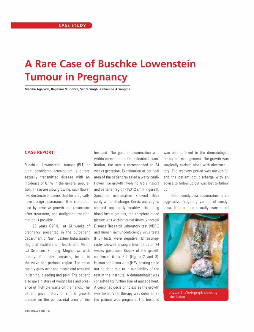
JPOG JANUARY 2013 • 30
CASE STUDY
CASE REPORT
Buschke Lowenstein tumour (BLT) or
giant condyloma acuminatum is a rare
sexually transmitted disease with an
incidence of 0.1% in the general popula-
tion. These are slow growing, cauliflower
like destructive lesions that histologically
have benign appearance. It is character-
ised by invasive growth and recurrence
after treatment, and malignant transfor-
mation is possible.
22 years G2P1L1 at 24 weeks of
pregnancy presented in the outpatient
department of North Eastern India Gandhi
Regional Institute of Health and Medi-
cal Sciences, Shillong, Meghalaya, with
history of rapidly increasing lesion in
the vulva and perianal region. The mass
rapidly grew over one month and resulted
in itching, bleeding and pain. The patient
also gave history of weight loss and pres-
ence of multiple warts on the hands. The
patient gave history of similar growth
present on the penoscrotal area of the
husband. The general examination was
within normal limits. On abdominal exam-
ination, the uterus corresponded to 24
weeks gestation. Examination of perineal
area of the patient revealed a warty cauli-
flower like growth involving labia majora
and perianal region (15X12 cm2) (Figure1).
Speculum examination showed thick
curdy white discharge. Cervix and vagina
seemed apparently healthy. On doing
blood investigations, the complete blood
picture was within normal limits. Venereal
Disease Research Laboratory test (VDRL)
and human immunodeficiency virus tests
(HIV) tests were negative. Ultrasonog-
raphy showed a single live foetus of 24
weeks gestation. Biopsy of the growth
confirmed it as BLT (Figure 2 and 3).
Human papilloma virus (HPV) testing could
not be done due to in availability of the
test in the institute. A dermatologist was
consulted for further line of management.
A combined decision to excise the growth
was taken. Viral therapy was deferred as
the patient was pregnant. The husband
was also referred to the dermatologist
for further management. The growth was
surgically excised along with electrocau-
tery. The recovery period was uneventful
and the patient got discharge with an
advice to follow up but was lost to follow
up.
Giant condyloma acuminatum is an
aggressive fungating variant of condy-
loma. It is a rare sexually transmitted
A Rare Case of Buschke Lowenstein Tumour in PregnancyManika Agarwal, Rajlaxmi Mundhra, Santa Singh, Kalkambe A Sangma
Figure 1. Photograph showing the lesion

CASE STUDY
JPOG JANUARY 2013 • 31
CASE STUDY
disease, triggered by HPV, usually geno-
type 6 or 11.1 It can be associated with
congenital or acquired immunodeficiency
including alcoholism, diabetes or immu-
nosuppressive therapy.2
Giant condyloma acuminatum pres-
ents with 2.7:1 male to female ratio. For
patients younger than 50 years, this ratio
is increased to 3.5:1. The most common
symptoms are perianal mass (47%), pain
(32%), abscess or fistula (32%) and bleed-
ing (18%).3
Chu et al analysed 42 known cases
of giant condyloma acuminatum in English
literature and reviewed their behaviour
and management. They concluded it as
highly aggressive tumour with propensity
for recurrence and malignant transforma-
tion but without metastatic potential.4
Surgery is considered the treatment
of choice for this disease.5 Complete exci-
sion is the preferred initial therapy when
feasible. Wide local excision, faecal
diversion or abdominoperineal resection
have been used. Chemotherapy with 5
fluorouracil and focused radiation therapy
may be used in certain cases of recur-
rence or extensive pelvic disease.
For unknown reasons, genital warts
frequently increase in number and size
during pregnancy. Acceleration of viral
replication by the physiological changes
of pregnancy might explain the growth of
perineal lesions and progression of some
to cervical neoplasm.6 Since our patient
was pregnant so probably the genital wart
grew into gigantic size. These lesions
sometimes grow to full vagina or cover
perineum, thus making vaginal delivery or
episiotomy difficult. Therapy in pregnancy
is mainly directed towards debulking
symptomatic large growths.
Giant condyloma acuminatum or BLT
of anorectal region and perineal region
is an uncommon entity that has not been
extensively reviewed hence more multi-
institutional studies are necessary to
further elaborate the nature and treat-
ment of this rare disease.
AcknowledgementI would like to thank Professor Vandana Raphel, Depart-ment of Pathology, NEIGRIHMS, Shillong for providing histopathological pictures of Condylomata acuminatum and Dr. Sharat Agarwal, Department of Orthopaedics and Trauma, NEIGRIHMS, Shillong for providing the help in preparing this manuscript.
About the AuthorsDr Manika Agarwal, Dr Rajlaxmi Mundhra, Dr Santa Singh is MD, Department of Gynaecology and Obstetrics and Dr Kalkambe A Sangma is a MD, Department Dermatol-ogy, North Eastern India Gandhi Regional Institute of Health and Medical Sciences, Shillong, Meghalaya.
REFERENCES
1. Buschke A, Lowenstein L. Uber carcinomahnliche condylomata acuminata des Penis. Klin Wochenschr 1925;4:1726−1728.2. Gillard P, Vanhooteghem O, Richert B, De La Brasine M. Tumour de Buschke-Lowenstein. Ann Dermatol Vene-reol. 2005;132:98−99.3. Trombetta LJ , Place RJ. Giant condyloma acuminatum of the anorectum trends in epidemiology and manage-ment: report of case and review of the literature. Dis Colon Rectum. 2001;44(12):1878−1886.4. Chu QD, Vezeridis MP, Libbey NP, Wanebo HJ. Giant condyloma acuminatum (Buschke – Lowenstein tumor)
of the anorectal and perineal region. Dis Colon Rectum 1994;37(9):950−957.5. Fernandez- Sanchez M, Espinosa–de La Monteros A, Saeb- Lima M, Vergara-Fernandez D. Surgical treat-ment of a perianal giant condyloma acuminate in a HIV patient . Rev Gastroenterol Mex 2011;76(2):178−181.6. Cunningham FG, Kenneth JL, Steven LB, John CH, Dwight JR, Catherine YS. Sexually Transmitted Diseases. In: Alyssa F, Karen D, editors. Williams Obstetrics. 23rd edition. USA:Mc Graw-Hill Medical.2010.pg 1245.
Figure 2. Condyloma acuminata showing complex papillae of stratifi ed squamous epithelium with fi bro-vascular core. (Hemat- oxylin & Eosin Stain, 40x)
Figure 3. Condyloma acuminata showing papillae of stratifi ed squamous epithelium with koilocytic changes. (Hematoxylin & Eosin Stain, 100x)

JPOG JANUARY 2013 • 32
INTRODUCTION
Placental insufficiency leading to intrau-
terine growth restriction (IUGR) in the
fetus affects up to 15% of pregnancies.1
IUGR can present diagnostic and manage-
ment challenges. The suspicion of IUGR
arises when fetal biometry indicates
small for gestational age (SGA), vari-
ously defined as abdominal circumference
below 2 standard deviations, abdominal
circumference at the 5th or 10th percen-
tile, or estimated fetal weight less than
the 10th percentile.2 SGA fetuses repre-
sent a heterogeneous group comprising
constitutional smallness and pathologi-
cal smallness. When pathologies, such
as chromosomal aberration and genetic
syndrome, are excluded, placental insuf-
ficiency is implied.
Management challenge lies in
timely intervention especially when IUGR
complicates a preterm pregnancy. On the
one hand, the obstetrician wants to avoid
unnecessary preterm delivery, which puts
the neonate at risk for complications of
prematurity such as cerebral palsy, neona-
tal death, respiratory distress, necrotiz-
ing enterocolitis, and intraventricular
haemorrhage. On the other hand, there
is the burden of possible in utero fetal
demise from delaying delivering. Surveil-
Intrauterine fetal growth restriction due to placental insufficiency affects up to 15% of pregnancies.
lance tools, such as electronic fetal heart
monitoring3 and Doppler velocimetry,4,5
have been evaluated for their predictive
strength of adverse perinatal outcomes
which may guide decision regarding timing
of delivery.
A synopsis of fetal growth regulation
and fetal responses to placental insuf-
ficiency will first be presented. This is
followed by a discussion on Doppler surveil-
lance, with attention to clinical signifi-
cance of the various arterial and venous
waveforms. Finally, a suggested approach
to management of IUGR is presented.
REGULATION OF FETAL GROWTH
The placenta acts as a conduit for trans-
fer of nutrients and oxygen from mother
Intrauterine Foetal Growth RestrictionMay Li Lim, MBBS, MRCOG, FRANZCOG; Kenneth Kwek, MBBS, MRCOG, MRANZCOG; Kok Hian Tan, MBBS, FRCOG, MMed, FAMS; George SH Yeo, MBBS, FRCOG, FAMS

Continuing Medical Education
JPOG JANUARY 2013 • 33
to fetus. Adequate substrate delivery
depends on normal uterine perfusion,
normal fetoplacental exchange area, and
normal umbilical–placental perfusion. The
placenta, being a metabolically active
compartment, utilizes approximately 70%
of the glucose and 45% of the oxygen being
transferred.6 Placental growth follows a
sigmoid-shaped curve, while fetal growth
is exponential at 1.5% per day to term.7
The development of the matched fetopla-
cental unit from early pregnancy is criti-
cal in ensuring optimal fetal growth and
development throughout pregnancy.
Following fertilization, cytotrophob-
last migration occurs, leading to the estab-
lishment of placental adherence through
anchoring the villi to the decidua and
uterus. The process is complete when a
low-resistance, high-capacitance placen-
tal compartment is achieved. Nutrient-
rich blood entering the fetal circulation is
delivered via the umbilical vein to the heart
and liver. The ductus venosus (DV) acts as
a shunt to direct a larger proportion of that
blood to the liver and the remaining to the
heart. Differential directionality of blood
entering the right atrium ensures that
well-oxygenated blood is distributed to
the left ventricle, myocardium and brain.
Low-nutrient venous return is forwarded
to the placenta for re-oxygenation and
nutrient-waste exchange via the umbili-
cal arteries. Through autoregulation, vari-
ous organs modify blood flow to meet the
oxygen and nutrient demands.6,8
FETAL RESPONSES TO PLACENTAL INSUFFICIENCY
In placental insufficiency, fetal responses
are seen in various systems. Metaboli-
cally, fetal hypoglycaemia leads to blunt-
ing of pancreatic insulin responses, which
in turn allows hepatic gluconeogenesis.
As the situation worsens, there is inabil-
ity to maintain oxidative metabolism. The
ensuing anaerobic metabolism leads to
the production of lactate, which is metab-
olized by the liver and used as alternative
substrate by fetal heart and brain. Acid-
base balance will be maintained as long
as fetal haemoglobin buffering capacity
is sufficient with a matching removal rate
by the liver, heart and brain.6 To maintain
oxygenation of the vital organs such as
the heart and brain, there is preferential
shunting of blood from the umbilical vein
through the DV. To facilitate preferential
distribution of cardiac output to the left
ventricle and thus coronary circulation and
brain, there is an increase in peripheral
vascular and placental blood flow resist-
ance (increased right ventricular afterload)
and a decline in cerebral flow resistance
(decreased left ventricular afterload).
Fetal growth is exponential at 1.5% per day to term whereas placental growth follows a sigmoid-shaped curve.

JPOG JANUARY 2013 • 34
Reduced liver perfusion from the increased
shunting through the DV to the heart leads
to the accumulation of blood lactate, thus
acidaemia. As hypoxaemia and acidaemia
worsen, the metabolic demands of the
heart can no longer be maintained and
myocardial dysfunction supervenes. With
declining cardiac function, there is failure
to accommodate the venous return and so
the central venous pressure rises. Progres-
sively, forward cardiac function fails. As
cardiac dysfunction reaches a critical
state, there is cardiac dilatation, indica-
tive of loss of cardiovascular homeosta-
sis.9 Typically, this starts in the right heart,
manifesting as dilated tricuspid annulus,
tricuspid regurgitation and reversed
a wave in the DV flow. Characteristic
responses are seen in the fetal behav-
iour when there is hypoxaemia. There is
delayed central nervous system matura-
tion and delayed central integration with
the fetal heart, resulting in reduced short-
and long-term fetal heart variation. As the
hypoxaemia worsens, fetal activity slows
down and fetal heart decelerations are
observed.8
FETAL SURVEILLANCE
There are multiple methods of fetal
assessment in IUGR. The biophysical
profile score, which incorporates fetal
tone, fetal movement, fetal breathing,
amniotic fluid volume and non-stress
test, has been shown to predict fetal
status and to produce improvement in
outcomes when employed in high-risk
pregnancies.10 Doppler ultrasound, which
provides information about placental
vascular resistance,11,12 preferential
cerebral blood flow13 and filling capac-
ity of fetal heart,14,15 allows the tracking
of progression of placental insufficiency.
The evidence for Doppler velocimetry in
clinical management has been evaluated
more thoroughly and more systematically
than evidence for any other techniques
in modern obstetrics. Doppler has been
applied to various vessels including umbil-
ical artery (UA), middle cerebral artery
(MCA), uterine artery, DV, aorta (isthmus)
and umbilical vein to predict placental
dysfunction, to assess fetal cardiovascu-
lar status in response to hypoxia, and to
predict adverse perinatal outcomes. Cardi-
otocography of the fetal heart also plays
an important role in assessing the fetal
condition in IUGR pregnancies, although it
is considered a late-stage disease when
there is abnormality such as deceleration
in the tracing.16
UA velocimetry gives information
about placental function. There is abun-
dant data linking abnormal UA Doppler
and adverse outcomes in IUGR pregnan-
cies5,17,18 This is hardly surprising because
Doppler ultrasound is the mainstay of fetal surveillance in pregnancies with IUGR.
In placental insufficiency,
fetal responses are seen
in various systems

Continuing Medical Education
JPOG JANUARY 2013 • 35
histological analysis has confirmed
placental vascular pathology in the pres-
ence of abnormal UA velocimetry, such
as reduction in volume of the intervillous
space, reduction in the elaboration of
distal villi, smaller exchange surface area,
and thicker trophoblastic epithelium.12 The
categories of abnormal UA waveforms—
increased pulsatility index (PI), absent
end-diastolic flow (AEDF) or reversed end-
diastolic flow (REDF)—represent progres-
sively worsening placental dysfunction.
Hence, fetuses can be assigned varying
degrees of risk according to these wave-
forms. The risk stratification can help
inform the obstetrician on the level of
fetal monitoring required.
McCowan et al found raised UA PI
to be associated with fetuses of smaller
proportions in terms of birthweight, head
circumference, and length.19 The utiliza-
tion of UA PI for predicting adverse peri-
natal outcome has been widely practised
for many years. The sensitivity and specif-
icity for any adverse outcome have been
reported to be 44.7% and 86.6%, respec-
tively.18 Comparing pregnancies with posi-
tive end-diastolic flow, those with AEDF or
REDF have been shown to be associated
with lower birthweight, earlier gestation
at birth, and higher frequency of severe
respiratory distress syndrome, cerebral
haemorrhage, hypoglycaemia and anae-
mia.20 The temporal changes on Doppler
ultrasound in IUGR are well documented.
Pregnancies with raised UA PI have been
seen to sequentially deteriorate to UA
AEDF and UA REDF.21 Perinatal mortality
rates of up to 28% are reported in such
cases.20 It is generally accepted that the
finding of these waveforms indicates that
the condition is severe and indeed should
prompt the obstetrician to re-assess
management, in particular, to make a deci-
sion regarding delivery.
Decision on the timing of delivery
should be considered with gestational
age in mind. Baschat et al investigated
the outcomes of fetuses with abnormal
Doppler velocimetry in arterial and venous
circulations.16 The outcome parameters
evaluated were perinatal mortality, respi-
ratory distress syndrome, bronchopulmo-
nary dysplasia, necrotizing enterocolitis,
intraventricular haemorrhage, and circula-
tory insufficiency. Although fetuses with
abnormal venous circulation generally
fared the worst, gestational age at deliv-
ery was the most significant contributor
to short-term outcomes after multivariate
analysis. This effect was most marked for
respiratory distress syndrome and least
for intraventricular haemorrhage and
necrotizing enterocolitis.16 In following
the progression of fetuses with UA AEDF
or UA REDF, Hartung et al also concluded
that while perinatal mortality was related
to abnormal UA velocimetry, gestational
age at delivery had a significant impact on
short-term morbidity.22 One recommenda-
tion by Hartung et al is that delivery should
be considered if the pregnancy is greater
than 32 weeks’ gestation as morbidity is
low.22 In the ascertainment of gestation-
specific survival and complication rates,
Baschat et al showed that the median
survival gained per day in utero was 2%
between 24 and 27 weeks’ gestation. With
more advanced gestation, major morbidity
fell from 56.6% at 24 weeks to 10.5% at
32 weeks, leading to significant neonatal
survival and intact survival.4
‘Brain-sparing’ is a compensatory
fetal response seen in placental insuf-
ficiency whereby blood is preferentially
distributed to cerebral structures by
autoregulation to maintain delivery of
oxygen and nutrients. MCA is the vessel
of choice because it is the most accessi-
ble and there is a high degree of observer
agreement in terms of PI measurement.
MCA velocimetry is expressed as either
MCA PI or a ratio such as MCA/UA PI
(cerebroplacental ratio [CPR]). CPR is
thought to be a better index because it
allows detection of blood flow redistribu-
tion from two potential mechanisms. As
well as detecting decreased cerebral blood
flow resistance due to brain-sparing, CPR
may be the first sign of elevated placen-
tal blood flow resistance.23 Longitudinal
studies have shown brain-sparing to be an
early phenomenon in placental dysfunc-
Although fetuses with
abnormal venous
circulation generally fared
the worst, gestational age
at delivery was the most
significant contributor to
short-term outcomes after
multivariate analysis

JPOG JANUARY 2013 • 36
tion. Persistent dilation of MCA was seen
in > 50% of fetuses 2 to 3 weeks before
any fetal heart tracing abnormality.1,24 The
consensus that cerebral redistribution
is a marker for early hypoxaemia is also
supported by findings of Dubiel et al who
demonstrated that fetuses with brain-
sparing effect still had reserves to cope
with the stress of normal labour.25
In IUGR, understanding of the vascu-
lar dynamics is enhanced by the incor-
poration of venous Doppler on vessels
such as umbilical vein, DV, inferior vena
cava, and portal vein. Abnormal DV
velocimetry has been shown to correlate
with acidaemia at cordocentesis26 and a
significant predictor of perinatal morbidity
and mortality.4 Longitudinal studies have
demonstrated abnormal DV to be a sign
of advanced stages of fetal compromise
and be indicative of the need for delivery
within the subsequent 7 days.1,24 Bilardo
and colleagues showed that at 2–7 days
before delivery, abnormal DV was present
in 79% of fetuses with adverse outcome.
On the day before delivery, this increased
to 93%.5 The significance of DV velocime-
try is supported by the work of Baschat et
al. Having corrected for birthweight and
gestation at delivery, DV velocity was the
only statistically significant predictor of
intact survival of IUGR babies.4
Doppler velocimetry at the level of
aortic isthmus has been evaluated as a
monitoring tool in IUGR. The isthmus is
the area between the origin of the left
subclavian artery (perfused by the left
ventricle) and aortic end of the ductus
arteriosus (perfused by the right ventricle).
Its unique position means that it is influ-
Late-onset IUGR is associated with a lesser degree of placental vascular abnormality and hence the lack of abnormal umbilical artery Doppler findings.
enced by both downstream impedance in
the lower body and that of the upper body
especially the brain. Waveform abnormali-
ties are classified as decreased, absent or
reversed flow. Reversed flow through the
aortic isthmus results in low-oxygen blood
from the right heart being directed to the
brain. This has been shown to be associ-
ated with long-term neurodevelopmental
impairment. The current thinking is that
aortic isthmus flow velocimetry has a low
sensitivity although it appears to be highly
specific as a sign of decompensation.27,28
There is increasing recognition of the
entity of late-onset IUGR. In contrast to
early-onset IUGR, late-onset IUGR is asso-
ciated with a lesser degree of placental
vascular abnormality and hence the lack
of abnormal umbilical artery Doppler find-
ings. The progressive cardiovascular dete-
rioration observed in early-onset IUGR is
also not seen. Oros et al monitored the
longitudinal Doppler changes in a cohort
of fetuses with suspected IUGR diagnosed
at a mean gestation of 34.1 weeks until
delivery at 38.7 weeks.29 They found no
significant difference in UA PI. There was,
however, progressive decrease in MCA PI
and CPR until delivery.29 The utility of MCA
Doppler as a prognostic indicator in late-
gestation IUGR is supported by findings
from studies showing abnormal neurobe-

Continuing Medical Education
JPOG JANUARY 2013 • 37
havioural performance in the neonatal
period and early childhood in fetuses with
abnormal MCA Doppler antenatally.30,31,32
SUGGESTED APPROACH TO MANAGEMENT
A suggested management approach is
as described in the Figure. It is useful to
bear in mind the time frame of progres-
sion. In early-onset IUGR, the anticipated
progression rate is defined by the rate
of progression of UA Doppler. Loss of
UA end-diastolic flow within 2 weeks is
suggestive of a rapidly progressive picture
with venous flow abnormality within the
subsequent 4 weeks. If, however, loss of
UA end-diastolic flow is more gradual over
4 weeks, there will be a longer latency
period of about 6 weeks to venous dete-
rioration.21 Delivery threshold needs to
be high if gestational age is less than 26
weeks owing to a high chance of mortal-
ity. Up until 28 weeks, each day in utero
potentially increases survival by 2% per
day. Abnormal DV is the most significant
predictor of neonatal mortality.
Figure. A suggested management approach for intrauterine fetal growth restriction.
Small for gestational age
Confirm dates correct
Exclude aneuploidy, infection
Is maternal status at risk
Is umbilical artery Doppler normal?
Is ductus venosusDoppler normal?
Continual discussion with coupleNeonatal counselling
Consideration to gestational ageOptions
-Expectant with risk of IUD-Steroids and deliver
Deliver
1-weekly Doppler2-weekly growth scan
Repeat daily
Is CTG normal?
Is umbilical artery
Doppler normal?
Repeat every 1 week
Deliver
Give steroidsthen deliver
< 28 weeks > 28 weeks
No
No
Yes
Yes
Yes
Yes
Yes
No
No
No
CTG = cardiotocograph; IUD = intrauterine fetal demise.

REFERENCES
1. Ferrazzi E, Bozzo M, Rigano S, et al. Temporal sequence of abnormal Doppler changes in the peripheral and central circulatory systems of the severely growth-restricted fetus. Ultra-sound Obstet Gynecol 2002;19:140–146.
2. Miller J, Turan S, Baschat A. Fetal growth restriction. Semin Perinatol 2008;32:274–280.
3. Torrance HL, Bloemen MCT, Mulder EJH, et al. Predictors of outcome at 2 years of age after early intrauterine growth restriction. Ultrasound Obstet Gynecol 2010;36:171–177.
4. Baschat A, Cosmi E, Bilardo CM, et al. Predictors of neonatal outcome in early-onset placental dysfunction. Obstet Gynecol 2007;109:253–261.
5. Bilardo CM, Wolf H, Stigter RH, et al. Rela-tionship between monitoring parameters and perinatal outcome in severe, early intrauterine growth restriction. Ultrasound Obstet Gynecol 2004;23:119–125.
6. Baschat A. Fetal responses to placental insuf-ficiency: an update. BJOG 2004;111:1031–1041.
7. Molteni RA, Stys SJ, Battaglia FC. Relation-ship of fetal and placental weight in human beings: fetal/placental weight ratios at various gestational ages and birth weight distributions. J Reprod Med 1978;21:327–334.
8. Martin CB Jr. Normal fetal physiology and behavior, and adaptive responses with hypox-emia. Semin Perinatol 2008;32:239–242.
9. Baschat A. Venous Doppler evaluation of the growth-restricted fetus. Clin Perinatol 2011;38:103–112.
10. Oyelese Y, Vintzileos AM. The uses and
limitations of the fetal biophysical profile. Clin Perinatol 2011;38:47–64.
11. Maulik D, Mundy D, Heitmann E, Maulik D. Umbilical artery Doppler in the assess-ment of fetal growth restriction. Clin Perinatol 2011;38:65–82.
12. Giles WB, Trudinger BJ, Baird PJ. Fetal umbilical artery flow velocity waveforms and placental resistance: pathological correlation. Br J Obstet Gynaecol 1985;92:31–38.
13. Mari G, Deter R. Middle cerebral artery flow velocity waveforms in normal and small-for-gestational age fetuses. Am J Obstet Gynecol 1992;166:1262–1270.
14. Kiserud T, Kessler J, Ebbing C, Rasmussen S. Ductus venosus shunting in growth-restricted fetuses and the effect of umbilical circula-tory compromise. Ultrasound Obstet Gynecol 2006;28:143–149.
15. Gudmundsson S, Tulzer G, Huhta JC, Marsal K. Venous Doppler in the fetus with absent end-diastolic flow in the umbilical artery. Ultrasound Obstet Gynecol 1996;7:262–267.
16. Baschat A, Gembruch U, Reiss I, Gortner L, Weiner CP, Harman CR. Relationship between arterial and venous Doppler and perinatal outcome in fetal growth restriction. Ultrasound Obstet Gynecol 2000;16:407–413.
17. Trudinger BJ, Cook CM, Giles WB, et al. Fetal umbilical artery velocity waveforms and subsequent neonatal outcome. Br J Obstet Gynaecol 1991;98:378–384.
18. Fong KW, Ohlsson A, Hannah ME, et al. Prediction of perinatal outcome in fetuses
suspected to have intrauterine growth restric-tion: Doppler US study of fetal cerebral, renal and umbilical arteries. Radiology 1999;213:681–689.
19. McCowan LME, Harding JE, Stewart AW. Umbilical artery Doppler studies in small for gestational age babies reflect disease severity. BJOG 2000;107:916–925.
20. Karsdorp VHM, van Vugt JM, van Geijn HP, et al. Clinical significance of absent or reversed end diastolic velocity waveforms in umbilical artery. Lancet 1994;344:1664–1668.
21. Turan OM, Turan S, Gungor S, et al. Progres-sion of Doppler abnormalities in intrauterine growth restriction. Ultrasound Obstet Gynecol 2008;32:160–167.
22. Hartung J, Kalache KD, Heyna C, et al. Outcome of 60 neonates who had ARED flow prenatally compared with a matched control group of appropriate-for-gestational age preterm neonates. Ultrasound Obstet Gynecol 2005;25:566–572.
23. Schenone MH, Mari G. The MCA Doppler and its role in the evaluation of fetal anemia and fetal growth restriction. Clin Perinatol 2011;38:83–102.
24. Hecher K, Bilardo CM, Stigter RH, et al. Monitoring of fetuses with intrauterine growth restriction: a longitudinal study. Ultrasound Obstet Gynecol 2001;18:564–570.
25. Dubiel M, Gudmundsson S, Gunnarsson G, Marsal K. Middle cerebral artery velocimetry as a predictor of hypoxemia in fetuses with increased resistance to blood flow in the umbili-cal artery. Early Hum Dev 1997;47:177–184.
26. Pardi G, Cetin I, Marconi AM, et al. Diag-nostic value of blood sampling in fetuses with growth retardation. N Engl J Med 1993;328:692–696.
27. Fouron JC, Gosselin J, Raboisson MJ, et al. The relationship between an aortic isthmus blood flow velocity index and the postnatal neurodevelopmental status of fetuses with placental circulatory insufficiency. Am J Obstet Gynecol 2005;192:497–503.
28. Tideman E, Marsal K, Ley D. Cognitive function in young adult following intrauter-ine growth restriction with abnormal fetal aortic blood flow. Ultrasound Obstet Gynecol 2007;29:614–618.
29. Oros D, Figueras F, Cruz-Martinez R, Meler E, Munmany M, Gratacos E. Longitudinal changes in uterine, umbilical and fetal cerebral Doppler indices in late-onset small-for-gesta-tional age fetuses. Ultrasound Obstet Gynecol 2011;37:191–195.
30. Eixarch E, Meler E, Iraola A, et al. Neurode-velopmental outcome in 2-year-old infants who were small-for-gestational age term fetuses with cerebral blood flow redistribution. Ultra-sound Obstet Gynecol 2008;32:894–899.
31. Cruz-Martinez R, Figueras F, Oros D, et al. Cerebral blood perfusion and neurobe-havioural performance in full-term small-for-gestational-age fetuses. Am J Obstet Gynecol 2009;201:474.e1–7.
32. Figueras F, Oros D, Cruz-Martinez R, et al. Neurobehavior in term, small-for-gestational age infants with normal placental function. Pediatrics 2009;124:e934–e941.
Acknowledgement
This paper was made possible through a collaboration between KK Women’s and Children’s Hospital (KKH) and the Journal of Paedi-atrics, Obstetrics and Gynaecology. KKH is the largest medical facility in Singapore which provides specialized care for women, babies and children.
In late-gestation IUGR, there is no
good evidence to guide the best manage-
ment. The clinician needs to balance the
risk of intrauterine fetal demise from not
delivering and the risk of delivery by either
labour induction or caesarean section. The
choice of delivery to avoid stillbirth is,
however, considered rational.
CONCLUSION
IUGR is associated with significant risk
of mortality and morbidity, which extends
into adult life. Doppler ultrasound is the
mainstay of fetal surveillance in these
pregnancies. There are sequential changes
in Doppler velocimetry, which can provide
clues to worsening of condition and hence
guide decision-making regarding delivery.
About the AuthorsDr Lim is Consultant, Dr Kwek is Head and Senior Consul-tant, Dr Tan is Clinical Associate Professor and Senior Consultant, and Dr Yeo is Chief of Obstetrics, Adjunct Professor and Senior Consultant, Department of Mater-nal Fetal Medicine, KK Women’s and Children’s Hospital, Singapore.