Embed Size (px)
Citation preview
Journal of the Egyptian Society of Parasitology, Vol. 51, No. 1, April 2020 J. Egypt. Soc. Parasitol. (JESP), 51(1), 2021: 213 - 226 (Online: 2090-2549)
213
IN VITRO EVALUATION OF THIOREDOXIN REDUCTASE INHIBITOR (AURANOFIN) ACTIVITY IN COMPARISON WITH TRICLABENDAZOLE
ON ADULT FASCIOLA GIGANTICA By
AMANY A. RADY1, NASHAAT E. NASSEF1, OMAIMA K. EL-SHAFEY1, ENGY V. BESHAY1*, SHEREEN F. MAHMOUD2, DOAA I. M. ABOUGALALAH1,
and SAMAR A. EL- REFAI1 Departments of Medical Parasitology1, and Pathology2, Faculty of Medicine,
Menoufia University, Egypt (*Correspondence: [email protected] ORCID: https://orcid.org/0000-0002-3556-2156)
Abstract Fasciola gigantica causes a worldwide waterborne/foodborne zoonotic disease in which hu-
mans are incidental hosts. Fascioliasis has a major impact on human health and its control
mainly depends on triclabendazole (TCBZ). Unfortunately, the effectiveness of this drug is de-
creased because of indiscriminate use resulting in development of resistance. Therefore, the
search for another effective anthelmintic is now compulsory. This work aimed to evaluate
the in vitro anthelmintic effects of auranofin (a thioredoxin reductase inhibitor) on adult F. gi-
gantica in comparison with the drug of choice; TCBZ. This study involved in vitro petri dish
incubation of seventy-five adult F. gigantica worms of nearly equal size with the tested drugs
and classified into five groups (fifteen worms each) as follows; G1 served as a control group,
G2 was exposed to TCBZ (20μL/mL), G3, G4, & G5 were exposed to auranofin (3, 5, &
10μg/mL, respectively). All adult worms were incubated and observed for three hours and sub-
jected to motility and egg hatchability assays, histopathological and ultrastructural studies, glu-
tathione-S-transferase and superoxide dismutase assay, and cathepsin-L gene expression anal-
ysis. Auranofin in all concentrations significantly decreased adult motility and egg hatchabil-
ity. It induced histopathological and ultrastructural deformities including apoptosis. Auranofin
in higher concentrations significantly suppressed the activity of the detoxifying enzyme; gluta-
thione-S-transferase, and significantly stimulated superoxide dismutase enzyme activity re-
flecting the oxidative stress. At all concentrations, it suppressed the expression of the cathep-
sin-L gene responsible for Fasciola invasive function.
Key words: Auranofin®, Fasciola gigantica; In-vitro, Triclabendazole
®.
Introduction
Fasciola hepatica (F. hepatica) and Fasc-
iola gigantica (F. gigantica) are two parasite
species that cause a foodborne zoonotic dis-
ease named fascioliasis (Rokni et al, 2018).
Fascioliasis is an emerging/re-emerging tre-
matodal disease in many countries with maj-
or impact on human health and development
(Mas-Coma et al, 2005), including Egypt
(Abo-Madyan et al, 2004). Animal and hu-
man fascioliases constitute major economic
and health problems in tropical and subtrop-
ical areas (Sarkari et al, 2017). Epidemio-
logical data proved that fascioliasis endemic
areas are expanding (Sarkari and Khabisi,
2017). The disease causes hepatic lesions
and chronic inflammation as well as fibrosis
of bile ducts (Ashrafi, 2015) with the devel-
opment of gall bladder stones (Mas-Coma et
al, 2019). It is also associated with helmi-
nth-induced hepatic cancer (Machicado and
Marcos, 2016). Besides, fascioliasis caused
ectopic lesions elsewhere with tissue da-ma-
ge and fibrosis (Taghipour et al, 2019). The
neurological and ocular disorders were rec-
orded in hepatic fascioliasis patients (Gonza-
lez-Miguel et al, 2019).
The control of fascioliasis relies on an an-
thelmintic drug, triclabendazole (TCBZ), a
member of benzimidazole derivatives (Fair-
weather, 2009). This drug is the most effec-
tive treatment for fascioliasis in humans and
animals. It penetrates the worm tegument by
diffusion, then, becomes metabolized by the
worm into an active sulfoxide metabolite
that binds to β-tubulin inhibiting the for-
214
mation of microtubules which are compo-
nents of the Fasciola cytoskeleton. The re-
sistance of TCBZ is recently reported in
humans and animals in many regions of the
world due to reduced diffusion and metabo-
lism with a change in efflux activity that re-
duces its efficacy (Merachew and Alemneh,
2020). Therefore, alternative effective fasci-
oliasis treatment became the research targets
(Marques et al, 2020). Among these targets,
cathepsin-L cysteine proteases, glutathione-
S-transferases (GST), superoxide dismutases
(SOD), and thioredoxin reductases (TrxR)
were recommended (Saccoccia et al, 2014).
These enzymes have different vital functions
in Fasciola worms including host tissue
penetration, worm survival and virulence,
detoxification reactions, and antioxidant de-
fense mechanisms (Ullah et al, 2017).
Redox enzymes are important in control-
ling the intracellular levels of reactive oxy-
gen species which are essential for the pre-
vention of DNA damage with subsequent
survival of all cell types, including parasites.
Auranofin is an FDA-approved treatment for
rheumatoid arthritis with a well-studied saf-
ety for humans (AbdelKhalek et al, 2018).
Auranofin acts mainly through the inhibition
of reduction/oxidation (Redox) enzymes as
thioredoxin reductase. The cells which ex-
press redox enzymes show an increase of
auranofin affinity towards them (Caroli et al,
2011; Tejman-Yarden et al, 2013). Inhibi-
tion of redox enzymes by auranofin chang-
es the redox state of the cell with increased
production of reactive oxygen species, hyd-
rogen peroxide, and oxidative stress causing
intrinsic apoptosis of affected cells (Roder
and Thomson, 2015).
Fortunately, the auranofin proved effective
against different parasites in previous studies
(Tejman-Yarden et al, 2013; da Silva et al,
2015; Capparelli et al, 2016). However, to
the best of the authors’ knowledge, it was
not tested before against adult F. gigantica.
This study aimed to evaluate in-vitro anth-
elmintic effects of different concentrations
of Auranofin® on F. gigantica adults as co-
mpared with the on drug TCBZ®.
Materials and Methods Fasciola worms: F. gigantica adult worms
were obtained from infected livers and bile
ducts of buffaloes slaughtered at El Bagur
Governmental Abattoir, Menoufia Governo-
rate. The liver was cut over the bile duct and
the flukes were collected in a laminar flow
cabinet under sterile conditions using non-
traumatic thumb forceps. Worms were put in
a warm 0.9% NaCl (37°C). They were clea-
ned with phosphate buffer saline (PBS) in
small sieves & placed in sterile RPMI-1640
medium (supplemented with (100µl/mL) Pe-
nicillin®, (160µ/mL) Gentamycin
®, (100µl/
mL) Streptomycin® and (30%) fetal calf ser-
um (Diab et al, 2010).
Tested drugs: Triclabendazole® was provi-
ded by Egaten, Novartis Pharma AG (USA).
Auranofin® was provided by Tocris Bio-Te-
chno Brand (USA) as a stock solution, and
was freshly prepared by dissolving 1mg aur-
anofin in 100ml dimethylsulfoxide (DMSO).
Experimental design: Anthelmintic effects
of the tested drugs were studied by in vitro
petri dish incubation method using RPMI-
1640 medium (Githiori et al, 2006). Seven-
ty-five adult F. gigantica worms of nearly
equal size were selected and classified into
five groups (fifteen worms each) as follows:
G1: Non-drug-exposed worms (Control).
G2: Worms were exposed to TCBZ at a con-
centration of 20μL/mL (TCBZ-exposed) aft-
er Nassef et al. (2014). G3: Worms were ex-
posed to auranofin at a concentration of 3μg/
ml (Aur 3μg/ml) after Song et al. (2012).
G4: Worms were exposed to auranofin at a
concentration of 5μg/ml (Aur 5μg/ml). G5:
Worms exposed to auranofin at a concentra-
tion of 10μg/ml (Aur 10μg/ml). All groups
were incubated for 3hrs at room temperat-
ure. Tested drugs were evaluated through
motility assay, egg hatchability test, histopa-
thological study for pathological and apop-
totic changes, ultrastructural scanning and
transmission electron microscopic studies,
evaluation of the antioxidant state by GST
and SOD assays, and finally, determination
215
of cathepsin-L gene expression by real-time
PCR (RT-PCR).
Parasitological study: a- Motility assay:
Motility of Fasciola adults of all groups was
recorded for 3hrs post-incubation at half an
hour intervals (Ullah et al, 2017). The motil-
ity was scored into the following (Jiraungk-
oorskul et al, 2005) score 3: Moving whole
body, score 2: Moving only parts of body
score 1: Grossly immobile but microscopic-
ally alive, and score 0: Microscopically de-
ad. b- Egg hatching study: Eggs of each
group were collected, washed five times
with dechlorinated water and then incubated
in 50ml tap water at 25ºC in the darkness for
15 days. Exposure to day light for 2hr were
done to test egg hatchability, where percent-
age of hatched and unhatched eggs were as-
sessed using dissecting microscope (Hanna
et al, 2006)
Histopathological study: From each group,
five worms were randomly picked and as-
signed for thistopathological studies. a- He-
matoxylin and Eosin (H&E) staining: Adults
from each group were fixed in 10% formal-
dehyde, dehydrated in ascending grades of
ethanol, cleared with xylene, and then em-
bedded in paraffin, for 5µm longitudinal sec-
tions to be stained in H&E stained (Carleton
et al, 1980). They were examined and pho-
tographed under an Olympus CX41 light
microscope at Pathology Department, Facul-
ty of Medicine, Menoufia University. b- Im-
munohistochemical (IHC) staining for casp-
ase- 3: Serial sections (4μm) were cut from
paraffin-embedded flukes of all groups and
mounted on positive slides (Sigma Aldrich,
UK), stained by IHC technique according to
the datasheet (Cat. #RP-096-05, Diagnostic
Biosystems, 6616 Owens drive Pleasanton,
CA, 94588). Finally, Mayer’s hematoxylin
was used as a counterstain (Sigma Aldrich,
UK).
Caspase-3 expression was confirmed in the
examined cells by cytoplasmic and/or nucle-
ar stain. Pattern of expression was catego-
rized as a patchy pattern in the form of ir-
regular distribution or diffuse pattern in fo-
rm of uniform distribution. While cellular
localization was assessed as cytoplasmic or
nucleo-cytoplasmic localization. The cells
stained intensity was evaluated using Histo-
score (H-score) with a score of 0-300 calcu-
lated according to the following equation:
H-score = (% of mildly stained cells ×1 +
% moderately stained cells ×2 +% of strong-
ly stained cells ×3)/300 and mean ± SD of
H-score was calculated for each group (Sm-
yth et al, 2007).
Electron microscopic study: From each
group, 5 worms were randomly picked and
assigned for the electron microscopic stud-
ies. a- SEM study: Following incubation,
control and drug-exposed flukes were rinsed
in fresh RPMI- 1640 medium, flat fixed for
30 minutes at room temperature in the fresh-
ly prepared 4% glutaraldehyde and thereaf-
ter free-fixed for another 3.5 hours in fresh
fixative at 4°C. Worms were then washed 4
times with sodium cacodylate buffer (pH
7.4), dehydrated in ascending grades of eth-
anol, critically point dried, mounted on alu-
minum stubs, and sputter-coated with 20nm
gold (Shareef et al, 2014). They were exam-
ined on JEOLSEM, (Japan) at 5 kV at Elec-
tron Microscope Unit, El- Mansoura Univer-
sity. b- TEM study: Flukes from all gro-ups
were fixed overnight in 2.5% glutaraldehyde
in phosphate buffer (pH 7.4). They were
sliced into transverse strips (l-2mm width).
Strips were transferred to fresh phos-phate
buffer for the rest of the fixation period and
washed in phosphate buffer (pH 7.4) then
again re-fixed in 1.0 % osmium tetroxide,
dehydrated in ethanol, and embedded in a
low-viscosity resin. Ultrathin sections were
cut using an ultra-microtome, mounted,
double-stained with aqueous lead citrate,
and alcoholic uranyl acetate (Soliman and
Taha, 2011), and viewed on JEOLSEM at
EMUnit, Mansoura University.
Antioxidant activity assays: From each
group, 5 worms were randomly picked and
assigned for the antioxidant activity assays
and determination of cathepsin-L gene exp-
ression by real-time PCR (RT-PCR). a- Glu-
216
tathione-S-transferase (GST) assay: Glutath-
ione- S- transferase assay kit (Biodiagnostic,
Egypt) was used. Five μl of each sample was
added to a reagent cocktail [(0.1 M phos-
phate buffer (pH 6.5) (880μL), 100 mM re-
duced glutathione (10μl) and one-chloro-2,
4-dinitrobenzene (10μl)] and PBS (5μl) and
mixed well. Absorbance was recorded for 3
minutes at 340 nm using a spectrophotome-
ter (GENESYS 10S UV-Vis, USA) after Ha-
big et al. (1974). b- Superoxide dismutase
(SOD) assay: It was measured using a sup-
eroxide dismutase assay kit (Biodiagnostic,
Egypt). Fifty μl of the sample were added to
2.85 mL of Triscacodylate buffer (pH 8.5)
followed by the addition of 0.13 mMpyro-
gallol (100μl). Change in absorbance was re-
corded for 3min at 420nm (Marklund and
Marklund, 1974).
Determination of cathepsin-L gene expres-
sion by real-time PCR (RT-PCR): Total
RNA from adult F. gigantica worms was
isolated using Directzol™ RNA Miniprep
Plus (Zymo Research) then reverse transcr-
ibed using Quanti-Tect Reverse Transcript-
ion Kit (Qiagen, Applied Biosystems, USA).
Briefly, 10μl of extracted RNA were added
to the reverse transcription master mix to
achieve a reverse-transcription reaction of
20μl total volume to obtain complementary
DNA (cDNA) needed for further amplifica-
tion. Reverse transcription reactions were
stored at -20°C. Amplification of the result-
ing cDNA was done using the Quanti-Tect
SYBR Green PCR Kit (Applied Biosystems,
USA). Gene expression of the cathepsin-L
gene was determined using forward and re-
verse primers (Clinilab, Egypt) according to
Ullah et al. (2017). The forward primer was
GATCGTTTGAGCCATGGAGT, and the
reverse primer was CACATGTTTCCTCGG
TTCCT. Forward and reverse primers were
used as an endogenous reference control.
Primers were reconstituted before use in the
labeled amount of Tris-EDTA buffer. For
each reaction, a mixture of 5µl of cDNA, 1
µl of each primer, 12.5µl of SYBR Green
master mix, and 5.5µl of RNase-free water
was prepared in an Eppendorf. An initial
denaturation step at 95°C for 15 min was
done followed by 35 PCR cycles of denatur-
ation at 94°C for 1 min, annealing at 58°C
for 1 min, and extension at 72°C for 1 min.
PCR was then terminated by a final exten-
sion at 72°C for 15 min with the cooling of
samples down to 4°C. Melting curve analy-
sis of the PCR yield was done using soft-
ware version 2.0.1 incorporated in the cycler
(Applied Biosystems, USA).
Statistical analysis: Data were collected,
tabulated and analyzed by Statistical Pack-
age of Social Science (SPSS) version 20 and
Epi Info 2000 programs, where the follow-
ing statistics were applied. Quantitative data
were presented in the form of the mean (x),
standard deviation (SD), range, and qualita-
tive data were presented in the form of num-
bers and percentages (%). Chi-square test, t-
test, Mann-Whitney, one-way ANOVA, and
Kruskal, Wallis tests were also used. P val-
ues ≤ 0.05 were considered statistically sig-
nificant.
Ethical approval: The study was conducted
following the International Animal Ethics
Committee, the Ethical Committee of Facul-
ty of Medicine, Menoufia University, and
the current Egyptian regulations for dealing
with experimental animal.
Results
Worm motility: Adult F. gigantica expo-
sed to either TCBZ (G2) or auranofin (G3,
G4, & G5) were less motile with time,
whereas no major loss of motility was ob-
served in control (G1). Both control (G1)
and TCBZ-exposed (G2) showed a non-sig-
nificant decrease in adult motility with time
throughout the study (P= 0.863 & 0.216, re-
spectively) while auranofin in all concentra-
tions (3, 5 & 10μg/ml) produced a highly
significant (P < 0.001) inhibition of the
worms motility over the incubation time in
time and concentration-dependent manners.
After incubation for 2hrs and at study end
(3hrs), all auranofin-exposed worms died
(Fig. 1).
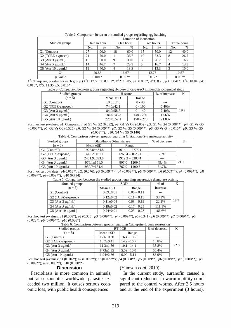
Egg hatchability: There was a significant
217
decrease in the percentages of hatched F.
gigantica eggs with the increase in the dura-
tion of incubation in control (G1), TCBZ-
exposed (G2), auranofin-exposed (G3, G4,
& G5) (P= 0.001, 0.003, 0.041, 0.013 &
0.010, respectively). There was a significant
difference in Fasciola egg hatching between
all the studied groups after half an hour, one
hour, two hours, and three hours of incuba-
tion (P= 0.001, 0.002, 0.012, and 0.032, re-
spectively).
Hematoxylin and eosin staining: The teg-
umental morphology of Fasciola adults of
the control specimens (G1) showed intact
tegument, spines, muscular layer, and paren-
chyma. The cytoplasmic syncytial layer of
the tegument showed numerous spines emb-
edded throughout its matrix, intact reticular
lamina, and deeply stained two muscular la-
yers. Regarding TCBZ-exposed flukes (G2);
they showed tegumental swelling with the
appearance of numerous vacuoles in the teg-
ument syncytium and mild apoptosis. Some
spines were dislodged with empty sockets
while others appeared embedded in the
swollen tegument. All flukes exposed to dif-
ferent concentrations of auranofin (3, 5, &
10μg/ml) showed tegumental swelling and
vacuolations with severe apoptosis in the te-
gument syncytium with sloughing and deta-
chment of parts of the tegument. Partial sep-
aration of some spines was also detected. At
highest concentration of auranofin (10μg/
ml) showed a complete separation of a large
number of spines with severe apoptosis and
necrosis. Tegument severity alterations in
auranofin-exposed worms (G3, G4, & G5)
were concentration-dependent (Fig. 2).
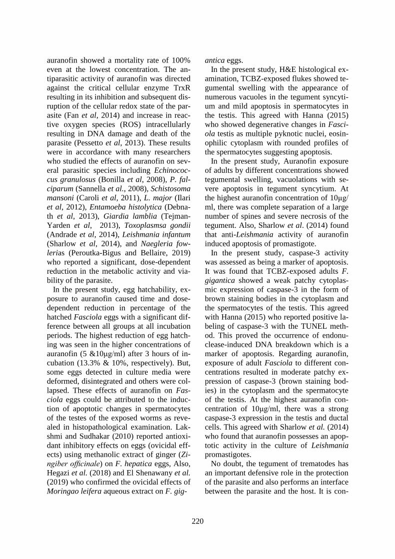
Immunohistochemical staining: Caspase-3
IHC staining of control (G1) showed mild
patchy cytoplasmic expression of caspase-3
of the tegumental cells (brown-stained bod-
ies) with a very mild expression in the sper-
matocytes of the testes. Some worms
showed a negative cytoplasmic expression
of caspase-3. The tegument of TCBZ- expo-
sed flukes (G2) showed a mild patchy cyto-
plasmic expression of caspase-3 and mild
expression in the spermatocytes of the testis.
All F. gigantica worms exposed to low aur-
anofin concentrations (G3) showed a moder-
ate patchy cytoplasmic expression of cas-
pase-3 and moderate expression in spermat-
ocytes of the testis. At higher auranofin con-
centration (5μg/ml), there was a strong pat-
chy expression of caspase-3 in the cytopla-
sm in the ductal cells. At 10μg/mL aurano-
fin, there was a strong diffuse expression of
caspase-3 in both cytoplasm and ductal cells
(Fig. 3). Only higher concentrations of the
auranofin (5 & 10μg/ml) induced significant
increases in caspase-3 expression H-scores
when compared to control (GI) (p3 & p4:
0.008, respectively) and the TCBZ (G2) (p6
& p7:0.009, respectively).
SEM: Control (G1) exhibited normal sur-
face morphology, both ventrally and dorsal-
ly. Tegument was covered with uniformly
distributed, tightly packed, posteriorly di-
rected, broad, and serrated tegumental fungi-
form spines. The apical cone spines showed
finger-like protrusions at their tips. Oral and
ventral suckers were smooth with normal
tegumental infoldings with intact and sharp-
ly pointed spines. Regarding TCBZ-exposed
flukes (G2), oral and ventral suckers were
smooth but surrounded by swollen tegum-
ental in-folding. There were disrupted spines
in some regions. However, some of the spin-
es showed distortions of the upper surfaces.
Some areas showed swelling and furrowing
of the tegument. All F. gigantica worms ex-
posed to auranofin different concentrations
G3, G4, & G5) showed obvious damages
everywhere. At an auranofin concentration
of 3μg/ml, tegument was severely swollen.
A distorted ventral sucker was seen. Some
spines were swollen with a bullous appear-
ance. Others were dislodged from their so-
ckets leaving pits. Exposure to 5μg/ml of au-
ranofin caused swelling of oral and ventral
suckers, extensive tegumental damage with
complete loss of the spines in some areas.
Moreover, other regions appeared sunken in
swollen tegument with blebbing. Exposure
to 10μg/ml of auranofin resulted in a marked
218
disruption of tegumental surface with ap-
peaarance of deep furrows, extensive spine
loss on the ventral surface of oral cone with
loss of finger-like spines, and sloughing da-
maged tegument with a widening of anterior
ventral sucker’s ring (Fig. 4).
TEM: Control (G1) exhibited a normal te-
gumental syncytium, intact apical plasma
membrane, and normal tightly close basal
in-folds. Secretory bodies T1 & T2 were
normal in shape and numbers of muscle lay-
ers beneath the basal lamina were intact.
Adult Fasciola worms incubated with TCBZ
(G2) showed areas with a normal tegumental
syncytium and other areas showed swelling
of the tegumental cells with accumulation
and swelling of T1 secretory bodies. Basal
lamina was swollen with swollen basal in-
folds. The muscle layers were surrounded by
large spaces. Some mitochondria were swol-
len and rounded. Adults exposed to different
concentrations of auranofin (G3, G4, & G5)
showed swollen basal in folds with swollen
mucopolysaccharide masses in between, sw-
ollen rounded mitochondria, numerous T1 &
T2 secretory bodies, and electron-lucent
vacuoles in syncytium below apical plasma
membrane. At higher auranofin concentra-
tions (5 & 10μg/ml), there were severe swe-
lling and disintegration of basal in-folds,
small blebs projecting from tegumental surf-
ace, some separated blebs, and multiple lar-
ge autophagic vacuoles filling tegumental
syncytium (Fig. 5).
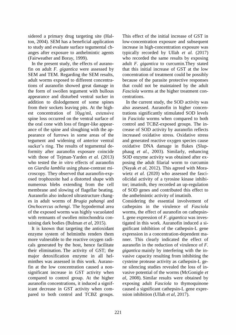
Glutathione S-transferase: Exposure to au-
ranofin initially stimulated activity of detox-
ifying enzyme; GST at low concentration
(G3) and inhibited at higher concentrations
(G4 & G5). Auranofin at a concentration of
3μg/ml (G3) caused a non-significant in-
crease in GST activity when compared to
control (G1) (p2: 0.076). Higher auranofin
concentrations (G4 & G5), induced a signif-
icant decrease in enzyme activity when com-
pared to control (G1) (p3 and p4: 0.009, re-
spectively)) and to TCBZ (G2) (p6 & p7:
0.009, respectively).
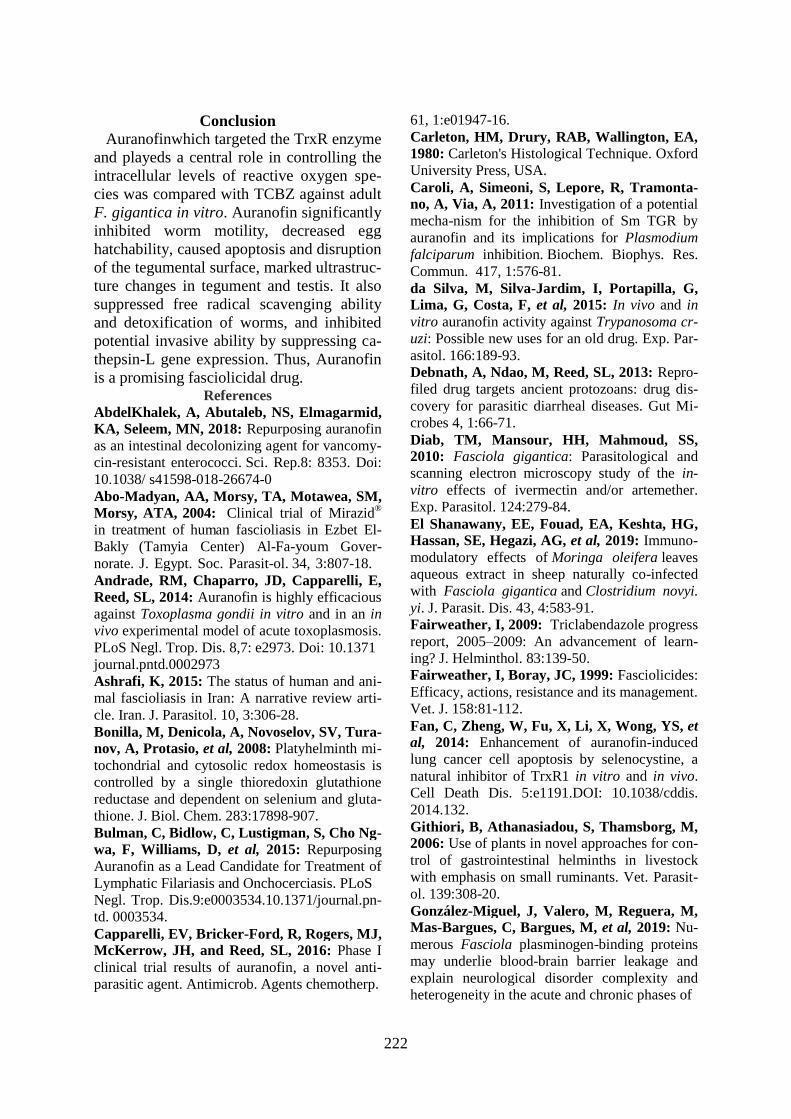
SOD activity increased significantly in hi-
gher concentrations of auranofin (G4 & G5)
when compared to control (G1) (p3 & p4:
0.009, respectively) and when compared to
TCBZ-exposed worms (G2) (p6 & p7:
0.009, respectively).
Cathepsin-L gene expression by RT-PCR
showed a significant inhibition of cathepsin-
L gene expression in a concentration depen-
dent manner in auranofin-exposed worms.
All concentrations (G3, G4 & G5) were re-
duced significant when compared to control
(G1) (p2&p3:0.009 & p4:0.008, respective-
ly) and to TCBZ-exposed worms (G2) (p5 &
p6: 0.009 & p7: 0.008, respectively).
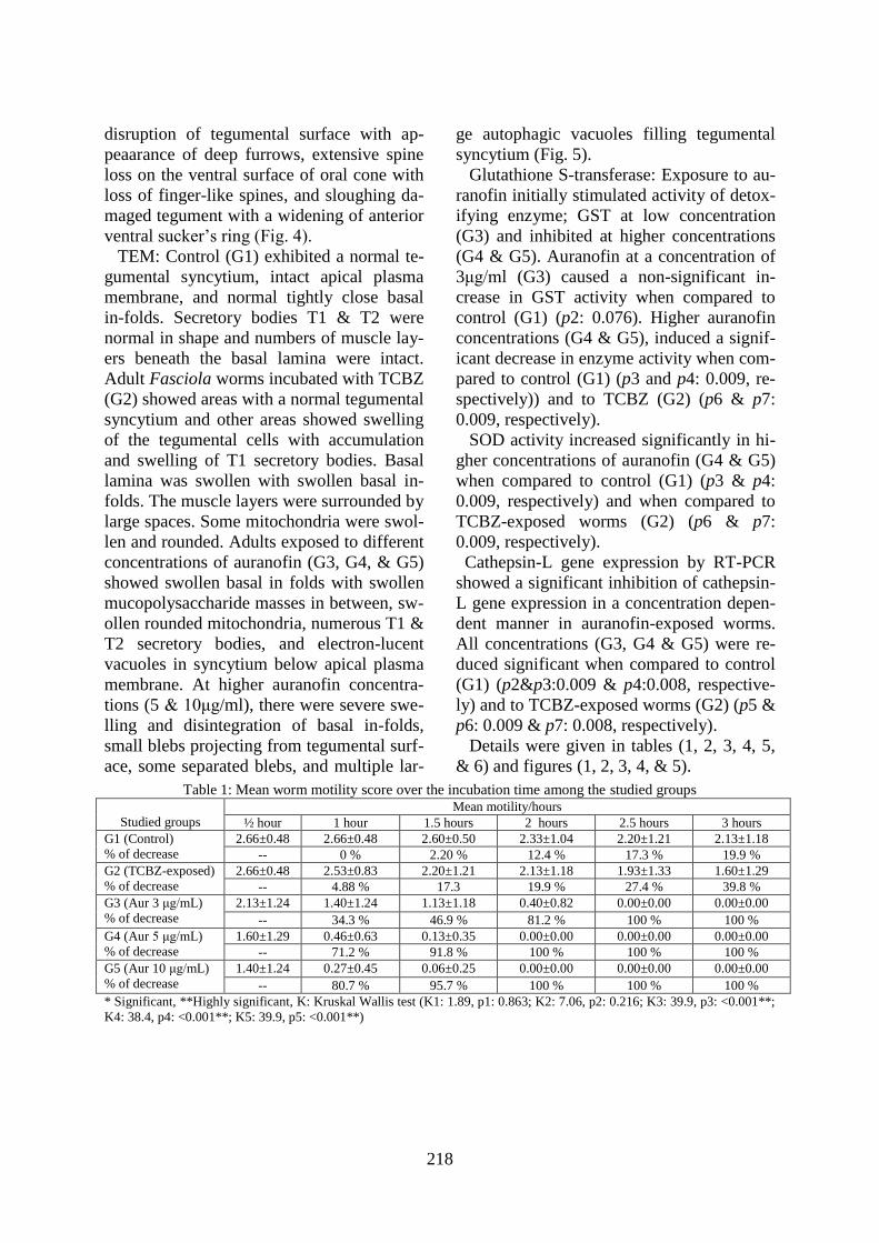
Details were given in tables (1, 2, 3, 4, 5,
& 6) and figures (1, 2, 3, 4, & 5).
Table 1: Mean worm motility score over the incubation time among the studied groups
Studied groups
Mean motility/hours
½ hour 1 hour 1.5 hours 2 hours 2.5 hours 3 hours
G1 (Control)
% of decrease
2.66±0.48 2.66±0.48 2.60±0.50 2.33±1.04 2.20±1.21 2.13±1.18
-- 0 % 2.20 % 12.4 % 17.3 % 19.9 %
G2 (TCBZ-exposed)
% of decrease
2.66±0.48 2.53±0.83 2.20±1.21 2.13±1.18 1.93±1.33 1.60±1.29
-- 4.88 % 17.3 19.9 % 27.4 % 39.8 %
G3 (Aur 3 μg/mL)
% of decrease
2.13±1.24 1.40±1.24 1.13±1.18 0.40±0.82 0.00±0.00 0.00±0.00
-- 34.3 % 46.9 % 81.2 % 100 % 100 %
G4 (Aur 5 μg/mL)
% of decrease
1.60±1.29 0.46±0.63 0.13±0.35 0.00±0.00 0.00±0.00 0.00±0.00
-- 71.2 % 91.8 % 100 % 100 % 100 %
G5 (Aur 10 μg/mL)
% of decrease
1.40±1.24 0.27±0.45 0.06±0.25 0.00±0.00 0.00±0.00 0.00±0.00
-- 80.7 % 95.7 % 100 % 100 % 100 %
* Significant, **Highly significant, K: Kruskal Wallis test (K1: 1.89, p1: 0.863; K2: 7.06, p2: 0.216; K3: 39.9, p3: <0.001**;
K4: 38.4, p4: <0.001**; K5: 39.9, p5: <0.001**)
219
Table 2: Comparison between the studied groups regarding egg hatching
Studied groups
Duration of incubation
Half an hour One hour Two hours Three hours
No. % No. % No. % No. %
G1 (Control) 27 90.0 18 60.0 15 50.0 12 40.0
G2 (TCBZ-exposed) 21 70.0 11 36.7 10 33.3 8 26.7
G3 (Aur 3 μg/mL) 15 50.0 9 30.0 8 26.7 5 16.7
G4 (Aur 5 μg/mL) 14 46.7 7 23.3 5 16.7 4 13.3
G5 (Aur 10 μg/mL) 12 40.0 4 13.3 4 13.3 3 10.0
X2 20.83 16.67 12.76 10.57
p. value 0.001* 0.002* 0.012* 0.032*
X2 Chi-square, p value for each group (X21: 17.5, p1: 0.001*; X22: 13.85, p2: 0.003*; X23: 8.25, p3: 0.041*; X24: 10.84, p4:
0.013*; X25: 11.35, p5: 0.010*)
Table 3: Comparison between groups regarding H-score of caspase-3 immunohistochemical study
Post hoc test p-values: p1: Comparison of G1 Vs G2 (0.052); p2: G1 Vs G3 (0.052); p3: G1 Vs G4 (0.008**); p4: G1 Vs G5
(0.008**); p5: G2 Vs G3 (0.525); p6: G2 Vs G4 (0.009**); p7: G2 Vs G5 (0.009**); p8: G3 Vs G4 (0.015*); p9: G3 Vs G5
(0.009**); p10: G4 Vs G5 (0.140)
Table 4: Comparison between groups regarding Glutathione S-transferase activity
Post hoc test p-values: p1(0.016*); p2: (0.076); p3 (0.009**); p4 (0.009**); p5 (0.009**); p6 (0.009**); p7 (0.009**); p8
(0.009**); p9 (0.009**); p10 (0.754)
Table 5: Comparison between the studied groups regarding superoxide dismutase activity
Studied groups
(n = 5)
SOD % of
increase
K
Mean ±SD Range
G1 (Control) 0.09±0.01 0.08 – 0.11 ---
18.9 G2 (TCBZ-exposed) 0.12±0.02 0.11 – 0.15 33.3%
G3 (Aur 3 μg/mL) 0.11±0.04 0.08 – 0.19 22.2%
G4 (Aur 5 μg/mL) 0.19±0.02 0.17 – 0.25 111.1%
G5 (Aur 10 μg/mL) 0.24±0.01 0.23 – 0.28 166.6%
Post hoc test p-values: p1 (0.036*); p2 (0.338); p3 (0.009**); p4 (0.009**); p5 (0.341); p6 (0.009**); p7 (0.009**); p8
(0.028*); p9 (0.009**); p10 (0.036*)
Table 6: Comparison between groups regarding Cathepsin–L gene expression
Studied groups
(n = 5)
RT-PCR % of decrease K
Mean ±SD Range
G1 (Control) 17.6±0.80 16.4 –18.5 ---
22.9 G2 (TCBZ-exposed) 15.7±0.41 14.2 –16.7 10.8%
G3 (Aur 3 μg/mL) 11.3±1.56 10.1 –14.1 35.8%
G4 (Aur 5 μg/mL) 8.73±1.85 5.59 –10.0 50.4%
G5 (Aur 10 μg/mL) 1.94±2.66 0.00 –5.11 88.9%
Post hoc test p-values: p1 (0.016*); p2 (0.009**); p3 (0.009**); p4 (0.008**); p5 (0.009**); p6 (0.009**); p7 (0.008**); p8
(0.009**); p9 (0.008**); p10 (0.008**)
Discussion Fascioliasis is more common in animals,
but also zoonotic worldwide parasite ex-
ceeded two million. It causes serious econ-
omic loss, with public health consequences
(Yamson et al, 2019).
In the current study, auranofin caused a
significant reduction in worm motility com-
pared to the control worms. After 2.5 hours
and at the end of the experiment (3 hours),
Studied groups
(n = 5)
H-score % of increase K
Mean ±SD Range
G1 (Control) 10.0±17.3 0 - 40 ---
19.9 G2 (TCBZ-exposed) 74.0±42.1 0 - 100 6.40%
G3 (Aur 3 μg/mL) 84.0±58.5 0 - 140 7.40%
G4 (Aur 5 μg/mL) 186.0±43.3 140 - 230 17.6%
G5 (Aur 10 μg/mL) 228.0±52.1 150 – 270 21.8%
Studied groups
(n = 5)
Glutathione S-transferase % of decrease K
Mean ±SD Range
G1 (Control) 1927.8±484.8 1614.1 – 2775.4 ----
21.1
G2 (TCBZ-exposed) 1445.2±161.1 1265.4 – 1625.1 25%
G3 (Aur 3 μg/mL) 2401.9±593.8 1912.3 – 3388.4 ----
G4 (Aur 5 μg/mL) 976.1±151.0 807.0 – 1209.5 49.4%
G5 (Aur 10 μg/mL) 930.7±664.4 762.0 – 1100.3 51.7%
220
auranofin showed a mortality rate of 100%
even at the lowest concentration. The an-
tiparasitic activity of auranofin was directed
against the critical cellular enzyme TrxR
resulting in its inhibition and subsequent dis-
ruption of the cellular redox state of the par-
asite (Fan et al, 2014) and increase in reac-
tive oxygen species (ROS) intracellularly
resulting in DNA damage and death of the
parasite (Pessetto et al, 2013). These results
were in accordance with many researchers
who studied the effects of auranofin on sev-
eral parasitic species including Echinococ-
cus granulosus (Bonilla et al, 2008), P. fal-
ciparum (Sannella et al., 2008), Schistosoma
mansoni (Caroli et al, 2011), L. major (Ilari
et al, 2012), Entamoeba histolytica (Debna-
th et al, 2013), Giardia lamblia (Tejman-
Yarden et al, 2013), Toxoplasmsa gondii
(Andrade et al, 2014), Leishmania infantum
(Sharlow et al, 2014), and Naegleria fow-
lerias (Peroutka-Bigus and Bellaire, 2019)
who reported a significant, dose-dependent
reduction in the metabolic activity and via-
bility of the parasite.
In the present study, egg hatchability, ex-
posure to auranofin caused time and dose-
dependent reduction in percentage of the
hatched Fasciola eggs with a significant dif-
ference between all groups at all incubation
periods. The highest reduction of egg hatch-
ing was seen in the higher concentrations of
auranofin (5 &10μg/ml) after 3 hours of in-
cubation (13.3% & 10%, respectively). But,
some eggs detected in culture media were
deformed, disintegrated and others were col-
lapsed. These effects of auranofin on Fas-
ciola eggs could be attributed to the induc-
tion of apoptotic changes in spermatocytes
of the testes of the exposed worms as reve-
aled in histopathological examination. Lak-
shmi and Sudhakar (2010) reported antioxi-
dant inhibitory effects on eggs (ovicidal eff-
ects) using methanolic extract of ginger (Zi-
ngiber officinale) on F. hepatica eggs, Also,
Hegazi et al. (2018) and El Shenawany et al.
(2019) who confirmed the ovicidal effects of
Moringao leifera aqueous extract on F. gig-
antica eggs.
In the present study, H&E histological ex-
amination, TCBZ-exposed flukes showed te-
gumental swelling with the appearance of
numerous vacuoles in the tegument syncyti-
um and mild apoptosis in spermatocytes in
the testis. This agreed with Hanna (2015)
who showed degenerative changes in Fasci-
ola testis as multiple pyknotic nuclei, eosin-
ophilic cytoplasm with rounded profiles of
the spermatocytes suggesting apoptosis.
In the present study, Auranofin exposure
of adults by different concentrations showed
tegumental swelling, vacuolations with se-
vere apoptosis in tegument syncytium. At
the highest auranofin concentration of 10μg/
ml, there was complete separation of a large
number of spines and severe necrosis of the
tegument. Also, Sharlow et al. (2014) found
that anti-Leishmania activity of auranofin
induced apoptosis of promastigote.
In the present study, caspase-3 activity
was assessed as being a marker of apoptosis.
It was found that TCBZ-exposed adults F.
gigantica showed a weak patchy cytoplas-
mic expression of caspase-3 in the form of
brown staining bodies in the cytoplasm and
the spermatocytes of the testis. This agreed
with Hanna (2015) who reported positive la-
beling of caspase-3 with the TUNEL meth-
od. This proved the occurrence of endonu-
clease-induced DNA breakdown which is a
marker of apoptosis. Regarding auranofin,
exposure of adult Fasciola to different con-
centrations resulted in moderate patchy ex-
pression of caspase-3 (brown staining bod-
ies) in the cytoplasm and the spermatocyte
of the testis. At the highest auranofin con-
centration of 10μg/ml, there was a strong
caspase-3 expression in the testis and ductal
cells. This agreed with Sharlow et al. (2014)
who found that auranofin possesses an apop-
totic activity in the culture of Leishmania
promastigotes.
No doubt, the tegument of trematodes has
an important defensive role in the protection
of the parasite and also performs an interface
between the parasite and the host. It is con-
221
sidered a primary drug targeting site (Hal-
ton, 2004). SEM has a beneficial application
to study and evaluate surface tegumental ch-
anges after exposure to anthelmintic agents
(Fairweather and Boray, 1999).
In the present study, the effects of aurano-
fin on adult F. gigantica were assessed by
SEM and TEM. Regarding the SEM results,
adult worms exposed to different concentra-
tions of auranofin showed great damage in
the form of swollen tegument with bullous
appearance and disturbed ventral sucker in
addition to dislodgement of some spines
from their sockets leaving pits. At the high-
est concentration of 10μg/ml, extensive
spine loss occurred on the ventral surface of
the oral cone with loss of finger-like appear-
ance of the spine and sloughing with the ap-
pearance of furrows in some areas of the
tegument and widening of anterior ventral
sucker’s ring. The results of tegumental de-
formity after auranofin exposure coincide
with those of Tejman-Yarden et al. (2013)
who tested the in vitro effects of auranofin
on Giardia lamblia using phase-contrast mi-
croscopy. They observed that auranofin-exp-
osed trophozoite had a distorted shape with
numerous blebs extending from the cell
membrane and slowing of flagellar beating.
Auranofin also induced ultrastructure chang-
es in adult worms of Brugia pahangi and
Onchocercus ochengi. The hypodermal area
of the exposed worms was highly vacuolated
with remnants of swollen mitochondria con-
taining dark bodies (Bulman et al, 2015).
It is known that targeting the antioxidant
enzyme system of helminths renders them
more vulnerable to the reactive oxygen radi-
cals generated by the host, hence facilitate
their elimination. The activity of GST; the
major detoxification enzyme in all hel-
minthes was assessed in this work. Aurano-
fin at the low concentration caused a non-
significant increase in GST activity when
compared to control group. At the higher
auranofin concentrations, it induced a signif-
icant decrease in GST activity when com-
pared to both control and TCBZ groups.
This effect of the initial increase of GST in
low-concentration exposure and subsequent
increase in high-concentration exposure was
typically recorded by Ullah et al. (2017)
who recorded the same results by exposing
adult F. gigantica to curcumin.They stated
that this initial increase of GST at the low
concentration of treatment could be possibly
because of the parasite protective responses
that could not be maintained by the adult
Fasciola worms at the higher treatment con-
centrations.
In the current study, the SOD activity was
also assessed. Auranofin in higher concen-
trations significantly stimulated SOD levels
in Fasciola worms when compared to both
control and TCBZ-exposed groups. The in-
crease of SOD activity by auranofin reflects
increased oxidative stress. Oxidative stress
and generated reactive oxygen species cause
oxidative DNA damage in flukes (Slup-
phaug et al., 2003). Similarly, enhancing
SOD enzyme activity was obtained after ex-
posing the adult filarial worm to curcumin
(Nayak et al, 2012). This agreed with Mora-
wietz et al. (2020) who assessed the fasci-
olicidal activity of a tyrosine kinase inhibi-
tor; imatinib, they recorded an up-regulation
of SOD genes and contributed this effect to
the anthelmintic activity of imatinib.
Considering the essential involvement of
cathepsins in the virulence of Fasciola
worms, the effect of auranofin on cathepsin-
L gene expression of F. gigantica was inves-
tigated in this work. Auranofin induced a si-
gnificant inhibition of the cathepsin-L gene
expression in a concentration-dependent ma-
nner. This clearly indicated the effect of
auranofin in the reduction of virulence of F.
gigantica mainly by interfering with the in-
vasive capacity resulting from inhibiting the
cysteine protease activity as cathepsin-L ge-
ne silencing studies revealed the loss of in-
vasive potential of the worms (McGonigle et
al, 2008). Similar results were obtained by
exposing adult Fasciola to thymoquinone
caused a significant cathepsin-L gene expre-
ssion inhibition (Ullah et al, 2017).
222
Conclusion
Auranofinwhich targeted the TrxR enzyme
and playeds a central role in controlling the
intracellular levels of reactive oxygen spe-
cies was compared with TCBZ against adult
F. gigantica in vitro. Auranofin significantly
inhibited worm motility, decreased egg
hatchability, caused apoptosis and disruption
of the tegumental surface, marked ultrastruc-
ture changes in tegument and testis. It also
suppressed free radical scavenging ability
and detoxification of worms, and inhibited
potential invasive ability by suppressing ca-
thepsin-L gene expression. Thus, Auranofin
is a promising fasciolicidal drug. References
AbdelKhalek, A, Abutaleb, NS, Elmagarmid,
KA, Seleem, MN, 2018: Repurposing auranofin
as an intestinal decolonizing agent for vancomy-
cin-resistant enterococci. Sci. Rep.8: 8353. Doi:
10.1038/ s41598-018-26674-0
Abo-Madyan, AA, Morsy, TA, Motawea, SM,
Morsy, ATA, 2004: Clinical trial of Mirazid®
in treatment of human fascioliasis in Ezbet El-
Bakly (Tamyia Center) Al-Fa-youm Gover-
norate. J. Egypt. Soc. Parasit-ol. 34, 3:807-18.
Andrade, RM, Chaparro, JD, Capparelli, E,
Reed, SL, 2014: Auranofin is highly efficacious
against Toxoplasma gondii in vitro and in an in
vivo experimental model of acute toxoplasmosis.
PLoS Negl. Trop. Dis. 8,7: e2973. Doi: 10.1371
journal.pntd.0002973
Ashrafi, K, 2015: The status of human and ani-
mal fascioliasis in Iran: A narrative review arti-
cle. Iran. J. Parasitol. 10, 3:306-28.
Bonilla, M, Denicola, A, Novoselov, SV, Tura-
nov, A, Protasio, et al, 2008: Platyhelminth mi-
tochondrial and cytosolic redox homeostasis is
controlled by a single thioredoxin glutathione
reductase and dependent on selenium and gluta-
thione. J. Biol. Chem. 283:17898-907.
Bulman, C, Bidlow, C, Lustigman, S, Cho Ng-
wa, F, Williams, D, et al, 2015: Repurposing
Auranofin as a Lead Candidate for Treatment of
Lymphatic Filariasis and Onchocerciasis. PLoS
Negl. Trop. Dis.9:e0003534.10.1371/journal.pn-
td. 0003534.
Capparelli, EV, Bricker-Ford, R, Rogers, MJ,
McKerrow, JH, and Reed, SL, 2016: Phase I
clinical trial results of auranofin, a novel anti-
parasitic agent. Antimicrob. Agents chemotherp.
61, 1:e01947-16.
Carleton, HM, Drury, RAB, Wallington, EA,
1980: Carleton's Histological Technique. Oxford
University Press, USA.
Caroli, A, Simeoni, S, Lepore, R, Tramonta-
no, A, Via, A, 2011: Investigation of a potential
mecha-nism for the inhibition of Sm TGR by
auranofin and its implications for Plasmodium
falciparum inhibition. Biochem. Biophys. Res.
Commun. 417, 1:576-81.
da Silva, M, Silva-Jardim, I, Portapilla, G,
Lima, G, Costa, F, et al, 2015: In vivo and in
vitro auranofin activity against Trypanosoma cr-
uzi: Possible new uses for an old drug. Exp. Par-
asitol. 166:189-93.
Debnath, A, Ndao, M, Reed, SL, 2013: Repro-
filed drug targets ancient protozoans: drug dis-
covery for parasitic diarrheal diseases. Gut Mi-
crobes 4, 1:66-71.
Diab, TM, Mansour, HH, Mahmoud, SS,
2010: Fasciola gigantica: Parasitological and
scanning electron microscopy study of the in-
vitro effects of ivermectin and/or artemether.
Exp. Parasitol. 124:279-84.
El Shanawany, EE, Fouad, EA, Keshta, HG,
Hassan, SE, Hegazi, AG, et al, 2019: Immuno-
modulatory effects of Moringa oleifera leaves
aqueous extract in sheep naturally co-infected
with Fasciola gigantica and Clostridium novyi.
yi. J. Parasit. Dis. 43, 4:583-91.
Fairweather, I, 2009: Triclabendazole progress
report, 2005–2009: An advancement of learn-
ing? J. Helminthol. 83:139-50.
Fairweather, I, Boray, JC, 1999: Fasciolicides:
Efficacy, actions, resistance and its management.
Vet. J. 158:81-112.
Fan, C, Zheng, W, Fu, X, Li, X, Wong, YS, et
al, 2014: Enhancement of auranofin-induced
lung cancer cell apoptosis by selenocystine, a
natural inhibitor of TrxR1 in vitro and in vivo.
Cell Death Dis. 5:e1191.DOI: 10.1038/cddis.
2014.132.
Githiori, B, Athanasiadou, S, Thamsborg, M,
2006: Use of plants in novel approaches for con-
trol of gastrointestinal helminths in livestock
with emphasis on small ruminants. Vet. Parasit-
ol. 139:308-20.
González-Miguel, J, Valero, M, Reguera, M,
Mas-Bargues, C, Bargues, M, et al, 2019: Nu-
merous Fasciola plasminogen-binding proteins
may underlie blood-brain barrier leakage and
explain neurological disorder complexity and
heterogeneity in the acute and chronic phases of
223
human fascioliasis. Parasitol. 146:284-98.
Habig, WH, Pabst, MJ, Jacoby, WB, 1974: Glutathione S-transferases: The first enzymatic
step in mercapturic acid formation. J. Biol. Ch-
em. 249:7130-9.
Halton, DW, 2004: Microscopy and the helm-
inth parasite. Micron 35:361-90.
Hanna, R, 2015: Fasciola hepatica: Histology
of the reproductive organs and differential ef-
fects of triclabendazole on drug-sensitive and
drug-resistant fluke isolates and on flukes from
selected field cases. Pathogens 4, 3:431-56.
Hanna, R, Cromie, L, Taylor, S, Couper, A,
2006: The effect of a parenteral ivermectin/
closantel combination on the growth and repro-
ductive development of immature Fasciola he-
patica in cattle. Vet. Parasitol. 142:78-90.
Hegazi, A, Abdel Megeed, K, Hassan, S, Abd-
elaziz, M, Toaleb, N, et al, 2018: Comparative
ovicidal activity of Moringao leifera leaf extra-
cts on Fasciola gigantica eggs. Vet. World DOI:
10.14202/ vetworld.215-20.
Ilari, A, Baiocco, P, Messori, L, Fiorillo, A,
Boffi, A, et al, 2012: A gold containing drug
against parasitic polyamine metabolism: The X-
ray structure of trypanothione reductase from
Leishmania infantum in complex with auranofin
reveals a dual mechanism of enzyme inhibition.
Amino Acids 42, 2/3:803-11.
Jiraungkoorskul, W, Sahaphong, S, Tansatit,
T, Kangwanragsan, N, Pipatshukiat, S, 2005:
Eurytremapan creaticum: The in vitro effect of
praziquantel and triclabendazole on the adult fl-
uke. Exp. Parasitol. 111, 3:172-7.
Lakshmi, BVS, Sudhakar, M, 2010: Attenua-
tion of acute and chronic restraint stress-induced
perturbations in experimental animals by Zingi-
ber officinale Roscoe. Food Chem. Toxicol. 48:
530-5.
Machicado, C, Marcos, LA, 2016: Carcinogen-
esis associated with parasites other than Schisto-
soma, Opisthorchis and Clonorchis: A systemat-
ic review. Int. J. Cancer138, 12:2915-21.
Marklund, SL, Marklund, G, 1974: Involve-
ment of the superoxide anion radical in the au-
toxidation of pyrogallol and a convenient assay
for superoxide dismutase. Eur. J. Biochem., 47:
469. DOI: 10.1111/j.1432-1033.1974.tb03714.x.
Marques, LT, Guedes, RA, Rodrigues, WD,
Archanjo, AB, Severi, JA, et al, 2020: Chemi-
cal composition of various plant extracts and
their in vitro efficacy in control of Fasciola he-
patica eggs. Ciência Rural 50, 5: e20190363.
https://doi.org/10.1590/0103-8478cr20190363
Mas-Coma, S, Bargues, M, Valero, M, 2005:
Fascioliasis and other plant borne trematode zo-
onosis. Inter. J. Parasitol. 35, 3:1255-78.
Mas-Coma, S, Valero, MA, Bargues, MD,
2019: Fascioliasis. Adv. Exp. Med. Biol. 1154:
71-103.
McGonigle, L, Mousley, A, Marks, NJ, Bren-
nan, GP, Dalton, JP, et al, 2008: The silencing
of cysteine proteases in Fasciola hepatica newly
excysted juveniles using RNA interference red-
uces gut penetration. Int. J. Parasitol. 38:149-55.
Merachew, W, Alemneh, T, 2020: Review on
triclabendazole resistance in Fasciola. J. Vet.
Sci. Med. 8, 1: 1-8.
Morawietz, CM, Houhou, H, Puckelwaldt, O,
Hehr, L, Dreisbach, D, et al, 2020: Targeting
kinases in Fasciola hepatica: Anthelminthic eff-
ects and tissue distribution of selected kinase in-
hibitors. Front. Vet. Sci. https://doi.org/10.3389 /
fvets.2020.611270.
Nassef, NE, El-Kersh, WM, El Sobky, MM,
Harba NM, Khalil, SA, et al, 2014: In-vitro as-
sessment of the effect of soybean extract on Fa-
sciola gigantica infection in comparison with tr-
iclabendazole. Menoufia Med. J. 27:93-102.
Nayak, A, Gayen, P, Saini, P, Mukherjee, N,
Babu, SP, 2012: Molecular evidence of curcum-
in-induced apoptosis in the filarial worm Setaria
cervi. Parasitol. Res. 111, 3:1173-86.
Peroutka-Bigusa, N, Bellairea, BH, 2019: Ant-
iparasitic Activity of Auranofin against Nae-
gleria fowleri. J. Eukar. Microbiol. 66:684-8.
Pessetto, ZY, Weir, SJ, Sethi, G, Broward, M
A, Godwin, AK, 2013: Drug repurposing for
gastrointestinal stromal tumor. Mol. Canc. Ther.
12, 7:1299-309.
Roder, C, Thomson, MJ, 2015: Auranofin: Re-
purposing an old drug for a golden new age.
Drugs RD. 15, 1:13-20.
Rokni, MB, Bozorgomid, A, Heydarian, P,
Aryaeipour, M, 2018: Molecular evidence of
human fasciolosis due to Fasciola gigantica in
Iran: A case report. Iran. J. Pub. Hlth. 47, 5:
750-4.
Saccoccia, F, Angelucci, F, Boumis, G, Carot-
ti, D, Desiato, G, et al, 2014: Thioredoxin redu-
ctase and its inhibitors. Curr. Prot. Peptide
Sci. 15, 6:621-46.
Sannella, AR, Casini, A, Gabbiani, C, Messo-
ri, L, Bilia, AR, et al, 2008: New uses for old
drugs. Auranofin, a clinically established antiar-
thritic metallodrug, exhibits potent antimalarial
224
effects in vitro: Mech. Pharmacol. Impli. FEBS
Lett. 582, 6:844-7.
Sarkari, B, and Khabisi SA, 2017: Immunodi-
agnosis of Human Fascioliasis: an update of co-
ncepts and performances of the serological as-
says. J. Clin. Diag. Res. 11, 6:OE05-10.
Sarkari, B, Parhoode, M, AbdolahiKhabisi,
S, Shafiei, R, Mohammadi-ghalehbin, B,
2017: Genetic diversity of Fasciola spp. isolates
from northern part of Iran: Comparison with
southwestern isolates. J. Parasit. Dis. 41, 2:768-
72.
Shareef, PA, Brennan, G, Mcveigh, Khan, M
A, Morphew, R, et al, 2014: Time-dependent
tegumental surface changes in juvenile Fasciola
gigantica in response to triclabendazole treat-
ment in goat. Acta Trop. 136:108-17.
Sharlow, E, Leimgruber, S, Murray, S, Lira,
A, Sciotti, R, et al, 2014: Auranofin is an apop-
tosis-simulating agent with in vitro and in vivo
anti-leishmanial activity. ACS Chem. Biol. 9, 3:
663-72.
Slupphaug, G, Kavli, B, Krokan, H, 2003:
Slupphaug G, Kavli B, Krokan HE: The interact-
ing pathways for prevention and repair of ox-
idative DNA damage. Mutat. Res. 531:231-51.
Smyth, JF, Gourley, C, Walker, G, MacKean,
MJ, Stevenson, A, et al, 2007: Antiestrogen
therapy is active in selected ovarian cancer cas-
es: the use of letrozole in estrogen receptor-
positive patients. Clin. Canc. Res. 13 ,12:3617-
22.
Soliman, M, Taha, H, 2011: Tegumental altera-
tions of Fasciola gigantica due to in vitro treat-
ment with Ro-354. Trop. Biomed. 28:283-92.
Song, L, Li, J, Xie, S, Qian, C, Wang J, et al,
2012: Thioredoxin glutathione reductase as a no-
vel drug target: Evidence from Schistosoma jap-
onicum. PloS One 7:e31456.https://doi.org/ 10.
1371/journal.pone.0031456.
Taghipour, A, Zaki , L, Rostami, A, Forout-
an, M, Ghaffarifar, F, 2019: Highlights of hu-
man ectopic fascioliasis: A systematic review.
Infect. Dis. 51, 11/12:785-92.
Tejman-Yarden, N, Miyamoto, Y, Leitsch, D,
Santini, J, Debnath, A, et al, 2013: A reprof-
iled drug, auranofin, is effective against metroni-
dazole-resistant Giardia lamblia. Antimicrob.
Agents Chemother. 57, 5:2029-35.
Ullah, R, Rehman, A, Zafeer, M F, Rehman,
L, Khan, YA, 2017: Anthelmintic Potential of
Thymoquinone and Curcumin on Fasciola gi-
gantica.PLoS One. 2:1-19.
Yamson, EC, Tubalinal, GA, Viloria1, VV,
Mingala, CN, 2019: Anthelmintic effect of betel
nut (Areca catechu) and neem (Azadira cht-
aindica) extract against liver fluke (Fasciola
spp.). J. Adv. Vet. Anim. Res. 6, 1:44-9. Explanation of figures
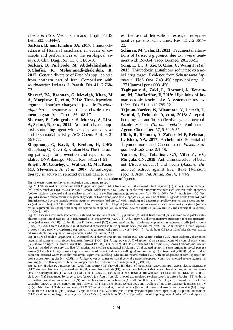
Fig. 1: Mean worm motility over incubation time among groups.
Fig. 2: H &E stained cut sections of adult F. gigantica: (a&b): Adult from control (G1) showed intact tegument (T), spine (s), muscular layer (m), and parenchyma (p) (a:×200,b: ×400). (c&d): Adult exposed to TCBZ (G2) showed numerous vacuoles (red arrows), mild apoptosis
(yellow circles), dislodged spines (yellow arrow), and a swollen tegument (green arrow). (c:×400, d:×200). (e&f): Adult from G3 (Aur
3μg/mL) showed vacuolations in tegument syncytium (red arrows) and severe apoptosis (yellow circle) (×400). (g&h): Adult from G4 (Aur 5μg/mL) showed severe vacuolations in tegument syncytium (red arrows) with sloughing and detachment (yellow arrows) and severe apopto-
sis (yellow circles) (g:×200, h:×400). (i&j): Adult from G5 (Aur 10μg/mL) showed numerous vacuolations in tegument syncytium (red ar-
rows), tegumental sloughing with complete separation of spines (yellow arrows), severe apoptosis (yellow circles), and necrosis (blue circles) (i: ×200, j:×400).
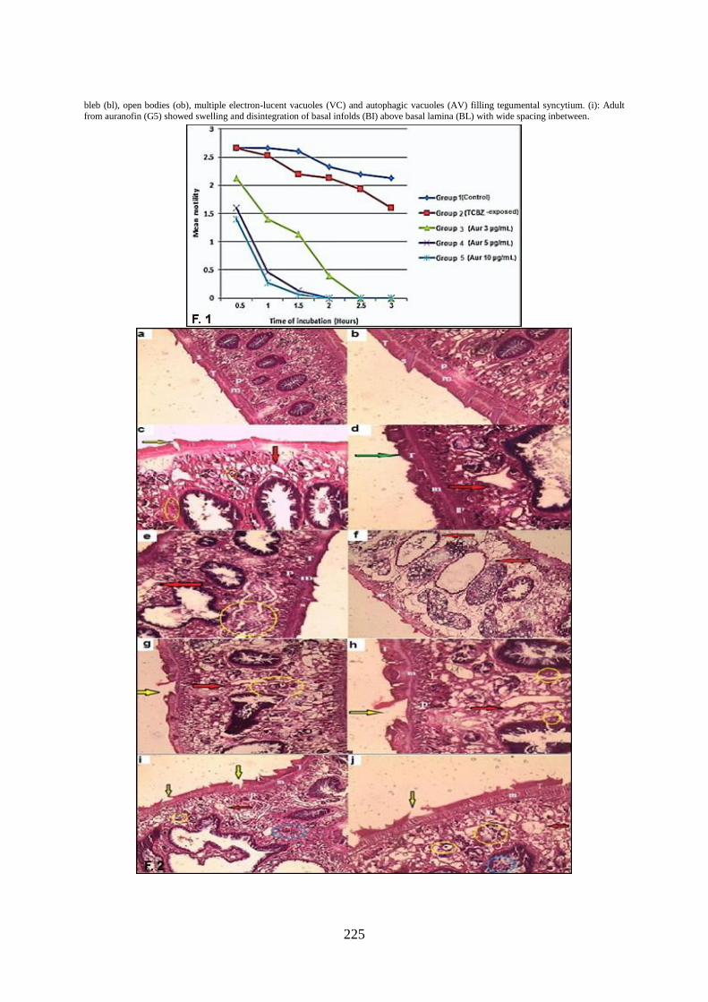
Fig. 3: Caspase-3 immunohistochemically stained cut sections of adult F. gigantica: (a): Adult from control (G1) showed mild patchy cyto-
plasmic expression of caspase -3 in tegumental cells (red arrows) (×200). (b): Adult from G1 showed negative expression in testes spermato-cytes (red arrows) (×200). (c): Adult from TCBZ-exposed (G2) showed mild patchy cytoplasmic expression in tegumental cells (red arrows)
(×400). (d): Adult from G2 showed mild caspase-3 expression in testes spermatocytes (red arrows) (×400). (e): Adult from G4 (Aur 5μg/mL)
showed strong patchy cytoplasmic expression in tegumental cells (red arrows) (×200). (f): Adult from G5 (Aur 10μg/mL) showed strong diffuse cytoplasmic expression in tegumental and ductal cells (×200).
Fig. 4: SEM of adult F. gigantica: (a): A control (G1) showed smooth oral sucker (OS) and ventral sucker (VS), intact uniformly distributed
tegumental spines (s) with ridged tegument (arrows) (×50). (b): A high power SEM of spines (s) on an apical cone of a control adult worm (G1) showed finger-like protrusions at tips (arrow) (×1000). (c): A SEM of a TCBZ-exposed adult from (G2) showed smooth oral sucker
(OS) surrounded by sensory papillae (b), moderately swollen tegumental infoldings (a), disrupted spines in some regions in apical part (s)
arrows (×150). (d): A high power of apical cone of adult from G2 showed swelling (a) and furrowing (b) of tegument (×1000). (e): A SEM of auranofin-exposed worm (G3) showed severe tegumental swelling (a,b) around ventral sucker (VS) with dislodgement of some spines from
their sockets leaving pits (c) (×200). (f): A high-power of spines on apical cone of auranofin-exposed worm (G3) showed severe tegumental
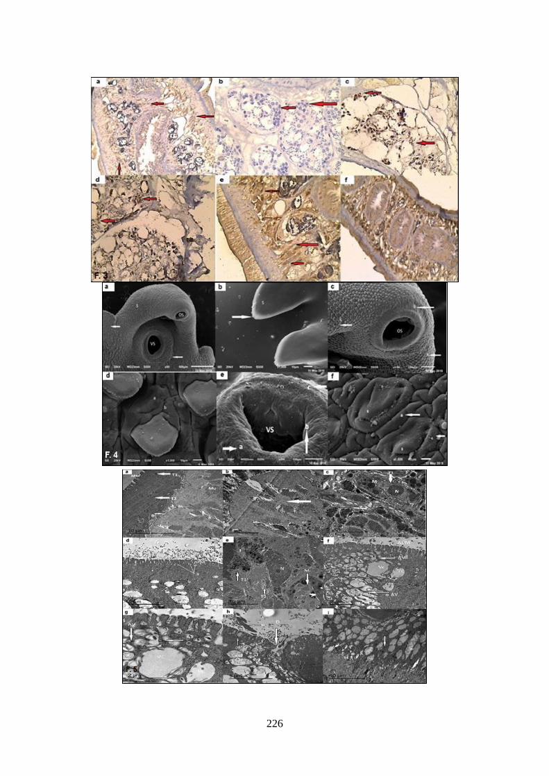
swelling (a), swollen spines with bulbous appearance (s), and some blebs in tegument (c) (×1000). Fig. 5:TEM of adult F. gigantica: (a): Adult from control (G1) showed a full depth of tegumental syncytium, from apical plasma membrane
(APM) to basal lamina (BL), normal and tightly closed basal infolds (BI), normal muscle layer (Mu) beneath basal lamina, and normal num-bers of secretory bodies (T1 & T2). (b): Adult from TCBZ-exposed (G2) showed basal lamina with swollen basal infolds (BL), normal mus-
cle layer (Mu) surrounded by large spaces (arrow). (c): Adult from G2 showed accumulated swollen type-1 secretory bodies (T1) within a
cell with a normal and round nucleus (N) and swollen rounded mitochondria (M). (d): Adult from G3 (Aur 3μg/mL) showed electron-lucent vacuoles (arrows a) in cell syncytium just below apical plasma membrane (APM) apex and swelling of mucopolysaccharide masses (arrow
b). (e): Adult from G3 showed numerous T1 & T2 secretory bodies, normal nucleus (N) morphology, and swollen mitochondria (M). (f&g):
Adult from G4 (Aur 5μg/mL) showed large electron-lucent vacuoles (Vc) in cell syncytium just below apex of apical plasma membrane (APM) and numerous large autophagic vacuoles (AV). (h): Adult from G5 (Aur 10μg/mL) showed large tegumental defect (D) and separated
225
bleb (bl), open bodies (ob), multiple electron-lucent vacuoles (VC) and autophagic vacuoles (AV) filling tegumental syncytium. (i): Adult
from auranofin (G5) showed swelling and disintegration of basal infolds (BI) above basal lamina (BL) with wide spacing inbetween.
226