Embed Size (px)
Citation preview
Journal of the Egyptian Society of Parasitology, Vol.46, No.1, April 2016J. Egypt. Soc. Parasitol. (JESP), 46(1), 2016: 185- 200
185
THE EFFICACY OF THREE MEDICINAL PLANTS; GARLIC, GINGERAND MIRAZID AND A CHEMICAL DRUG METRONIDAZOLE AGAINST
CRYPTOSPORIDIUM PARVUM: II- HISTOLOGICAL CHANGESBy
MOHAMED F. ABOUEL-NOUR1, DINA MAGDY M. EL-SHEWEHY1,SHADIA F. HAMADA1 AND TOSSON A. MORSY2
Department of Zoology, Faculty of Science, Mansoura University1, Mansoura andDepartment of Parasitology, Faculty of Medicine, Ain Shams University, Cairo
115662, EgyptAbstract
Cryptosporidiosis parvum is a zoonotic protozoan parasite infects intestinal epithelial cells ofman and animals causing a major health problem. This study was oriented to evaluate the pro-tective and curative capacity of garlic, ginger and mirazid in comparison with metronidazoledrug (commercially known) against Cryptosporidium in experimental mice. Male Swiss Albinomice experimentally infected with C. parvum were treated with medicinal plants extracts (Gin-ger, Mirazid, and Garlic) as compared to chemical drug Metronidazole. Importantly, C. par-vum-infected mice treated with ginger, Mirazid, garlic and metronidazole showed a completeelimination in shedding oocysts by 9th day PI. The reduction and elimination of shedding oo-cysts in response to the treatments might be attributable to a direct effect on parasite growth inintestines, sexual phases production and/ or the formation of oocysts. The results were evaluat-ed histopathological examination of ileum section of control mice (uninfected, untreated) dis-played normal architecture of the villi. Examination of infected mice ileum section (infected,untreated) displayed histopathological alterations from uninfected groups. Examination of ile-um section prepared from mice treated with garlic, ginger, mirazid, and metronidazole dis-played histopathological alterations from that of the control groups, and showed marked histo-logic correction in the pattern with the four regimes used in comparison to control mice. Garlicsuccessfully eradicated oocysts of infected mice from stool and intestine. Supplementation ofginger to infected mice markedly corrected elevation in the inflammatory risk factors and im-plied its potential antioxidant, anti-inflammatory and immunomodulatory capabilities. Infectedmice treated with ginger, mirazid, garlic and metronidazole showed significant symptomaticimprovements during treatment.Key words: Cryptosporidiosis, Histopathology, Garlic, Ginger, Mirazid, Metronidazole, mice
IntroductionProtozoan parasites of the genus Cryptos-
poridium belong to the class Sporozoasida,family Cryptosporidiidae and phylum Api-complexa. They are often referred to as coc-cidia. While some coccidia can undergo ex-tra-intestinal development and are tissue-cyst forming (Sarcocystis, Toxoplasma) oth-ers develop in the gastrointestinal or respira-tory tract, without formation of tissue cysts(i.e. Eimeria, Isospora & Cryptosporidium).Like others, it was thought that Cryptospor-idium was highly host specific and almost20 species were named according to speciesof infected host from which they were iso-lated (Tyzzer, 1912; Levine, 1984; Current
et al. 1986; Fayer and Ungar 1986). Later,cross-transmission studies with mammalianisolates of Cryptosporidium indicated lowhost specificity, that first prompted (Tziporiet al, 1994) to consider Cryptosporidium asa single species genus and Levine (1984)suggested that only four species may be val-id. The valid number of species was in-creased to six species with C. parvum causesrespiratory and intestinal infections but, C.muris causes stomach infections. Respirato-ry and intestinal cryptosporidiosis in birdswas attributed to C. baileyi, C. meleagridis,C. serpentis infects reptiles & C. nasoruminfected fish (Fayer et al, 1997; Koudela andModry, 1998; Lindsay et al, 2000).
186
In man, cryptosporidiosis, infections areacquired by ingestion or inhalation (feco-oral route, foodborne, waterborne…etc.) ofthe parasite’s transmissive stage known asthe oocyst. The time between acquisition ofinfection and manifestation of symptoms(pre-patent period) is dependent on variousfactors (i.e. host susceptibility, virulence ofthe infecting strain of oocysts, oocyst infec-tivity…etc.) but may be somewhat from 5-28 days mean 7.2 (Anderson et al, 1982;Current et al. 1983; Højlyng et al. 1987).
Cryptosporidium infections are associatedwith acute and clinical disease characterizedby diarrhoea in man and various domesticand wild animals including birds. Cryptos-poridium infections are most pathogenic inneonatal hosts, however in humans infec-tions were reported from a 3 day old infant,from a mother with cryptosporidiosis, to a95 year old individual. Disease often mani-fests itself with profuse, watery diarrhea,abdominal cramping, nausea, vomiting andlow grade fever. In well nourished, immu-nocompetent patients, cryptosporidiosis maylast 2-12 days but, usually self-limiting. Oc-casionally, infections may continue for twoweeks or more and may require fluid re-placement therapy. Nowadays, no effectivechemotherapeutic treatment is available andduration of disease is dependent upon thepatient’s immune status. In patients withcongenital or acquired immune deficienciesor malnourished ones, infection can be con-siderably prolonged, resulting in malabsorp-tion, severe dehydration and death (Ander-son et al, 1982; Current et al, 1983; Højlynget al, 1987, O’Donoghue, 1995; Fayer et al,2000; Xiao, 2010).
The work aimed to study the effect ofsome natural products on the control andtreatment of the infection in comparisonwith a chemotherapeutic drug already used.Study the effect of the infection and thetreatment and protection on the histology ofthe small intestine and if there is any effecton the small intestine by different treatme-nt’s strategies.
Treatment: The search for bioactive plantswhich can be used as non-conventional anti-parasitic treatment has received considerableattention in recent times because of the in-creasing worldwide development of re-sistance to chemical drugs in parasitic popu-lations. However, scientific evidence to val-idate the use of plants remains limited(Hoste et al. 2008) Thus, this study orientedto evaluate the protective and curative ca-pacity of garlic, ginger, mirazid and metro-nidazole against cryptosporodial infection.Mirazid: Mirazid is an oleo-resin extract de-rived from Myrrh which is obtained fromthe stem of Commiphora molmol, a thornytree that grows in Somalia and Arabian Pen-insula (Massoud et al, 2001). The antisepticand antineoplastic properties of myrrh arethought to be attributed to terpenoids(Nomicus, 2007). Myrrh was approved byUS food and Drug Administration (Ford etal., 1992). Mirazid has been reported in sev-eral clinical and experimental trials to be asafe and effective natural herbal drug. Evi-dent anti-trematode activity was demonstrat-ed in schistosomiasis (Massoud, 1999;Badria et al, 2001), in fascioliasis (Massoudet al, 2001; Abo-Madyan et al, 2004), in ex-perimental and human heterophyidiasis(Fathy et al, 2005; Massoud et al, 2007) andas anti-cestode in monisziasis expansa (El-Shazly et al, 2004) and Bertiella studeri (Al-Mathal et al, 2010) and as anti-nematode instrongyloidiasis stercoralis (Massoud et al,2006). Anti-protozoal activity of Mirazidhas been proved in human against Cryptos-poridium parvum (Massoud et al., 2008), inhepatic coccidiosis produced by Eimeriastidae in rabbits (Baghaddi and Al-Mathal,2010) and also against Trichomonasvaginalis in women with infection resistantto metronidazole. Therapeutic efficacy ofmirazid on experimental Giardia lambliainfection in Albino rats as indicated bya100% reduction in parasite-load of bothintestinal and fecal parasitic count by usingtinidazole as control (Fathy, 2011).
187
Ginger: Zingiber officinale Roscoe (gin-ger, Zingiberaceae) is a widely used spicesand it is a common additive in large numberof compounded foods and beverages due toits flavor and pungency. Its rhizome is oneof the most commonly used medicinal herbsas well as one of the most commonly usedcondiments in Chinese cuisine. Severalpharmacological effects of Zingiber plantwas reported such as antiulcer effect (Yo-shikawa et al, 1994), antioxidant effect, po-tent antibacterial activity (Mahady et al,2003), potent antifungal activity (Ficker etal, 2003) and anthelmintic activity (Iqbal etal, 2001). Z. officinale extracts have beenextensively studied for a broad range of bio-logical activities including antibacterial, anticonvulsant, analgesic, antiulcer, gastric anti-secretory, antitumor, antifungal, antispasmo-dic, antiallergenic, and other activities suchas the ability to increase digestive fluids,plus absorb and neutralize toxins and stom-ach acid. Z. officinale increased bile secre-tion, as well as increase the action and toneof the bowels (Bradley, 1992). The anti-giardial activity of Z. officinale was reportedusing experimental infected Balb/c micewith Giardia lamblia. The extract of Z. offi-cinale was more active specially whenmixed with honey the watery extract of Z.officinale reduced the G. lamblia trophozo-ites (Al-masoudi, 2011).
Garlic: Allium sativum (A. sativum) or gar-lic is used as food and medicine in manycultures for thousands of years, dating atleast as far back as the time that the Gizapyramids were built. It has been recognizednot only as a spice but also as a substanceexerted a control on microorganisms (Soffarand Mokhtar, 1991; Ayaz et al, 2008; Masa-mha et al, 2010). A. sativum is remarkablefor a number of potentially active chemicalconstituents. It contains seventeen aminoacids as arginine, at least 33 organosulphatecompounds as allin and allicin, eight miner-als (germanium, calcium, copper, iron, po-tassium, magnesium, selenium and zinc),enzymes as allinase, and the vitamins A, B1
& C. The physiological activity of dietary A.sativum is attributed to allicin (diallyl thio-sulphinate) is one of the organosulphatecompounds found in the bulb. It is responsi-ble for the anti-microbial properties and thecharacteristic flavor of fresh garlic (Ayaz etal, 2008; Masamha et al, 2010; Thompsonand Ali, 2003). Ancient Egyptians realizedthe benefits of garlic; its medical and magi-cal powers were described on the walls ofancient temples and on papyri dating to1500 BC. Garlic proved to have antimicro-bial, antithrombotic, hypolipidemic, hypo-glycemic and antitumor activities (Thomp-son and Ali, 2003). Lately, garlic has widelybeen used to treat intestinal parasites. Anti-helminthic effect of garlic is a matter of in-terest of researchers. Their results showedthat treatment with garlic evoked a signifi-cant reduction in the worm load (Soffar andMokhtar, 1991; Abdel-Rahman et al, 1998;Sutton and Haik, 1999; Ayaz et al, 2008;Riad et al, 2009). Also, garlic was used suc-cessfully in a single uncontrolled study inChina applied on 20 AIDS patients to treatCryptosporidium (Fareed et al, 1996) Besi-des, garlic compounds were purified andtried as complementary medicine in theleishmaniasis management (Wabwoba et al,2010). Many microorganisms were signifi-cantly susceptible to garlic extract and gavegood results broad-spectrum therapeutic ag-ent (Adetumbi and Lau, 1983).
Metronidazole: Also known as: Flagyl,Metronidazol, Gineflavir, Meronidal, Met-ronidaz, Trichazol, Trichopol, Danizol,Trivazol. Metronidazole 250mg and 500mgtablets are for oral administration. The dis-covery of metronidazole and its long actingderivative, secnidazole first synthesized inthe early 1960s by Jacobs et al. completelychanged the treatment of some protozoaninfections such as urogenital trichomoniasis,amebiasis and giardiasis. Second generationderivatives, generally long acting com-pounds, quickly appeared with tinidazole(Miller et al, 1970) and ornidazole synthe-sized (Hoffer, 1969). They were highly ef-
188
fective in vitro and in vivo against these pro-tozoa and quickly underwent clinical trialsworldwide. Metronidazole received regula-tory approvals in a large number of coun-tries in the developed and in the developingworld and became the treatment of choicefor trichomoniasis and amebiasis, both tissu-lar such as in amebic liver abscess and intes-tinal. Darbon et al. (1962) showed that met-ronidazole could be used in giardiasis. Met-ronidazole is completely absorbed after oraladministration and penetrates body tissuesand fluids such as saliva, breast milk, se-men, and vaginal secretions. The drug is me-tabolized mainly in the liver and is excretedin the urine (Lau et al, 1992).
Materials and MethodsAnimals used were male Swiss Albino
mice, aged three to five weeks, weighing 25-30 grams. They were housed in well venti-lated cages with perforated covers, suppliedwith standard pellet food and water. Bed-ding was changed every day. The mice wereallowed to adapt to the laboratory environ-ment for one week before the experiment(El-Fakhry et al, 1998) and their stools wereexamined by direct wet saline smear, iodineand Sheather’s sugar flotation method toexclude the presence of parasites (Garciaand Brucker, 1997).
Cryptosporidium parvum oocysts werepurchased from Waterborne™, Inc. (NewOrleans, Louisiana) and stored in shippingmedium (Phosphate-buffered saline with pe-nicillin, streptomycin, gentamycin, amphote-ricin B & 0.01% Tween 20) at 4°C until use.
Experimental design: The experimentalanimals (male Swiss Albino mice) were di-vided into the following groups (5 mice/group) as follow: Control group (uninfect-ed- untreated) Mice of this group did notreceive any treatments. Infected group (in-fected-untreated) Mice were inoculated oral-ly with the isolated Cryptosporidium oocystsat a dose of 104oocysts/mouse (Gaafar,2007). Ingestion was performed by gastricgavage, using a 23-gauge needle tipped withplastic tubing (Riad et al, 2009). Experim-
ental Prophylactic group, which was subdi-vided as following: a- Subgroup I- Prophy-lactic 1 (P1): received garlic two days beforeinfection then these mice continued to re-ceive garlic daily for 12 days post-infection.b- Subgroup II - Prophylactic 2 (P2): re-ceived ginger two days before infection thenthese mice continued to receive ginger dailyfor 12 days post-infection. c- Subgroup III -Prophylactic 3 (P3): received mirazid twodays before infection then these mice con-tinued to receive mirazid daily for 12 dayspost-infection. d- Subgroup IV - Prophylac-tic 4(P4): received metronidazole two daysbefore infection then mice continued to re-ceive metronidazole daily for 12 days post-infection. Experimental Therapeutic group,was subdivided as following: a- Subgroup I-Treatment 1 (T1): received garlic one dayafter the infection then these mice continuedto receive garlic daily for 2 weeks. b- Sub-group II- Treatment 2 (T2): received gingerone day after the infection then these micecontinued to receive ginger daily for 2weeks. c- Subgroup III - Treatment 3 (T3):received mirazid one day after the infectionthen these mice continued to receive miraziddaily for 2 weeks. d- Subgroup IV- Treat-ment 4 (T4): received metronidazole oneday after infection then these mice continuedto receive metronidazole daily for 2 weeks.Each mouse of the infected, experimentalProphylactic, and experimental therapeuticgroups was inoculated orally with the isolat-ed Cryptosporidium oocysts at a dose of 104
oocysts/ mouse (Gaafar, 2007) Ingestionwas performed by gastric gavage, using a23-gauge needle tipped with plastic tubing(Riad et al, 2009).
Garlic dose was 50mg/kg body-weight/dayan hr before breakfast. Preparation: Freshgarlic bulbs were separated, peeled, andwashed with distilled water. After drying;about 500g of garlic bulbs were crushed in ablender until a uniform consistency wasachieved. The resulting paste was dilutedwith distilled Water to obtain a 1 g/ml aque-ous solution. Raw garlic juice was aliquoted
189
and was stored at -20⁰C until use (Burke etal, 2009). Working solution was made fromstock one by dilution with distilled water.The selected dose for the present work was50 mg/kg body weight (Masamha et al,2010), Ginger dose was 50mg/kg body wei-ght/ day, an hr before breakfast. Miraziddose was 10 mg/kg body weight/day, two hrbefore breakfast. Metronidazole dose was50mg/kg body weight /day an hr beforebreakfast.
Stool analysis: Undiluted stool samplesfrom all mice were stained by MZN (Modi-fied Ziel-Nelseen) and examined by lightmicroscope.
Histological investigations: Using overdose of ether, the sacrifice of animals wasachieved 12 days post infection for the sub-groups (P1), (P2), (P3), and (P4), while sac-
rifice of mice of the subgroups, (T1), (T2),(T3) & (T4) was achieved 15 days post in-fection. For light microscopy, autopsy sam-ples were taken from the intestine of mice indifferent groups and fixed in 10% neutralformalin for 24 h. Then washing was donein tap water then serial dilutions of alcohol(absolute ethyl) were used for dehydration.Specimens were cleared in xylene and em-bedded in paraffin at 56°C in hot air ovenfor 24 hr. Paraffin bees wax tissue blockswere prepared for sectioning at a thicknessof 4μm by sledge microtome. The obtainedtissue sections were collected on glassslides, deparaffinized, stained by hematoxy-lin & eosin stain for routine examinationthen examination was done through the lightelectric microscope (Banchroft et al, 1996).
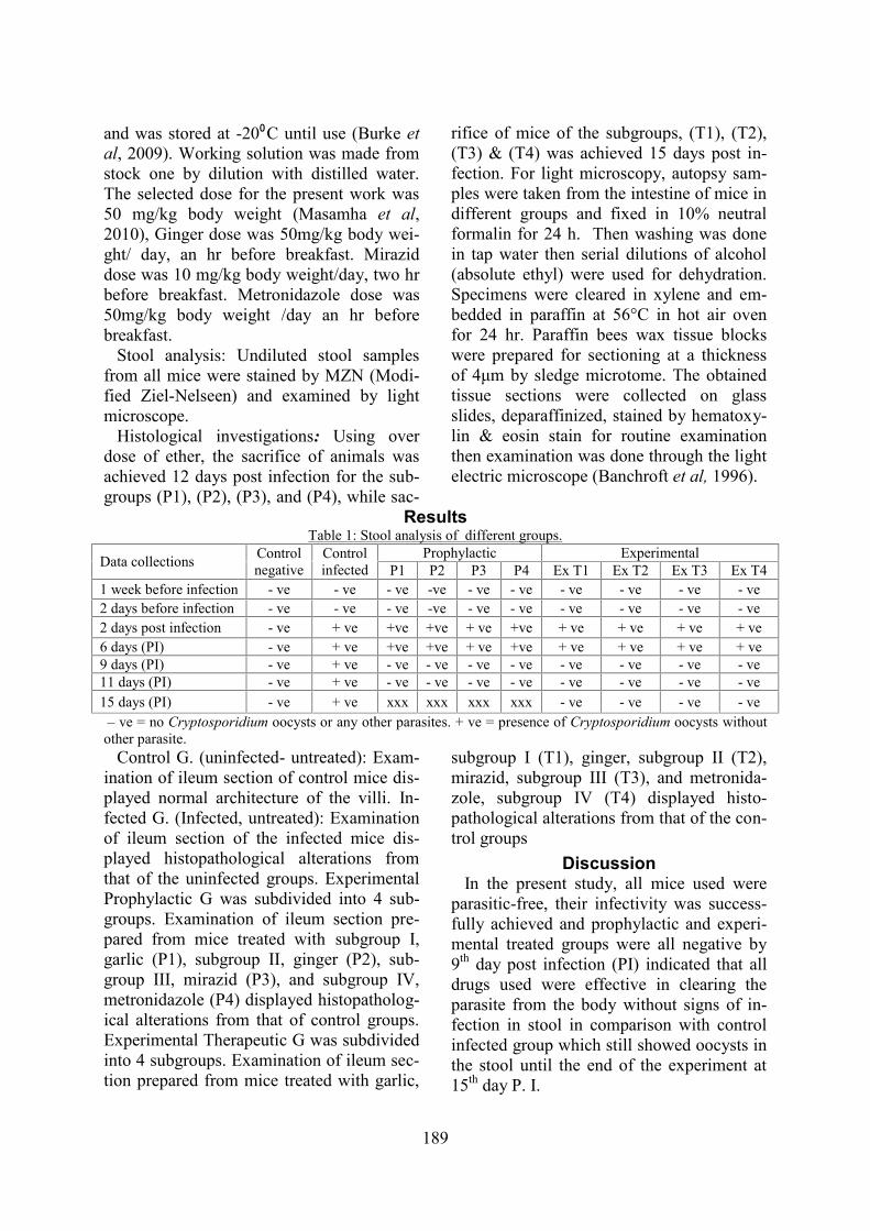
ResultsTable 1: Stool analysis of different groups.
Data collections Controlnegative
Controlinfected
Prophylactic ExperimentalP1 P2 P3 P4 Ex T1 Ex T2 Ex T3 Ex T4
1 week before infection - ve - ve - ve -ve - ve - ve - ve - ve - ve - ve2 days before infection - ve - ve - ve -ve - ve - ve - ve - ve - ve - ve2 days post infection - ve + ve +ve +ve + ve +ve + ve + ve + ve + ve6 days (PI) - ve + ve +ve +ve + ve +ve + ve + ve + ve + ve9 days (PI) - ve + ve - ve - ve - ve - ve - ve - ve - ve - ve11 days (PI) - ve + ve - ve - ve - ve - ve - ve - ve - ve - ve15 days (PI) - ve + ve xxx xxx xxx xxx - ve - ve - ve - ve
– ve = no Cryptosporidium oocysts or any other parasites. + ve = presence of Cryptosporidium oocysts withoutother parasite.
Control G. (uninfected- untreated): Exam-ination of ileum section of control mice dis-played normal architecture of the villi. In-fected G. (Infected, untreated): Examinationof ileum section of the infected mice dis-played histopathological alterations fromthat of the uninfected groups. ExperimentalProphylactic G was subdivided into 4 sub-groups. Examination of ileum section pre-pared from mice treated with subgroup I,garlic (P1), subgroup II, ginger (P2), sub-group III, mirazid (P3), and subgroup IV,metronidazole (P4) displayed histopatholog-ical alterations from that of control groups.Experimental Therapeutic G was subdividedinto 4 subgroups. Examination of ileum sec-tion prepared from mice treated with garlic,
subgroup I (T1), ginger, subgroup II (T2),mirazid, subgroup III (T3), and metronida-zole, subgroup IV (T4) displayed histo-pathological alterations from that of the con-trol groups
DiscussionIn the present study, all mice used were
parasitic-free, their infectivity was success-fully achieved and prophylactic and experi-mental treated groups were all negative by9th day post infection (PI) indicated that alldrugs used were effective in clearing theparasite from the body without signs of in-fection in stool in comparison with controlinfected group which still showed oocysts inthe stool until the end of the experiment at15th day P. I.
190
In the present study, explosive, chronic,fatal, non-bloody diarrhea is considered avery serious management problem in immu-nosuppressed patients as well as in normalpersons in both developed and developingcountries. Pathogenic intestinal protozoarepresent the main causes of this diarrhea,among which Cryptosporidium producesregularly occurred outbreaks throughout theworld (Ma, 1989; Farthing, 2006). Most ofthe immunodeficient patients got failure ofthe available drugs used for the treatment,besides the multiple adverse effects that theyproduced (Miao et al, 2000). Thus, new ef-fective drug became consequently urgentlyneeded. The search for bioactive plantswhich can be used as nonconventional anti-parasitic treatment has received considerableattention in recent times because of the in-creasing worldwide development of re-sistance to chemical drugs in parasitic popu-lations. However, scientific evidence to val-idate the use of plants remains limited(Hoste et al, 2008).
In the present study, examination of ileumsection of control mice (uninfected, untreat-ed) displayed normal architecture of the villiand examination of ileum section of infectedmice (infected, untreated) were displayedhistopathological alterations.
In the present study, experimental treat-ment group was subdivided into 4 sub-groups. Examination of ileum section pre-pared from mice treated with garlic, sub-group I (T1), ginger, subgroup II (T2),mirazid, subgroup III (T3), and metronida-zole, subgroup IV (T4) displayed histo-pathological alterations from that of controlgroups.
The allicin was obtained from crushedfresh garlic bulbs (Ankri and Mirelman1999; Sasaki et al, 1999; Lemar et al, 2002).The dose selected for the present work was50 mg/kg body weight. Riad et al. (2009)suggested that this dose is equivalent to thedaily amount of garlic recommended for anaverage human to maintain good health (4g).Garlic successfully eradicated Cryptosporid-
ium oocysts from stool and intestinal sec-tions of infected mice. Abouel-Nour et al.(2015) proved this immunologically.Ginger is one of the most commonly used
fresh herbs and spices. Ginger is among the20 top-selling herbal supplements in theUnited States (Blumenthal, 1998), pharma-copoeias from different countries list thatginger extract for various digestive diseases(Blumenthal et al, 2000). .In this study, thefirst time that, in addition to an antiemeticeffect, ginger extracts exhibit antidiarrhealactivities. Administration of ginger extractmodulated the harmful effect in liver hy-droxyproline and serum AFP induced underthe effect of S. mansoni infection, indicatingtheir strong anti-fibrotic effect in reducinggranuloma formation associated with liverfibrosis. The present results are in harmonywith previous data revealing ginger anthel-mintic activity against S. mansoni (Ade-wunmi et al, 1990; Sanderson et al, 2002)and gastrointestinal nematodes (Iqbal et al,2006) by killing parasites through its bind-ing to parasite beta-tubulin and inhibitingglucose uptake. This effect of ginger may berelated to the significant anthelmintic activi-ty of its constituents, shogaol (Ali et al,2008) and gingerol (Lin et al, 2010). Theseactive components of ginger completelyabolished the infectivity of S. mansoni mira-cidia and cercariae of Biomphalaria glabra-ta and mice respectively indicating its’ mol-luscicidal and schistosomicidal activities.
Supplementation of ginger to the infectedmice markedly corrected elevation in theinflammatory risk factors in the presentstudy, implying its potential antioxidant, an-ti-inflammatory and immunomodulatory ca-pabilities. The beneficial effects of gingerwere previously reported due to its activeconstituents such as zingerone, paradol, gin-gerols and shogaols (Adewusi et al, 1996).Also, Abouel-Nour et al. (2015) proved thisimmunologically
Metronidazole, a heterocyclic compoundwith a nitro group on the fifth position of animidazole ring, derived from the Streptomy-
191
ces antibiotic azomycin. Developed in 1959,metronidazole was approved for the treat-ment of trichomoniasis in the early 1960sand was the first drug to have a cure rate ap-proaching 100% with systemic treatment(Cosar and Julou, 1959). Metronidazole is asmall molecule, does not bind to serum pro-teins, and is well distributed through bodilytissue and fluids. Therapeutic levels of thedrug have been found in blood, cerebrospi-nal fluid, pulmonary exudates, bile, andseminal fluid, as well as bone, brain, andpelvic tissue. Metronidazole is a small mol-ecule that enters T. vaginalis via passive dif-fusion. The drug itself is inactive, but anaer-obic reduction results in the formation of acytotoxic nitro radical anion. This nitro radi-cal is hypothesized to bind transiently toDNA, disrupting or breaking the strands andleading to cell death (Lloyd and Kristensen,1985; Edwards, 1993).
Mirazid (Oleo-resin extract from Myrrh ofCommiphora molmol tree, family: Bursera-ceae) is a product by Pharco PharmaceuticalCompany, Alexandria. Administered doseswere two equal 600mg oral doses of purifiedCommiphora extract for 3 consecutive days(Haridy et al, 2003) on empty stomach, atleast two hours before eating, oral doses of10μg/mL (Nogata et al, 2001; Manthey andGuthrie, 2002).
Haridy et al. (2003), Hegab and Hassan(2003) and Massoud et al. (2008) reportedthat Mirazid proved as an effective fasci-olicidal drug, without clinical side effect.Also, Hassan et al. (2003) stated thatMirazid contracted S. mansoni worms mus-cle and affected its surface by ultra-struc-ture causing tegument disruption and tuber-cles collapse El-Baz et al. (2003) provedthat Mirazid was very effective and safe intreatment of Schistosoma haematobium.
In the present study, infected mice thatwere treated with ginger, mirazid, garlic andmetronidazole showed significant sympto-matic improvements during the treatmentperiod. So, the treatments may have a directand powerful impact on the parasite that
consequently minimizes the pathology asso-ciated with C. parvum infection.
C. parvum- infected mice treated with gin-ger, mirazid, garlic and metronidazole show-ed a complete elimination of shed- ding oo-cyst by 9th day PI. Reduction and elimina-tion of shedding oocyst in response to thetreatments might be attributable to a directeffect on parasite growth in the intestines,the production of the sexual phases, and/ orthe formation of oocysts. The present histo-pathological changes in the infected/untreat-ed group ileum tissues were reported (Tzipo-ri et al, 1994; Capet et al, 1999; Leitch andHe, 1999; Motta et al, 2002; Guitard et al,2006; Maruyama et al, 2007; Robinson andSmyth, 2008). Pathological changes wereattributed to C. parvum displacing brushborders causing an asymmetrical loss of epi-thelial cells resulting in shortening and fus-ing of villi. Cryptosporidiosis of villi atro-phy might be due to secreted toxins that di-rectly damage epithelial cells (Heine et al,1984; Tzipori, 2002). C. parvum infectionalso resulted in T-cell migration to laminapropria. Abouel-Nour et al. (2015) used C.parvum immunological response to evaluatethe efficacy of anti-cryptosporidisis agentsof Garlic, Ginger, Mirazid and Metronida-zole in experimentally infected mice. Exper-imentally the immunologic mediated elimi-nation of C. parvum required CD4+ T cellsand IFN-gamma. But, the innate immuneresponses also have a significant protectiverole in both man and animals. The mucosalimmune response to C. parvum in C57BL/6neonatal and GKO mice showed a concomi-tant Thl & Th2 cytokine mRNA expression,with a crucial role for IFN-gamma in infec-tion resolution. NK cells & IFN-gammaproved important components in immunityin T & B cell-deficient mice, but the IFN-gamma-dependent resistance was detected inalymphocytic mice. Epithelial cells played avital role in immunity as once infected cellsincreased expression of inflammatory chem-okines and cytokines and demonstrated anti-infection killing mechanisms.
192
In Egypt, many authors dealt with zoono-tic cryptoaporidiosis, the selected ones areof interest from epidemiological point ofview. Shalaby and Shalaby (2015) deter-mined cryptosporidiosis among 120 ran-domly chosen school children aged 4-16years. Medical sheets were filled out on eachchild. They found that watery and loose di-arrhea was more significant among infectedchildren. Positive stool samples were among37 (30.8 %), while ELISA and IFA detected30 (25%) and 33 (27.5%) respectively. Thevalidity test of ELISA declared sensitivityand specificity with 93.3% and 90% whileIFA declared 90.9% and 91.1% respectively.El-Shazly et al. (2015) in Egypt examined50 children with the chronic liver diseases(CLD) of different etiology and 50 non-CLDchildren with gastrointestinal complaintsserved as controls. They found the common-est intestinal protozoa in CLD patients were:Entamoeba histolytica/E. dispar (16 %), Gi-ardia lamblia (14 %), Blastocystis hominis(14%), C. parvum (10%), E. histolytica andG. lamblia (2%), E. histolytica and B. homi-nis (2%), G. lamblia and B. hominis (2%),B. hominis and E. coli (2%) and Microspor-idium (2%). As compared to controls, inci-dence of these organisms in CLD patientswas significantly higher (p<0.045) as re-gards stool examination by unstained tech-niques and no significant difference betweenboth groups as regards stool examination bystained techniques (p<0.478). They conclud-ed that CLD affect the immunity of the pa-tients showed significant increase in the in-cidence of intestinal parasites in cases com-pared to controls. El-Badry et al. (2015)studied the molecular prevalence and sea-sonality of Cryptosporidium over a period ofone year in a cohort of Egyptian diarrhoeicpatients. Stool samples were collected from865 diarrhoeic patients attending outpatientclinics of Cairo University Hospitals, fromall age groups over one year, examined mi-croscopically for oocysts by the acid-faststaining method and for copro-DNA detec-tion using nPCR. Cryptosporidium copro-
DNA was detected in 19.5% of allover year,with a major peak in summer (August) and asmall rise in spring (April). Infection wasmainly C. hominis (95.8%) followed by C.parvum (3.0%), affecting all age groups,with predominance in the pre-school agegroup, and decrease with age. They con-cluded that infection in diarrheic Egyptianswas of distinct endemicity, with the bi-model mostly influenced by population dy-namics, with a clear high prevalence in pre-school children and predominating anthro-ponotic (C. hominis) transmission through-out the year and that Cryptosporidium was awater contaminant and an important cause ofhealth problems in Egypt, necessitating fur-ther studies of the risk factors. El-Shabrawiet al. (2015) stated that diarrhea continues tocause significant morbidity in Egypt. Theydetected Rotavirus in 11% of patients. En-terotoxigenic Escherichia coli (ETEC),Campylobacter, Shigella, and Salmonella in7%, 3.7%, 1.1% &1.4% of patients, respec-tively; and in 11.1%, 3.1%, 0.6% & 0.6% ofcontrols, respectively. Cryptosporidium wasdetected in 3.9% of cases. Mixed infectionwas detected in 5.9% of cases and 0.9% ofcontrols, with a significant difference (p <0.001). Ibrahim et al. (2016) investigated theepidemiology and public health significanceof Cryptosporidium species and genotypeswere in Beni-Suef Governorate. By micro-scopic examination, the overall estimatedprevalence in cattle, buffaloes, and humanswas 10.2, 12.3, and 19 %, respectively. Thehighest rates were in the calves less than 2months of age (17.1 %) and diarrheic ani-mals (13.0 %). In man, the highest preva-lence was in infants (31.3 %) and diarrheicindividuals (21.1 %), infection was com-monest in males (21.7 %) than females(14.5 %). Based on molecular characteriza-tion, Cryptosporidium oocyst wall protein(COWP) and gp60 genes were successfullyamplified in 36/50 samples subjected togenotyping. The restriction fragment lengthpolymorphism (RFLP) analysis of COWPfragments revealed that C. parvum was de-
193
tected only in cattle (12 isolates) and buffa-loes (4 isolates), while in man, species wereC. hominis (15 isolates) and C. parvum (5isolates). Sequence analysis of the gp60gene identified the subtype IIdA20G1 withinC. parvum isolated from both animals andhumans. The common occurrence of zoono-tic subtypes of C. parvum in cattle and buf-faloes highlights the potential role of theseanimals as significant reservoirs of infectionto humans.
ConclusionNo doubt, cryptosporidiosis is a world-
wide problem particularly among childrenand immnunocompromised individuals.
The histopathological patterns were cor-rected with the drugs used and the best resultin descending order was as follow ginger,mirazid and garlic respectively. These threeplant extracts proved to have a direct andpowerful impact on C. parvum and mini-mized its pathology complications as com-pared to Metronidazole.
ReferencesAbdel-Rahman, EH, Kandil, OM, Abdel- Me-geed, KN, 1998: Comparative studies of lethaleffects of Bacillus thuringiensis, Allium sativumand Nerium oleander on Trichostrongylidae par-asites. Egypt. J. Zool. 30:6-79.Abouel-Nour, MF, El-Shewehy, DMM, Ha-mada, SF, Morsy, TA, 2015: The Efficacy ofthree medicinal plants: garlic, ginger and mira-zid and a chemical drug metronidazole againstCryptosporidium parvum. 1- Immunological res-ponse. J. Egypt. Soc. Parasitol. 45, 3:559-70.Adetumbi, MA, Lau, BH, 1983: Allium sati-vum (garlic), a natural antibiotic. Med. Hypothe-ses 12:227-37.Adewunmi, CO, Oguntimein, BO, Furu, P,1990: Molluscicidal and anti-schistosomal activ-ities of Zingiber officinale. Planta Med. 56:374-6.Adewusi, OI, Nix, NA, Lu, X, Colley, DG, Se-cor, WE, 1996: Schistosoma mansoni: relation-ship of tumor necrosis factor-alpha to morbidityand collagen deposition in chronic experimentalinfection. Exp. Parasitol. 84: 115-23.Al-Masoudi, HK, 2011: Antigiardial activity ofZingiber officinale in combination with honey invivo. J. Babylon Univ/Pure Appl. Sci. 2, 19:450-
4.Al-Mathal, EM, Saleh, NMK, Morsy, TA,2010: Human infection with Bertiella studeri(Cestode: Anoploocephalidae) in an Egyptianworker retuning back from Saudi Arabia. J.Egypt. Soc. Parasitol. 40, 1:89-92.Anderson, BC, Donndelinger, T, Wilkins, RM, Smith, J, 1982: Cryptosporidiosis in a veter-inary student. J. Am. Vet. Med. Assoc. 180:408-9.Ankri, S, Mirelman, D, 1999: Antimicrobialproperties of allicin from garlic. Microb. Infect.J. 1:125-9.Ayaz, E, Türel, I, Gül, A, Yilmaz, O, 2008:Evaluation of the antihelmintic activity of garlic(Allium sativum) in mice naturally infected withAspiculuris tetraptera. Rec. Pat. Anti-infect.Drug Disc. 3:149-52.Badria, F, Abou-Mohamed, G, El-Mowafy, A,Massoud, A, Salama, O, 2001: Mirazid: A newschistosomicidal drug. Pharmaceut. Biol. 93:127-31.Baghaddi, HB, Al-Mathal, EM, 2010: Anti-coccidial effect of Commiphora molmol in thedomestic rabbit (Oryctolagus cuniculus domesti-cus). J. Egypt. Soc. Parasitol.40, 3:653-68.Banchroft, J, Stevens, A, Turner, D, 1996:Theory and Practice of Histological Techniques,fourth ed. Churchill Livingstone, New York,London, San Francisco, Tokyo.Blumenthal, M, 1998: The Complete Commis-sion E Monographs. Boston: Integrative Medi-cine Publications.Blumenthal, M, Goldberg, A, Brinckmann, J,2000: Herbal Medicine: Expanded CommissionE Monographs. Boston (MA): Integrative Medi-cine Communications.Bradley, PR, 1992: British Herbal Compendi-um: British Herbal Medicine Association, Bour-nemouth, UK.Burke, JM, Wells, A, Casey, P, Miller, JE,2009: Garlic and papaya lack control over gas-trointestinal nematodes in goats and lambs. Vet.Parasitol. 159:171-4.Capet, C, Kapel, N, Huneau, JF, Magne, D,Laikuen, R, et al, 1999: Cryptosporidium par-vum infection in suckling rats: Impairment ofmucosal permeability and Na (+) -glucose cotransport. Exp. Parasitol. 91:119-25.Cosar, C, Julou L, 1959: Activity of 1-(2-Darbon, A, Portal, A, Girier, L, Pantin, J, Le-claire, C, 1962: Traitement de la giardiase (la-
194
mbliase) par le métronidazole. La Presse Méd.70:15-6.Edwards, DI, 1993: Nitroimidazole drugs ac-tion and resistance mechanisms: Two mechani-sms of resistance. J. Antimicrob. Chemother. 31:201-10.El-Badry, AA, Al-Antably, AS, Hassan, MA,Hanafy, NA, Abu-Sarea, EY, 2015: Molecularseasonal, age and gender distributions of Cryp-tosporidium in diarrhoeic Egyptians: distinctendemicity. Eur. J. Clin. Microbiol. Infect. Dis.34, 12:2447-53El-Baz, MA, Morsy, TA, El Bandary, MM,Motawea, SM, 2003: Clinical and parasitologi-cal studies on the efficacy of mirazid in treat-ment of schistosomiasis haematobium in TatoonEtsa Center El Fayoum Governorate. J. Egypt.Soc. Parasitol. 33:761-76.El-Fakhry, Y, Achbarou, A, Desportes, I, Ma-zier, D, 1998: Encephalitozoon intestinalis: Hu-moral responses in interferon-gamma receptorknockout mice infected with a Microsporidiumpathogenic in AIDS patients. Exp. Parasitol. 89:113-21.El-Shabrawi, M, Salem, M, Abou-Zekri, M,El-Naghi, S, Hassanin, F, et al, 2015: The bur-den of different pathogens in acute diarrhoealepisodes among a cohort of Egyptian childrenless than five years old. Prz. Gastroenterol. 10,3:173-80El-Shazly, LB, El-Faramawy, AA, El-Sayed,NM, Ismail, KA, Fouad, SM, 2015: Intestinalparasitic infection among Egyptian children withchronic liver diseases. J. Parasit. Dis. 39, 1:7-12El-Shazly, AM, Morsy, TA, Dawoud, HA,2004: Human monieziasis expansa: the firstEgyptian parasitic zoonosis. J. Egypt. Soc. Para-sitol. 34, 2:515-8.Fareed, G, Scolaro, M, Jordan, W, Sanders,N, Chesson, C, et al, 1996: The use of a high-dose garlic preparation for the treatment of Cry-ptosporidium parvum diarrhea. Int. Conf. AIDS11:288-92.Farthing, MJG, 2006: Treatment options forthe eradication of intestinal protozoa. Nat. Clin.Pract. Gastrenterol. Hepatol. 3:436-45.Fathy, FM, 2011: Effect of Mirazid (Commi-phora molmol) on experimental giardiasis. J.Egypt. Soc. Parasitol. 41, 1:155-78.Fathy, FM, Salama, O, Massoud, A, 2005:Effect of mirazid (commiphora molmol) on ex-perimental heterophyidiasis. J. Egypt. Soc. Para-sitol. 35, 3:137-50.
Fayer, R Speer, CA, Dubey, JP, 1997: TheGeneral Biology of Cryptosporidium. In: Cryp-tosporidium and Cryptosporidiosis. CRC Press,Florida.Fayer, R, Ungar, BL, 1986: Cryptosporidiumspp. and cryptosporidiosis. Microbiol. Rev. 50,4:458-63.Ficker, CE, Smith, ML, Leaman, DL, Irawa-ti, C, Arnason, JT, 2003: Inhibition of humanpathogenic fungi by member of Zingiberaceae.used by kenyah (Indonesian Borneo). J. Eth-nopharmacol. 85:289-93.Ford, JK, Quinones, MA, Sego, DJ, Sorra, S,1992: Factors affecting the opportunity to per-form trained tasks on the job. Personnel Psychol.45:511-27.Gaafar, MR, 2007: Effect of solar disinfectionon viability of intestinal protozoa in drinkingwater. J. Egypt. Soc. Parasitol. 37:65-86.Garcia, LS, Bruckner, DA, 1997: Macroscopicand microscopic examination of fecal speci-mens: Diagnostic Medical Parasitology 3rd ed.Washington D.C. AMS Press.Guitard, J, Menotti, J, Desveaux, A, Alima-rdani, P, Porcher, R, et al, 2006: Experimentalstudy of effects of probiotics on Cryptosporidi-um parvum infection in neonatal rats. Parasitol.Res. 99:522-7.Haridy, FM, El-Garhy, MF, Morsy, TA,2003: Efficacy of mirazid Commiphora molmolagainst fascioliasis Egyptian sheep. J. Egypt.Soc. Parasitol. 33, 3:917-24.Hassan, M, El-Motaiem, M, Afify H, Abaza,B, El-Shafei, M, et al, 2003: In vitro Effect ofmirazid on Schistosoma mansoni worms. J.Egypt. Soc. Parasitol. 33, 3:999-1008.Hegab, MHA, Hassan, RM, 2003: Role of cir-culating fasciola antigens and IGG4 isotype inassessment of cure from fascioliasis. J. Egypt.Soc. Parasitol. 33:561-70.Heine, J, Moon, HW, Woodmansee, DB,1984: Persistent Cryptosporidium infection incongenitally athymic (nude) mice. Infect. Im-munol. 43:856-9.Hoffer, M, 1969: Nitoimidazole derivatives.United States Patent No. 3435049.Højlyng, N, Anderson, W, Jepson, S, 1987:Cryptosporidiosis: a case of airborne transmis-sion. Lancet 2:271-2.Hoste, H, Torres-Acosta, JF, Alonso-Diaz, MA, Brunet, S, Sandoval-Castro, C, et al, 2008:Identification and validation of bioactive plantsfor the control of gastrointestinal nematodes in
195
small ruminants. Proceedings of 5th Internation-al Workshop: Novel Approaches to the Controlof Helminth Parasites of Livestock. hydroxyeth-yl)-2-methyl-5-nitroimidazole (8823 RP) againstexperimental Trichomonas vaginalis infection.Ann. Inst. Pasteur 96:238-41.Ibrahim, MA, Abdel-Ghany, AE, Abdel-La-tef, GK, Abdel-Aziz, SA, Aboelhadid, SM,2016: Epidemiology and public health signifi-cance of Cryptosporidium isolated from cattle,buffaloes, and humans in Egypt. Parasitol. Res.2016 Apr 5. [Epub ahead of print]Iqbal, M, Ashraf, M, Jamil, A, 2006: Seed en-hancement with cytokinins: Changes in growthand grain yield in salt stressed wheat plants.Plant Grow. Reg. 50:29-39.Iqbal, Z, Nadeem, QK, Kkan, MN, Akhtar, MS, Waraich, FN, 2001: In vitro antihelminticactivity of Allium sativum, Zingiber officinale,Curcurbita mexicana and Ficus religiosa. Int. J.Agric. Biol. 3:454-7.Koudela, B, Modry, D, 1998: New species ofCryptosporidium (Apicomplexa, Cryptosporidi-idae) from lizards. Folia Parasitol. 45:93-100.Lau, AH, Lam, NP, Piscitelli, SC, Wilkes, L,Danzinger, LH, 1992: Clinical pharmacokinet-ics of metronidazole and other nitro-imidazoleanti-infections. Clin. Pharmacokine 23:328-64.Leitch, GJ, He, Q, 1999: Reactive nitrogen andoxygen species ameliorate experimental crypto-sporidiosis in the neonatal BALB/c mouse mod-el. Infect. Immun. 67:5885-91.Lemar, KM, Turner, MP, Lioyd, D, 2002: Ga-rlic (Allium sativum) as an anti-Candida agent: acomparison of the efficacy of fresh garlic andfreeze-dried extracts. J. Appl. Microbiol. 93:398-405.Levine, N.D. (1984): Taxonomy and review ofthe coccidian genus Cryptosporidium (Protozo-an: Apicomplexa). J. Protozool. 31:94-98.Lindsay, DS, Upton, SJ, Owens, DS, Morgan,UM, Mead, JR, et al, 2000: Cryptosporidiumandersoni n. sp. (Apicomplexa: Cryptosporiid-ae) from cattle, Bos taurus. J. Eukary. Microbi-ol. 47, 1: 91-5.Lloyd, D, Kristensen, B, 1985: Metronidazoleinhibition of hydrogen production in vivo indrug-sensitive and resistant strains of Trichomo-nas vaginalis. J. General Microbiol. 131:849-53.Mahady, GB, Pendl, SL, Yun, GS, Lu, ZZ,Stoia, A, 2003: Ginger (Zingiber officinale) andthe gingerols inhibit the growth of Cag A+
strains of Helicobacter pylori. Anticancer Res.23:3699-702.Manthey, JA, Guthrie, N, 2002: Antiprolifera-tive activities of citrus flavonoids against sixhuman cancer cell lines. J. Agric. Food Chem.50:5837-43.Maruyama, H, Tanaka, M, Hashimoto, M,Inoue, M, Sasahara, T, 2007: The suppressiveeffect of Mekabu fucoidan on an attachment ofCryptosporidium parvum oocysts to the intesti-nal epithelial cells in neonatal mice. Life Sci.80:775-81.Masamha, B, Gadzirayi, CT, Mukutirwa, I,2010: Efficacy of Allium sativum (garlic) in con-trolling nematode parasites in sheep. J. Appl.Res. Vet. Med. 8:161-9.Massoud, A, 1999: myrrh a schistosomicide forhuman Schistosoma mansoni. Ain-Shams Med.J. 50, 10:1401-17.Massoud, A, El-Shazly, A, Morsy, TA, 2007:Mirazid (commiphora molmol) in treatment ofheterophyiasis. J. Egypt. Soc. Parasitol. 37, 2:395-410.Massoud, A, El-Sisy, S, Salama, O, 2001: Pre-liminary study of the therapeutic efficacy of anew fasciolicidal drug derived from commipho-ra molmol (myrrh). Am. Soc. Trop. Med. Hyg.65, 2:96-9.Massoud, A, Hafez, AO, Abdel-Gawad, A, El-Shazly, A, Morsy, TA, 2008: Mirazid alone orcombined with paromomycin in treating cryp-tosporidiosis parvum in immuno-competent hos-pitalized patients. J. Egypt. Soc. Parasitol. 38, 1:399-418.Massoud, AM, El-Shazly, AM, Awad, SE,Morsy, ATA, Sadek, GS, Morsy, TA, 2006:New trends in diagnosis and treatment of chron-ic intestinal strongyloidiasis stercoralis in Egyptin patients. J. Egypt. Soc. Parasitol. 36, 3:827-44.Miao, YM, Bjarnson, I, Crane, R, Hayes, PJ,Gazzard, BG, 2000: Normalization of intestinalpermeability in AIDS following successful an-tiretroviral therapy. 6th Ann. Conf. Brit. HIVAssoc. (BHIVA), Edinburgh.Miller, MW, Howes, HL, Kasubick, RV, Eng-lish, AR, 1970: Alkylation of 2-methyl-5-nitro-imidazole: Some potent antiprotozoal agents. J.Med. Chemist. 13:849-54.Motta, I, Gissot, M, Kanellopoulos, J, Ojcius,D, 2002: Absence of weight loss during Crypto-sporidium infection in susceptible mice deficient
196
in Fas-mediated apoptosis. Microb. Infect. 4:821-7.Nogata, Y, Sekiya, K, Ohta, H, Kusumoto, K,Ishizu, T, 2001: Inhibitors of platelet lipoxyg-enase from Ponkan fruit. Phytochem. 56:729-32.Nomicus, EY, 2007: Myrrh: medical marvel ormyth of the magi. Holistic Nurs. Pract. 21, 6:308-23.O'Donoghue, PJ, 1995: Cryptosporidium andcryptosporidiosis in man and animals. Int. J.Parasitol. 25, 2:139-55.Riad, NHA, Taha, HA, Mahmoud, YI, 2009:Effects of garlic on albino mice experimentallyinfected with Schistosoma mansoni: A parasito-logical and ultrastructural study. Trop. Biomed.26:40-50.Robinson, MD, Smyth, GK, 2008: Small sam-ple estimation of negative binomial dispersion,with applications to SAGE data. Biostatics 9:321-32.Sanderson, EW, Redford, KH, Vedder, A,Coppolillo, PB, Ward, SE, 2002: A conceptualmodel for conservation planning based on land-scape species requirements. Landscape UrbanPlan. 58:41-56.Sasaki, JI, Kita, T, Ishita, K, Uchisawa, H,Matsue, H, 1999: Antibacterial activity of garlicpowder against Escherichia coli 0-157. J. Nutr.Sci. Vitaminol. 45:785-90Shalaby, NM, Shalaby, NM, 2015: Cryptos-poridium parvum infection among Egyptianschool children. J. Egypt. Soc. Parasitol. 45,1:125-31.Soffar, SA, Mokhtar, GM, 1991: Evaluation ofthe antiparasitic effect of aqueous garlic (Alliumsativum) extract in hymenolepiasis nana and gi
ardiasis. J. Egypt. Soc. Parasitol. 21, 2:497-502Sutton, GA, Haik, R, 1999: Efficacy of garlicas an anthelminthic in donkeys. Isra. J. Vet.Med. 54: 66-78Thompson, M, Ali, M, 2003: Garlic (Alliumsativum): a review of its potential use as an anti-cancer agent. Curr. Cancer Drug Targ. 3:67-81.Tyzzer EE, 1912: Cryptosporidium parvum (sp.nov), a coccidium found in the small intestine ofthe common mouse. Arch. Protistenkunde26:394-412.Tzipori, S, 2002: Introduction. Cryptosporidio-sis: current trends and challenges. Microb. In-fect. 4: 1045-9.Tzipori, S, Rand, W, Griffiths, J, Widmer, G,Crabb, J, 1994: Evaluation of an animal modelsystem for cryptosporidiosis: therapeutic effica-cy of paromomycin and hyperimmune bovinecolostrum-immunoglobulin. Clin. Diag. Lab.Immunol. 1:450-63.Wabwoba, BW, Anjili, CO, Ngeiywa, MM,Ngure, PK, Kigondu, EM, et al, 2010: Exper-imental chemotherapy with Allium sativum (Lil-iaceae) methanolic extract in rodents infectedwith Leishmania major and Leishmania do-novani. J. Vector Borne Dis. 47:160-7.Xiao, L, 2010: Molecular epidemiology of cryp-tosporidiosis: an update. Exp. Parasitol. 124,1:80-9.Yoshikawa, MS, Yamagashi, K, Kumini, H,Matsuda, Y, Okuno, J, et al, 1994: Stomachicprinciple in ginger. Anti-ulcer principle, 6-ging-esulfonicacid and three mono acyl digalactosyglycerols ginger glycolipids A, B & C, from Zi-ngiber rhizome originating in Taiwan. Chem.Pharmaceu. Bull. 2:226-30.
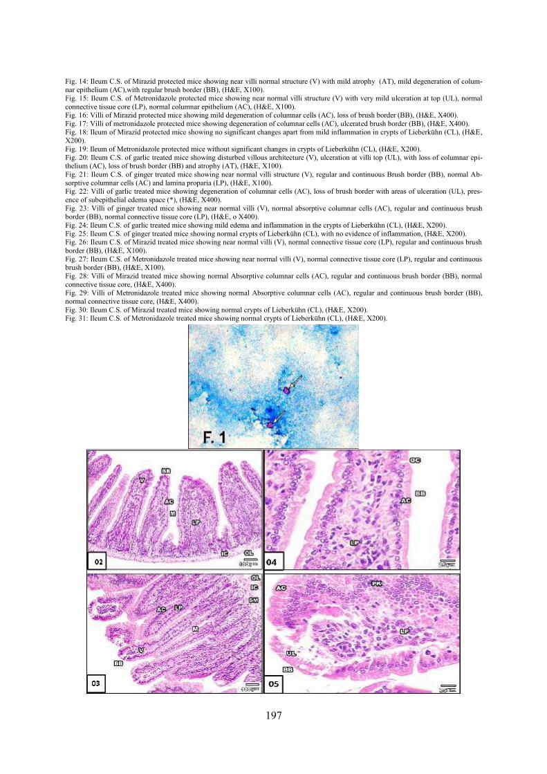
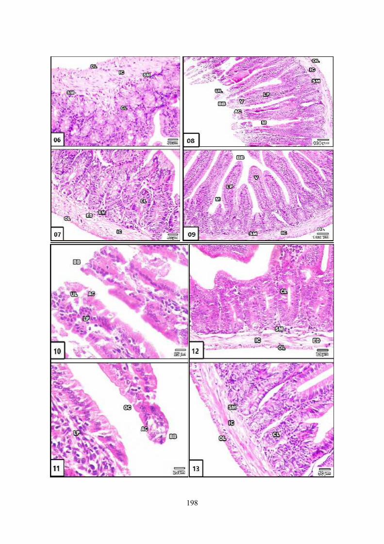
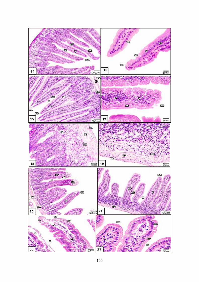
Explanation of figuresFig. 1: Cryptosporidial oocysts in stool of infected mice stained with MZN acid fast stain (x400)Fig. 2: A cross section (C.S.) of ileum of control uninfected mice showing normal structure of villi (V), regular and continuous Brush border(BB), Absorptive columnar cells (AC) and lamina proparia (LP), (H&E, X100).Fig. 3: Ileum C.S. of control infected mice showing disturbed villous architecture, hyperplasia of villi (V) with local ulceration (UL), (H&E,X100).Fig. 4: Villi of control uninfected mice showing normal structure of villi (V), regular and continuous brush border (BB), presence of gobletcells between enterocytes (GC), normal connective tissue core of villi (LP), (H&E, X400).Fig. 5: Villi of control infected mice showing degeneration of columnar epithelium (AC) with many pyknotic nuclei (PK) and appearance ofoocyst (OO) at denucleated brush border (BB), (H&E, X400).Fig. 6: Ileum of control uninfected mice showing normal structure of crypts of Lieberkühn (CL), (H&E, X200).Fig. 7: Ileum C.S. of control infected mice showing mild edema with very mild inflammatory infiltrate in crypts of Lieberkühn (CL), (H&E,X200).Fig. 8: An ileum of garlic protected mice showing disturbed villous architecture (V), ulceration at villi top (UL), with loss of columnar epi-thelium (AC), loss of brush border (BB), (H&E, X100).Fig. 9: Ileum C.S. of ginger protected mice showing near normal villi (V) apart from mild atrophy (AT), (H&E, X100).Fig. 10: Villi of garlic protected mice showing degeneration of columnar cells (AC), no brush border (BB) with areas of ulceration (UL),atrophy at villi top (AT), (H&E, X400).Fig. 11: Villi of ginger protected mice showing very mild degeneration of columnar epithelium (AC) with preserved brush border (BB),(H&E, X400).Fig. 12: Ileum of garlic protected mice showing mild edema (ED) and inflammation of crypts of Lieberkühn (CL), (H&E, X200).Fig. 13: Ileum of ginger protected mice showing normal crypts of Lieberkühn (CL) without evidence of inflammation, (H&E, X200).
197
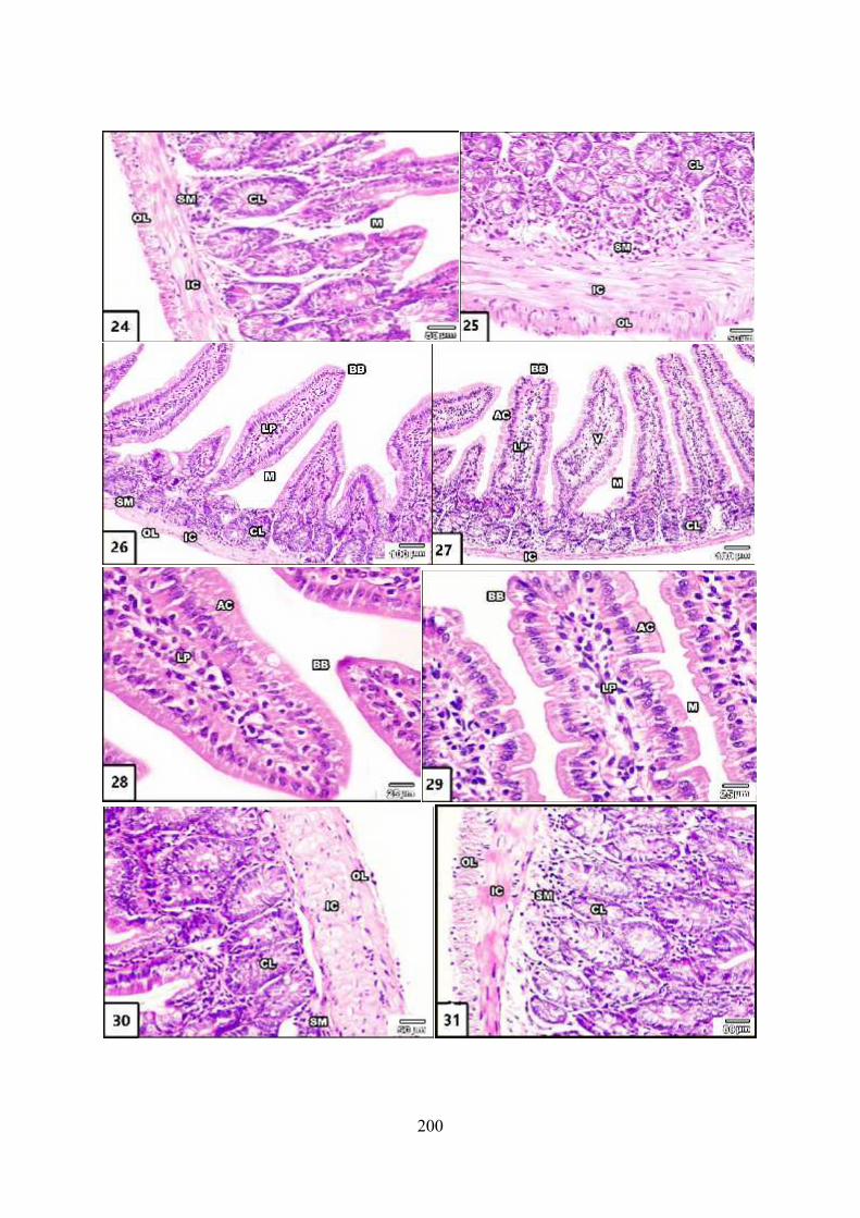
Fig. 14: Ileum C.S. of Mirazid protected mice showing near villi normal structure (V) with mild atrophy (AT), mild degeneration of colum-nar epithelium (AC),with regular brush border (BB), (H&E, X100).Fig. 15: Ileum C.S. of Metronidazole protected mice showing near normal villi structure (V) with very mild ulceration at top (UL), normalconnective tissue core (LP), normal columnar epithelium (AC), (H&E, X100).Fig. 16: Villi of Mirazid protected mice showing mild degeneration of columnar cells (AC), loss of brush border (BB), (H&E, X400).Fig. 17: Villi of metronidazole protected mice showing degeneration of columnar cells (AC), ulcerated brush border (BB), (H&E, X400).Fig. 18: Ileum of Mirazid protected mice showing no significant changes apart from mild inflammation in crypts of Lieberkühn (CL), (H&E,X200).Fig. 19: Ileum of Metronidazole protected mice without significant changes in crypts of Lieberkühn (CL), (H&E, X200).Fig. 20: Ileum C.S. of garlic treated mice showing disturbed villous architecture (V), ulceration at villi top (UL), with loss of columnar epi-thelium (AC), loss of brush border (BB) and atrophy (AT), (H&E, X100).Fig. 21: Ileum C.S. of ginger treated mice showing near normal villi structure (V), regular and continuous Brush border (BB), normal Ab-sorptive columnar cells (AC) and lamina proparia (LP), (H&E, X100).Fig. 22: Villi of garlic treated mice showing degeneration of columnar cells (AC), loss of brush border with areas of ulceration (UL), pres-ence of subepithelial edema space (*), (H&E, X400).Fig. 23: Villi of ginger treated mice showing near normal villi (V), normal absorptive columnar cells (AC), regular and continuous brushborder (BB), normal connective tissue core (LP), (H&E, o X400).Fig. 24: Ileum C.S. of garlic treated mice showing mild edema and inflammation in the crypts of Lieberkühn (CL), (H&E, X200).Fig. 25: Ileum C.S. of ginger treated mice showing normal crypts of Lieberkühn (CL), with no evidence of inflammation, (H&E, X200).Fig. 26: Ileum C.S. of Mirazid treated mice showing near normal villi (V), normal connective tissue core (LP), regular and continuous brushborder (BB), (H&E, X100).Fig. 27: Ileum C.S. of Metronidazole treated mice showing near normal villi (V), normal connective tissue core (LP), regular and continuousbrush border (BB), (H&E, X100).Fig. 28: Villi of Mirazid treated mice showing normal Absorptive columnar cells (AC), regular and continuous brush border (BB), normalconnective tissue core, (H&E, X400).Fig. 29: Villi of Metronidazole treated mice showing normal Absorptive columnar cells (AC), regular and continuous brush border (BB),normal connective tissue core, (H&E, X400).Fig. 30: Ileum C.S. of Mirazid treated mice showing normal crypts of Lieberkühn (CL), (H&E, X200).Fig. 31: Ileum C.S. of Metronidazole treated mice showing normal crypts of Lieberkühn (CL), (H&E, X200).
198
199
200