Embed Size (px)
Citation preview
This document consists of 15 printed pages and 1 blank page.
DC (NF/SW) 68412/4© UCLES 2013 [Turn over
*3144561450*
BIOLOGY 9790/03
Paper 3 Practical Examination May/June 2013
2 hours 30 minutes
Candidates answer on the Question Paper.
Additional Materials: As listed in the Confidential Instructions.
READ THESE INSTRUCTIONS FIRST
Write your Centre number, candidate number and name on all the work you hand in.Write in dark blue or black pen.You may use a soft pencil for any diagrams, graphs or rough working.Do not use staples, paper clips, highlighters, glue or correction fluid.DO NOT WRITE IN ANY BARCODES.
Section AAnswer all questions.Write your answers in the spaces provided on the Question Paper.
Section BAnswer all questions.Write your answers in the spaces provided on the Question Paper.
Electronic calculators may be used.You may lose marks if you do not show your working or if you do not use appropriate units.
At the end of the examination, fasten all your work securely together.The number of marks is given in brackets [ ] at the end of each question or part question.
UNIVERSITY OF CAMBRIDGE INTERNATIONAL EXAMINATIONSCambridge International Level 3 Pre-U CertificatePrincipal Subject
For Examiner’s Use
Section A
Section B
Total
2
9790/03/M/J/13© UCLES 2013
ForExaminer’s
Use
Section A
Answer all the questions.
You are recommended to spend no longer than 90 minutes on question 1.
1 You should read through the whole of this question carefully and then plan your use of the time to make sure that you finish all the work that you would like to do.
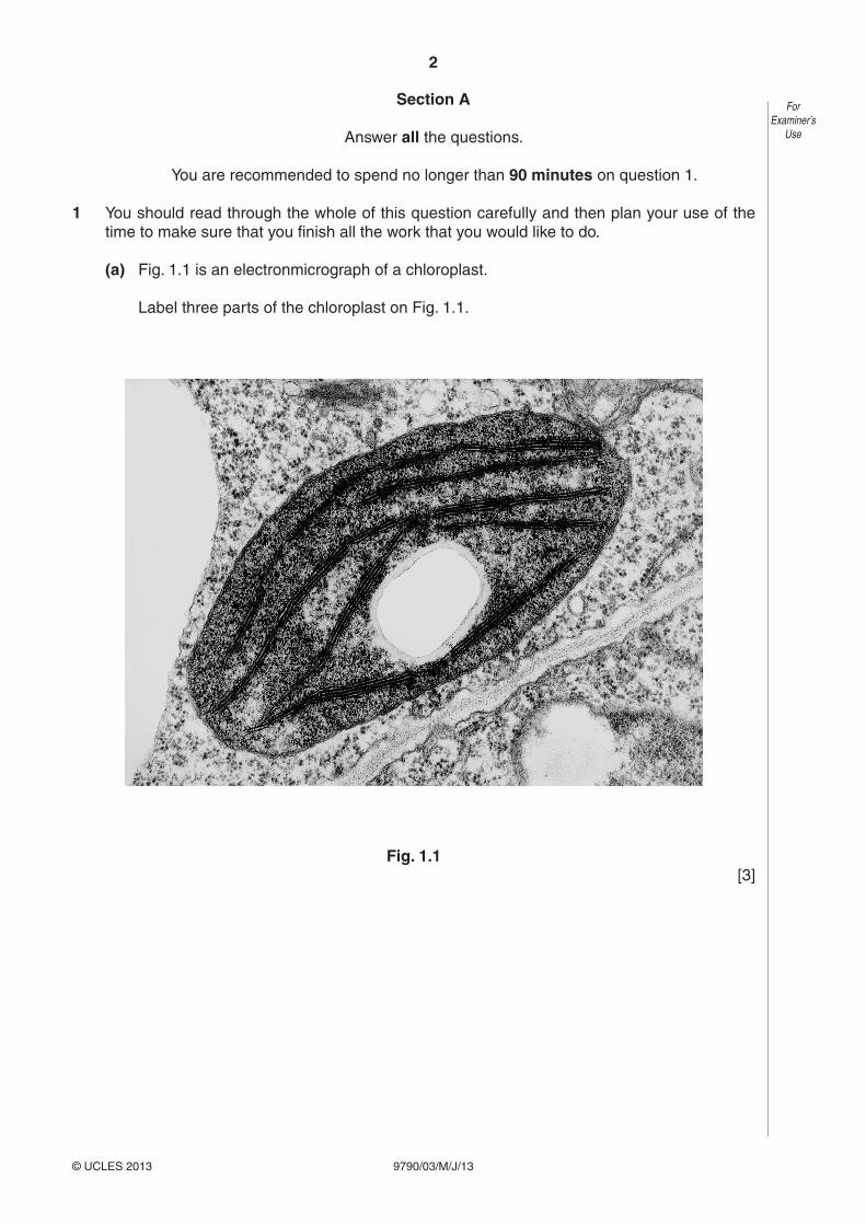
(a) Fig. 1.1 is an electronmicrograph of a chloroplast.
Label three parts of the chloroplast on Fig. 1.1.
Fig. 1.1 [3]

3
9790/03/M/J/13© UCLES 2013 [Turn over
ForExaminer’s
Use
(b) For this investigation you are supplied with a suspension of chloroplasts. The suspension was prepared in the following way.
1 Some leaves were cut into small pieces and placed in a blender.
2 The pieces were homogenised in an ice-cold sucrose solution buffered at pH 7.0.
3 The homogenised leaf tissue was filtered to remove cell debris.
Explain why chloroplasts are suspended in a medium which: • is ice-cold • is buffered • contains sucrose.
..........................................................................................................................................
..........................................................................................................................................
..........................................................................................................................................
..........................................................................................................................................
..........................................................................................................................................
..........................................................................................................................................
..........................................................................................................................................
..........................................................................................................................................
..................................................................................................................................... [4]
4
9790/03/M/J/13© UCLES 2013
ForExaminer’s
Use
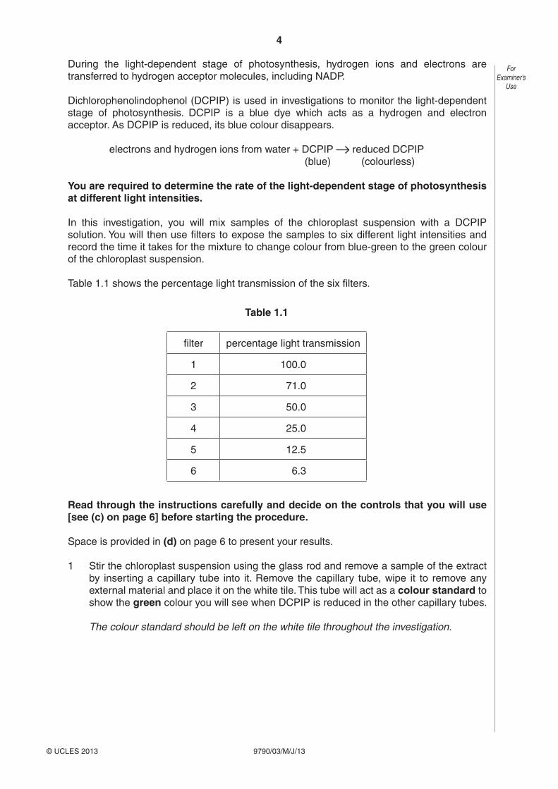
During the light-dependent stage of photosynthesis, hydrogen ions and electrons are transferred to hydrogen acceptor molecules, including NADP.
Dichlorophenolindophenol (DCPIP) is used in investigations to monitor the light-dependent stage of photosynthesis. DCPIP is a blue dye which acts as a hydrogen and electron acceptor. As DCPIP is reduced, its blue colour disappears.
electrons and hydrogen ions from water + DCPIP reduced DCPIP (blue) (colourless)
You are required to determine the rate of the light-dependent stage of photosynthesis at different light intensities.
In this investigation, you will mix samples of the chloroplast suspension with a DCPIP solution. You will then use filters to expose the samples to six different light intensities and record the time it takes for the mixture to change colour from blue-green to the green colour of the chloroplast suspension.
Table 1.1 shows the percentage light transmission of the six filters.
Table 1.1
filter percentage light transmission
1 100.0
2 71.0
3 50.0
4 25.0
5 12.5
6 6.3
Read through the instructions carefully and decide on the controls that you will use [see (c) on page 6] before starting the procedure.
Space is provided in (d) on page 6 to present your results.
1 Stir the chloroplast suspension using the glass rod and remove a sample of the extract by inserting a capillary tube into it. Remove the capillary tube, wipe it to remove any external material and place it on the white tile. This tube will act as a colour standard to show the green colour you will see when DCPIP is reduced in the other capillary tubes.
The colour standard should be left on the white tile throughout the investigation.
5
9790/03/M/J/13© UCLES 2013 [Turn over
ForExaminer’s
Use
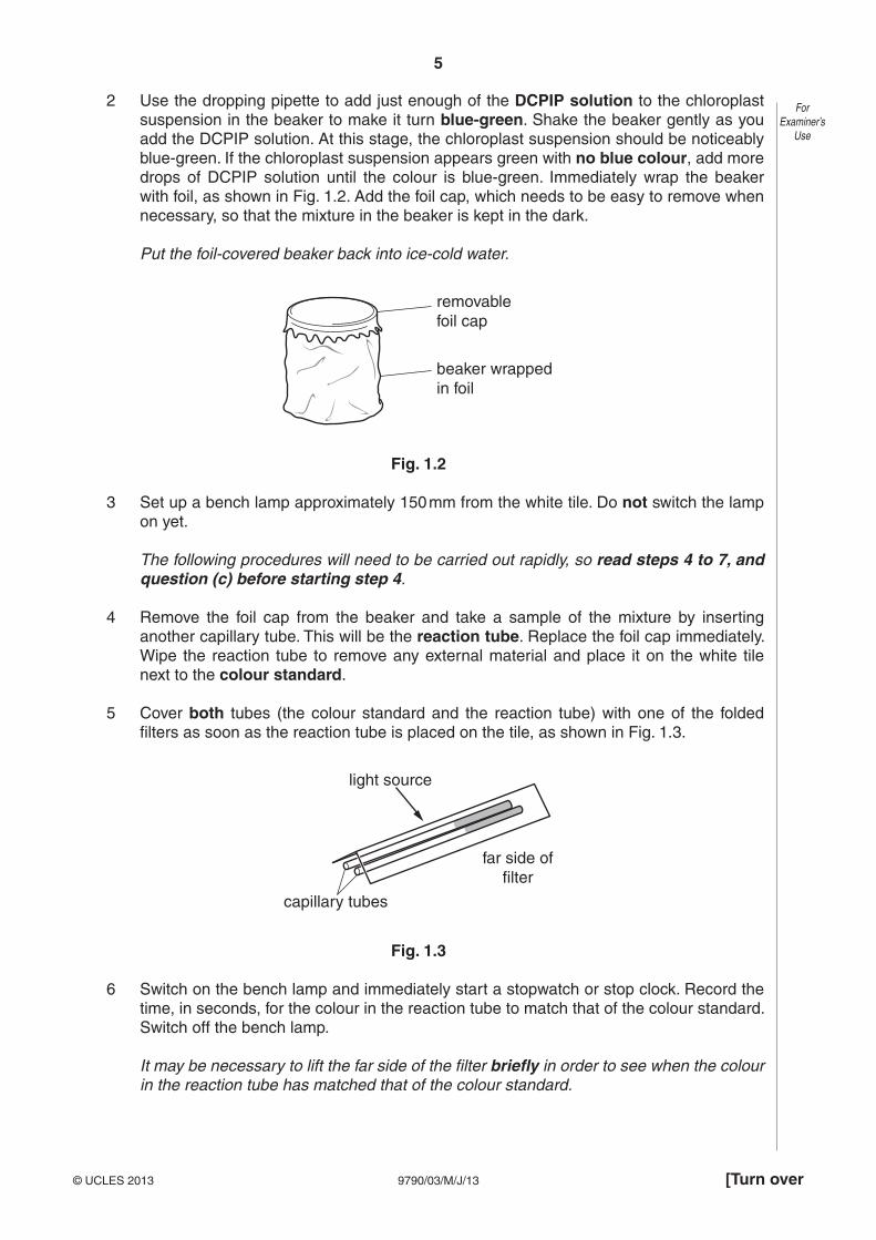
2 Use the dropping pipette to add just enough of the DCPIP solution to the chloroplast suspension in the beaker to make it turn blue-green. Shake the beaker gently as you add the DCPIP solution. At this stage, the chloroplast suspension should be noticeably blue-green. If the chloroplast suspension appears green with no blue colour, add more drops of DCPIP solution until the colour is blue-green. Immediately wrap the beaker with foil, as shown in Fig. 1.2. Add the foil cap, which needs to be easy to remove when necessary, so that the mixture in the beaker is kept in the dark.
Put the foil-covered beaker back into ice-cold water.
removablefoil cap
beaker wrappedin foil
Fig. 1.2
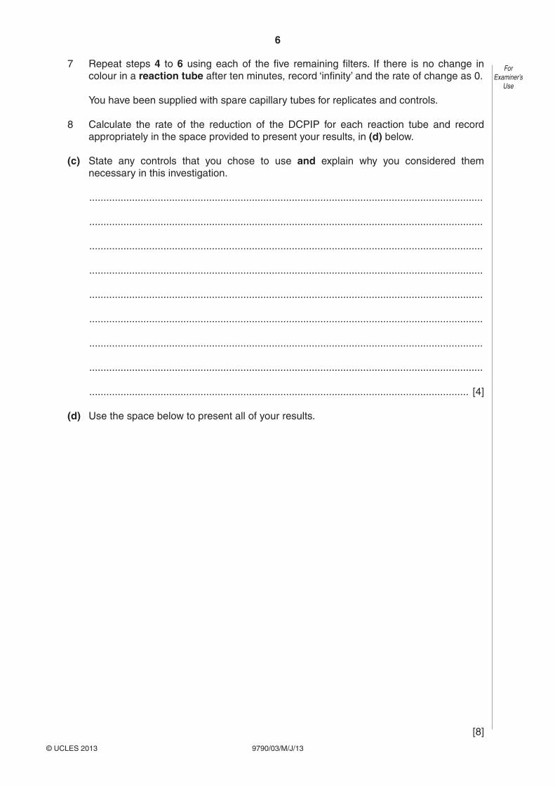
3 Set up a bench lamp approximately 150 mm from the white tile. Do not switch the lamp on yet.
The following procedures will need to be carried out rapidly, so read steps 4 to 7, and question (c) before starting step 4.
4 Remove the foil cap from the beaker and take a sample of the mixture by inserting another capillary tube. This will be the reaction tube. Replace the foil cap immediately. Wipe the reaction tube to remove any external material and place it on the white tile next to the colour standard.
5 Cover both tubes (the colour standard and the reaction tube) with one of the folded filters as soon as the reaction tube is placed on the tile, as shown in Fig. 1.3.
light source
capillary tubes
far side offilter
Fig. 1.3
6 Switch on the bench lamp and immediately start a stopwatch or stop clock. Record the time, in seconds, for the colour in the reaction tube to match that of the colour standard. Switch off the bench lamp.
It may be necessary to lift the far side of the filter briefly in order to see when the colour in the reaction tube has matched that of the colour standard.
6
9790/03/M/J/13© UCLES 2013
ForExaminer’s
Use
7 Repeat steps 4 to 6 using each of the five remaining filters. If there is no change in colour in a reaction tube after ten minutes, record ‘infinity’ and the rate of change as 0.
You have been supplied with spare capillary tubes for replicates and controls.
8 Calculate the rate of the reduction of the DCPIP for each reaction tube and record appropriately in the space provided to present your results, in (d) below.
(c) State any controls that you chose to use and explain why you considered them necessary in this investigation.
..........................................................................................................................................
..........................................................................................................................................
..........................................................................................................................................
..........................................................................................................................................
..........................................................................................................................................
..........................................................................................................................................
..........................................................................................................................................
..........................................................................................................................................
..................................................................................................................................... [4]
(d) Use the space below to present all of your results.
[8]
7
9790/03/M/J/13© UCLES 2013 [Turn over
ForExaminer’s
Use
(e) Draw a graph of your results on the grid below to show the effect of light intensity on the rate of the light-dependent stage of photosynthesis.
[5]
8
9790/03/M/J/13© UCLES 2013
ForExaminer’s
Use
(f) Describe and explain the pattern of results shown by your graph.
..........................................................................................................................................
..........................................................................................................................................
..........................................................................................................................................
..........................................................................................................................................
..........................................................................................................................................
..........................................................................................................................................
..........................................................................................................................................
..........................................................................................................................................
..........................................................................................................................................
..........................................................................................................................................
..........................................................................................................................................
..........................................................................................................................................
..........................................................................................................................................
..........................................................................................................................................
..........................................................................................................................................
..........................................................................................................................................
..........................................................................................................................................
..........................................................................................................................................
..........................................................................................................................................
..........................................................................................................................................
..........................................................................................................................................
..........................................................................................................................................
..........................................................................................................................................
..........................................................................................................................................
..........................................................................................................................................
..........................................................................................................................................
..........................................................................................................................................
..................................................................................................................................... [8]
9
9790/03/M/J/13© UCLES 2013 [Turn over
ForExaminer’s
Use
(g) DCPIP easily crosses chloroplast membranes. Some other electron acceptors used in investigations of the light-dependent stage of photosynthesis do not.
Suggest how a chloroplast suspension would be treated in order to be used with one of these other electron acceptors.
..........................................................................................................................................
..........................................................................................................................................
..........................................................................................................................................
..................................................................................................................................... [2]
(h) DCPIP accepts electrons from the electron transport chain between photosystem II (PSII) and photosystem I (PSI).
Explain how this allows the light-dependent stage of photosynthesis to be studied without any influence from the action of the light-independent stage (Calvin cycle).
..........................................................................................................................................
..........................................................................................................................................
..........................................................................................................................................
..........................................................................................................................................
..........................................................................................................................................
..........................................................................................................................................
..................................................................................................................................... [3]
(i) Identify the limitations and sources of error in the investigation that may have affected the quality of the results.
Explain how you would improve the method to overcome the limitations that you have identified.
..........................................................................................................................................
..........................................................................................................................................
..........................................................................................................................................
..........................................................................................................................................
..........................................................................................................................................
..........................................................................................................................................
..........................................................................................................................................

10
9790/03/M/J/13© UCLES 2013
ForExaminer’s
Use ..........................................................................................................................................
..........................................................................................................................................
..........................................................................................................................................
..........................................................................................................................................
..........................................................................................................................................
..........................................................................................................................................
..........................................................................................................................................
..........................................................................................................................................
..........................................................................................................................................
..........................................................................................................................................
..........................................................................................................................................
..........................................................................................................................................
..........................................................................................................................................
..........................................................................................................................................
..........................................................................................................................................
..........................................................................................................................................
..........................................................................................................................................
..........................................................................................................................................
..........................................................................................................................................
..........................................................................................................................................
..........................................................................................................................................
..........................................................................................................................................
..........................................................................................................................................
..........................................................................................................................................
..................................................................................................................................... [8]
[Total: 45]
11
9790/03/M/J/13© UCLES 2013 [Turn over
ForExaminer’s
Use
Section B
Answer all the questions.
You are recommended to spend no longer than 60 minutes on question 2.
2 You should read through the whole of this question carefully and then plan your use of the time to make sure that you finish all the work that you would like to do.
Fish gills are composed of gill filaments (primary lamellae). These support large numbers of secondary lamellae that form the gas exchange surface.
(a) Specimen K1 is a gill from a mackerel, Scomber scombrus.
Use the instruments and hand lens provided to help you examine the structure of the gills.
Make a drawing of specimen K1 in the space below.
Annotate your drawing to describe the appearance of the gill.
Indicate the magnification of your drawing.
magnification = ....................................................... [7]
12
9790/03/M/J/13© UCLES 2013
ForExaminer’s
Use
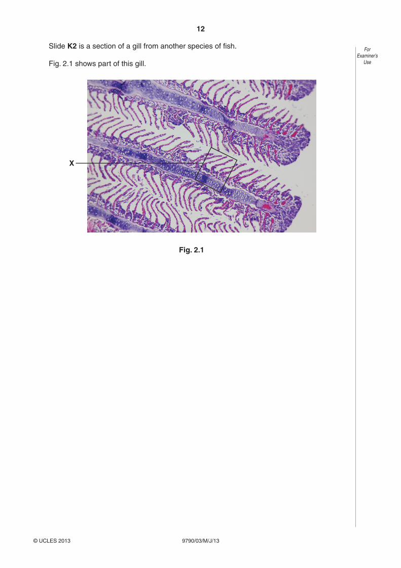
Slide K2 is a section of a gill from another species of fish.
Fig. 2.1 shows part of this gill.
X
Fig. 2.1

13
9790/03/M/J/13© UCLES 2013 [Turn over
ForExaminer’s
Use
(b) Use the high power of your microscope to make a drawing of the gill from slide K2, in an area similar to that indicated by X on Fig. 2.1.
Indicate the magnification of your drawing and explain how you calculated it.
magnification = .......................................................
explanation
..........................................................................................................................................
..........................................................................................................................................
..........................................................................................................................................
..........................................................................................................................................
..........................................................................................................................................
..................................................................................................................................... [8]
(c) Suggest how the surface area of one secondary lamella could be determined.
..........................................................................................................................................
..........................................................................................................................................
..........................................................................................................................................
..........................................................................................................................................
..........................................................................................................................................
..................................................................................................................................... [3]
14
9790/03/M/J/13© UCLES 2013
ForExaminer’s
Use
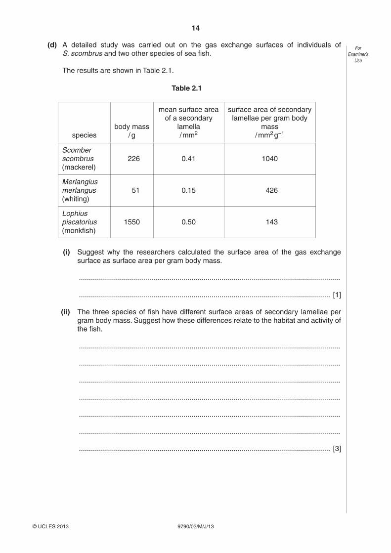
(d) A detailed study was carried out on the gas exchange surfaces of individuals of S. scombrus and two other species of sea fish.
The results are shown in Table 2.1.
Table 2.1
speciesbody mass
/ g
mean surface area of a secondary
lamella/ mm2
surface area of secondary lamellae per gram body
mass/ mm2 g−1
Scomber scombrus(mackerel)
226 0.41 1040
Merlangius merlangus(whiting)
51 0.15 426
Lophius piscatorius(monkfish)
1550 0.50 143
(i) Suggest why the researchers calculated the surface area of the gas exchange surface as surface area per gram body mass.
..................................................................................................................................
............................................................................................................................. [1]
(ii) The three species of fish have different surface areas of secondary lamellae per gram body mass. Suggest how these differences relate to the habitat and activity of the fish.
..................................................................................................................................
..................................................................................................................................
..................................................................................................................................
..................................................................................................................................
..................................................................................................................................
..................................................................................................................................
............................................................................................................................. [3]
15
9790/03/M/J/13© UCLES 2013
ForExaminer’s
Use
Slide K3 is a section of a lung from a small mammal.
Use the high power of your microscope to examine the lung tissue carefully.
(e) Lung tissue consists of airways, blood vessels and alveoli.
Compare the features of these three structures that are visible in slide K3.
Present your comparison as a table in the space below.
[7]
(f) Use the space below to compare gas exchange in mackerel with that in a small mammal.
[6]
[Total: 35]
16
9790/03/M/J/13© UCLES 2013
BLANK PAGE
Copyright Acknowledgements:
Question 1 Fig. 1.1 © DR JEREMY BURGESS/SCIENCE PHOTO LIBRARY.
Permission to reproduce items where third-party owned material protected by copyright is included has been sought and cleared where possible. Every reasonable effort has been made by the publisher (UCLES) to trace copyright holders, but if any items requiring clearance have unwittingly been included, the publisher will be pleased to make amends at the earliest possible opportunity.
University of Cambridge International Examinations is part of the Cambridge Assessment Group. Cambridge Assessment is the brand name of University of Cambridge Local Examinations Syndicate (UCLES), which is itself a department of the University of Cambridge.