Embed Size (px)
Citation preview
Keratoprosthesis (Artificial Cornea)
Policy Number: 9.03.01 Last Review: 6/2017 Origination: 6/2007 Next Review: 6/2018
Policy Blue Cross and Blue Shield of Kansas City (Blue KC) will provide coverage for
keratoprostheses when it is determined to be medically necessary because the
criteria shown below are met.
When Policy Topic is covered The Boston (Dohlman-Doane) Keratoprosthesis (Boston KPro) may be considered
medically necessary for the surgical treatment of severe corneal opacification in situations where cadaveric corneal transplants have failed or have a very low
likelihood of success (see Considerations).
When Policy Topic is not covered A permanent keratoprosthesis for all other conditions is considered investigational.
All other types of permanent keratoprostheses are considered investigational
Considerations Implantation of a keratoprosthesis is considered a high-risk procedure associated
with numerous complications and probable need for additional surgery. Therefore,
the likelihood of regaining vision and the patient’s visual acuity in the contralateral
eye should be taken into account when considering the appropriateness of this procedure. Treatment should be restricted to centers experienced in treating this
condition and staffed by surgeons adequately trained in techniques addressing
implantation of this device.
Conditions under which cadaveric corneal transplants have a likelihood of failure include but are not limited to the following:
The cornea is severely opaque and vascularized AND
Best-corrected visual acuity is 20/400 or less in the affected eye and 20/40 or less in the contralateral eye AND
No end-stage glaucoma or retinal detachment is present AND
Keratoprosthesis (Artificial Cornea) 9.03.01
The patient has one of the following indications:
o History of 1 or more corneal transplant graft failures
o Stevens-Johnson syndrome o Ocular cicatricial pemphigoid
o Autoimmune conditions with rare ocular involvement
o Ocular chemical burns
o An ocular condition unlikely to respond favorably to primary corneal transplant surgery (eg, libel stem cell compromise or postherpetic
anesthesia)
Note that patients should be able and expected to comply with postoperative care.
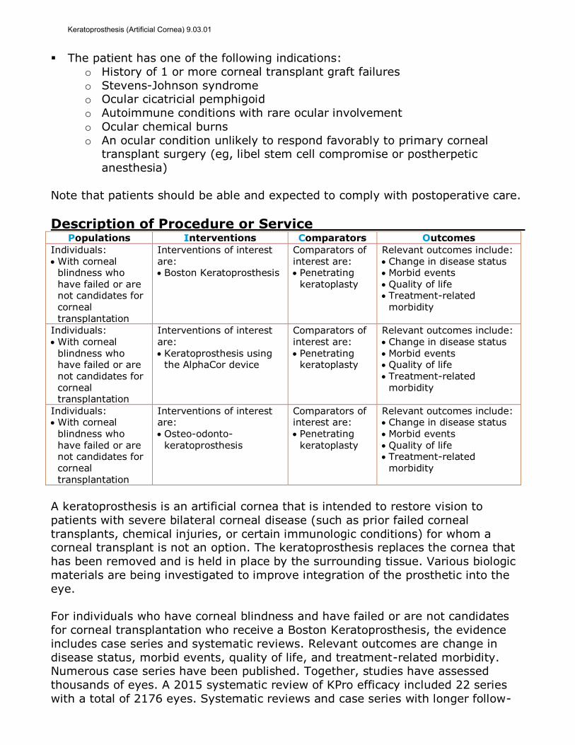
Description of Procedure or Service Populations Interventions Comparators Outcomes
Individuals: With corneal
blindness who
have failed or are not candidates for corneal transplantation
Interventions of interest are: Boston Keratoprosthesis
Comparators of interest are: Penetrating
keratoplasty
Relevant outcomes include: Change in disease status Morbid events
Quality of life Treatment-related
morbidity
Individuals: With corneal
blindness who have failed or are not candidates for corneal transplantation
Interventions of interest are:
Keratoprosthesis using the AlphaCor device
Comparators of interest are:
Penetrating keratoplasty
Relevant outcomes include: Change in disease status
Morbid events Quality of life Treatment-related
morbidity
Individuals: With corneal
blindness who have failed or are not candidates for corneal transplantation
Interventions of interest are:
Osteo-odonto-keratoprosthesis
Comparators of interest are:
Penetrating keratoplasty
Relevant outcomes include: Change in disease status
Morbid events Quality of life Treatment-related
morbidity
A keratoprosthesis is an artificial cornea that is intended to restore vision to
patients with severe bilateral corneal disease (such as prior failed corneal
transplants, chemical injuries, or certain immunologic conditions) for whom a corneal transplant is not an option. The keratoprosthesis replaces the cornea that
has been removed and is held in place by the surrounding tissue. Various biologic
materials are being investigated to improve integration of the prosthetic into the
eye.
For individuals who have corneal blindness and have failed or are not candidates
for corneal transplantation who receive a Boston Keratoprosthesis, the evidence
includes case series and systematic reviews. Relevant outcomes are change in
disease status, morbid events, quality of life, and treatment-related morbidity. Numerous case series have been published. Together, studies have assessed
thousands of eyes. A 2015 systematic review of KPro efficacy included 22 series
with a total of 2176 eyes. Systematic reviews and case series with longer follow-
Keratoprosthesis (Artificial Cornea) 9.03.01
up (ie, at least 2 years) have shown improvement in visual outcomes in a
substantial percentage of patients with Boston KPro. This procedure is high risk
and associated with numerous complications (eg, growth of retro prosthetic membranes) and a probable need for additional surgery, thus careful patient
selection is important. The evidence is sufficient to determine that the technology
results in a meaningful improvement in the net health outcome.
For individuals who have corneal blindness and have failed or are not candidates
for corneal transplantation who receive a keratoprosthesis using the AlphaCor
device, the evidence includes case series. Relevant outcomes are change in
disease status, morbid events, quality of life, and treatment-related morbidity. Only a few published case series have evaluated the AlphaCor device. There are
insufficient data on improvement in vision outcomes using the AlphaCor device.
Moreover, the device has been associated with complications, including thinning or
melting of the anterior corneal surface and corneal necrosis. The evidence is insufficient to determine the effects of the technology on health outcomes.
For individuals who have corneal blindness and have failed or are not candidates
for corneal transplantation who receive an osteo-odonto-keratoprosthesis (OOKP),
the evidence includes case series and a systematic review. Relevant outcomes are change in disease status, morbid events, quality of life, and treatment-related
morbidity. A 2012 systematic review of case series, all conducted outside of the
United States, found high anatomic survival rates at 5 and 20 years, but vision
outcomes were not well-described. OOKP is a complex surgical procedure and has been associated with a number of complications, including extrusion of the
keratoprosthesis, retinal detachment, and vitreoretinal complications. The
evidence is insufficient to determine the effects of the technology on health
outcomes.
Background
The cornea, a clear, dome-shaped membrane that covers the front of the eye, is a
key refractive element of the eye. Layers of the cornea consist of the epithelium
(outermost layer); Bowman’s layer; the stroma, which comprises approximately 90% of the cornea; Descemet’s membrane; and the endothelium. The established
surgical treatment for corneal disease is penetrating keratoplasty (PK), which
involves making a large central opening through the cornea and then filling the
opening with full-thickness donor cornea. In certain conditions, such as Stevens-Johnson syndrome; cicatricial pemphigoid; chemical injury; or prior failed corneal
transplant, survival of transplanted cornea is poor. The keratoprosthesis has been
developed to restore vision in patients for whom a corneal transplant is not an
option.
Keratoprosthetic devices consist of a central optic held in a cylindrical frame. The
keratoprosthesis replaces the section of cornea that has been removed, and, along
with being held in place by the surrounding tissue, may be covered by a membrane to further anchor the prosthesis. A variety of biologic materials are
being investigated to improve the integration of prosthetic corneal implants into
the stroma and other corneal layers.
Keratoprosthesis (Artificial Cornea) 9.03.01
The Dohlman-Doane Keratoprosthesis, most commonly referred to as the Boston
Keratoprosthesis (KPro), is manufactured under the auspices of the Harvard Medical School‒affiliated Massachusetts Eye and Ear Infirmary. The Boston type I
KPro uses a donor cornea between a central stem and a back plate. The Boston
type II prosthesis is a modification of the type I prosthesis and is designed with an
anterior extension to allow implantation through surgically closed eyelids. The AlphaCor, previously known as the Chirila keratoprosthesis (Chirila KPro) consists
of a polymethylmethacrylate (PMMA) device with a central optic region fused with
a surrounding sponge skirt; the device is inserted in a 2-stage surgical procedure.
Autologous keratoprostheses use a central PMMA optic supported by a skirt of
either tibia bone or the root of a tooth with its surrounding alveolar bone. The
most common is the OOKP, which uses osteodental lamina derived from an
extracted tooth root and attached alveolar bone that has been removed from the patient’s jaw. Insertion of the OOKP device requires a complex staged procedure,
in which the cornea is first covered with buccal mucosa. The prosthesis itself
consists of a PMMA optical cylinder, which replaces the cornea, and is held in place
by a biological support made from a canine tooth extracted from the recipient. A
hole is drilled through the dental root and alveolar bone, and the PMMA prosthesis is placed within. This entire unit is placed into a subcutaneous ocular pocket and is
then retrieved 6 to 12 months later for final insertion.
Hydroxyapatite, with a similar mineral composition to both bone and teeth (phosphate and calcium), may also be used as a bone substitute and as a
bioactive prosthesis with the orbit. Collagen coating and scaffolds have also been
investigated to improve growth and biocompatibility with the cornea epithelial
cells, which form the protective layer of the eye. Many of these materials and devices are currently being tested in vitro or in animal models.
Regulatory Status
In January 1992, the Boston KPro (Dohlman-Doane keratoprosthesis;
Massachusetts Eye and Ear Infirmary) was approved by the U.S. Food and Drug Administration (FDA) through the premarket approval process for use in patients
with severe corneal opacity. The device is used when standard corneal transplant
has failed or would be unlikely to succeed. There are 2 types of Boston KPro. Type
1 is used in eyes when eyelids, blink mechanism, and tear film are intact. Type 2 is used with severe dry eye and in eyes with mucosal keratinization and
obliteration of normal conjunctival fornices.
In August 2002, the AlphaCor® (Chirila keratoprosthesis) was cleared for marketing by FDA through the 510(k) process. FDA determined that this device
was substantially equivalent to the Dolman-Doane keratoprosthesis. The
AlphaCor® device is indicated as a keratoprosthesis in adults with corneal opacity
when standard penetrating keratoplasty with donor tissue is not suitable, when patients have declined standard penetrating keratoplasty, or when adjunctive
procedures to prevent graft rejection are contraindicated.
Keratoprosthesis (Artificial Cornea) 9.03.01
Rationale This evidence review was originally created in March 1996 and has been updated
regularly with searches of the MEDLINE database. The most recent literature review was performed through January 25, 2017.
The keratoprosthesis is intended for the relatively small number of patients with
severe corneal damage who have lost vision and for whom a corneal transplant is not expected to result in satisfactory outcomes. These criteria generally refer to
the population of patients who have failed 1 or more corneal transplants and who
therefore have very few options to prevent blindness. Because this surgery is
considered a salvage procedure with no acceptable alternative treatments,
comparative studies are limited and/or lacking. The available literature primarily consists of retrospective case series. This evidence review examines the types of
devices currently being tested in humans, focusing on reports that permit
assessment of integration within the eye, durability, visual outcomes, and adverse
events following implantation.
Boston (Dohlman-Doane) Keratoprosthesis (KPro)
Systematic Reviews A 2015 systematic review from the American Academy of Ophthalmology identified
22 studies on the efficacy and safety of the Boston (Dohlman-Doane)
Keratoprosthesis (Boston KPro).1 Studies were published in English and
retrospective series had to include at least 25 eyes. The 22 studies included a total
of 2176 eyes; sample sizes in individual studies ranged from 30 to 300 eyes. The proportion of patients with visual acuity of 20/200 after surgery ranged from 54%
to 84% in the 10 studies reporting this outcome. Five articles reported that 11%
to 39% of treated eyes attained visual acuities of 20/40 or better. Reviewers noted
that published data were skewed toward visual improvement. Fourteen articles reported retention rates (eyes retaining the KPro device without loss, extrusion or
dehiscence of the device), and these rates ranged from 65% to 100% (mean,
88%). The most common reasons for KPro loss were corneal melts with device
exposure or extrusion, endophthalmitis, infectious keratitis, or corneal ulceration. The most common complication was retroprosthetic membrane formation, which
ranged from 1% to 65% (mean, 30%) in the 13 studies reporting complications.
A 2016 systematic review by Ahmad et al examined 26 studies on repeat penetrating keratoplasty (PK) versus Boston KPro implantation after failed PK.2
Studies selected focused on patients with corneal opacity who had failed 1 or more
PKs. Studies were excluded if they only selected patients with ocular surface
disease. The primary outcome of interest was the proportion of patients with visual
acuity of 20/200 or better at 2 or more years postsurgery. In a meta-analysis of 9 studies, the likelihood of 20/200 vision or better at least 2 years after repeat PK
surgery was 42% (95% confidence interval [CI], 30% to 56%). A total of 104
eyes from 98 patients underwent KPro after failed PK surgery; 31 patients had
only 1 previous PK. In a meta-analysis of data on KPro implantation after failed PK surgery, the probability of maintaining visual acuity of 20/200 or better at 2 years
Keratoprosthesis (Artificial Cornea) 9.03.01
was 80% (95% CI, 68% to 88%). Among patients with a history of 1 failed PK,
the probability of maintaining a visual acuity of 20/200 or better at 2 years was
74% (95% CI, 45% to 89%). (Reviewers did not specify the number of patients receiving KPro who were included in the analysis of 20/200 vision at 2 years.) In
terms of complications after KPro following failed PK, at 2 years 29% of patients
had elevated intraocular pressure (IOP) and 8% needed surgery for glaucoma. In
an analysis limited to patients undergoing KPro after 1 failed KP, complication rates ranging from 29% to 10% (which did not differ significantly from patients
with KPro after >1 failed KPs). Reviewers did not report the number of patients
included in the complication analyses.
Case Series
Representative larger series include a 2013 report from the Boston Type 1
Keratoprosthesis Study Group assessed retention of the KPro device in 300 eyes of
300 patients.3 At a mean follow-up of 17.1 months (range, 1 week to 6 years), 93% of the keratoprostheses were retained. The probability of retention was 94%
at 1 year and 89% at 2 years. Mean device durability was 3.8 years. Risk factors
for keratoprosthesis loss were autoimmune disease, ocular surface exposure, and
number of prior failed PK procedures. Additional data on this cohort were
published in 2016.4 Preoperative visual acuity, available for 47% of eyes, was 20/1205. During a mean follow-up of 17 months (range, 1 week to 6 years), visual
acuity improved significantly for 85% of eyes to a final mean of 20/150. Median
time to achieve visual acuity of 20/200 was 1 month, and this level of acuity lasted
for a mean of 48 months among patients with sufficient follow-up.
In 2014, Srikumaran et al reported mean follow-up of 46.7 months (range, 6
weeks to 8.7 years) for 139 eyes of 133 patients who had received a Boston KPro
at 1 of 5 tertiary referral centers in the United States.5 Twenty-seven percent of eyes underwent a primary KPro procedure while 73% had a prior donor graft
failure. Postoperatively, visual acuity improved to at least 20/200 in 70% of eyes.
The probability of maintaining visual acuity of at least 20/200 was 50%, and
device retention was estimated at 67% at 7 years. The 7-year cumulative
incidence of complications was 49.7% for retroprosthetic membrane formation, 21.6% for glaucoma surgery, 18.6% for retinal detachment, and 15.5% for
endophthalmitis.
In 2010, Dunlap et al retrospectively analyzed 122 patients (126 eyes) at 2 centers who received a Boston type 1 keratoprosthesis between 2004 and 2007.6
For most patients, the affected eye had a visual acuity of less than 20/400, and
the contralateral eye did not have better vision. Of the 126 eyes, 112 had a
history of multiple failed corneal grafts, and 14 had received the keratoprosthesis as a primary procedure due to the presence of limbal stem cell deficiency or
significant ocular surface diseases. Following implantation, 96 (76%) eyes had
improved vision, 22 (17.4%) eyes did not improve, and 8 (6.3%) eyes lost vision.
At 3-month follow-up, 54% of eyes had 20/200 vision or better, with 18% achieving 20/40 or better. In approximately 45% of the eyes, visual acuity
remained less than 20/400. The percentage of patients with improved visual
Keratoprosthesis (Artificial Cornea) 9.03.01
outcomes was lower than in other published studies, due in part to the presence of
comorbid conditions (eg, glaucoma, retinal detachment).
Complications
In 2015, Odorcic et al published a literature review on fungal infections after
Boston type 1 KPro.7 They identified 15 relevant publications, primarily
retrospective case series. Annual rates of fungal infections reported in these studies ranged from 0.9 to 2 per 100 patients. The largest case series assessed
291 eyes, and the cumulative incidence of fungal endophthalmitis was 2.4% over
10 years.
In 2016, Chan et al retrospectively reviewed 110 patients (128 eyes) who received
a Boston type 1 KPro, focusing on corneal melts, leaks, and extrusions.8 Mean
follow-up was 29 months (range, 3-77 months). Melt-related complications
requiring surgical repair occurred in 16% (20/128) of eyes; 7 of these eyes had multiple episodes. The average time to a melt complication was 13 months after
KPro implantation. Risk factors significantly associated with melt-related
complications were previous infectious keratitis, and conjunctival deficiency caused
by Stevens-Johnson syndrome, mucous membrane pemphigoid, or previous
chemical injury.
A 2012 prospective series of 265 eyes (265 patients) from 18 medical centers,
published by the Boston Type 1 Keratoprosthesis Study Group, focused on the
time to development of retroprosthetic membranes.9 Most eyes (85.4%) had undergone an average of 2.2 (range, 1-8) PKs before keratoprosthesis
implantation. The remaining eyes (14.6%) were considered at high risk for PK
failure and had received a primary keratoprosthesis. At a mean follow-up of 17.8
months, retroprosthetic membranes had formed in 31.7% of eyes. The mean time to development of retroprosthetic membranes was 216.7 days (range, 7 days to 4
years). Risk factors were the indication for the keratoprosthesis, specifically,
infectious keratitis had a hazard ratio of 3.2 (95% CI, 1.7 to 6.2) and aniridia had
a hazard ratio of 3.1 (95% CI, 1.1 to 8.9).
Posterior segment complications were reported by Goldman et al in 2013.10 Of 83
eyes (93 procedures) with follow-up of at least 6 months (range, 6-84 months),
38 (40.9%) eyes had at least 1 postoperative posterior segment complication,
which included retinal detachment (16.9%), choroidal detachment (16.9%), and sterile vitritis (14.5%). Visual acuity was worse in eyes that experienced posterior
segment complications than in eyes that did not.
Section Summary: Boston Keratoprosthesis Numerous case series and systematic reviews of these series have assessed
thousands of eyes implanted with the KPro device. A 2015 systematic review of
KPro efficacy included 22 series with a total of 2176 eyes. Studies with longer
follow-up (ie, at least 2 years) have shown improved visual outcomes in a substantial percentage of patients with Boston KPro. This procedure is high risk
and is associated with numerous complications (eg, growth of retroprosthetic
Keratoprosthesis (Artificial Cornea) 9.03.01
membranes) and a probable need for additional surgery, thus careful patient
selection is important.
AlphaCor Device
Studies have suggested that, with the AlphaCor device, thinning or “melting” of
the anterior corneal surface can lead to loss of biointegration.11,12 This
complication appears most prevalent in patients with ocular herpes, hence, the AlphaCor device is contraindicated in these patients.
Several case series evaluating the AlphaCor have been published. One of the
larger series was published in 2003 by Hicks et al.11 It included 40 devices implanted in 38 patients.
At an average 30-month follow-up, 42% of eyes had a visual acuity better than
20/200. In 2015, Hoffart et al evaluated the AlphaCor device implanted in 12 patients.13 At a mean follow-up of 25 months, 8 (67%) of devices were retained
and patients had a mean gain in BCVA of 2.5 lines. The most common
complication was corneal necrosis, observed in 7 (59%) patients, 2 of whom had a
history of ocular herpes.
Section Summary: AlphaCor Device
Only a few published case series have evaluated the AlphaCor device, and hence
there are insufficient data on improvements in vision outcomes this device.
Moreover, the device has been associated with complications, including thinning or melting of the anterior corneal surface and corneal necrosis.
Osteo-Odonto-Keratoprosthesis
A 2012 systematic review by Tan et al included 8 case series describing surgical outcomes and complication rates of the osteo-odonto-keratoprosthesis (OOKP).14
Sample sizes ranged from 4 to 181 eyes. None of the studies was conducted in the
United States. At 5 years, the pooled anatomic survival rate was 87.8% (range,
67%-100%) and, at 20 years, based on pooled data from 3 series, the anatomic
survival rate was 81.0% (range, 65%-98%). About half of the patients obtained visual acuity better than 6/18. Visual acuity in the other patients was not
described.
One of the largest case series (included in Tan) is that by Falcinelli et al, who reported on OOKP in 181 patients.15 At a median follow-up of 12 years, survival
analysis estimated that the probability of retaining an anatomically intact OOKP 18
years after surgery with reasonable visual acuity was 85%.
In 2008, investigators from Spain retrospectively reviewed 227 patients who
underwent OOKP (n=145) or osteokeratoprosthesis (OKP; n=82) using tibial bone
in patients who lacked canine teeth to assemble the prosthesis.16 A second
publication in 2011 from the same study examined the impact of clinical factors on long-term functional and anatomic outcomes.17 The primary diagnosis was
chemical or thermal burn (48%), Steven-Johnson syndrome and Lyell syndrome
(13%), cicatricial pemphigoid (11%), trachoma (11%), and other or not
Keratoprosthesis (Artificial Cornea) 9.03.01
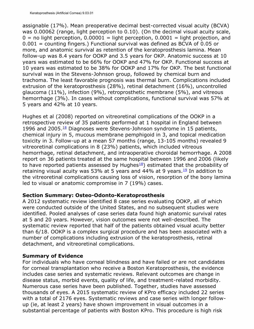
assignable (17%). Mean preoperative decimal best-corrected visual acuity (BCVA)
was 0.00062 (range, light perception to 0.10). (On the decimal visual acuity scale,
0 = no light perception, 0.00001 = light perception, 0.0001 = light projection, and 0.001 = counting fingers.) Functional survival was defined as BCVA of 0.05 or
more, and anatomic survival as retention of the keratoprosthesis lamina. Mean
follow-up was 8.4 years for OOKP and 3.5 years for OKP. Anatomic success at 10
years was estimated to be 66% for OOKP and 47% for OKP. Functional success at 10 years was estimated to be 38% for OOKP and 17% for OKP. The best functional
survival was in the Stevens-Johnson group, followed by chemical burn and
trachoma. The least favorable prognosis was thermal burn. Complications included
extrusion of the keratoprosthesis (28%), retinal detachment (16%), uncontrolled glaucoma (11%), infection (9%), retroprosthetic membrane (5%), and vitreous
hemorrhage (3%). In cases without complications, functional survival was 57% at
5 years and 42% at 10 years.
Hughes et al (2008) reported on vitreoretinal complications of the OOKP in a
retrospective review of 35 patients performed at 1 hospital in England between
1996 and 2005.18 Diagnoses were Stevens-Johnson syndrome in 15 patients,
chemical injury in 5, mucous membrane pemphigoid in 3, and topical medication
toxicity in 3. Follow-up at a mean 57 months (range, 13-105 months) revealed 9 vitreoretinal complications in 8 (23%) patients, which included vitreous
hemorrhage, retinal detachment, and intraoperative choroidal hemorrhage. A 2008
report on 36 patients treated at the same hospital between 1996 and 2006 (likely
to have reported patients assessed by Hughes18) estimated that the probability of retaining visual acuity was 53% at 5 years and 44% at 9 years.19 In addition to
the vitreoretinal complications causing loss of vision, resorption of the bony lamina
led to visual or anatomic compromise in 7 (19%) cases.
Section Summary: Osteo-Odonto-Keratoprosthesis
A 2012 systematic review identified 8 case series evaluating OOKP, all of which
were conducted outside of the United States, and no subsequent studies were
identified. Pooled analyses of case series data found high anatomic survival rates
at 5 and 20 years. However, vision outcomes were not well-described. The systematic review reported that half of the patients obtained visual acuity better
than 6/18. OOKP is a complex surgical procedure and has been associated with a
number of complications including extrusion of the keratoprosthesis, retinal
detachment, and vitreoretinal complications.
Summary of Evidence
For individuals who have corneal blindness and have failed or are not candidates
for corneal transplantation who receive a Boston Keratoprosthesis, the evidence includes case series and systematic reviews. Relevant outcomes are change in
disease status, morbid events, quality of life, and treatment-related morbidity.
Numerous case series have been published. Together, studies have assessed
thousands of eyes. A 2015 systematic review of KPro efficacy included 22 series with a total of 2176 eyes. Systematic reviews and case series with longer follow-
up (ie, at least 2 years) have shown improvement in visual outcomes in a
substantial percentage of patients with Boston KPro. This procedure is high risk
Keratoprosthesis (Artificial Cornea) 9.03.01
and associated with numerous complications (eg, growth of retro prosthetic
membranes) and a probable need for additional surgery, thus careful patient
selection is important. The evidence is sufficient to determine that the technology results in a meaningful improvement in the net health outcome.
For individuals who have corneal blindness and have failed or are not candidates
for corneal transplantation who receive a keratoprosthesis using the AlphaCor device, the evidence includes case series. Relevant outcomes are change in
disease status, morbid events, quality of life, and treatment-related morbidity.
Only a few published case series have evaluated the AlphaCor device. There are
insufficient data on improvement in vision outcomes using the AlphaCor device. Moreover, the device has been associated with complications, including thinning or
melting of the anterior corneal surface and corneal necrosis. The evidence is
insufficient to determine the effects of the technology on health outcomes.
For individuals who have corneal blindness and have failed or are not candidates
for corneal transplantation who receive an osteo-odonto-keratoprosthesis (OOKP),
the evidence includes case series and a systematic review. Relevant outcomes are
change in disease status, morbid events, quality of life, and treatment-related
morbidity. A 2012 systematic review of case series, all conducted outside of the United States, found high anatomic survival rates at 5 and 20 years, but vision
outcomes were not well-described. OOKP is a complex surgical procedure and has
been associated with a number of complications, including extrusion of the
keratoprosthesis, retinal detachment, and vitreoretinal complications. The evidence is insufficient to determine the effects of the technology on health
outcomes.
Supplemental Information
Clinical Input From Physician Specialty Societies and Academic Medical
Centers
While the various physician specialty societies and academic medical centers may
collaborate with and make recommendations during this process, through the provision of appropriate reviewers, input received does not represent an
endorsement or position statement by the physician specialty societies or
academic medical centers, unless otherwise noted.
In response to requests, input was received from 1 specialty society and 4
academic medical centers while this policy was under review in 2009. Reviewers
generally supported a limited role for the Boston Keratoprosthesis (Boston KPro)in
select patients. Some reviewers recommended use without first attempting a transplant under specific conditions that have a poor prognosis for corneal
transplant; however, others found this controversial. Some reviewers
recommended use only in patients with limited visual acuity in the contralateral
eye. Overall, input indicated that the Boston KPro should be reserved for cases in which no other alternative (ie, corneal transplantation) is available for treatment
of corneal opacification.
Keratoprosthesis (Artificial Cornea) 9.03.01
Practice Guidelines and Position Statements
A 2013 Preferred Practice Parameter on ocular edema and opacification by the
American Academy of Ophthalmology did not provide specific recommendations on the keratoprosthesis, but included this discussion of the technology20:
“…Significant improvements in the design and postoperative management of
the Boston type 1 keratoprosthesis has resulted in a steady rise in the number of these procedures performed both in the United States and
abroad. Reduced incidence of postoperative stromal necrosis and bacterial
endophthalmitis due to the chronic use of protective soft contact lenses and
topical antibiotics has resulted in improved retention and visual outcomes and has had a positive impact on surgeons’ perceptions of when to
recommend keratoprosthesis. Once considered a procedure of last resort in
patients with severe bilateral visual impairment, it is now being used for a
variety of unilateral and bilateral indications, such as ocular trauma, herpetic keratitis, aniridia, Stevens-Johnson syndrome, and congenital corneal
opacification. More recently, as corneal surgeons have gained a greater
appreciation of the failure rate of repeat corneal transplantation, a role for a
keratoprosthetic in cases of multiple graft failure has become clearer.”
“Patients with severe dry eye and autoimmune ocular surface diseases
(particularly Stevens-Johnson syndrome and OMMP [ocular mucous
membrane pemphigoid]) remain a difficult management group despite the
other successes of the Boston type 1 keratoprosthetic. Epithelial defects, scleral necrosis, extrusion, and endophthalmitis are the principal concerns.
This group of patients has had some success with a Boston type 2
keratoprosthetic designed to be used through the lid and the osteo-odonto-
keratoprosthesis.”
U.S. Preventive Services Task Force Recommendations
Not applicable.
Medicare National Coverage There is no Medicare national coverage policy. In 2006, Medicare has established
an Ambulatory Payment Classification 0293 for level V anterior segment eye
procedures that includes CPT code 65770 (keratoprosthesis) and a HCPCS code for
the prosthesis (C1818 - integrated keratoprosthesis OR L8609 - artificial cornea).21
Ongoing and Unpublished Clinical Trials
Some currently unpublished trials that might influence this review are listed in
Table 1.
Table 1. Summary of Key Trials NCT No. Trial Name Planned
Enrollment Completion Date
Ongoing NCT02084745 Timing of Glaucoma Drainage Device With Boston
KPro Surgery (GDD-KPro) 60 Mar 2017
Keratoprosthesis (Artificial Cornea) 9.03.01
NCT: national clinical trial.
References 1. Lee WB, Shtein RM, Kaufman SC, et al. Boston Keratoprosthesis: outcomes and complications:
a report by the American Academy of Ophthalmology. Ophthalmology. Jul 2015;122(7):1504-1511. PMID 25934510
2. Ahmad S, Mathews PM, Lindsley K, et al. Boston Type 1 Keratoprosthesis versus repeat donor keratoplasty for corneal graft failure: a systematic review and meta-analysis. Ophthalmology. Jan 2016;123(1):165-177. PMID 26545318
3. Ciolino JB, Belin MW, Todani A, et al. Retention of the Boston keratoprosthesis type 1: multicenter study results. Ophthalmology. Jun 2013;120(6):1195-1200. PMID 23499061
4. Rudnisky CJ, Belin MW, Guo R, et al. Visual acuity outcomes of the Boston Keratoprosthesis
Type 1: multicenter study results. Am J Ophthalmol. Feb 2016;162:89-98 e81. PMID 26550696 5. Srikumaran D, Munoz B, Aldave AJ, et al. Long-term outcomes of boston type 1
keratoprosthesis implantation: a retrospective multicenter cohort. Ophthalmology. Nov 2014;121(11):2159-2164. PMID 25017414
6. Dunlap K, Chak G, Aquavella JV, et al. Short-term visual outcomes of Boston type 1 keratoprosthesis implantation. Ophthalmology. Apr 2010;117(4):687-692. PMID 20096462
7. Odorcic S, Haas W, Gilmore MS, et al. Fungal infections after Boston Type 1 Keratoprosthesis Implantation: literature review and in vitro antifungal activity of hypochlorous acid. Cornea. Dec 2015;34(12):1599-1605. PMID 26488624
8. Chan CC, LoVerde L, Qiang J, et al. Incidence, risk factors, and surgical management of Boston Type 1 Keratoprothesis corneal melts, leaks, and extrusions. Cornea. Aug 2016;35(8):1049-1056. PMID 27391092
9. Rudnisky CJ, Belin MW, Todani A, et al. Risk factors for the development of retroprosthetic membranes with Boston keratoprosthesis type 1: multicenter study results. Ophthalmology.
May 2012;119(5):951-955. PMID 22361316 10. Goldman DR, Hubschman JP, Aldave AJ, et al. Postoperative posterior segment complications in
eyes treated with the Boston type I keratoprosthesis. Retina. Mar 2013;33(3):532-541. PMID 23073339
11. Hicks CR, Crawford GJ, Lou X, et al. Corneal replacement using a synthetic hydrogel cornea, AlphaCor: device, preliminary outcomes and complications. Eye (Lond). Apr 2003;17(3):385-392. PMID 12724702
12. Crawford GJ, Hicks CR, Lou X, et al. The Chirila Keratoprosthesis: phase I human clinical trial. Ophthalmology. May 2002;109(5):883-889. PMID 11986092
13. Hoffart L, Carles G, Matonti F. Lamellar corneal lenticule graft to treat keratolysis after AlphaCor keratoprosthesis implantation. Eur J Ophthalmol. Jan-Feb 2015;25(1):1-7. PMID 25198171
14. Tan A, Tan DT, Tan XW, et al. Osteo-odonto keratoprosthesis: systematic review of surgical outcomes and complication rates. Ocul Surf. Jan 2012;10(1):15-25. PMID 22330056
15. Falcinelli G, Falsini B, Taloni M, et al. Modified osteo-odonto-keratoprosthesis for treatment of
corneal blindness: long-term anatomical and functional outcomes in 181 cases. Arch Ophthalmol. Oct 2005;123(10):1319-1329. PMID 16219722
16. Michael R, Charoenrook V, de la Paz MF, et al. Long-term functional and anatomical results of osteo- and osteoodonto-keratoprosthesis. Graefes Arch Clin Exp Ophthalmol. Aug 2008;246(8):1133-1137. PMID 18491123
17. De La Paz MF, De Toledo JA, Charoenrook V, et al. Impact of clinical factors on the long-term functional and anatomic outcomes of osteo-odonto-keratoprosthesis and tibial bone keratoprosthesis. Am J Ophthalmol. May 2011;151(5):829-839. PMID 21310387
18. Hughes EH, Mokete B, Ainsworth G, et al. Vitreoretinal complications of osteoodontokeratoprosthesis surgery. Retina. Oct 2008;28(8):1138-1145. PMID 18779721
19. Liu C, Okera S, Tandon R, et al. Visual rehabilitation in end-stage inflammatory ocular surface disease with the osteo-odonto-keratoprosthesis: results from the UK. Br J Ophthalmol. Sep 2008;92(9):1211-1217. PMID 18511541
20. American Academy of Ophthalmology. Prefered Practice Parameter: Corneal Edema and
Opacification. 2013 October; https://www.aao.org/preferred-practice-pattern/corneal-edema-opacification-ppp--2013. Accessed February 20, 2017.
Keratoprosthesis (Artificial Cornea) 9.03.01
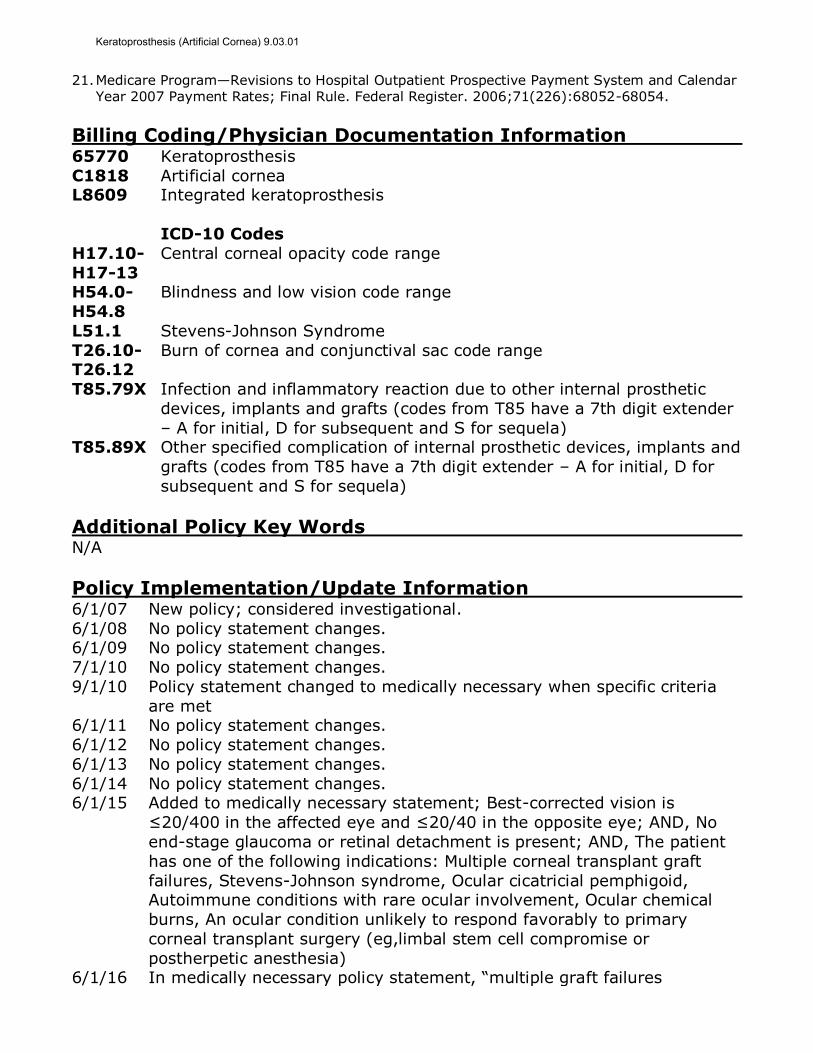
21. Medicare Program—Revisions to Hospital Outpatient Prospective Payment System and Calendar Year 2007 Payment Rates; Final Rule. Federal Register. 2006;71(226):68052-68054.
Billing Coding/Physician Documentation Information 65770 Keratoprosthesis
C1818 Artificial cornea L8609 Integrated keratoprosthesis
ICD-10 Codes
H17.10-
H17-13
Central corneal opacity code range
H54.0-
H54.8
Blindness and low vision code range
L51.1 Stevens-Johnson Syndrome
T26.10-T26.12
Burn of cornea and conjunctival sac code range
T85.79X Infection and inflammatory reaction due to other internal prosthetic
devices, implants and grafts (codes from T85 have a 7th digit extender
– A for initial, D for subsequent and S for sequela) T85.89X Other specified complication of internal prosthetic devices, implants and
grafts (codes from T85 have a 7th digit extender – A for initial, D for
subsequent and S for sequela)
Additional Policy Key Words N/A
Policy Implementation/Update Information 6/1/07 New policy; considered investigational.
6/1/08 No policy statement changes. 6/1/09 No policy statement changes.
7/1/10 No policy statement changes.
9/1/10 Policy statement changed to medically necessary when specific criteria
are met 6/1/11 No policy statement changes.
6/1/12 No policy statement changes.
6/1/13 No policy statement changes.
6/1/14 No policy statement changes. 6/1/15 Added to medically necessary statement; Best-corrected vision is
≤20/400 in the affected eye and ≤20/40 in the opposite eye; AND, No
end-stage glaucoma or retinal detachment is present; AND, The patient
has one of the following indications: Multiple corneal transplant graft
failures, Stevens-Johnson syndrome, Ocular cicatricial pemphigoid, Autoimmune conditions with rare ocular involvement, Ocular chemical
burns, An ocular condition unlikely to respond favorably to primary
corneal transplant surgery (eg,limbal stem cell compromise or
postherpetic anesthesia) 6/1/16 In medically necessary policy statement, “multiple graft failures
Keratoprosthesis (Artificial Cornea) 9.03.01
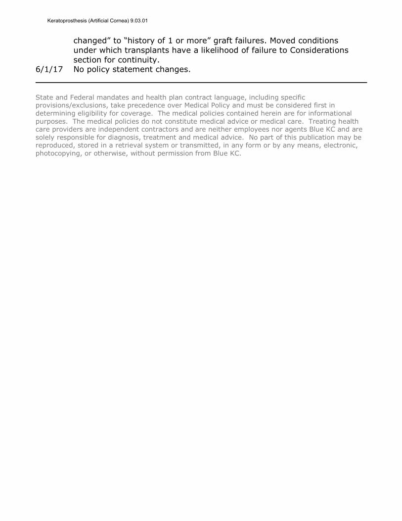
changed” to “history of 1 or more” graft failures. Moved conditions
under which transplants have a likelihood of failure to Considerations
section for continuity. 6/1/17 No policy statement changes.
State and Federal mandates and health plan contract language, including specific provisions/exclusions, take precedence over Medical Policy and must be considered first in determining eligibility for coverage. The medical policies contained herein are for informational purposes. The medical policies do not constitute medical advice or medical care. Treating health care providers are independent contractors and are neither employees nor agents Blue KC and are
solely responsible for diagnosis, treatment and medical advice. No part of this publication may be reproduced, stored in a retrieval system or transmitted, in any form or by any means, electronic, photocopying, or otherwise, without permission from Blue KC.
Keratoprosthesis (Artificial Cornea) 9.03.01