Embed Size (px)
Citation preview
8/6/2019 ketosis promt
http://slidepdf.com/reader/full/ketosis-promt 1/23
Syndromes of Ketosis-Prone Diabetes MellitusAshok Balasubramanyam, Ramaswami Nalini, Christiane S. Hampe, and Mario MaldonadoTranslational Metabolism Unit (A.B., R.N., M.M.), Division of Diabetes, Endocrinologyand Metabolism, Baylor College of Medicine, Houston, Texas 77030; Endocrine Service(A.B., R.N.), Ben Taub General Hospital, Houston, Texas 77030; Robert H. Williams
Laboratory (C.S.H.), University of Washington, Seattle, Washington 98195; and Novartis,Inc. (M.M.), CH-4002 Basel, SwitzerlandAddress all correspondence and requests for reprints to: Ashok Balasubramanyam, M.D.,Translational Metabolism Unit, Division of Diabetes, Endocrinology and Metabolism,Baylor College of Medicine, Room 700B, One Baylor Plaza, Houston, Texas 77030. E-mail: [email protected] Received August 13, 2007; Accepted January 9, 2008.
This article has been cited by other articles in PMC.
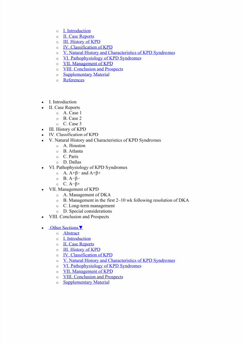
y Other Sections o Abstract o I. Introduction o II. Case Reports o III. History of KPD o IV. Classification of KPD o V. Natural History and Characteristics of KPD Syndromes o VI. Pathophysiology of KPD Syndromes o VII. Management of KPD o VIII. Conclusion and Prospects o Supplementary Material o References
AbstractKetosis-prone diabetes (KPD) is a widespread, emerging, heterogeneous syndromecharacterized by patients who present with diabetic ketoacidosis or unprovoked ketosis butdo not necessarily have the typical phenotype of autoimmune type 1 diabetes. Multiple,severe forms of -cell dysfunction appear to underlie the pathophysiology of KPD. Untilrecently, the syndrome has lacked an accurate, clinically relevant and etiologically usefulclassification scheme. We have utilized a large, longitudinally followed, heterogeneous,multiethnic cohort of KPD patients to identify four clinically and pathophysiologicallydistinct subgroups that are separable by the presence or absence of -cell autoimmunity andthe presence or absence of -cell functional reserve. The resulting ³A ´ classificationsystem of KPD has proven to be highly accurate and predictive of such clinically importantoutcomes as glycemic control and insulin dependence, as well as an aid to biochemical andmolecular investigations into novel causes of -cell dysfunction. In this review, we describethe current state of knowledge in regard to the natural history, pathophysiology, andtreatment of the subgroups of KPD, with an emphasis on recent advances in understandingtheir immunological and genetic bases.
y Other Sections o Abstract
8/6/2019 ketosis promt
http://slidepdf.com/reader/full/ketosis-promt 2/23
o I. Introduction o II. Case Reports o III. History of KPD o IV. Classification of KPD o V. Natural History and Characteristics of KPD Syndromes o
VI. Pathophysiology of KPD Syndromes o VII. Management of KPD o VIII. Conclusion and Prospects o Supplementary Material o References
y I. Introductiony II. Case Reports
o A. Case 1o B. Case 2o C. Case 3
y III. History of KPDy IV. Classification of KPDy V. Natural History and Characteristics of KPD Syndromes
o A. Houstono B. Atlantao C. Pariso D. Dallas
y VI. Pathophysiology of KPD Syndromeso A. A+ í and A+ +o B. Aí ío C. Aí +
y VII. Management of KPDo A. Management of DKAo B. Management in the first 2±10 wk following resolution of DKAo C. Long-term managemento D. Special considerations
y VIII. Conclusion and Prospects
y Other Sections o Abstract o I. Introduction o II. Case Reports o III. History of KPD o IV. Classification of KPD o V. Natural History and Characteristics of KPD Syndromes o VI. Pathophysiology of KPD Syndromes o VII. Management of KPD o VIII. Conclusion and Prospects o Supplementary Material
8/6/2019 ketosis promt
http://slidepdf.com/reader/full/ketosis-promt 3/23
8/6/2019 ketosis promt
http://slidepdf.com/reader/full/ketosis-promt 4/23
was unremarkable except for signs of volume depletion; specifically, she had no lesions of acanthosis nigricans, and the ocular and neurological examinations were normal.Laboratory tests revealed no evidence of acute infection, cardiac ischemia, cerebrovascular disease, renal or liver dysfunction, or recent alcohol use. The arterial pH was acidemic,anion gap 24, serum bicarbonate 12 mmol/liter, and serum glucose 344 mg/dl. The pat ient
was admitted to the hospital and received standard treatment for DKA with iv fluids andinsulin. She recovered uneventfully and was discharged on the third hospital day on aregimen of NPH insulin twice daily with insulin lispro before meals. She was followedclosely in our KPD research clinic thereafter.2. Baseline biochemistry and serology.Glycosylated hemoglobin (HbA1c) at presentation was 13.8%. Autoantibodies to -cellantigens were measured at the Robert H. Williams Laboratory (University of Washington,Seattle, WA) ( 1), utilizing highly sensitive and specific assays in which the upper limit of normal was defined as the ethnic-specific value at the 99th percentile of the levelsmeasured in large groups of Caucasians, Hispanics, and African-Americans living inHouston. Autoantibodies to glutamic acid decarboxylase (GAD) 65 and IA-2 were absentin the serum. -Cell functional reserve was assessed 1 wk after resolution of DKA byserum C-peptide levels after an overnight fast and after stimulation with glucagon. Thefasting C-peptide level was undetectable (<0.5 ng/ml) at baseline and did not change after glucagon stimulation.3. Evolution of -cell function.
-Cell functional reserve was assessed annually in our clinic. C-peptide secretion wasconsistently absent, both at baseline and after glucagon stimulation.4. Clinical course.he patient was initially treated with a basal (ultralente)/bolus (regular) insulin regimen,which was later changed to insulin glargine ( 55 U/d) with insulin lispro ( 10 U) beforemeals. She suffered a miscarriage in the first trimester with a relapse of DKA in November 2002 but completed an uncomplicated term pregnancy with continuous sc insulin infusiontherapy in 2003. She developed peripheral sensory neuropathy but has been free of other microvascular or macrovascular complications. There have been no episodes of DKA since2002. Nonpregnant HbA1c levels have ranged from 7.9 to 11.2%, and nonpregnant weighthas ranged from 132 to 144 lb. She adheres to a diabetic diet intermittently and does notexercise. Blood pressure and serum lipid levels are at desired goals without medications.She remains on continuous sc insulin infusion therapy.5. Conclusion.This young, lean Hispanic patient with childhood-onset diabetes and a family history of relatively early-onset diabetes has no evidence of -cell autoimmunity, was unable to comeoff insulin therapy without relapse of ketoacidosis, and has had difficulty achieving optimalglycemic control. Shortly after the index episode of DKA, she had virtually no -cellfunctional reserve, and this has not improved over time with intensive insulin therapy. Shehas the phenotype of what we have recently defined as ³Aí í´ KPD ( 1,2).B. Case 21. Clinical presentation.Patient 2, a 56-yr-old African-American man with no prior history of diabetes, presentedwith DKA in January 2000. He complained of polyuria, polydipsia, blurred vision, fatigue,and a 25-lb weight loss developing over the preceding 2 months. Evaluation in the
8/6/2019 ketosis promt
http://slidepdf.com/reader/full/ketosis-promt 5/23
emergency center revealed DKA with no clinical evidence of other precipitating illnesses or stressful events.The past medical history was significant for hypertension. One brother was known to havediabetes and was being treated with a oral medication. The patient did not smoke or abusealcohol.
His weight was 198 lb, height 5 ft-11 in, and BMI 28 kg/m2
. Mild acanthosis nigricans was present on the neck. The remainder of the physical examination was unremarkable exceptfor increased abdominal girth. There was no clinical evidence of diabetic retinopathy,neuropathy, or nephropathy. Laboratory tests revealed no evidence of acute infection,cardiac ischemia, or cerebrovascular disease, renal or liver dysfunction, or recent alcoholuse. The arterial pH was 7.22, anion gap 35, bicarbonate 12 mmol/liter, and glucose 765mg/dl. The patient was admitted to the hospital and received standard treatment for DKAwith iv fluids and insulin. He recovered uneventfully and was discharged on the thirdhospital day on a regimen of NPH insulin twice daily with insulin lispro before meals. Hewas followed closely in our KPD research clinic thereafter.2. Baseline biochemistry and serology.HbA1c at the time of hospital admission was 12%. Evaluation of -cell autoantibodies asdescribed above revealed GAD65 autoantibodies in high titer, but not IA-2 autoantibodies.
-Cell functional reserve assessed 2 wk after the acute episode of DKA revealed a fastingC-peptide level of 1.15 ng/ml and a peak C-peptide level after glucagon stimulation of 1.62ng/ml.3. Evolution of -cell function.Fasting C-peptide levels and peak responses to glucagon increased over time, then reacheda plateau. In early 2007, the fasting C-peptide was 2.6 ng/ml, and the peak postglucagonlevel was 5.2 ng/ml.4. Clinical course.Following a protocol described previously ( 1), we gradually withdrew insulin therapy andsubstituted oral diabetic medications with close monitoring for recurrence of ketoacidosisor decline in glycemic control. The patient tolerated the withdrawal without mishap, andinsulin was totally discontinued by the end of 2000. Pioglitazone was added to the regimein May 2000. He has had no recurrence of DKA. HbA1c levels since 2001 have ranged
between 6.2 and 7.8%, and the most recent in 2007 is 7.4%. His weight has ranged between218 and 232 lb, and the current BMI is 30.8 kg/m 2. Blood pressure and fasting lipid levelsare within desired ranges. His current medications are pioglitazone 15 mg/d, glipizide XL10 mg/d, metformin 1000 mg twice daily, simvastatin 40 mg/d, metoprolol XL 50 mg/d,candesartan 32 mg/d, hydrochlorothiazide 25 mg/d, and aspirin 81 mg/d.5. Conclusion.This middle-aged, overweight African-American patient, presenting with DKA as the firstmanifestation of diabetes, has evidence of -cell autoimmunity, is able to come off insulintherapy without relapse of ketoacidosis, and has experienced significant glycemicimprovement. Shortly after the acute episode of DKA, he had partially preserved -cellfunctional reserve, and this improved further while he was being treated with oralantidiabetic agents. He has the phenotype of A+ + KPD ( 1,2).C. Case 31. Clinical presentation.Patient 3, a 44-yr-old Hispanic man, presented with DKA in June 2004. He hadexperienced polyuria, polydipsia, and fatigue for 1 month preceding the DKA episode and
8/6/2019 ketosis promt
http://slidepdf.com/reader/full/ketosis-promt 6/23
had lost 30 lb of weight over 3 to 4 months. These symptoms prompted him to consult a physician 3 wk before presentation to the emergency center, and he was placed on an oralantidiabetic agent whose name he was unable to recall. There was no evidence of acuteillness or severe stress provoking the DKA. There was no significant past medical history.Both parents and a sister had type 2 diabetes. He denied smoking or illicit drug use, and
used alcohol occasionally. His weight was 252 lb, height 5 ft-10 in, and BMI 36 kg/ m2
.Examination revealed obesity with increased abdominal girth but was otherwiseunremarkable.Laboratory tests revealed no evidence of acute infection, cardiac ischemia, or cerebrovascular disease, renal or liver dysfunction, or recent alcohol use. The arterial pHwas 7.31, anion gap 21, bicarbonate 14 mmol/liter, and glucose 359 mg/dl. The patient wasadmitted to the hospital and received standard treatment for DKA with iv fluids and insulin.He recovered uneventfully and was discharged on the second hospital day on a regimen of
NPH insulin twice daily with regular insulin before meals. He was followed closely in our KPD research clinic thereafter.2. Baseline biochemistry and serology.HbA1c at presentation with DKA was 12.4%. -Cell autoantibodies were absent in theserum. The fasting C-peptide level measured 2 wk after recovery from DKA was 3.6 ng/ml,and peak C-peptide after glucagon stimulation was 6.6 ng/ml.3. Evolution of -cell function.
-Cell functional reserve was assessed at 6-month intervals in our clinic. Fasting C-peptidelevels remained stable. In February 2006, the fasting C-peptide level was 3.2 ng/ml.4. Clinical course.We gradually withdrew insulin therapy and substituted oral antidiabetic medications withclose monitoring for recurrence of ketoacidosis or decline in glycemic control. The patienttolerated the withdrawal without mishap, and insulin was totally discontinued by March2006. He has maintained excellent glycemic control, with HbA1c levels ranging between5.2 and 6.1%, no recurrence of ketoacidosis, and no episodes of hypoglycemia. He has beentreated with metformin since September 2006. Blood pressure has remained at the desiredgoal without specific medications. The weight has ranged from 248 to 272 lb, and thecurrent BMI is 38 kg/m 2. His current medications are metformin 500 mg twice daily,simvastatin 40 mg/d, slow release niacin 500 mg/d, and aspirin 81 mg/d.5. Conclusion.This middle-aged, obese Hispanic patient, presenting with unprovoked DKA very soonafter initial diagnosis of type 2 diabetes, has no evidence of -cell autoimmunity, was ableto come off insulin therapy without relapse of ketoacidosis, and has achieved sustainedeuglycemia. Shortly after the acute episode of DKA, he had partially preserved -cellfunctional reserve, which has remained stable over time with oral antidiabetic therapy. Hehas the phenotype of Aí + KPD ( 1,2).
y Other Sections o Abstract o I. Introduction o II. Case Reports o III. History of KPD o IV. Classification of KPD o V. Natural History and Characteristics of KPD Syndromes

8/6/2019 ketosis promt
http://slidepdf.com/reader/full/ketosis-promt 7/23
o VI. Pathophysiology of KPD Syndromes o VII. Management of KPD o VIII. Conclusion and Prospects o Supplementary Material o References
III. History of KPDOccasional reports of African or African-American patients whose clinical features seemedto be intermediate between those of type 1 and type 2 diabetes have appeared since the late1960s. In a review of tropical diabetes published in 1967, Dodu ( 3) noted that some patientsrequired revision of their type of diabetes over time. In 1968, Adadevoh ( 4) described a few
Nigerian patients with ³reversible´ diabetes who displayed only transient insulindependence. A decade later, Oli ( 5) described a series of seven Nigerian patients who
presented with ketosis and initially required insulin therapy but later experienced³remission´ of diabetes. In a classic paper in 1987, Winter et al. (6) described a cohort of obese African-American children who were atypical because they lacked islet cellautoantibodies, presented with DKA as the initial manifestation of diabetes, and becameinsulin independent over time. The elegant studies of Banerji et al. (7) in 1994 described asomewhat different atypical syndrome in overweight, adult Afro-Caribbean patients whohad clinical characteristics of type 2 diabetes but presented with DKA; the term Flatbushdiabetes entered the literature at this point. The following year, Umpierrez et al. (8)carefully characterized obese African-American patients in Atlanta, Georgia, who had late-onset diabetes presenting with DKA. They noted that -cell functional reserve was higher at baseline in these obese patients than in typical lean patients who developed DKA, andthat it improved further after 12 wk of treatment. These investigators introduced theconcept of BMI as a means to distinguish two phenotypes (obese or lean) of patients
presenting with DKA, based on their immunological and -cell functional differences ( 9).These earlier studies suggested that such atypical forms of diabetes were uncommon and
perhaps restricted to persons of African ancestry. However, from 1995 to 2003, case seriesand retrospective reviews reported (often large) numbers of patients from a wide array of geographic areas and ethnic backgrounds, with the common themes of absent islet cellautoantibodies, presentation with unprovoked ketosis or ketoacidosis, and frequentevolution to insulin independence. These included reports of Japanese patients ( 10);Apache Indians ( 11 ); African-Americans in Ohio ( 12); multiethnic U.S. populationsincluding Hispanics, Caucasians, and Native Americans ( 13,14,15); Europeans ( 16);Pakistanis ( 17); and Chinese ( 18). The term ³ketosis-prone diabetes´ was introduced in2002 by Sobngwi et al. in a review of diabetes in West Africans ( 19).In 2003, Maldonado et al. (1) reported a large, longitudinal, prospective study of multiethnic patients with four different forms of KPD in Houston, Texas, and introduced aclassification scheme based on two criteria: autoantibodies and -cell functional reserve. In2004, Mauvais-Jarvis et al. (20) published a 10-yr longitudinal study of KPD in immigrantsfrom sub-Saharan Africa living in Paris. These authors classified autoantibody-negativeKPD patients according to insulin dependence and contrasted their natural history to that of
patients with typical type 1 and type 2 diabetes. In 2006, Ramos-Roman et al. (21) reported4-yr outcomes in patients with atypical diabetes compared with those in patients withtypical type 1 and type 2 diabetes in Dallas, Texas.
8/6/2019 ketosis promt
http://slidepdf.com/reader/full/ketosis-promt 8/23
y Other Sections o Abstract o I. Introduction o II. Case Reports o III. History of KPD o
IV. Classification of KPD o V. Natural History and Characteristics of KPD Syndromes o VI. Pathophysiology of KPD Syndromes o VII. Management of KPD o VIII. Conclusion and Prospects o Supplementary Material o References
IV. Classification of KPDTo date, attempts to differentiate patients with KPD into clinically distinct and relevantsubgroups have resulted in four different classification schemes: the ADA classification, aBMI-based system, a modified ADA classification, and the A system.The first is contained within the ADA¶s most recent classification of diabetes in general(15) and has been adopted by investigators at the University of Texas SouthwesternMedical School (Dallas, TX). All patients who experience DKA are defined as having type1 diabetes, and among this group those who lack autoantibodies are referred to as³idiopathic type 1´ or ³type 1b.´ Strictly interpreted, the ADA scheme would define
patients with both type 1a and type 1b diabetes as insulin dependent, because it does notmention possible reversion to insulin independence in either category; however, the Dallasgroup considers patients with type 1b to behave more like patients with type 2 diabetes,with some becoming insulin-independent. A second scheme is that developed byinvestigators at Emory University (Atlanta, GA) who separate KPD patients into lean or obese ( 9). ³Lean KPD´ patients are those with clinical characteristics of type 1 diabeteswith low -cell function, whereas ³obese KPD´ patients are those with clinicalcharacteristics of type 2 diabetes with some preservation of -cell function. A modificationof the ADA scheme is used by investigators at the University of Paris who divide KPD
patients into three groups ( 20). Patients with -cell autoantibodies are classified as type 1a just as in the ADA scheme, whereas those who lack autoantibodies are distinguishedretroactively, based on long-term insulin dependence, into ³KPD insulin-dependent´ (KPD-ID) and ³KPD non-insulin dependent´ (KPD-NID). Both type 1a and KPD-ID patientshave clinical characteristics of type 1 diabetes with poor -cell function, whereas subjectswith KPD-NID have clinical characteristics of type 2 diabetes with preserved -cellfunction for a prolonged duration.Our collaborative group at Baylor College of Medicine and the University of Washingtonhas used a classification system that distinguishes four KPD subgroups based on the
presence or absence of autoantibodies and the presence or absence of -cell functionalreserve (A classification) ( 1). The four subgroups are: A+ í (patients with autoantibodiesand absent -cell function); A+ + (those with autoantibodies but preserved -cellfunctional reserve); Aí í (those without autoantibodies but absent -cell function); andAí + (those without autoantibodies and preserved -cell functional reserve). A+ í andAí í patients are immunologically and genetically distinct from each other but shareclinical characteristics of type 1 diabetes with very low -cell function, whereas A+ + and
8/6/2019 ketosis promt
http://slidepdf.com/reader/full/ketosis-promt 9/23
Aí + patients are immunologically and genetically distinct from each other but shareclinical characteristics of type 2 diabetes with preserved -cell functional reserve (Fig. 1 1 and Table 1).
Figure 1 Frequency distribution of patients in the four A groups in a multiethnic adult U.S.
urban population. [Reproduced with permission from M. Maldonado et al. : J Clin Endocrinol Metab 88:5090±5098, 2003 ( 1). Copyright The Endocrine (more ...)
Figure 1 Frequency distribution of patients in the four A groups in a multiethnic adult U.S. urban
population. [Reproduced with permission from M. Maldonado et al. : J Clin Endocrinol Metab 88:5090±5098, 2003 ( 1). Copyright The Endocrine Society.]
Tabl e 1 A groups: clinical characteristics
The value of a nosological system depends on the accuracy with which it predicts clinical behavior or distinguishes pathophysiological mechanisms. In the case of KPD, a keydeterminant of clinical behavior is long-term -cell functional reserve, which is necessaryfor such important outcomes as glycemic control ( 22) and insulin dependence ( 23). Tospecify the most accurate classification method, we compared the ability of the four existing systems to predict long-term -cell functional reserve in a large, multiethnic cohortof 294 KPD patients (138 new onset), followed for at least 12 months and tested repeatedly(2). The A system was the most accurate in predicting preserved -cell function 12 monthsafter the index DKA, with 99.4% sensitivity and 95.9% specificity; positive and negative
predictive values of 97.1 and 99.2%, respectively; positive and negative likelihood ratios of 24.55 and 0.01, respectively; and area under the receiver operator characteristic (ROC)curve of 0.972 ( 2,24). The modified ADA system had slightly greater specificity, positive
predictive value and positive likelihood ratio, but significantly lower sensitivity, negative predictive value, negative likelihood ratio, and ROC area under the curve (AUC). The other two schemes were generally less accurate, with the ADA system being the least reliable.Recently, the term ³ketosis-prone type 2 diabetes´ has entered the literature. In general, thisterm refers to the Aí + KPD subgroup, or in some instances, even further restricted tothose Aí + patients who present with ³unprovoked´ DKA or ketosis and new onsetdiabetes ( 25,26). Aí + patients comprise the largest subgroup of KPD patients and are theones who most commonly come to the notice of physicians because they present with DKAyet have all the clinical features and subsequent behavior of type 2 diabetes; hence, ketosis-
prone type 2 diabetes is certainly a fitting description for them. However, we believe thatfrom a heuristic standpoint, in the interest of defining and investigating novel syndromes of
-cell dysfunction, the broader terminology of ketosis-prone diabetes with its four subgroups subsumed under the A classification is more useful and unbiased, because itdoes not presume to define a syndrome a priori . Moreover, in cohorts of clinicallyheterogeneous, multiethnic patients, the four subgroups of KPD are useful as controls for one another in investigations of etiology and natural history.
y Other Sections

8/6/2019 ketosis promt
http://slidepdf.com/reader/full/ketosis-promt 10/23
o Abstract o I. Introduction o II. Case Reports o III. History of KPD o IV. Classification of KPD o
V. Natural History and Characteristics of KPD Syndromes o VI. Pathophysiology of KPD Syndromes o VII. Management of KPD o VIII. Conclusion and Prospects o Supplementary Material o References
V. Natural History and Characteristics of KPD SyndromesSeveral small series and retrospective reports have described patients with different
phenotypes of KPD, as detailed earlier ( 11 ,12,14). However, the natural histories of thesesyndromes are best detailed in studies utilizing large cohorts with longitudinal follow-up.Data from four such cohorts (in Houston, Atlanta, Paris, and Dallas) have been published.A. HoustonThe longitudinal Houston cohort comprises over 500 multiethnic patients (44% African-American, 40% Hispanic, 15% Caucasian, 1% Asian), including 185 patients who have
been followed for a mean of 5.5 yr ( 27). A comprehensive database compiles informationon the natural history of all four groups of KPD. A+ í KPD, identical to early-onset,autoimmune type 1 diabetes, displays a typical course of complete insulin dependence anddifficulty in attaining and achieving excellent long-term glycemic control. Aí í KPD
patients follow a very similar course. The more novel aspects of the natural history of KPDare revealed in the two + groups.The majority of A+ + KPD patients have new onset diabetes and follow one of twocourses. About 50% are able to maintain long-term -cell functional reserve and come off insulin therapy successfully shortly after resolution of DKA, whereas the others remaininsulin dependent. The relative level of initial -cell functional reserve by fasting C-peptidemeasurement or C-peptide response to glucagon does not predict which A+ + KPD
patients will become insulin dependent and which ones will not. However, twoimmunological markers offer significant predictive value. The first is epitope specificity of the GAD65 autoantibody (88% of A+ + KPD patients are GAD Ab positive). Patientswhose GAD65 Ab is directed toward an amino-terminal epitope in GAD defined bymonoclonal antibody DPD ( 28) have greater -cell functional reserve than those who donot have this characteristic, both initially and upon 12-month follow-up. These patients arealso significantly more likely to be insulin independent after 2 yr. The other marker lies inthe pattern of histocompatibility locus antigen (HLA) alleles associated with susceptibilityor resistance to autoimmune type 1 diabetes. New-onset A+ + KPD patients whose -cellfunction deteriorates over 2 yr tend to possess one or more susceptibility alleles (DQB1*02,DRB1*03, DRB1*04, DQB1*0302), whereas those with preserved -cell functionalreserve have a lower frequency of this pattern. Longer follow-up of A+ + KPD patientswill be necessary to delineate precisely their natural history with respect to glycemiccontrol and -cell function.Aí + KPD patients comprise the largest subgroup of KPD; approximately 50% of these
patients have new-onset diabetes and develop DKA without a clinically evident
8/6/2019 ketosis promt
http://slidepdf.com/reader/full/ketosis-promt 11/23
precipitating factor (unprovoked Aí + KPD), whereas the remainder have had long-standing diabetes before presentation with DKA and develop ketoacidosis in associationwith acute illness or noncompliance with antidiabetic treatment (provoked Aí + KPD).Unprovoked Aí + KPD patients display a striking male predominance (2.6:1,male:female) which is quite distinct from provoked Aí + KPD patients (0.7:1); this gender
imbalance has been noted also in patients with the unprovoked Aí + KPD phenotype inthe Atlanta, Paris, and Dallas cohorts ( 8,20,29). Longitudinal, prospective assessment of 113 unprovoked and 103 provoked Aí + KPD patients revealed that despite equivalentdegrees of hyperglycemia and -cell functional reserve at initial testing after the indexDKA episode, the former group had significantly greater improvement in -cell functionafter 12 months of treatment (2-fold greater), associated with significantly better glycemiccontrol (twice the frequency of attaining HbA1c < 7%) and twice the rate of insulindiscontinuation. Differences in -cell function at 12 months and rates of insulindiscontinuation persisted in a subgroup analysis of patients who achieved excellentglycemic control. These data suggest that patients with unprovoked or provoked Aí +KPD may have distinct underlying mechanisms of -cell dysfunction.B. AtlantaUmpierrez et al. (8) originally reported a short-term longitudinal study of 35 obese patientswho presented with new-onset, unprovoked DKA, comparing their clinical characteristicsand course to 10 lean patients with DKA, 22 obese nonketotic hyperglycemic patients, and10 obese nondiabetic patients. -Cell functional reserve was preserved shortly after theindex DKA episode in the obese DKA patients (although significantly less than in the other two obese groups), but not in the lean DKA patients. After 12 wk of intensive glycemicmanagement with insulin, -cell functional reserve improved significantly in the obeseDKA patients (to the extent that insulin therapy could be withdrawn in a majority), but notin the lean DKA patients. The baseline group differences in -cell function were confirmedin a larger set of similar patients in a subsequent study ( 9). In this study, 17% of the obeseDKA patients had autoantibodies, compared with 48% of the lean DKA patients,suggesting that the obese DKA patients probably represented a mixture of the Aí + andA+ + KPD phenotypes, whereas the lean DKA patients probably represented a mixture of A+ í and Aí í KPD phenotypes.C. ParisMauvais-Jarvis et al. (20) reported long-term follow-up of 111 patients of West Africanorigin who presented with DKA or unprovoked ketosis without a prior history of diabetes.Over 10 yr, 84 patients were found retrospectively to have a phenotype corresponding toAí + KPD (designated KPD-NID by the investigators), whereas 27 were found to have a
phenotype corresponding to Aí í KPD (designated KPD-ID). Their natural history wascompared with that of 21 patients with newly diagnosed autoimmune type 1 diabetes and88 patients with type 2 diabetes. During the observation period, KPD patients with the
probable Aí + phenotype became insulin independent at a mean of 14 wk after presentation with ketosis or ketoacidosis and achieved significant glycemic control withoral antidiabetic agents. The mean duration until relapse to insulin dependence was 40months, but 40% were still insulin independent at the end of the follow-up period. Relapseto insulin dependence was heralded by ketosis, usually preceded by breakthroughhyperglycemia, and some patients experienced a second remission to insulin independence.This description of relapsing and remitting ketosis and insulin requirement, with ultimate
8/6/2019 ketosis promt
http://slidepdf.com/reader/full/ketosis-promt 12/23
insulin dependence in a little over half the cases, represents the longest follow-up of patients with a probable phenotype of unprovoked Aí + KPD.D. DallasPiñero-Piloña et al. (15) reported 5-yr clinical follow-up of 54 multiethnic patients (65%African-American, 30% Hispanic, 5% Native-American) who presented with DKA and
lacked -cell autoantibodies (idiopathic type 1b diabetes according to the ADAclassification system). -Cell function was not measured, but insulin could be successfullywithdrawn over time in 21 patients. The investigators reported a second longitudinal study(4-yr follow-up) of insulin sensitivity and -cell function in 12 patients with idiopathic type1b diabetes compared with 10 patients with autoimmune type 1a diabetes ( 21). Theidiopathic type 1b patients were reported to be less insulin-sensitive and have higher degrees of -cell function than the type 1a patients. It is probable that the idiopathic type 1b
patients represented a mixture of the Aí í and Aí + KPD phenotypes.
y Other Sections o Abstract o I. Introduction o II. Case Reports o III. History of KPD o IV. Classification of KPD o V. Natural History and Characteristics of KPD Syndromes o VI. Pathophysiology of KPD Syndromes o VII. Management of KPD o VIII. Conclusion and Prospects o Supplementary Material o References
VI. Pathophysiology of KPD SyndromesA. A+ í and A+ +Inclusion of patients with islet cell autoantibodies in a longitudinal cohort analysis of
patients with KPD permits identification of distinct phenotypes among patients with a presumed autoimmune basis for severe -cell dysfunction. There is clearly a spectrum of clinical phenotypes among patients with islet autoantibodies who do not present withketosis, including those termed ³latent autoimmune diabetes in adults´ (LADA) ( 30), ³type1.5 diabetes´ ( 31,32,33), and ³slowly progressing type 1 diabetes´ ( 34). A similar spectrumexists in KPD that includes the very different phenotypes of A+ í and A+ + KPD. A+ íKPD is synonymous with classic, early onset autoimmune type 1 diabetes; A+ + KPD mayoverlap with LADA. However, there are differences between LADA, as recently defined bythe Immunology of Diabetes Society, and A+ + KPD patients; most importantly, thedefinition of LADA excludes patients who require insulin within the first 6 months after diagnosis, whereas the majority (90%) of A+ + KPD patients present with DKA as the firstmanifestation of diabetes and therefore require insulin at the start. Regardless of thesedistinctions, distinguishing A+ í from A+ + KPD permits investigators to exploredifferent autoimmune pathways leading to clinically distinct patterns of -cell loss, such asdifferent latencies and variable degrees of -cell destruction.Hampe et al. (28) investigated the role of epitope-specific autoantibodies to GAD65 inspecifying the clinical phenotypes of longitudinally followed A+ í and A+ + KPD

8/6/2019 ketosis promt
http://slidepdf.com/reader/full/ketosis-promt 13/23
patients. Five GAD65-specific Fab fragments were used to characterize the specificity of the titers of epitope-specific antibodies in relation to longitudinal measures of -cellfunction and clinical outcomes. A specific amino-terminal epitope defined by monoclonalantibody DPD correlated strongly with higher -cell functional reserve, both at baseline andafter 1 yr of follow-up, and was associated with A+ + KPD rather than A+ í KPD. Hence,
the later onset and more moderate clinical course (ability to discontinue insulin for over 2yr after the index episode of DKA in 50% of the patients) of A+ + KPD compared withA+ í KPD appears to be associated with a specific GAD65Ab epitope pattern. Themechanisms that result in this autoantibody specificity and give rise to variable -cellfunctional reserve remain to be elucidated.B. Aí íIn the Houston cohort, only 10% of these patients had new-onset diabetes when identifiedat presentation with DKA; the majority had had insulin-dependent diabetes for many years
previously ( 1). Furthermore, Zhang et al. (35) have noted that about 6% of patients meetingthe ADA criteria for type 1b diabetes (ketosis-prone, GAD65/IA-2 autoantibody-negative,likely with phenotypes of Aí í or Aí + KPD) have circulating antibodies against SOX-13(SRY-related high mobility group box antigen 13), another presumed marker of islet cellautoimmunity. Also, these patients have not been tested for antibodies to the cation effluxtransporter ZnT8, a recently identified islet autoantibody present in a small percentage of
patients with type 1 diabetes who lack GAD65 and IA-2 antibodies ( 36). These data raisethe concern of whether some Aí í KPD pat ients are misclassified as Aí because of adecline in autoantibody titers over time, or because they possess untested autoantibodies.Although it may be impossible to completely rule out an autoimmune component to Aí íKPD, this is probably not a significant issue in characterizing patients accurately. In thefirst place, GAD autoantibodies are quite durable, with only 10±20% of patients showingdeclines in titer to undetectable levels over 10 yr of follow-up ( 37,38,39). IA-2autoantibodies are less durable; about 50% of patients who test positive for IA-2-Ab at thetime of diagnosis have undetectable levels after 10 yr ( 37,38). GAD65Abs do not have arelationship to age at diagnosis; in contrast, presence of IA-2-Ab correlates negatively withage at diagnosis ( 40), possibly explaining our finding of only 36% IA-2-Ab-positive adult
patients (only 25% exclusively IA-2-Ab-positive) in the A+ KPD subgroups. Hence,absence of GAD65 or IA-2 Abs, even in patients with long-standing diabetes, is probablyquite reliable in classifying them as Aí. To limit further the possibility of misclassifyingA+ í KPD patients as Aí í, we have performed extensive HLA typing ( 1,27) and havefound that the frequencies of major class II alleles associated with susceptibility toautoimmune type 1 diabetes are not significantly higher in Aí í KPD patients than inethnic-matched population controls, whereas they are significantly higher in A+ í KPD
patients.The strong family history of diabetes in near relatives of Aí í KPD patients also suggeststhat there is a familial trait and that variants in genes required for -cell development,regeneration, or function may contribute to the phenotype. Preliminary data suggest that
potentially significant variants in TCF1 and PDX-1 , encoding the key -cell transcriptionfactors HNF1 (hepatocyte nuclear factor-1 ) and PDX-1 (pancreas-duodenum homeobox-1), are enriched in Aí í KPD patients compared with ethnic-specific population controls(W. Haaland, D. Mansouri, A. Balasubramanyam, M. Metzker, unpublished data).C. Aí +
8/6/2019 ketosis promt
http://slidepdf.com/reader/full/ketosis-promt 14/23
Umpierrez et al. (41) have examined the roles of glucotoxicity and lipotoxicity in inducingthe severe but partially reversible -cell functional defect in an obese African-American
patient with the phenotype of unprovoked Aí + KPD shortly after resolution of the indexepisode of DKA. The investigators measured the effects of exposure to 20 h of hyperglycemia and 48 h of hyperlipidemia (by lipid infusion) on C-peptide secretion. Acute
hyperglycemia but not acute hyperlipidemia caused severe blunting of the C-peptideresponse to glucose stimulation, and chronic hyperglycemia was associated with reducedexpression and insulin-stimulated threonine-308 phosphorylation of Akt2 in skeletalmuscle. These data suggest that severe glucotoxic blunting of an intracellular pathwayleading to insulin secretion may contribute to the reversible -cell dysfunctioncharacteristic of Aí + KPD patients, and that hyperglycemia may be exacerbated bydefects in skeletal muscle glucose uptake resulting from glucotoxic down-regulation of skeletal muscle insulin signaling. One mechanism of glucotoxic -cell dysfunction isincreased oxidant stress in the islets. Sobngwi et al. (42) investigated the possibility of X-linked glucose-6-phosphate dehydrogenase (G6PD) deficiency as a genetic basis for themale-predominant Aí + KPD phenotype in West African patients. They found a higher
prevalence of functional G6PD deficiency in the KPD patients compared with patients withtype 2 diabetes and a relationship between -cell functional reserve and erythrocyte G6PDactivity; however, the functional G6PD deficiency was not matched by a higher prevalenceof G6PD gene mutations. Hence, G6PD dysfunction may contribute to depressed -celldefense against oxidant stress in the face of acute hyperglycemia, but its cause does notappear to be a genetic mutation.Variants in key -cell developmental genes may also contribute to the phenotype of Aí +KPD. Mauvais-Jarvis et al. (43) found high frequency of a polymorphism leading to anamino acid substitution (R133W) in PAX4 , a transcription factor essential for isletmorphogenesis -cell development, among patients with phenotypes of Aí + or Aí íKPD. Because this variant is found in a high percentage of West Africans and African-Americans with and without type 2 diabetes, but not in Caucasians, its pathophysiologicalsignificance in the specific context of KPD is unclear.
y Other Sections o Abstract o I. Introduction o II. Case Reports o III. History of KPD o IV. Classification of KPD o V. Natural History and Characteristics of KPD Syndromes o VI. Pathophysiology of KPD Syndromes o VII. Management of KPD o VIII. Conclusion and Prospects o Supplementary Material o References
VII. Management of KPDClinical management of KPD includes: 1) acute management of DKA; 2) outpatientmanagement shortly after resolution of DKA, including classification of the patient
8/6/2019 ketosis promt
http://slidepdf.com/reader/full/ketosis-promt 15/23
according to KPD subgroup and evaluation of predictive factors; and 3) long-termmanagement.A. Management of DKAAll patients who present with DKA should be treated according to established principles of acute management of the metabolic decompensation. Standard inpatient hospital protocols
requiring aggressive fluid replacement; continuous insulin therapy; assessment for andtreatment of precipitating factors; monitoring for resolution of hyperglycemia, ketoacidosis,and electrolyte disorders; and transition from iv insulin to sc insulin regimens have beenwell described ( 26,44). It is important to note that inpatient treatment during the episode of DKA should be the same regardless of the apparent phenotype of the KPD patient, and thatall KPD patients should be discharged from the hospital on a regimen that provides 24-hinsulin coverage. Any attempt to withdraw insulin treatment should be based on preciseclassification of the KPD subgroup and assessment of the predictive factors, which should
be performed at the first outpatient visit 1±3 wk after hospital discharge.B. Management in the first 2±10 wk following resolution of DKAAssessment of -cell secretory reserve and -cell autoimmunity should be performed after complete resolution of DKA to minimize any acute effects of glucose toxicity or desensitization on -cell function, generally 1±3 wk after resolution of ketoacidosis. Themethods for performing these tests, as well as receiver-operator curve analysis to establishthe C-peptide cutoffs that distinguish í from + status, have been previously establishedand published ( 1).Patients are classified as í if the fasting serum C-peptide concentration is less than 1ng/ml (0.33 nmol/liter) and the peak serum C-peptide response to glucagon (measured at 5and 10 min after iv injection of 1 mg glucagon) is less than 1.5 ng/ml (0.5 nmol/liter), andthey are classified as + if the fasting serum C-peptide concentration is at least 1 ng/ml(0.33 nmol/liter) or the peak serum C-peptide response to glucagon is at least 1.5 ng/ml (0.5nmol/liter) (ROC AUC for peak serum C-peptide concentration after glucagon stimulation= 0.96751, ROC AUC for fasting C-peptide = 0.97776, and ROC AUC for C-
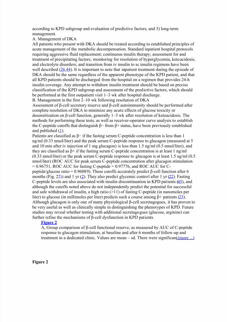
peptide/glucose ratio = 0.96089). These cutoffs accurately predict -cell function after 6months (Fig. 2 2)) and 1 yr ( 2). They also predict glycemic control after 1 yr ( 22). FastingC-peptide levels are also associated with insulin discontinuation in KPD patients ( 45), andalthough the cutoffs noted above do not independently predict the potential for successfuland safe withdrawal of insulin, a high ratio (>11) of fasting C-peptide (in nanomoles per liter) to glucose (in millimoles per liter) predicts such a course among + patients ( 23).Although glucagon is only one of many physiological -cell secretagogues, it has proven to
be very useful as well as clinically simple in distinguishing the phenotypes of KPD. Futurestudies may reveal whether testing with additional secretagogues (glucose, arginine) canfurther refine the mechanisms of -cell dysfunction in KPD patients.
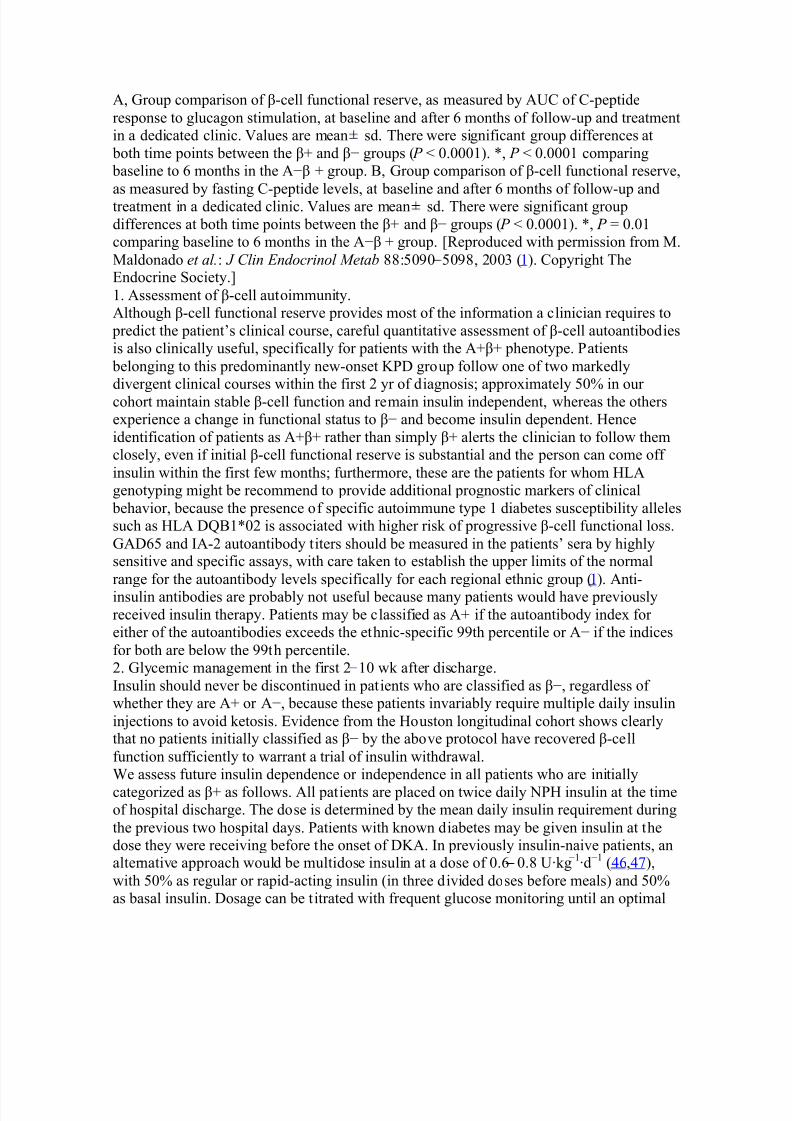
Figure 2 A, Group comparison of -cell functional reserve, as measured by AUC of C-peptideresponse to glucagon stimulation, at baseline and after 6 months of follow-up andtreatment in a dedicated clinic. Values are mean ± sd. There were significant (more ...)
Figure 2
8/6/2019 ketosis promt
http://slidepdf.com/reader/full/ketosis-promt 16/23
A, Group comparison of -cell functional reserve, as measured by AUC of C-peptideresponse to glucagon stimulation, at baseline and after 6 months of follow-up and treatmentin a dedicated clinic. Values are mean sd. There were significant group differences at
both time points between the + and í groups ( P < 0.0001). *, P < 0.0001 comparing baseline to 6 months in the Aí + group. B, Group comparison of -cell functional reserve,
as measured by fasting C-peptide levels, at baseline and after 6 months of follow-up andtreatment in a dedicated clinic. Values are mean sd. There were significant groupdifferences at both time points between the + and í groups ( P < 0.0001). *, P = 0.01comparing baseline to 6 months in the Aí + group. [Reproduced with permission from M.Maldonado et al. : J Clin Endocrinol Metab 88:5090 5098, 2003 ( 1). Copyright TheEndocrine Society.]1. Assessment of -cell autoimmunity.Although -cell functional reserve provides most of the information a clinician requires to
predict the patient¶s clinical course, careful quantitative assessment of -cell autoantibodiesis also clinically useful, specifically for patients with the A+ + phenotype. Patients
belonging to this predominantly new-onset KPD group follow one of two markedlydivergent clinical courses within the first 2 yr of diagnosis; approximately 50% in our cohort maintain stable -cell function and remain insulin independent, whereas the othersexperience a change in functional status to í and become insulin dependent. Henceidentification of patients as A+ + rather than simply + alerts the clinician to follow themclosely, even if initial -cell functional reserve is substantial and the person can come off insulin within the first few months; furthermore, these are the patients for whom HLAgenotyping might be recommend to provide additional prognostic markers of clinical
behavior, because the presence of specific autoimmune type 1 diabetes susceptibility allelessuch as HLA DQB1*02 is associated with higher risk of progressive -cell functional loss.GAD65 and IA-2 autoantibody titers should be measured in the patients¶ sera by highlysensitive and specific assays, with care taken to establish the upper limits of the normalrange for the autoantibody levels specifically for each regional ethnic group ( 1). Anti-insulin antibodies are probably not useful because many patients would have previouslyreceived insulin therapy. Patients may be classified as A+ if the autoantibody index for either of the autoantibodies exceeds the ethnic-specific 99th percentile or Aí if the indicesfor both are below the 99th percentile.2. Glycemic management in the first 2 10 wk after discharge.Insulin should never be discontinued in patients who are classified as í, regardless of whether they are A+ or Aí, because these patients invariably require multiple daily insulininjections to avoid ketosis. Evidence from the Houston longitudinal cohort shows clearlythat no patients initially classified as í by the above protocol have recovered -cellfunction sufficiently to warrant a trial of insulin withdrawal.We assess future insulin dependence or independence in all patients who are initiallycategorized as + as follows. All patients are placed on twice daily NPH insulin at the timeof hospital discharge. The dose is determined by the mean daily insulin requirement duringthe previous two hospital days. Patients with known diabetes may be given insulin at thedose they were receiving before the onset of DKA. In previously insulin-naive patients, analternative approach would be multidose insulin at a dose of 0.6 0.8 U·kg í1 ·d í1 (46,47),with 50% as regular or rapid-acting insulin (in three divided doses before meals) and 50%as basal insulin. Dosage can be titrated with frequent glucose monitoring until an optimal
8/6/2019 ketosis promt
http://slidepdf.com/reader/full/ketosis-promt 17/23
8/6/2019 ketosis promt
http://slidepdf.com/reader/full/ketosis-promt 18/23
close monitoring for at least 2 yr, because the evolution of their -cell function is the least predictable of the KPD groups. The presence of specific HLA susceptibility allelesassociated with autoimmune type 1 diabetes may predict a more aggressive course andinsulin dependence within 1±2 yr ( 1). Hence, HLA typing may play a useful role in themanagement of this group of KPD patients, because it may help to identify those who are
likely to experience a more aggressive course or may be candidates for futureimmunomodulatory therapy.Upon successful insulin withdrawal in Aí + patients, we frequently use insulin-sensitizingagents such as metformin or a thiazolidinedione because these patients have the highestfrequency of the metabolic syndrome among KPD groups ( 50). If blood glucose levels donot achieve therapeutic targets within 8 wk, we add low doses of a sulfonylurea, or ameglitinide or -glucosidase inhibitor. We have very limited experience with newer agentssuch as glucagon-like peptide-1 mimetics or inhibitors of dipeptidyl peptidase-IV, but theyare of potential interest because of their -cell trophic properties and ability to reducecirculating glucagon levels. Aggressive management of the metabolic syndrome (includingstrategies to decrease intraabdominal fat) and cardiovascular risks are important in allsubgroups of KPD patients.In summary, management of KPD patients requires some special considerations. Becausethese patients are heterogeneous and the type of diabetes is unclear at presentation withDKA, they should all be maintained on insulin initially. Further management can be guidedrationally by accurate classification based on assessment of -cell functional reserve, -cellautoantibodies, and in some instances HLA allelotyping. It is also instructive to note that inurban, indigent populations, treatment of KPD patients carries a significant financial cost,due in significant part to misunderstanding regarding the type of diabetes in a patient who
presents with DKA and subsequent confusion in management ( 51,52). We have found thatan informed, systematic approach, utilizing the principles outlined above and deliveredthrough a dedicated program, reduces the burden of cost and improves clinical outcomes inKPD patients ( 53,54).
y Other Sections o Abstract o I. Introduction o II. Case Reports o III. History of KPD o IV. Classification of KPD o V. Natural History and Characteristics of KPD Syndromes o VI. Pathophysiology of KPD Syndromes o VII. Management of KPD o VIII. Conclusion and Prospects o Supplementary Material o References
VIII. Conclusion and ProspectsAlthough proper epidemiological surveys remain to be conducted, syndromes of KPDappear to be increasingly recognized worldwide, especially among urban, multiethnic
populations. They offer challenges to both clinicians and researchers, but also offer theexciting prospect of revealing novel mechanisms of -cell dysfunction in common forms of
8/6/2019 ketosis promt
http://slidepdf.com/reader/full/ketosis-promt 19/23
type 2 diabetes. It is our view that KPD patients (especially those with Aí forms of KPD)represent only the ³tip of the iceberg´; below the surface is likely to be a much larger poolof patients who have early or primary -cell defects in development, expansion in the faceof insulin resistance, regeneration in response to injury, or insulin secretion.Molecular investigations into KPD syndromes should take advantage of clinical samples
and databases derived from existing patient cohorts and utilize multiple approaches(genomic, metabolomic, proteomic) to generate etiological hypotheses. Animal models thatrecapitulate the characteristic -cell defects of some of the syndromes would be extremelyvaluable. In the clinical arena, pressing questions include how to classify similar syndromesin children, how to screen asymptomatic patients, and how best to apply new and emerging
-cell trophic therapies to patients with the different KPD subgroups. It is clear thatrecognition of KPD syndromes and careful phenotypic classification of the patients willfacilitate investigations of pathophysiology, the results of which could be relevant to thescreening, early diagnosis, and rational treatment of a broader group of patients withnonketotic early -cell dysfunction.Supplementary Material[RPHR Note] Click here to view. AcknowledgmentsThe authors acknowledge the invaluable collaborative efforts of the following members of the KPD investigative team at Baylor College of Medicine (Houston, TX) and theUniversity of Washington (Seattle, WA): Drs. Lakshmi Gaur, Ake Lernmark, Dinakar Iyer,Michael Metzker, and Wade Haaland, Ms. Lucille Rodriguez, and Mr. Jesus Villanueva.FootnotesDisclosure Statement: The authors have nothing to disclose.Grant Support: RO1 HL73969 and the Alkek Foundation (to A.B.).First P u bl ished On line Fe b ru a ry 21, 2008 Abbreviations: Ab, Autoantibody; AUC, area under the curve; BMI, body mass index;DKA, diabetic ketoacidosis; GAD, glutamic acid decarboxylase; G6PD, glucose-6-
phosphate dehydrogenase; HbA1c, glycosylated hemoglobin; HLA, histocompatibilitylocus antigen; IA-2, tyrosine phosphatase-like protein IA-2; KPD, ketosis-prone diabetes;KPD-ID, KPD insulin dependent; KPD-NID, KPD non-insulin dependent; LADA, latentautoimmune diabetes in adults; ROC, receiver operator characteristic.
y Other Sections o Abstract o I. Introduction o II. Case Reports o III. History of KPD o IV. Classification of KPD o V. Natural History and Characteristics of KPD Syndromes o VI. Pathophysiology of KPD Syndromes o VII. Management of KPD o VIII. Conclusion and Prospects o Supplementary Material o References
8/6/2019 ketosis promt
http://slidepdf.com/reader/full/ketosis-promt 20/23
References
1. M al don ado M , H amp e CS, G aur LK, D¶A m ico S, Iyer D, H amm er le L P ,Bo lgiano D, R odriguez L, R aja n A, Lern ma rk A, B ala su b r ama ny am A 2003Ketosis-prone diabetes: dissection of a heterogeneous syndrome using an
immunogenetic and -cell functional classification, prospective analysis, andclinical outcomes. J Clin Endocrinol Metab 88:5090±5098. [ PubMed ]2. Bala su b r ama ny am A, G a rz a G, R odriguez L, H amp e CS, G aur L, Lern ma rk
A, M al don ado M R 2006 Accuracy and predictive value of classification schemesfor ketosis-prone diabetes. Diabetes Care 29:2575±2579. [ PubMed ]
3. Dodu S R 1967 Diabetes in the tropics. Br Med J 2:747±750. [ PMC free article ][PubMed ]
4. Ad adevoh BK 1968 ³Temporary diabetes´ in adult Nigerians. Trans R Soc TropMed Hyg 62:528±530. [ PubMed ]
5. O li J M 1978 Remittent diabetes mellitus in Nigeria. Trop Geogr Med 30:57±62.[PubMed ]
6. W inter WE , M acla ren NK, R iley W J, C la rke D W , K app y M S, S p illa r RP 1987Maturity-onset diabetes of youth in black Americans. N Engl J Med 316:285±291.[PubMed ]
7. Baner ji M A, Ch a iken R L, H uey H , Tuo m i T , Norin AJ, M ack ay I R , R ow leyM J, Zi mm et P Z, Le b ovitz H E 1994 GAD antibody negative NIDDM in adult
black subjects with diabetic ketoacidosis and increased frequency of humanleukocyte antigen DR3 and DR4. Flatbush diabetes. Diabetes 43:741±745.[PubMed ]
8. U mp ierrez G E , C asal s MM , Ge b ha rt S P , M ixon P S, C la rk W S, P hi llip s LS 1995 Diabetic ketoacidosis in obese African-Americans. Diabetes 44:790±795.[PubMed ]
9. U mp ierrez G E , W oo W , H agop ian W A, Is aa cs SD, P alm er J P , G aur LK,Nep om G T , C la rk W S, M ixon P S, Kit ab chi A E 1999 Immunogenetic analysissuggests different pathogenesis for obese and lean African-Americans with diabeticketoacidosis. Diabetes Care 22:1517±1523. [ PubMed ]
10. Aizawa T , K a takur a M , Ta guchi N, Ko ba yashi H , Aoy agi E , H ashizu m e K,Yoshiz a wa K 1995 Ketoacidosis-onset noninsulin dependent diabetes in Japanesesubjects. Am J Med Sci 310:198±201. [ PubMed ]
11. W ilson C, Kr akoff J, Gohdes D 1997 Ketoacidosis in Apache Indians with non-insulin-dependent diabetes mellitus. Arch Intern Med 157:2098±2100. [ PubMed ]
12. P inh as-H am ie l O, Do la n L M , Zeit ler P S 1997 Diabetic ketoacidosis among obeseAfrican-American adolescents with NIDDM. Diabetes Care 20:484±486. [ PubMed ]
13. Bala su b r ama ny am A, Zern J W , H yma n DJ, P avlik V 1999 New profiles of diabetic ketoacidosis: type 1 vs type 2 diabetes and the effect of ethnicity. ArchIntern Med 159:2317±2322. [ PubMed ]
14. W est ph al SA 1996 The occurrence of diabetic ketoacidosis in non-insulin-dependent diabetes and newly diagnosed diabetic adults. Am J Med 101:19±24.[PubMed ]
15. P inero- P ilon a A, Liton jua P , Avi les-S ant a L, R askin P 2001 Idiopathic type 1diabetes in Dallas, Texas: a 5-year experience. Diabetes Care 24:1014±1018.[PubMed ]
8/6/2019 ketosis promt
http://slidepdf.com/reader/full/ketosis-promt 21/23
16. P itte loud N, P hi lipp e J 2000 Characteristics of Caucasian type 2 diabetic patientsduring ketoacidosis and at follow-up. Schweiz Med Wochenschr 130:576±582.[PubMed ]
17. J abba r A, F a rooqui K, H ab ib A, Is lam N, H aque N, Akhter J 2004 Clinicalcharacteristics and outcomes of diabetic ketoacidosis in Pakistani adults with type 2
diabetes mellitus. Diabet Med 21:920±923. [ PubMed ]18. Ta n KC, M ack ay I R , Zi mm et P Z, H awkins B R , L am KS 2000 Metabolic andimmunologic features of Chinese patients with atypical diabetes mellitus. DiabetesCare 23:335±338. [ PubMed ]
19. Sobngwi E , M auv a is-J a rvis F, Vexi au P , M ba ny a JC, G autier JF 2002 Diabetesin Africans. Part 2: Ketosis-prone atypical diabetes mellitus. Diabetes Metab 28:5± 12. [ PubMed ]
20. M auv a is-J a rvis F, So b ngwi E , P orcher R , R ive line J P , Kevorki an J P , V a isse C,Ch a r p entier G, Gui lla usse au P J, Vexi au P , G autier JF 2004 Ketosis-prone type2 diabetes in patients of sub-Saharan African origin: clinical pathophysiology andnatural history of -cell dysfunction and insulin resistance. Diabetes 53:645±653.[PubMed ]
21. R am os- R oma n M A, P inero- P ilon a A, Ad am s-H uet B, R askin P 2006Comparison of type 1, type 2, and atypical ketosis-prone diabetes at 4 years of diabetes duration. J Diabetes Complications 20:137±144. [ PubMed ]
22. M al don ado M , D¶A m ico S, Otini ano M , B ala su b r ama ny am A, R odriguez L,Cuev as E 2005 Predictors of glycaemic control in indigent patients presenting withdiabetic ketoacidosis. Diabetes Obes Metab 7:282±289. [ PubMed ]
23. M al don ado M R , Otini ano ME , Chee ma F, R odriguez L, B ala su b r ama ny am A 2005 Factors associated with insulin discontinuation in subjects with ketosis-pronediabetes but preserved -cell function. Diabet Med 22:1744±1750. [ PubMed ]
24. Baner ji M A, Dh am S 2007 A comparison of classification schemes for ketosis- prone diabetes. Nat Clin Pract Endocrinol Metab 3:506±507. [ PubMed ]
25. U mp ierrez G E 2006 Ketosis-prone type 2 diabetes: time to revise the classificationof diabetes. Diabetes Care 29:2755±2757. [ PubMed ]
26. U mp ierrez G E , S m iley D, Kit ab chi A E 2006 Narrative review: ketosis-prone type2 diabetes mellitus. Ann Intern Med 144:350±357. [ PubMed ]
27. Nal ini R , G aur L, M al don ado M , H amp e C, R odriguez L, G a rz a G, Lern ma rk A, B ala su b r ama ny am A 3 March 2008 HLA class II alleles specify phenotypes of ketosis-prone diabetes (KPD). Diabetes Care 10.2337/dc07-1971.
28. H amp e CS, N al ini R , M al don ado M R , H all T R , G a rz a G, Iyer D,Bala su b r ama ny am A 2007 Association of amino-terminal-specific antiglutamatedecarboxylase (GAD65) autoantibodies with -cell functional reserve and a milder clinical phenotype in patients with GAD65 antibodies and ketosis-prone diabetesmellitus. J Clin Endocrinol Metab 92:462±467. [ PubMed ]
29. P inero- P ilon a A, R askin P 2001 Idiopathic type 1 diabetes. J DiabetesComplications 15:328±335. [ PubMed ]
30. Tuo m i T , Groo p LC, Zi mm et P Z, R ow ley M J, Know les W , M ack ay I R 1993Antibodies to glutamic acid decarboxylase reveal latent autoimmune diabetesmellitus in adults with a non-insulin-dependent onset of disease. Diabetes 42:359± 362. [ PubMed ]
8/6/2019 ketosis promt
http://slidepdf.com/reader/full/ketosis-promt 22/23
31. P ietro pa olo M , B a rin as-M itche ll E , P ietro pa olo SL, Ku ller L H , T rucco M 2000Evidence of islet cell autoimmunity in elderly patients with type 2 diabetes.Diabetes 49:32±38. [ PubMed ]
32. Groo p L, Groo p PH , Koski m ies S 1986 Relationship between B-cell function andHLA antigens in patients with type 2 (non-insulin-dependent) diabetes.
Diabetologia 29:757±760. [ PubMed ]33. Na ik R G, P alm er J P 2003 Latent autoimmune diabetes in adults (LADA). RevEndocr Metab Disord 4:233±241. [ PubMed ]
34. Ko ba yashi T 1994 Subtype of insulin-dependent diabetes mellitus (IDDM) inJapan: slowly progressive IDDM±the clinical characteristics and pathogenesis of thesyndrome. Diabetes Res Clin Pract 24 Suppl:S95±S99. [ PubMed ]
35. Zh ang Z, A p se K, P ang J, St anton R C 2000 High glucose inhibits glucose-6- phosphate dehydrogenase via cAMP in aortic endothelial cells. J Biol Chem275:40042±40047. [ PubMed ]
36. W enz la u J M , Juh l K, Yu L, M ou a O, S a rk a r SA, Gott lieb P , R ewers M ,E isen ba rth GS, Jensen J, D avidson H W , H utton JC 2007 The cation effluxtransporter ZnT8 (Slc30A8) is a major autoantigen in human type 1 diabetes. Proc
Natl Acad Sci USA 104:17040±17045. [ PMC free article ] [PubMed ]37. Decochez K, T its J, Coo lens JL, V an G aal L, Krzentowski G, W innock F,
Anck aert E , W eets I, P ipeleers DG, Gorus FK 2000 High frequency of persistingor increasing islet-specific autoantibody levels after diagnosis of type 1 diabetes
presenting before 40 years of age. The Belgian Diabetes Registry. Diabetes Care23:838±844. [ PubMed ]
38. Borg H , Gotts a ter A, Fern lund P , Sundkvist G 2002 A 12-year prospective studyof the relationship between islet antibodies and -cell function at and after thediagnosis in patients with adult-onset diabetes. Diabetes 51:1754±1762. [ PubMed ]
39. R ow ley M J, M ack ay I R , Chen QY, Know les W J, Zi mm et P Z 1992 Antibodiesto glutamic acid decarboxylase discriminate major types of diabetes mellitus.Diabetes 41:548±551. [ PubMed ]
40. Gr ah am J, H agop ian W A, Kocku m I, Li LS, S an jeevi CB, Lowe R M , Sch aeferJB, Z a rgh am i M , D ay H L, L andin-O lsson M , P alm er J P , J aner-Vi lla nuev a M ,H ood L, Sundkvist G, Lern ma rk A, Bres low N, D ah lquist G, B loh m e G 2002Genetic effects on age-dependent onset and islet cell autoantibody markers in type 1diabetes. Diabetes 51:1346±1355.
41. U mp ierrez G E , S m iley D, Gos ma nov A, Tho ma son D 2007 Ketosis-prone type 2diabetes: effect of hyperglycemia on -cell function and skeletal muscle insulinsignaling. Endocr Pract 13:283±290. [ PubMed ]
42. Sobngwi E , G autier JF, Kevorki an J P , Vi llette J M , R ive line J P , Zh ang S,Vexi au P , Le al SM , V a isse C, M auv a is-J a rvis F 2005 High prevalence of glucose-6-phosphate dehydrogenase deficiency without gene mutation suggests anovel genetic mechanism predisposing to ketosis-prone diabetes. J Clin EndocrinolMetab 90:4446±4451. [ PubMed ]
43. M auv a is-J a rvis F, S m ith SB, Le M ay C, Le al SM , G autier JF, M olokhi a M ,R ive line J P , R aja n AS, Kevorki an J P , Zh ang S, Vexi au P , Ger ma n M S, V a isseC 2004 PAX4 gene variations predispose to ketosis-prone diabetes. Hum MolGenet 13:3151±3159. [ PubMed ]
8/6/2019 ketosis promt
http://slidepdf.com/reader/full/ketosis-promt 23/23
44. Kit ab chi A E , U mp ierrez G E , M ur p hy M B, B a rrett E J, Kreis berg R A, M al oneJI, W all BM 2001 Management of hyperglycemic crises in patients with diabetes.Diabetes Care 24:131±153.
45. Sobngwi E , Vexi au P , Levy V, Le pa ge V, M auv a is-J a rvis F, Le bla nc H ,M ba ny a JC, G autier JF 2002 Metabolic and immunogenetic prediction of long-
term insulin remission in African patients with atypical diabetes. Diabet Med19:832±835. [ PubMed ]46. U mp ierrez G E , S m iley D, Zis ma n A, P rieto L M , P ala cio A, Ceron M , P uig A,
M e jia R 2007 Randomized study of basal-bolus insulin therapy in the inpatientmanagement of patients with type 2 diabetes (RABBIT 2 trial). Diabetes Care30:2181±2186. [ PubMed ]
47. Kit ab chi A E , U mp ierrez G E , M ur p hy M B, Kreis b erg R A 2006 Hyperglycemiccrises in adult patients with diabetes: a consensus statement from the AmericanDiabetes Association. Diabetes Care 29:2739±2748. [ PubMed ]
48. H sin Y, Guo H , W u T 2001 Factors associated with discontinuing insulin therapyafter diabetic ketoacidosis in adult diabetic patients. Diabet Med 18:895±899.[PubMed ]
49. R asou li N, E lb ein SC 2004 Improved glycemic control in subjects with atypicaldiabetes results from restored insulin secretion, but not improved insulin sensitivity.J Clin Endocrinol Metab 89:6331±6335. [ PubMed ]
50. Otini ano ME , B ala su b r ama ny am A, M al don ado M 2005 Presence of themetabolic syndrome distinguishes patients with ketosis-prone diabetes who have atype 2 diabetic phenotype. J Diabetes Complications 19:313±318. [ PubMed ]
51. M al don ado M R , Chong E R , Oeh l M A, B ala su b r ama ny am A 2003 Economicimpact of diabetic ketoacidosis in a multiethnic indigent population: analysis of costs based on the precipitating cause. Diabetes Care 26:1265±1269. [ PubMed ]
52. J avor KA, Kots anos JG, M cDon al d R C, B a ron AD, Kesterson JG, T ierneyWM 1997 Diabetic ketoacidosis charges relative to medical charges of adult
patients with type I diabetes. Diabetes Care 20:349±354. [ PubMed ]53. M al don ado M R , D¶A m ico S, R odriguez L, Iyer D, B ala su b r ama ny am A 2003
Improved outcomes in indigent patients with ketosis-prone diabetes: effect of adedicated diabetes treatment unit. Endocr Pract 9:26±32. [ PubMed ]
54. Levet an CS, P a ssa ro M D, J abl onski KA, R a tner R E 1999 Effect of physicianspecialty on outcomes in diabetic ketoacidosis. Diabetes Care 22:1790±1795.[PubMed ]