Embed Size (px)
Citation preview
Copyright 0 1996 by the Genetics Society of America
Kinesin Mutations Cause Motor Neuron Disease Phenotypes by Disrupting Fast Axonal Transport in Drosophila
Daryl D. Hurd and William M. Saxton
Department of Biology, Indiana University, Bloomington, Indiana 47405
Manuscript received June 18, 1996 Accepted for publication July 29, 1996
ABSTRACT Previous work has shown that mutation of the gene that encodes the microtubule motor subunit
kinesin heavy chain (Khc) in Drosophila inhibits neuronal sodium channel activity, action potentials and neurotransmitter secretion. These physiological defects cause progressive distal paralysis in larvae. To identify the cellular defects that cause these phenotypes, larval nerves were studied by light and electron microscopy. The axons of Khc mutants develop dramatic focal swellings along their lengths. The swellings are packed with fast axonal transport cargoes including vesicles, synaptic membrane proteins, mitochondria and prelysosomal organelles, but not with slow axonal transport cargoes such as cytoskeletal elements. Khcmutations also impair the development of larval motor axon terminals, causing dystrophic morphology and marked reductions in synaptic bouton numbers. These observations suggest that as the concentration of maternally provided wild-type KHC decreases, axonal organelles transported by kinesin periodically stall. This causes organelle jams that disrupt retrograde as well as anterograde fast axonal transport, leading to defective action potentials, dystrophic terminals, reduced transmitter secretion and progressive distal paralysis. These phenotypes parallel the pathologies of some vertebrate motor neuron diseases, including some forms of amyotrophic lateral sclerosis (ALS), and suggest that impaired fast axonal transport is a key element in those diseases.
T HE asymmetric organization and long projections of neurons place special demands on cytoplasmic
motility processes. The axon of a motor neuron can be a meter in length and contain >99% of the cell’s total cytoplasm. Because the axon supports little synthesis of proteins or membrane, materials must be constantly imported from the synthetically active cytoplasm of the cell body. This anterograde transport is balanced by retrograde transport that returns old, damaged compo- nents and endocytic material to the cell body for recy- cling. Axonal transport is essential for axon develop- ment and the maintenance of neuronal function. When it is blocked by disease o r experimental manipulation, axons lose their signalling capacity and eventually de- generate.
Axonal transport proceeds in two rate components. Cytoskeletal elements such as neurofilaments, tubulin and actin move in the slow component (0.5-2 mm/ day), while membrane-bounded organelles and synap- tic membrane proteins move in the fast component (50-200 mm/day) (BRADY 1991; VALLEE and BLOOM 1991; OCHS and BRIMIJOIN 1993; HIROKAWA 1996). The molecular mechanism of slow axonal transport is not well understood. However, a general understanding of the mechanism of fast axonal transport has evolved from studies of microtubule polarity within axons
Corresponding author; William M . Saxton, Department of Biology, Jordan Hall, Indiana University, Bloomington, IN 47405. E-mail: [email protected]
Genetics 144 1075-1085 (November, 1996)
(HEIDEMANN et al. 1981) and from the characterization of families of microtubule motor proteins (reviewed by BLOOM and ENDOW 1994; HIROKAWA 1996; SCHOLEY 1996). Axonal microtubules, which are oriented with their minus ends toward the cell body and their plus ends toward the axon terminal, serve as polarized tracks for the ATP-driven movement of various kinesins and cytoplasmic dyneins. Plus-end directed (anterograde) kinesins carry cargo toward the axon terminal. Minus- end directed (retrograde) motors, such as cytoplasmic dynein and perhaps NCD-related kinesins, carry cargo back toward the cell body.
The likelihood that multiple members of the kinesin superfamily participate in fast anterograde transport has raised the issue of which kinesin-related proteins (KRPs) transport what cargoes. The possibility that dif- ferent kinesins transport different axonal cargoes is sup- ported by the observation that the predicted cargo- binding regions of kinesin superfamily members have little sequence similarity (GOLDSTEIN 1993). Genetic studies of UNC-104, a nematode KRP, suggest that it associates with and moves synaptic vesicle precursors (HALL and HEDGECOCK 1991). Cell fractionation and localization studies of its homologue from mouse (KIFlA) support that conclusion (OKADA et al. 1995). Further, localization and in vitro motility studies suggest that KIFlB and perhaps its homologues associate with and transport mitochondria (NANGAKU et al. 1995). In contrast, studies of conventional kinesin suggest that it can bind a variety of organelles in neurons and other
1076 D. D. Hurd and W. M. Saxton
cell types. Furthermore, experiments designed to dis- rupt kinesin function using antibodies and antisense oligonucleotides affect the movements of a wide range of organelles and proteins (reviewed by BLOOM and ENDOW 1994).
To study the roles of microtubule motors and cyto- plasmic transport in a complex organism, we have used a genetic approach to disrupt kinesin function in Dro- sophila. Kinesin is abundant and ubiquitously ex- pressed in Drosophila (SAXTON et al. 1988, 1991). Null mutations in the gene that encodes kinesin heavy chain (Khc), the force-producing subunit of kinesin, cause lethality during the second larval instar. This observa- tion does not address the question of whether or not kinesin has important functions at earlier stages of de- velopment because mutant oocytes from heterozygous mothers contain maternally provided wild-type kinesin. The lethal phase of the null mutations suggests that protein turnover and dilution eliminate most of the maternal load of wild-type kinesin by the second larval instar.
Before dying, Khc mutant larvae become paralyzed. The paralysis is more severe in posterior segments than in anterior segments. This is particularly evident with hypomorphic Khc genotypes that allow development to progress through the third larval instar (SAXTON et al. 1991). Electrophysiological tests of Khc mutant larvae and interactions between mutations in Khc and muta- tions in ion channel genes indicate that the distal paral- ysis is due to two separate defects in neuron function (GHO et al. 1992; HURD et al. 1996). Impaired sodium channel activity reduces compound action potential amplitudes by a factor of four and makes them difficult to stimulate. Independent of the action potential de- fect, excitatoryjunctional current (EJC) amplitudes are reduced by a factor of three in anterior segments and by a factor of five in posterior segments. These EJC data indicate that the capacity of motor axon terminals to secrete neurotransmitter is reduced, and that the termi- nals of the long axons that innervate posterior segments are most severely affected. The distal paralysis behav- ioral phenotype and the length-dependent neurosecre- tion defect support the hypothesis that kinesin is a mo- tor for axonal transport.
This report describes the effects of Khc mutations on the structure and ultrastructure of larval neurons. Khc mutations cause axonal swellings that are filled with the cargoes of fast axonal transport, including many membrane-bounded organelles and synaptic mem- brane proteins. In addition, Khc mutations inhibit mo- tor axon terminal development. It appears that im- paired kinesin function causes a general disruption of fast axonal transport that in turn leads to dystrophic neuron development, length-dependent defects in neu- rotransmission and progressive distal paralysis. These effects are similar to those caused by some motor neu- ron diseases in vertebrates.
MATERIALS AND METHODS
Drosophila culture and mutant and control genotypes: Flies were cultured at 25" with a 12 hr light and 12 hr dark cycle on standard soft medium (0.5% agar, 7% molasses, 6% cornmeal, and 0.8% killed yeast) seeded with live yeast. To obtain the primary hemizygous test genoty e (y'w'; Khc6/ Df(ZR)Jpb), males with the genotype y'w'; Khc /Cy0 P(y'} were mated to females with the genotype y'w'; Df(2R)Jph/CyO P(y+). The use of y' on the first chromosome and a y f transgene on the second chromosome balancer allowed the recognition of larvae carrying the mutant genotype based on light pigmenta- tion of their mouthparts rather than on behavioral pheno- types. All of the mutant animals used in this study were y'w'; Khc6/Df(2RjJp6, except in tests for alelle specificity. Three types of controls were used: wild-type Oregon R, heterozygous siblings of the hemizygous test animals, and animals carrying the test genotype with the addition of a wild-type Khc transgene on chromosome 3 (transgenic rescue). The transgenic rescue genotype was constructed by mating y'7~'; Khc6/CyO Ply') males to y'w'; Df(2R)Jpb; P(w', Khc'}/T(2, 3) CyO, TM6b Tb females. Larvae bearing this control genotype were then picked based on light mouthpart pigmentation and normal body shape (nontubby). The genotypes of the controls shown in the various experiments are noted in the figure legends. Detailed descriptions of the genetics of the Khc mutations used can be found in SAXTON et al. (1991) and HURD et al. (1996). The other markers and chromosomes used are described by LINDSLEY and ZIMM (1992).
Immunocytochemistry and confocal microscopy: Larvae were dissected in Ca++ free dissection buffer (128 mM NaC1, 36 mM sucrose, 2 mM KC1, 1 mM EGTA, 5 mM HEPES, 4 mM MgC12)in a Sylgard coated depression dish using microdissec- tion scissors and spines from a ball cactus (Notocactus). Fixa- tion was done by rapidly exchanging the dissecting buffer with 4% formaldehyde in dissecting buffer. The fixative was replaced several times over 30-60 min. Samples were then rinsed rapidly with three changes of 0.2% Triton X-100 in standard phosphate buffered saline (PBST), followed by sev- eral more changes over 30-60 min. Antibodies were diluted in PBST. The types, concentrations, and sources of the pri- mary antibodies used were as follows: rabbit polyclonal anti- horseradish peroxidase at 1:250 (Jackson Immunoresearch), rabbit polyclonal anti-synaptotagmin at 1:500 (LITTI.ETON et al. 1993), rabbit polyclonal anti-KHC at 1:500 (SAXTON et al. 1988), affinity-purified rabbit polyclonal anti-KHC at 1:500 (RODIONOV et al. 1993), mouse monoclonal anti-/?-tubulin at 1 : l O (DETTMAN et al. 1996), mouse monoclonal anti-cysteine- string protein at 1:500 (ZINSMAIER et al. 1994), rabbit poly- clonal anti-syntaxin at 1:500 (HATA et al. 1993), rabbit poly- clonal anti-ankyrin at 1:500 (DUBREUII. and YU 1994), and mouse monoclonal anti-Fasciclin I1 at 1:lO (LIN et al. 1993). Fluorescein isothiocyanate- or TRITGgoat anti-rabbit or goat anti-mouse (Jackson Immunoresearch) secondary antibodies were typically used at 1:500. TRITC-phalloidin (Molecular Probes, Inc.) was used at 1:500 to stain F-actin. Primary anti- bodies were typically applied overnight at 4" and were fol- lowed by multiple rinses in PBST for a total of 60 min at room temperature. Secondary antibodies were typically applied for 30-60 min at room temperature and followed by multiple rinses in PBST. Samples were then mounted in buffered glyc- erol (10% 0.1 M NaH2C03 pH 8.4) and examined using a Bio- Rad MRC-600 laser scanning confocal microscope. Extended focus images were collected at a miminal confocal aperature using Kalman averaging. They were prepared for publication using a Macintosh Quadra 800 (Apple Computer Co.) run- ning Image v1.49b (W. RASBAND, National Institutes of Health), Photoshop v2.5.1 (Adobe Systems, Inc.) and Canvas v3.5 (Deneba Software). Final images were printed on a Phaser IISDX dye-sublimation printer (Tektronix, Inc.).
R
Kinesin and Axonal Transport 1077
Electron microscopy and video image collection: Transmis- sion elrctron microscopy was r~setl to examine segmental nenes from four control and three mutant ~vantlcring third instar larvae. (hntrols consisted of two heterozygotes, one Oregon R and one transgcnically rescued mr~tant. Lanae were dissected in Schneider's tissue culture medium. They w r c thcn fixed by multiple rapid exchanges with 2% glutaral- dchycle/2% p;~l-;~lorm~~Idehytlr in 0 . 1 51 c;~codylatc brllf'er (pH 7.2-'i.4) followed by four changes in a moist scaled container over ( 5 0 min. After rinsing in cacodylate buffer, postfixation W;IS done in 0..5% OsO,, 0.8% K., F C ( C N ) ~ in cacodylate buffer. Samplcs w r c thcn rinsed in distilled water, stained in
and fl;~tembedtletl in Epox-Araltlitr. Serial ultrathin (73- I00 nm) tr;tnsvcrse sections of lanA segmental n e n r s were cut with a Leica UCT ultramicrotome and transferred t o Formvar- co;~tcd singlc-slot grids. Grids were examined and photomi- crographs ~vcre ohtainctl using :IJEOLJKM I 0 1 0 running at (30 k\'. More detailed descriptions of these techniques can be found elsewhere ( M ( : I ) ~ s A I . I ) 1994). 1;anae wcrc prepared for sc;mning electron microscopy (SEM) using the same dis- section and fixation mcthotls. Further sample preparation ;mtl SFM were clone by F. R. TL'KSICK as dcscril)etl (M'AKIW)TO r/ ( I / . 1984).
I .an . ;~ l crawling behavior \vas vitlcotapecl as previously rle- scrihctl (S..\sTos cl a / . 1991 ) using a Dagc 68 Ne\\vicon cam- era fitted w i t h a Nikon Micrtrnikkor .% mm 12.8 lens. Single fr;~mcs were captured and digitized on a Macintosh Quatlra 840AV (Apple Computer Co. ) running Premier v4.2 (Atlohe Systems, Inc.). They were processed and printed as described al,ove.
' ( 2 X aqrlcorls uranyl acetate, dehydrated in an acetone series
RESULTS
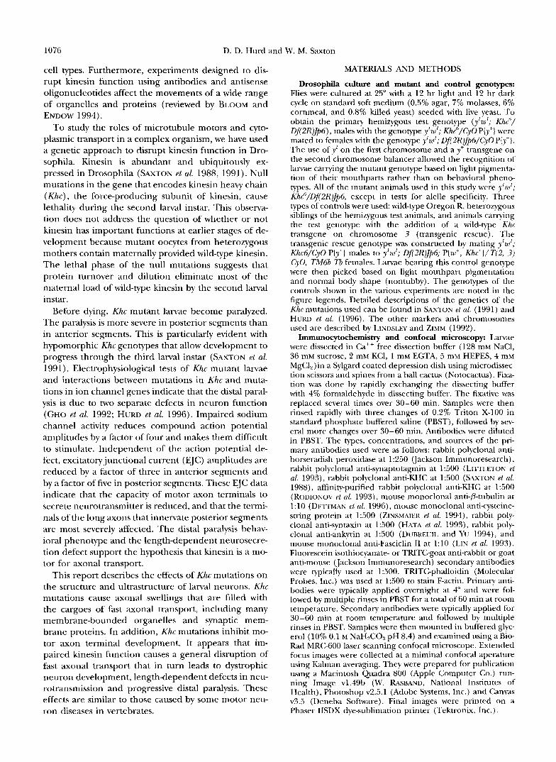
Kinesin mutations cause posterior-ventral paraly- sis: The crawling motion o fa Drosophila larva is driven by the contraction of segmentally repeated sets of hody- wall muscles that are attached to the inner surface of the cuticle. The pattern of contractions is coordinated by motor neurons whose cell bodies reside in the ven- tral ganglion. These neurons extend axons to the ap- propriate muscles via compound segmental nerves, each ofwhich contains 60-80 motor and sensory axons. Scanning electron microscopy shows that mutations in Khc do not cause major defects in the organization or morphology of the l a n d neuromuscular system (com- pare Figure 1A to Figure 3 o f J . 4 ~ a n d J m 1976). Never- theless, striking behavioral phenotypes appear in Klzc mlltant larvae. The anterior-posterior gradient of paral- ysis that was originally reported for Khc mutants (SAX- w s PI 01. 1991) is preceded by a dorsal-ventral gradient of paralysis (Figure 1, R and C ) . Early loss of motor activity in the ventral posterior segments causes an im- balance in hodyall contractions such that larvae rhyth- mically flip their tails upward during locomotion. Vari- ous degrees of the tail-flipping phenotype are caused by most Khc mutant genotypes. Dorsal and ventral motor axons that innervate the same posterior segment are not very different in length. Therefore, the tail-flipping phenotype suggests that while axon length is important, other structural o r physiological factors can also influ- ence the sensitivity of neurons to a loss of kinesin func- tion.
I
I
. .,.
F I ( ; ( ' I < I , : 1 .-lliincsin mrltations c a ~ w wrly posterior vcntral paralysis. (A) Scanning elrctron micrograph of a ~ C I I I - O ~ L I S -
cular preparation from a mutant 1a1-w (Kitc"/l!/(2R)J/,I;). The ventral ganglion (VG), located in the anterior end, constitutes the l m ~ A central ncnous system. Outside the ventral gan- glion, most motor and sensory axons are brlntlled together in the mqjor segmental nerves (SNs). The rectangul;~r body w a l l mrtscles are arranged on either side of the ventral midline in a bilaterally symmetrical, segmentally repeated pattern. Each lateral half-segment is i n n e ~ w t e d by a single major seg- mental nene. Motor neuron RP8 and a second motor neuron i n n e ~ ~ a t e t h e 6/7 pair of ventral longitudinal muscles, and they form synaptic contacts in and around the cleft between the muscles (m6/7: the arrow points to the cleft; see also Figures 4 and .5). Abdominal segments 2, 4, and 6 are indi- cated (A2, A4, and A(;). While the general structure of the lanral neuromuscular system is normal in kinesin mutants, its frlnction is not. A heterozygous control (R) and a mrltant (C) lama are shown in the act ol'crawling. The mutant periodically flips its tail upward indicating early paralysis of postcrior-ven- tral hodv wall muscles.
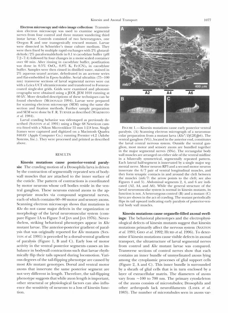
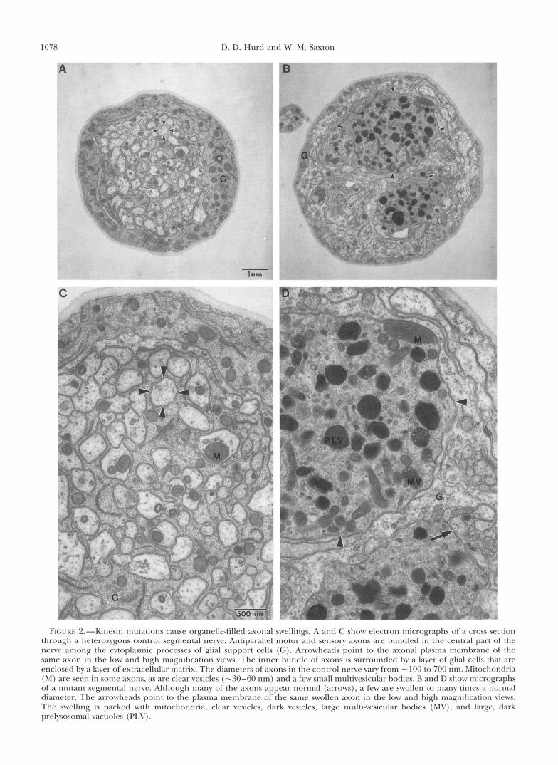
Kinesin mutations cause organelle-filled axonal swell- ings: The behavioral phenotypes and the electrophysi- ological defect$ of kinesin mutants suggest that kinesin mutations primarily affect the nervous system (SAXTON PI 01. 1991; GHO P t c d . 1992; HURD Pt (11. 1996). To deter- mine if kinesin mutations cause visible defects in axonal transport, the ultrastructure of l a n d segmental nerves from control and Khc mutant larvae was compared. Transverse sections of control nerves show that each contains an inner bundle of unmyelinated axons lying among the cytoplasmic processes of glial srlpport cells (Figure 2, A and C). This inner bundle is surrounded by a sheath of glial cells that is in turn enclosed by a layer of extracellular matrix. The diameters of axons vary from -100 to 700 nm. The primary cytoskeleton of the axons consists of microtubules; Drosophila and other arthropods lack neurofilaments (LWK Pt nl. 1983). The number of microtubules seen in axons var-
1078 D. D. Hurd and Mr. M. Saxton
C
i i
FI(; l .KI . ""Kinesin mutations cause organclle-filled axonal swellings. A ;~nd (; shon. rlrctron mirrogr;lphs o f ' ;I cross section through a heterozygous control segmental nene. Antiparallel motor and sensory axons are hundled i n thc central part of the nerve among the cytoplasmic proccsscs of glial support cells (G). Arrowheads point to the axonal plasma membrane of the same axon in the low and high magnification views. The inner bundle of axons is surrounded hv a laver of glial cells that are enclosed hv a laver of extracellular matrix. The diameters of axons in the control nenre vary from -100 to 700 nm. Mitochondria (M) are seen in some axons, as arc clear vesicles (-30-60 nm) and a few small multivesicular hodies. R and D show micrographs of a mutant segmental nerve. Although many of the axons appear normal (arrows), a few are swollen to many times a normal diameter. The arrowheads point to the plasma membrane of the same swollen axon in the low and high magnification views. The swelling is packed with mitochondria, clear vesicles, dark vesicles, large multi-vesicular hodies (MV), and large, dark prelvsosomal vacuoles (PLV).
Kinesin and Axonal Transport 1079
ies from one to 15 depending on axon diameter. Mito- chondria and small, clear vesicles (30-60 nm) are often seen in axons. Irregular cisternal membranes and small mutivesicular bodies are occasionally seen.
In some sections of Khc mutant nerves all axons have normal diameters and appear to contain normal popu- lations of membrane-bounded organelles. However, in other sections one or more axons are dramatically swol- len and packed with membrane-bounded organelles (Figure 2, B and D) . Serial section analysis revealed that the diameter of an axon of normal appearance could increase more than 10-fold within a micron. The organ- elles seen in the swellings include a multitude of mito- chondria, small vesicles, cisternal membranes and multivesicular bodies. These types of membrane- bounded organelles are the normal cargoes of fast axo- nal transport.
Large, dark-staining organelles, including large multivesicular bodies and vacuoles (Figure 2, B and D) are a conspicuous feature of the swellings. Their morphologies suggest that they represent early stages in the lysosomal pathway (NIXON and CATALDO 1995). Prelysosomal organelles of this size have not been seen in unswollen regions of mutant axons or in control axons. Therefore, they probably represent products of the fusion and maturation of smaller prelysosomal or- ganelles. Stress-driven autophagy of cytoplasm within the swellings may also contribute to the dark organelles (NIXON and CATALDO 1995).
Slow axonal transport does not make a substantial contribution to the swellings: Axonal swellings con- taining disorganized neurofilaments and membrane- bounded organelles are associated with some forms of amyotrophic lateral sclerosis (ALS) and other verte- brate motor neuron diseases (HIRANO 1991; DYCK et al. 1993; CLEVELAND 1996). Experiments with transgenic mice have shown that expression of a mutant neurofil- ament subunit or overexpression of wild-type subunits causes tangles of disorganized neurofilaments, axonal swellings, axonal dystrophy and motor neuron disease (reviewed by BRADY 1995; CLEVELAND 1996). Recent experiments suggest that the neurofilament disorder causes swellings by focal inhibition of the slow axonal transport of cytoskeletal proteins and perhaps of the fast axonal transport of membrane-bounded organelles (COLLARD et al. 1995).
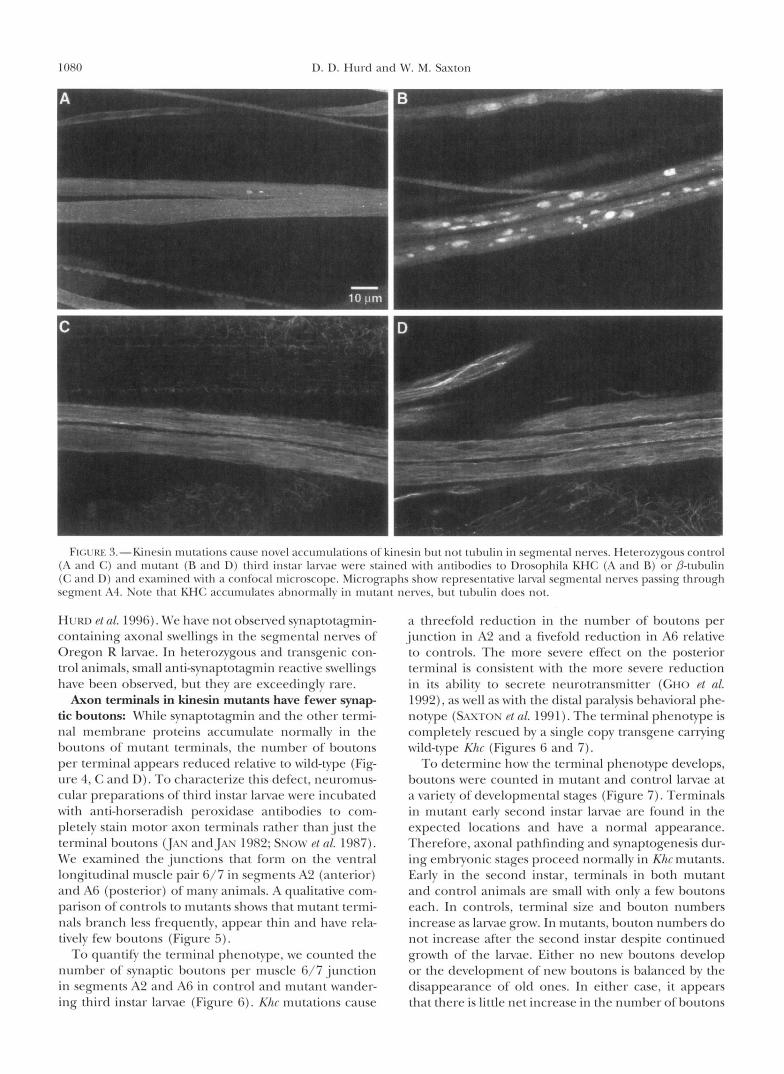
To test the contribution of the slow component of axonal transport to the organelle-filled swellings caused by Khc mutations, the distributions of tubulin, ankyrin and actin were compared to that of the mutant fast transport motor itself (KHC). In control nerves KHC is evenly distributed (Figure 3A), and microtubules ap- pear to lie parallel to the long axis of the segmental nerve (Figure 3C). Ankyrin and actin are also evenly distributed, although their faint staining patterns sug- gest that these proteins are not major components of the axonal cytoskeleton (not shown). In mutant nerves, KHC is concentrated in numerous elongated swellings
(Figure 3B). Tubulin (Figure 3D), ankyrin and F-actin have normal distributions and clearly do not accumu- late in axonal swellings. The Khc allele used (Khc6) pro- duces a partially functional full length KHC protein (SAXTON et al. 1991). By the third instar, little maternal KHC should remain, so the anti-KHC staining in mu- tants represents the distribution of the mutant protein. These staining patterns indicate that the swellings form with little contribution from the fibrous cytoskeletal ele- ments that move in the slow component of axonal trans- port.
Kinesin mutations cause abnormal accumdation of nerve terminal proteins in axon swellings: Membrane proteins required in the axon terminal for the synthesis of synaptic vesicles and plasma membrane are synthe- sized in the cell body. It is thought that small transport vesicles bearing these proteins are formed by the trans- Golgi network and then are carried to the terminal by fast anterograde axonal transport. If the abundant vesi- cles in the axonal swellings of Khc mutants are deposited by fast anterograde transport, they should carry proteins that are normally destined for the axon terminal.
To determine if anterograde transport vesicles con- tribute to the axonal swellings of kinesin mutants, the distributions of a number of terminal membrane pro- teins were compared in control and Khc mutant third instar larvae, Synaptotagmin is an integral membrane protein of synaptic vesicles, which are concentrated in terminal boutons (neurite varicosities). Anti-synapto- tagmin staining of control larvae shows the expected accumulation of synaptotagmin in terminal boutons (Figure 4C). Significant quantities of synaptotagmin are not found in control segmental nerves (Figure 4A). In contrast, striking accumulations of synaptotagmin are seen in the segmental nerves of Khc mutants (Figure 4B). Similar staining patterns were seen with antibodies to three other terminal membrane proteins: cysteine- string protein (synaptic vesicle membranes), syntaxin (intracellular, presynaptic plasma membrane), and Fas- ciclin I1 (extracellular, presynaptic plasma membrane) (HATA et al. 1993; LIN and GOODMAN 1993; ZINSMAIER et al. 1994; SCHULZE et al. 1995). Double-label experi- ments suggest that the distributions of these proteins and of KHC in the swellings are coincident. The accu- mulation of the various synaptic membrane proteins, mutant KHC and membrane-bounded organelles in ax- onal swellings contrasts with the lack of accumulation of cytoskeletal elements. These observations suggest that the axonal swellings of Khc mutants are caused by a direct inhibition of fast axonal transport.
The axonal swellings observed in Khc6 hemizygotes are not allele specific. Three other alleles tested, includ- ing Khc' (amorphic), Khc'lS (hypomorphic), and Khct" (mild hypomorphic), also cause accumulation of synap- totagmin in segmental nerves. The extent of synaptotag- min accumulation caused by each of these four alleles parallels the relative severities of the behavioral and other phenotypes that they cause (SAXTON et al. 1991;
1080 D. D. Hurd and Mr. M. Saxton
HUN) rt d . 1996). We have not obsened svnaptotagmin- containing axonal swellings in the segmental nerves of Oregon R larvae. In heterozvgous and transgenic con- trol animals, small anti-synaptotagmin reactive swellings have been observed, but they are exceedingly rare.
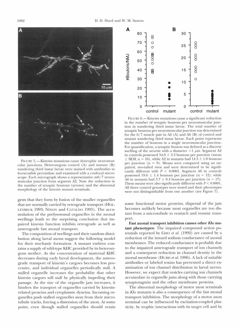
Axon terminals in kinesin mutants have fewer q m a p tic boutons: While svnaptotagmin and the other termi- nal membrane proteins accumulate normally in the boutons of mutant terminals, the number of boutons per terminal appears reduced relative to wild-type (Fig- ure 4, C and D). To characterize this defect, neuromus- cular preparations of third instar lanrae were incubated with anti-horseradish peroxidase antibodies to com- pletelv stain motor axon terminals rather than just the terminal borttons (JAS a n d J x s 1982; Ssow PI 01. 1987). We examined the junctions that form on the ventral longitudinal muscle pair 6/7 in segments A2 (anterior) and A6 (posterior) of many animals. A qualitative com- parison of controls to mutants shows that mutant termi- nals branch less freqttently, appear thin and have rela- tively few boutons (Figure 5).
To quanti& the terminal phenotype, we counted the number of synaptic boutons per muscle 6/7 junction in segments A2 and A6 in control and mutant wander- ing third instar larvae (Figure 6). Kltr mutations cause
a threefold reduction in the number of boutons per junction in A2 and a fivefold reduction in A6 relative to controls. The more severe effect on the posterior terminal is consistent with the more severe reduction in its ability to secrete neurotransmitter (CHO Pt 01. 1992), a s well as with the distal paralysis behavioral phe- notype (SAXTOS PI 01. 1991). The terminal phenotype is completelv rescued by a single copv transgene carrying wild-type Khc (Figures 6 and 7).
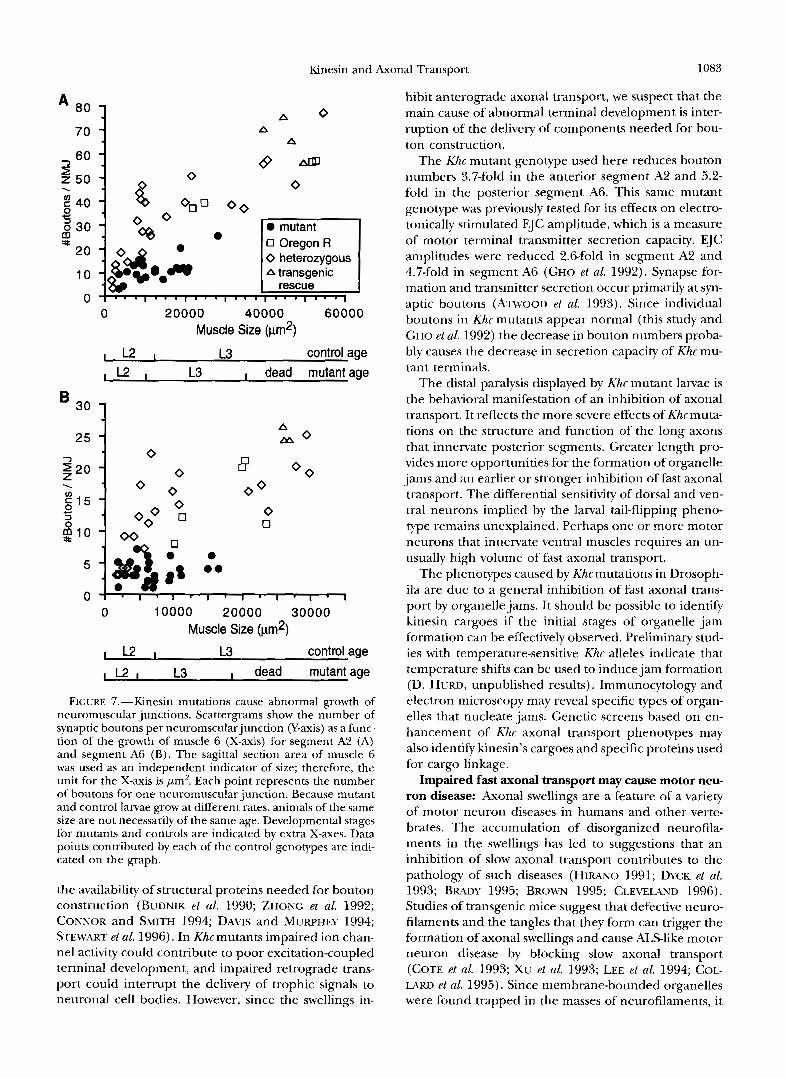
To determine how the terminal phenotype develops, boutons were counted in mutant and control larvae at a variety of developmental stages (Figure 7). Terminals in mutant earlv second instar larvae are found in the expected locations and have a normal appearance. Therefore, axonal pathfinding and synaptogenesis dur- ing embryonic stages proceed normallv in Klzc mutants. Early in the second instar, terminals in both mutant and control animals are small with only a few boutons each. In controls, terminal size and bouton numbers increase as larvae grow. In mutants, bouton numbers do not increase after the second instar despite continued growth of the larvae. Either no new boutons develop or the development of new boutons is balanced by the disappearance of old ones. In either case, it appears that there is little net increase in the number of boutons
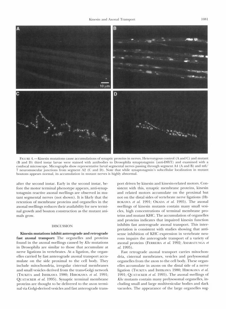
FI(;I.KI.: -l.-Kinc.sin mtlt ;1tiolls cause ;~cct lmrll;~~iol~s ol’synaptic protcins i n ncnw. I lc~cro~ygotls control (;I ; ~ n t l (;) and mutan t (R and D) third instar Ianac were stained w i t h antil)odies to lhosophila synaptotagmin (anti-DSW) antl examined w i t h a confocal microscopc. Micrographs show representative lanal segmental nenes passing through segment A4 (A antl R) and m6/ 7 neuromuscular junctions from segment A2 (C and D). Note that while synaptotagmin’s suhcellular localization in m u t m t houtons appears normal, its accumulation in mutant nerves is highly abnormal.
after the second instar. Early in the second instar, be- fore the motor terminal phenotype appears, anti-svnap totagmin reactive axonal swellings are observed in mu- tant segmental nerves (not shown). It is likely that the retention of membrane proteins and organelles in the axonal swellings reduces their availability for new termi- nal growth and bouton construction as the mutant ani- mals grow.
DISCUSSION
Kinesin mutations inhibit anterograde and retrograde fast axonal transport: The organelles and proteins found in the axonal swellings caused by Klu- mutations in Drosophila are similar to those that accumulate at nerve ligations in vertebrates. At a ligation, the organ- elles carried by fast anterograde axonal transport accu- mulate on the side proximal to the cell body. They include mitochondria, irregular cisternal membranes and small vesicles derived from the trans-Golgi network (TSUKITA and ISMIMM’A 1980; HIROKAWA P/ nl. 1991; QUATACKER P/ nl. 1995). Synaptic terminal membrane proteins are thought to be delivered to the axon termi- nal via Golgiderived vesicles and fast anterograde trans-
port driven by kinesin and kinesin-related motors. Con- sistent with this, synaptic membrane proteins, kinesin and related motors accumulate on the proximal hut not on the distal sides of vertebrate nerve ligations (HI- ROKAM’A P/ nl. 1991 ; O M ~ A P/ nl. 199.5). The axonal swellings of kinesin mutants contain many small vesi- cles, high concentrations of terminal membrane pro- teins antl mutant KHC. The accumulation of organelles and proteins indicates that impaired kinesin function inhibits fast anterograde axonal transport. This inter- pretation is consistent with studies showing that anti- sense inhibition of KHC expression in vertebrate neu- rons impairs the anterograde transport o f a variety of axonal proteins (FERREIRA P/ nl. 1992; AMARATUNGA P/
(11. 199.5). Fast retrograde axonal transport carries mitochon-
dria, cisternal membranes, vesicles and prelysosomal organelles from the axon to the cell body. These organ- elles accumulate in axons on the distal side of a nerve ligation (TS~~KITA and ISI-IIKAWA 1980; HIKOKAM’A Pt nl. 1991; QUATACKER P/ nl. 1995). The axonal swellings of Khc mutants contain many prelysosomal organelles, in- cluding small and large multivesicular bodies and dark vacuoles. The appearance of the large organelles sug-
1082 D. D. Hurt1 and M r . M. Saxton
LIS-
R) wanrIiring third instar Ianae were stainetl wit11 antihotIies to horseradish peroxidase and examined with a confocal micro- scope. Each micrograph shows a representative m6/7 neuro- muscular junction from segment A'L. Note the reduction in the numbcr of synaptic bolltons (arrows) and the abnormal morpholou of the kinesin mutant terminals.
gests that they form by fusion of the smaller organelles that are normally carried by retrograde transport (Hot,- LESRECK 1993; NIWS and ChT,u.m 199.5). The accu- mulation of the prelysosomal organelles in the axonal swellings leads to the surprising conclusion that im- paired kinesin fhc t ion inhibits retrograde as well as anterograde fast axonal transport.
The composition of swellings and their random distri- bution along larval axons suggest the following model for their stochastic formation. A mutant embryo con- tains a supply of wild-type KHC provided by its heterozy- gous mother. As the concentration of maternal KHC decreases during early l a n d development, the antero- grade transport of kinesin's cargoes becomes less pro- cessive, and individual organelles periodically stall. A stalled organelle increases the probability that other kinesin cargoes will stall by physically impeding their passage. As the size of the organelle jam increases, it hinders the transport of organelles carried by kinesin- related proteins and cytoplasmic dvneins. Incoming or- ganelles push stalled organelles away from their micro- tubule tracks, forcing a distension of the axon. At some point, even though stalled organelles should retain
A B
: 0
-1
2o 1 10
0
2o 1 0
10 0 0
0
0 0
0- control mutant
30 1 25
20
15
10
5
0 control mutant
FI(;L~RE 6.--Kinesin mutations cause a significant reduction in the number of synaptic houtons per neuromuscularjllnc- tion in wandering third instar Iawae. The total number of synaptic boutons per neuromrlscular.junction was determined for the 6/7 muscle pair in A2 (A) and Ab (R) of control and mutant wandering third instar larvae. Each point represents the number of borltons in a single neuromuscdar junction. For quantification, a synaptic bouton was defined as a discrete swelling of the neurite with a diameter > 1 pm. Segment A2 in controls possessed .54.0 2 3.3 houtons per.junction (mean 2 SEM, n = I6 ) , while A:! in mutants had 14.3 2 1.8 boutons per junction (11 = 9). Means were compared using an un- paired, two-tailed /-test and were determined to he signifi- cantly different with I-' < 0.0001. Segment A6 in controls possessed 19.6 2 1.4 boutons per junction (n = 12), while A6 in mutants had 3.7 2 0.4 boutons perjunction (n = 17). These means were also significantlv different with P < 0.0001. All three control genotypes were tested and their phenotypes were not distinguishable from one another (see Figure 7) .
some functional motor proteins, dispersal of the jam becomes unlikely because most organelles are too dis- tant from a microtubule to reattach and resume trans- port.
Fast axonal transport inhibition causes other Khc mu- tant phenotypes: The impaired compound action po- tentials reported by GHO PI d . (1992) are caused by a reduction of the inward sodium conductance of axonal membranes. The reduced conductance is probably due to the impaired anterograde transport of ion channels and a consequent reduction in channel density in the axonal membrane (HUKII PI nl. 1996). A lack of suitable antibodies or labeled toxins has prevented a direct ex- amination of ion channel distribution in land nerves. However, we expect that vesicles carrying ion channels accumulate in organellejams along with those carrying synaptotagmin and the other membrane proteins.
The abnormal morphology of motor axon terminals in Khc mutants is also a consequence of the fast axonal transport inhibition. The morphology of a motor axon terminal can be influenced by excitation-coupled plas- ticity, by trophic interactions with its target cell and by
Kinesin and Axonal Transport 1083
A 80
70 1 -7 60
5 50 40
2 30 20
10
0
\
c
%
A o A
A
8 0
0 mutant 0 Oregon R 0 heterozygous A transgenic -
0 20000 40000 60000 Muscle Size (pm2)
1 L 2 1 L3 control age ,L2 I L3 I dead mutant age
A M o
f 20
g 10
1
i_i 5 0
0 0 0 17
0 0
8' 7
B 0
l-
O O 0
0 0
I
0 10000 20000 30000 Muscle Size (pm2)
, L2 I L3 control age 1 1 2 1 L3 I dead mutant age
FIGURE 7.-Kinesin mutations cause abnormal growth of neuromuscular junctions. Scattergrams show the number of synaptic boutons per neuromscularjunction (Y-axis) as a func- tion of the growth of muscle 6 (X-axis) for segment A2 (A) and segment A6 (B) . The sagittal section area of muscle 6 was used as an independent indicator of size; therefore, the unit for the X-axis is pm2. Each point represents the number of boutons for one neuromuscular junction. Because mutant and control larvae grow at different rates, animals of the same size are not necessarily of the same age. Developmental stages for mutants and controls are indicated by extra X-axes. Data points contributed by each of the control genotypes are indi- cated on the graph.
the availability of structural proteins needed for bouton construction (BUDNIK et al. 1990; ZHONG et al. 1992; CONNOR and SMITH 1994; DAVIS and MUWHEY 1994; STEWART et al. 1996). In Khcmutants impaired ion chan- nel activity could contribute to poor excitationcoupled terminal development, and impaired retrograde trans- port could interrupt the delivery of trophic signals to neuronal cell bodies. However, since the swellings in-
hibit anterograde axonal transport, we suspect that the main cause of abnormal terminal development is inter- ruption of the delivery of components needed for bou- ton construction.
The Khc mutant genotype used here reduces bouton numbers 3.7-fold in the anterior segment A2 and 5.2- fold in the posterior segment A6. This same mutant genotype was previously tested for its effects on electro- tonically stimulated EJC amplitude, which is a measure of motor terminal transmitter secretion capacity. EJC amplitudes were reduced 2.6-fold in segment A2 and 4.7-fold in segment A6 (GHO et al. 1992). Synapse for- mation and transmitter secretion occur primarily at syn- aptic boutons (ATWOOD et al. 1993). Since individual boutons in Khc mutants appear normal (this study and GHO et al. 1992) the decrease in bouton numbers proba- bly causes the decrease in secretion capacity of Khc mu- tant terminals.
The distal paralysis displayed by Khc mutant larvae is the behavioral manifestation of an inhibition of axonal transport. It reflects the more severe effects of Khc muta- tions on the structure and function of the long axons that innervate posterior segments. Greater length pro- vides more opportunities for the formation of organelle jams and an earlier or stronger inhibition of fast axonal transport. The differential sensitivity of dorsal and ven- tral neurons implied by the larval tail-flipping pheno- type remains unexplained. Perhaps one or more motor neurons that innervate ventral muscles requires an un- usually high volume of fast axonal transport.
The phenotypes caused by Khc mutations in Drosoph- ila are due to a general inhibition of fast axonal trans- port by organelle jams. It should be possible to identify kinesin cargoes if the initial stages of organelle jam formation can be effectively observed. Preliminary stud- ies with temperature-sensitive Khc alleles indicate that temperature shifts can be used to induce jam formation (D. HURD, unpublished results). Immunocytology and electron microscopy may reveal specific types of organ- elles that nucleate jams. Genetic screens based on en- hancement of Khc axonal transport phenotypes may also identify kinesin's cargoes and specific proteins used for cargo linkage.
Impaired fast axonal transport may cause motor neu- ron disease: Axonal swellings are a feature of a variety of motor neuron diseases in humans and other verte- brates. The accumulation of disorganized neurofila- ments in the swellings has led to suggestions that an inhibition of slow axonal transport contributes to the pathology of such diseases (HIRANO 1991; DYCK et al. 1993; BWY 1995; BROWN 1995; CLEVELAND 1996). Studies of transgenic mice suggest that defective neuro- filaments and the tangles that they form can trigger the formation of axonal swellings and cause ALS-like motor neuron disease by blocking slow axonal transport (COTE et al. 1993; XU et al. 1993; LEE et al. 1994; COL- LARD et al. 1995). Since membrane-bounded organelles were found trapped in the masses of neurofilaments, it
1084 D. D. Hurd and W. M. Saxton
has been suggested that a secondary inhibition of fast axonal transport may also contribute to the ALSlike phenotypes (BRADY 1995; COLLARD et al. 1995).
The axonal swellings in Drosophila Khc mutants are caused by a disruption of fast axonal transport. They are filled with membrane-bounded organelles and con- tain no masses of cytoskeletal filaments. This specific disruption of fast axonal transport causes motor neuron disease-like phenotypes. The parallels include defective compound action potentials, reduced transmitter secre- tion by motor terminals, axonal dystrophy and progres- sive distal paralysis (CORNBLATH et al. 1992; HAHN 1993; LAMBERT and DYCK 1993; MASELLI et al. 1993; ROWLAND 1994; BOSTOCK et al. 1995; EISEN 1995). This suggests that an inhibition of fast axonal transport is a major factor in some vertebrate motor neuron diseases. Fur- thermore, our data support the hypothesis that a sec- ondary inhibition of fast transport is an important pathological effect of neurofilament tangles.
The authors thank SUSAN STROME, EI.I%ABETIi IL\FF and MARYANN
MARTIN for critical evaluation of the manuscript. We also thank H. BEI.I.EN, V. GEI.FAND, R. DUBREIUII., C . G o o ~ ~ t u v , E. RAFF, K. ZINS MAlER and T. SCIDHOF for their generous gifts of antibodies. Further, we thank F. R. TURNER for the scanning electron microscopy, R. BRENIYLA for help with the transmission electron microscopy, M. MAR- TIN for help with the video microscopy, and J. LENAVITT for assistance with image processing. This work was supported by National Institutes of Health (NIH) grant GM-46295 (W.M.S.), NIH predoctoral training grant T32GM-07227 (D.D.H.), and by an Established Investigatorship from the Arnerican Heart Association (AHA) with funds contributed in part by the AHA Indiana Affiliate, Inc. (W.M.S.).
LITERATURE CITED
AURATVNGA, A,, S. E. I.EEMN, K. S. KOSIK and R. E. FINE, 1995 Inhibition of kinesin synthesis in vivo inhihie the rapid transport of representative proteins for three transport vesicle classes into the axon. J. Neurochem. 64: 2374-2376.
ATMVOD, H. L., C. K. GWIND and C.-F. WU, 1993 Differential ultra- structure of synaptic terminals on ventral longitudinal abdominal muscles in Drosophila larvae. J. Neurobiol. 2 4 1008-1024.
BLOOM, G., and S. ENDOW, 1994 MotorProt~ins I : Kinesins. Academic
BOSTO(:K, H., M. K. SHARIEF, G. -11) and N. M. F. MVRKAY, 1995 Press, London.
Axonal ion channel dysfunction in amyotrophic lateral sclerosis. Brain 118: 217-225.
BRADY, S. T., 1991 Molecular motors in the nervous system. Neuron 7: 521-533.
BRADY, S. T., 1995 Interfering with the runners. Nature 375: 12- 13.
BROWN, R. H., 1995 Amyotrophic lateral sclerosis: recent insights from genetics and transgenic mice. Cell 80: 687-692.
BUDNIK, V., Y. ZHONC and C.-F. Wv, 1990 Morphological plasticity of motor axons in Dmsophila mutants with altered excitibility. J. Neurosci. 10: 3754-3768.
CI.EVEIAND, D. W., 1996 Neuronal growth and death: order and disorder in the axoplasm. Cell 84: 663-666.
C O I . I ~ , J.-F., F. COTE andJ.-P.Jul.IEN, 1995 Defective axonal trans- port in a transgenic mouse model of amyotrophic lateral sclero- sis. Nature 375: 61-64.
CONNOR, E. A,, and M. A. SMITII, 1994 Retrograde signalling in the formation and maintenance of the neuromuscular junction. J. Neurobiol. 25: 722-739.
(:IA\$'SON rt al., 1992 Nerve conduction studies in amyotrophic lateral sclerosis. Muscle Nerve 15: 1111-1115.
COTE, F., J.-F.COI.IAR~ and J:P. JL~I.IEN, 1993 Progressive neuropa- thy in transgenic mice expressing the human neurofilament
CORNRI.ATH, D. R., R. W. KUNCI., E. D. MEL.LITS, S. A. QVASKEY, L.
heavy chain gene: a mouse model of amyotrophic lateral sclero- sis. Cell 73: 35-46.
DAWS, G. W., and R. K. MURPHEY, 1994 Long-term regulation of short-term transmitter release proerties: retrograde signalling and synaptic development. Trends Neurosci. 17: 9-13.
DE'IT'MAN, R. W., F. R. TURNER and E. C. RMF, 1996 Genetic analysis of the Ilrosophila P3tubulin gene demonstrates that the rnicrotu- bule cytoskeleton in the cells of the visceral mesoderm is re- quired for morphogenesis of the midgut endoderm. Dev. Biol.
DLJBRE~JII., R. R., and J. Y I ~ , 1994 Ankyrin and P-spectrin accumulate independently of a-spectrin in Drosophila. Proc. Natl. Acad. Sci.
DYc:K, P. J., P. CHANCF., R. LEBO and J. A. CARNEY, 1993 Hereditary motor and sensory neuropathies, pp. 1094-1136 in Pm$hrral ,Wur@athy, edited by P. J. DYC:K, P. K. THOMAS, J. W. GRIFFIN, P. A. LOW and J. F. PODUSLO. W. B. Saunders, Philadelphia.
EISEN, A,, 1995 Amyotrophic lateral sclerosis is a multifactorial dis- ease. Muscle Nerve 16: 741 -752.
FERKEIKA, A,, J. N ~ c ~ n s , R. D. VAIX, G. BANKKR and K. S. KOSIK, 1992 Suppression of kinesin expression in cultured hippocampal neu- rons using antisense oligonucleotides. J. Cell Biol. 117: 595-606.
GHO. M., K. MC:DONAI.D, B. GANErLhT and W. M. SAXTON, 1992 Ef- fects of kinesin mutations on neuronal functions. Science 258: 313-316.
GOI.I)STFIN, L. S. R . , 1993 With apologies to scheherazade: tails of 1001 kinesin motors. Annu. Rev. Genet. 27: 319-351.
HAHK, X. F., 1993 Hereditary sensory and motor neuropathy: HMSN type I1 (neuronal type) and X-linked HMSN. Brain Pa- thol. 3: 147-155.
HAI I . , D. H., and E. M. HEDC:E(:O(:K, 1991 Kinesin-related gene unr- 104 is required for axonal transport of synaptic vesicles in C. elegans. Cell 65: 837-847.
HATA, Y., C . L. SIALIGHTER and T. C . SLWHOF, 1993 Synaptic vesicle fusion complex contains unr-18 homologue hound to syntaxin. Nature 366: 347-350.
HEIDEMASN, S. R., J. M. LANDERS and M. A. HAhlBORC, 1981 Polarity orientation of axonal microtubules. J. Cell Biol. 91: 661 -665.
H I R ~ N O , A,, 1991 Cytopathology of amyotrophic lateral sclerosis. Xdv. Neurol. 56: 91-101.
HIROK~WA, N., 1996 Organelle transport along microtubules-the role of KIFs. Trends Cell Biol. 6: 135-140.
BI.OOM ~t al., 1991 Kinesin associates with anterogradely trans- ported membranous organelles in vivo. J. Cell Biol. 114: 295- 302.
HOI.I.ENBE(:K, P. J., 1993 Producuj of endocytosis and autophagy are
J. Cell Biol. 121: 305-315. retrieved from axons by regulated retrograde axonal transport.
HuRI), D. D., M. STEKK and W. M. SAXTON, 1996 Mutation of the axonal transport motor kinesin enhances para and suppresses Skakrr in Drosophila. Genetics 1 4 2 195-204.
JAK, L.. Y., and Y. N.JAN, 1982 Antibodies to horseradish peroxidase as specific neuronal markers in Drosophila and in grasshopper embryos. Proc. Natl. Acad. Sci. USA 79: 2700-2704.
JAN, Y. N., and L . Y. J m , 1976 Properties of the larval neuromuscular junction in Drosophila melanogasler. J. Physiol. 262: 189-214.
IAMBF.RI, E. H., and P. J. DYCK, 1993 Compound action potentials of sural nerve in vitro in peripheral neuropathy, pp. 672-685 in I'priphrral Ahropathy, edited by P. J. DYCK, P. K. THOMAS, J. W. GRIFFIN. P. A. L , o w and,J. F. PoDr:sI.o. W. B. Saunders, Philadel- phia.
LSEK, R. J, , M. M. OFXINGER and P. F. DRAKE, 1983 Molecular biology of neuronal geometry: expression of neurofilament genes influences axonal diameter. Cold Spring Harbor Symp. Quant. Biol. 4 8 731-744.
LFX, M. K., J. R. MARSL.4I.EK and D. W. CI.EVF,IANI~, 1994 A mutant neurofilament subunit causes massive, selective motor neuron death: implications for the pathogenesis of human motor neuron disease. Neuron 13: 975-988.
LIN, D. M., and C. S. GOOL)MAK, 1993 Ectopic and increased expres- sion of Fasciclin 11 alters motoneuron growth cone guidance. Neuron 13: 507-523.
LIN, D. M., R. D. FETTER, C:. KOP(:ZYNSI(I, G. GIZF-NNINGI.OI{ and C . s. C;oon>ms, 1993 Genetic analyis of Fasciclin I1 in Drosophila: defasciculation, refasciculation, and altered fasciculation. Neu- ron 13: 1055-1069.
177: 117-135.
USA 91: 10285-10289.
HIROKAWA, N., R. %TO-YOSHITAKE, N. KOBAYASHI, K. K. PFISTER, G. s.
Kinesin and Axonal Transport 1085
LINDSIXY, D. L., and G. G. ZIMM, 1992 The Genolnc of Drosophila melanogarter. Academic Press, San Diego.
LITTLETON, J. T., H. J. BELLEN and M. S. PERIN, 1993 Expression of synaptotagmin in Drosophila reveals transport and localization of synaptic vesicles to the synapse. Development 118: 1077-1088.
MASEILI, R. A,, R. L. WOILMAN, C. LEUNC;, B. DISTAD, S. PALOMBI pt al., 1993 Neuromuscular transmission in amyotrophic lateral sclerosis. Muscle Nerve 16: 1193-1203.
MCDONALD, K. L., 1994 Electron microscopy and EM immunocyto- chemistry, pp. 411-444 in Drosophila melanogaster: Practical Uses in Cell and Molecular Biology, edited by L. S. B. GOI.DSTEIN and E. A. FRYBEKC;. Academic Press, San Diego.
NANGAKU, M., R. SATO-YOSHITAKE, Y. OKADA, Y. NODA, R. TAKEMURA tal., 1995 KIFIB, a novel microtubule plus enddirected mono- meric motor protein for mitochondria transport. Cell 79: 1209- 1220.
NIXON, R. A., and A. M. CATAI.DO, 1995 The endosomal-lysosomal system of neurons: new roles. Trends Neurosci. 18: 489-496.
OCHS, s., and W. s. BRIMIIOIN, 1993 Axonal transport, pp. 331-360 in Peri>herd Neuropathy, edited by P. J. DYCK, P. K. THOMAS, J. W. GKIFFIN, P. A. LOW and J. F. PODUSIX). W. B. Saunders, Philadel- phia.
OKADA, Y., H. YAMUAKI, Y. SEKINE-AIZAWA and N. HIKOIL+WA, 1995 The neuronspecific kinesin superfamily protein KIFlA is a unique monomeric motor for anterograde transport of synaptic vesicle precursors. Cell 81: 769-780.
QUATACKER, J., W. ANNAERT and W. DEPOTTER, 1995 The organiza- tion of the axonal reticulum at a ligation, in in vilro incubated bovine splenic nerves. Brain Res. 680: 36-42.
RODIONOV, V. I., F. K. GYOEVA, E. TANAM, A. D. BERSHADSKY, J . M. VAS11.1Ev ( 4 nl.; 1993 Microtubule-dependent control of. cell shape and pseudopodial activity is inhibited by the antibody to kinesin motor domain. J. Cell Biol. 123: 1811-1820.
ROWAND, L. P., 1994 Natural history and clinical features of amyo- trophic lateral sclerosis, pp. 507-522 in A;rurorlegpnrrativeDisf~asps, edited by D. B. CNX.E. W. B. Saunders Co., Philadelphia.
SAXTON, W. M . , M. E. PORTER, S. A. COHN, J. M. SCHOI.EY, E. C. RAFF
et al., 1988 Drosophila kinesin: characterization of microtubule motility and ATPase. Proc. Natl. Acad. Sci. USA 85: 1109-11 13.
SAXTON, W. M., J. HI~:LS, L. S. B. GOLDSTEIN and E. C. WF, 1991 Kinesin heavy chain is essential for viability and neuromuscular functions in Drosophila, but mutants show no defects in mitosis. Cell 64: 1098-1 102.
SCHOI.EY,.J. M., 1996 Kinesin-11, a membrane traffic motor in axons, axonemes, and spindles. .J. Cell Biol. 133: 1-4.
SCHLUE, K. L., K. BROADIE, M. S. PERIN and H. ,J. BELLEN, 1995 Genetic and electrophysiological studies of Drosophila syntaxin- 1A demostrate its role in nonneuronal secretion and neurotrans- mission. Cell 80: 31 1-320.
SNOW, P. M., N. H. PATEI, A. L. HARREISON and C. S. GOODMAN, 1987 Neural-specific carbohydrate moiety shared by many sur- face glycoproteins in Drosophilaand grasshopper embryos. J. Neu- rosci. 7 4137-4144.
1996 Homeostatsis of synaptic transmission in Drosophila with genetically altered nerve terminal morphology. J. Neurosci. 1 6 3877-3886.
TSUKITA, S., and H. 1SHlli4M7A, 1980 The movement of membranous organelles in axons. J. Cell Biol. 84: 513-530.
VAI.LE:k:, R. B., and G. S. BLOOM, 1991 Mechanisms of fast and slow axonal transport. Annu. Rev. Neurosci. 14 59-92.
WANMO'Io, B. T., F. R. TCKNER and T. C. K ~ \ ~ ~ F ~ I A N , 1984 Defects in embryogenesis in mutants associated writh the antennapedia gene complex o f Ihomphila melunogastrr. Dev. Biol. 102: 147-172.
XC, Z., L. C. CORK, J. W. GRIFFIN and D. W. CI.EVEIAN[I, 1993 In- creased expression of neurofilament subunit NF-L produces morphological alterations that resemble the pathology of human motor neuron disease. Cell 73: 23-33.
ZHONG, Y., \I. BI'I)NIK and C:F. WL', 1992 Synaptic plasticity in Dro suphila memory and hyperexcitihle mutants: role o f CAMP cas- cade. .J. Neurosci. 12: 644-651.
ZINSMAIER, K. E., K. K. EBERLE, E. BIIC~INER, N. WALTER and S. BEN- XEK, I994 Paralysis and early death in cysteine string protein mutants of Drosophilu. Science 263: 977-980.
%rEWART, B. A,, C. M. SCIII'STER, c. S. GOODMAN and H. L, AWOOD,
Communicating editor: R. S. HAWIXY