Embed Size (px)
Citation preview
RESEARCH Open Access
krCRISPR: an easy and efficient strategy forgenerating conditional knockout ofessential genes in cellsBei Wang1, Zishi Wang1, Daqi Wang1, Baolong Zhang2, Sang-Ging Ong3,4, Mingqing Li5, Wenqiang Yu2 andYongming Wang1*
Abstracts
Background: CRISPR/Cas9 system is a powerful tool for knocking out genes in cells. However, genes essential forcell survival cannot be directly knocked out. Traditionally, generation of conditional knockout cells requires multiplesteps.
Results: In this study, we developed an easy and efficient strategy to generate conditional knockout cells by usingdouble episomal vectors – one which expresses gRNA and Cas9 nuclease, and the other expresses an induciblerescue gene. Using this system which we named “krCRISPR” (knockout-rescue CRISPR), we showed that essentialgenes, HDAC3 and DNMT1, can be efficiently knocked out. When cells reach a desired confluency, the exogenousrescue genes can be silenced by the addition of doxycycline. Furthermore, the krCRISPR system enabled us to studythe effects of the essential gene mutations on cells. We showed that the P507L mutation in DNMT1 led todownregulation of global DNA methylation in cells, indicating that it is a disease-causing mutation.
Conclusions: The krCRISPR system offers an easy and efficient platform that facilitates the study of essential genes’function.
Keywords: Episomal vector, CRISPR/Cas9, Essential gene, Knockout
BackgroundDeletion of a target gene in cells and observation of theresulting phenotype is a common strategy to determinethe function of a gene in biological research [1–3]. How-ever, numerous genes that are essential for cell viabilityresult in cell death when they are ablated [3–5]. Recentgenome-wide screening has revealed that essential genesaccount for ~ 10% of total human genes [6]. To study es-sential genes in cells, conditional knockout strategieshave been developed.The Cre/loxP recombination system is the most com-
monly used technique to knockout essential genes. Thistechnique requires insertion of a pair of 34 bp loxP sitesflanking the target genes, and expression of Cre recom-binase enzyme will cause a deletion of the genes between
the two loxP sites [7, 8] (Additional file 1: Figure S1A).Other conditional strategies have also been developed.Liao et al. generated a transgenic cell line that expressesan essential gene under the control of Ptet promoter(Doxycycline-inducible gene expression), and thenknocked out the endogenous target gene [3]. Upondesirable cell density, the expression of the exogenousgene was shut down [3] (Additional file 1: Figure S1B).Matsunaga et al. inserted a tetracycline-regulated indu-cible gene promoter (tet-OFF/TRE-CMV) upstream ofthe endogenous target gene in cells that express tetra-cycline transactivator (tTA) [9]. The inserted promoterdisrupted the endogenous promoter and controlled en-dogenous gene expression by doxycycline (Dox) [9](Additional file 1: Figure S1C). Nevertheless, both strat-egies required multiple steps to generate stable integra-tion cell lines. The combination of the CRISPR/Cas9and episomal vector technology represent an alternativestrategy to knockout essential genes.
* Correspondence: [email protected] Key Laboratory of Contemporary Anthropology at School of LifeSciences and Zhongshan Hospital, Fudan University, Shanghai 200438, ChinaFull list of author information is available at the end of the article
© The Author(s). 2019 Open Access This article is distributed under the terms of the Creative Commons Attribution 4.0International License (http://creativecommons.org/licenses/by/4.0/), which permits unrestricted use, distribution, andreproduction in any medium, provided you give appropriate credit to the original author(s) and the source, provide a link tothe Creative Commons license, and indicate if changes were made. The Creative Commons Public Domain Dedication waiver(http://creativecommons.org/publicdomain/zero/1.0/) applies to the data made available in this article, unless otherwise stated.
Wang et al. Journal of Biological Engineering (2019) 13:35 https://doi.org/10.1186/s13036-019-0150-y

The RNA-guided CRISPR/Cas9 system is a powerfultool for genome editing in diverse organisms and celltypes [10–12]. CRISPR/Cas9 system consists of twocomponents: a Cas9 nuclease and a 100 nucleotide guideRNA (gRNA) which directs Cas9 to cleave the targetsites and generate double-strand breaks (DSBs) [13]. Asan RNA-guided DNA endonuclease, Cas9 can be easilyprogrammed to target new sites by altering its gRNA se-quence. The DSBs can be repaired by the cell’s endogen-ous DNA repair machinery through homology-directedrepair (HDR) using an introduced DNA repair template,such as a double-stranded DNA donor plasmid or asingle-stranded oligo DNA nucleotide (ssODN), enablingknock-in of precise mutations or reporters [14–16]. TheDSBs can also be repaired by non-homologousend-joining (NHEJ), resulting in nonspecific small inser-tions and deletions (indels) useful for generatingloss-of-function mutations [1, 3].We have previously used an episomal vector to express
gRNA and Cas9 nuclease, and achieved high efficiencyof gene knockout [17]. Episomal vector allows forlong-term genome editing with the puromycin resistancegene on the episomal vector enabling enrichment oftransfected cells. In this study, we developed an easy andefficient strategy to generate gene knockout-rescue sys-tems with two episomal vectors, allowing conditionalknockout of essential genes. Using this system, one plas-mid encodes Cas9 and gRNA for essential gene knock-out, and the other plasmid encodes the target genecontrolled by Tet-Off system. This system is designatedas krCRISPR (knockout-rescue CRISPR). We showedtwo examples of essential gene knockout using this sys-tem. We further showed that the effects of specific mu-tation can be studied by replacing wild-type (WT)essential gene with mutant versions. Our system will fa-cilitate functional studies of essential genes.
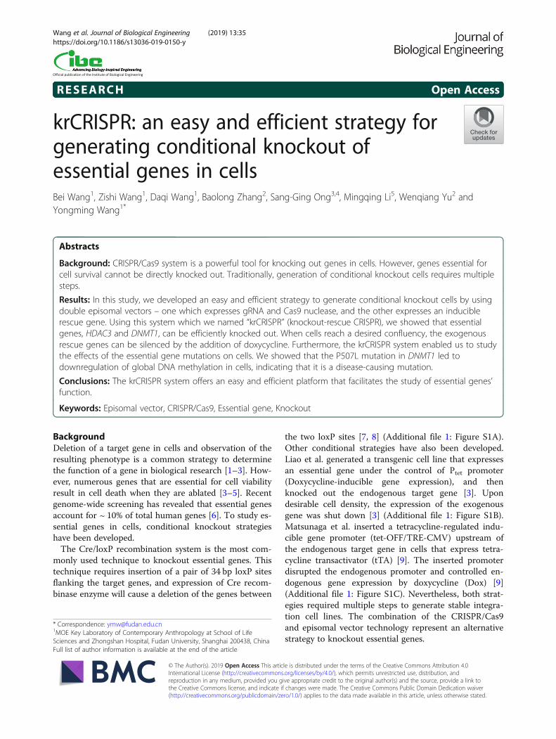
ResultsEstablishment of a knockout-rescue system by usingdouble episomal vectorsIn order to knockout essential genes in cells, we de-signed a gene knockout-rescue system with doubleepisomal vectors (Fig. 1a). Double vectors can accom-modate multiple genetic components. One plasmidwhich encodes Cas9 and gRNA for gene knockoutwas designated as KO (knockout) plasmid and theother plasmid which encodes the rescue gene wasdesignated as Rescue plasmid. The advantage of theepisomal vectors is that they can replicate ineukaryotic cells, allowing long-term Cas9 and gRNAexpression [17]. The episomal vector used in thisstudy was derived from Epstein-Barr virus (EBV)which contains two components essential for the epi-somal maintenance in cells: the latent origin oriP and
its binding protein Epstein-Barr-associated nuclearantigen 1 (EBNA1) [18]. In order to simultaneouslyretain two episomal plasmids in cells, the EBNA1coding sequence was removed from the Rescue plas-mid, resulting in the episomal maintenance of theRescue plasmid solely dependent on the KO plasmid(Fig. 1b). The Rescue plasmid encodes three genes:rescue gene, GFP and puromycin resistant gene(Puro), separated by self-cleaving T2A peptide. Allthree genes’ expression was controlled by the Tet-Offsystem. In order to reduce leaky expression, the tTAgene was encoded by the KO plasmid. Under puro-mycin selection, cells’ survival depends on the Rescueplasmid that expresses puromycin resistant gene.Therefore, cells’ survival requires the episomal main-tenance of both plasmids.We first tested whether both plasmids can be simul-
taneously maintained in cells. Both plasmids contain acommon sequence that has an MfeI restriction site. Todifferentiate between these plasmids by restriction frag-ment length polymorphism (RFLP) assay, the MfeI re-striction site on the Rescue plasmid was destroyed bydigestion and religation (Fig. 1c). We co-transfected thesame amount of each plasmid into different cell typesand PCR-amplified the common region for the RFLPassay. Twenty-four hours post-transfection, a similaramount of both plasmids was detected (Fig. 1d, e andAdditional file 1: Figure S2A). After 10 days of puro-mycin selection, higher amount of the KO plasmid wasdetected. A possible reason is that the KO plasmidretained the intact oriP/EBNA1 sequence which favorsplasmid replication. To rule out the possibility that onlyone plasmid was present in a portion of cells, we trans-fected individual plasmid into cells and selected withpuromycin for three days. As expected, neither KO plas-mid nor Rescue plasmid could support cell’s survival(Fig. 1f ). Co-transfection of the two plasmids couldsupport cells’ survival (Fig. 1f ). GFP expression increasedover time with puromycin selection (Additional file 1:Figure S2B and C). We next tested the capacity of theTet-Off system for regulation of GFP expression. Threedays after addition of Dox, expression of GFP was efficientlyshut down (Fig. 1f, Additional file 1: Figure S2B and C).We next investigated the capacity of the knockout-rescue
system for genome editing. We cloned a gRNA targetingexon1 of poly (ADP-ribose) polymerase 1 (PARP1) geneinto the KO plasmid. The KO plasmid and Rescue plasmidwere co-transfected into HEK293Tcells with puromycin se-lection. At day 5, 10 and 15 after transfection, the indel fre-quency was analyzed by T7E1 assay (Fig. 2a). As expected,the indel frequency increased over time (Fig. 2b and c). Weanalyzed twenty single cell-derived clones by Sanger se-quencing and all of them were biallelic knockout (Fig. 2d,Additional file 1: Figure S3 and Table 1). In summary, we
Wang et al. Journal of Biological Engineering (2019) 13:35 Page 2 of 13
Fig. 1 Establishment of the knockout-rescue system with double episomal vectors. a Schematic of the krCRISPR system design. This systemconsists of two plasmids, KO and Rescue. KO plasmid is for gene knockout, and Rescue plasmid is for gene rescue. b Schematic of the plasmiddesign. KO plasmid contains a hU6 promoter-driven gRNA and an EF1α promoter-driven Cas9 nuclease for gene knockout, a tTA gene forinducible gene expression, and OriP/EBNA1 elements for the episomal maintenance of the plasmid in the cells. Cas9 and the tTA gene used thesame promoter but were separated by a P2A peptide. Rescue plasmid contains a TRE promoter-driven rescue gene, puromycin resistance geneand copGFP separated with P2A peptides, and an OriP element for the plasmid replication. c An MfeI restriction site was removed from theRescue plasmid by digestion and religation. d Representative gel pictures of RFLP analysis of double plasmids at day 1 and day 10 aftertransfection in A375 and H9 cells. KO and Rescue plasmids contain a common region that can be amplified by a pair of primers. An MfeIrestriction site is only present on the KO plasmid and digestion of the PCR products resulted in two bands (481 + 148 bp). A 633 bp fragmentamplified from Rescue plasmid could not be digested by MfeI. Two lanes on the left are the PCR products amplified from plasmid DNA and usedas a control. Each lane on the middle and right gels presented an independent transfection. e The ratio of the two plasmids was quantifiedbased on RFLP analysis (n = 3, error bars showed mean ± SEM). f Representative images of the cells transfected with single or double plasmids.Cells transfected with single plasmid could not survive with puromycin selection, while cells transfected with double plasmids could survive andexpress GFP. GFP expression was inhibited by addition of Dox for 3 days
Wang et al. Journal of Biological Engineering (2019) 13:35 Page 3 of 13

successfully established a double episomal vector systemthat enabled efficient genome editing. Hereafter, the doubleepisomal vector system was designated krCRISPR (knock-out-rescue CRISPR).
The krCRISPR enabled knockout of HDAC3 geneTo investigate the capacity of the krCRISPR for essentialgene knockout, we used this system to knockout histonedeacetylase 3 (HDAC3) gene in human HEK293T cells.HDAC3 is involved in apoptosis, cellular proliferationand DNA damage [19, 20]. Due to the overexpression of
Fig. 2 The krCRISPR system enables efficient gene knockout for the PARP1 gene. a Schematic of the experimental workflow. b Representative gelpictures of T7E1 assay for detection of indels at PARP1 sites in HEK293T cells. The indel frequency was labeled below. Ctr is the PCR band fromunmodified cells with T7 enzyme digestion. c Quantification for the T7E1 assay for Fig. 2b (n = 3, error bars showed mean ± SEM). d Examples ofindel sequences for four single cell-derived clones. Schematic of the gRNA target site was shown above. PAM sequence is marked in orange.Cas9 cutting site is indicated by red arrow. Insertions are indicated by red letter
Table 1 Efficiency of genome editing for single cell-derivedclones
Heterozyote Homozygous WT Total
PARP1 5 12 3 20
DNMT1 3 16 1 20
HDAC3 3 15 2 20
Wang et al. Journal of Biological Engineering (2019) 13:35 Page 4 of 13
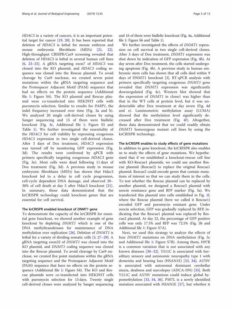
HDAC3 in a variety of cancers, it is an important poten-tial target for cancer [19, 20]. It has been reported thatdeletion of HDAC3 is lethal for mouse embryos andmouse embryonic fibroblasts (MEFs) [21, 22].High-throughput CRISPR/Cas9 screening revealed thatdeletion of HDAC3 is lethal in several human cell lines[6, 23–25]. A gRNA targeting exon7 of HDAC3 wascloned into the KO plasmid, and HDAC3 coding se-quence was cloned into the Rescue plasmid. To avoidcleavage by Cas9 nuclease, we created seven pointmutations within the gRNA targeting sequence andthe Protospacer Adjacent Motif (PAM) sequence thathad no effects on the protein sequence (Additionalfile 1: Figure S4). The KO plasmid and Rescue plas-mid were co-transfected into HEK293T cells withpuromycin selection. Similar to results for PARP1, theindel frequency increased over time (Fig. 3a and b).We analyzed 20 single cell-derived clones by usingSanger sequencing and 15 of them were biallelicknockout (Fig. 3c, Additional file 1: Figure S5 andTable 1). We further investigated the essentiality ofthe HDAC3 for cell viability by repressing exogenousHDAC3 expression in two single cell-derived clones.After 3 days of Dox treatment, HDAC3 expressionwas turned off by monitoring GFP expression (Fig.3d). The results were confirmed by qPCR withprimers specifically targeting exogenous HDAC3 gene(Fig. 3e). Most cells were dead following 11 days ofDox treatment (Fig. 3d). A previous study in mouseembryonic fibroblasts (MEFs) has shown that Hdac3knockout led to a delay in cell cycle progression,cell-cycle dependent DNA damage, and observed 20–30% of cell death at day 5 after Hdac3 knockout [21].In summary, these data demonstrated that thekrCRISPR technology could knockout genes that areessential for cell survival.
The krCRISPR enabled knockout of DNMT1 geneTo demonstrate the capacity of the krCRISPR for essen-tial gene knockout, we showed another example of geneknockout by depleting DNMT1 which is one of theDNA methyltransferases for maintenance of DNAmethylation over replication [26]. Deletion of DNMT1 islethal for a variety of dividing somatic cells [3, 27–29]. AgRNA targeting exon32 of DNMT1 was cloned into theKO plasmid, and DNMT1 coding sequence was clonedinto the Rescue plasmid. To avoid cleavage by Cas9 nu-clease, we created five point mutations within the gRNAtargeting sequence and the Protospacer Adjacent Motif(PAM) sequence that have no effects on the protein se-quence (Additional file 1: Figure S4). The KO and Res-cue plasmids were co-transfected into HEK293T cellswith puromycin selection for 15 days. Twenty singlecell-derived clones were analyzed by Sanger sequencing
and 16 of them were biallelic knockout (Fig. 4a, Additionalfile 1: Figure S6 and Table 1).We further investigated the effects of DNMT1 repres-
sion on cell survival in two single cell-derived clones.After 3 days of Dox treatment, DNMT1 expression wasshut down by indication of GFP expression (Fig. 4b). Atday seven after Dox treatment, the cells started undergo-ing apoptosis (Fig. 4b). A previous study in human em-bryonic stem cells has shown that all cells died within 9days of DNMT1 knockout [3]. RT-qPCR analysis withprimers specifically targeting exogenous DNMT1 generevealed that DNMT1 expression was significantlydownregulated (Fig. 4c). Western blot showed thatthe expression of DNMT1 in clone1 was higher thanthat in the WT cells at protein level, but it was un-detectable after Dox treatment at day seven (Fig. 4dand e). Luminometric methylation assay (LUMA)showed that the methylation level significantly de-creased after Dox treatment (Fig. 4f ). Altogether,these data demonstrated that we could readily obtainDNMT1 homozygous mutant cell lines by using thekrCRISPR technology.
The krCRISPR enables to study effects of gene mutationsIn addition to gene knockout, the krCRISPR also enablesus to study the effects of gene mutations. We hypothe-sized that if we established a knockout-rescue cell linewith KO-Rescue1 plasmids, we could use another Res-cue plasmid (Rescue2) to replace the original Rescue1plasmid. Rescue2 could encode genes that contain muta-tions of interest so that we can study them in the cells.To test whether the Rescue plasmid can be replaced byanother plasmid, we designed a Rescue2 plasmid withzeocin resistance gene and RFP marker (Fig. 5a). Wetransfected this plasmid into cells established in Fig. 1f,where the Rescue plasmid (here we called it Rescue1)encoded GFP and puromycin resistant gene. Underzeocin selection, GFP was gradually replaced by RFP, in-dicating that the Rescue1 plasmid was replaced by Res-cue2 plasmid. At day 22, the percentage of GFP positivecells was only 17.5% and RFP was 72.5% (Fig. 5b andAdditional file 1: Figure S7A).Next, we used this strategy to analyze the effects of
four DNMT1 mutations on DNA methylation (Fig. 5cand Additional file 1: Figure S7B). Among them, H97Ris a common variation that is not associated with anyknown diseases [30–32]. Y511C is associated with her-editary sensory and autonomic neuropathy type 1 withdementia and hearing loss (HSAN1E) [33, 34]; A570Vis associated with autosomal dominant cerebellarataxia, deafness and narcolepsy (ADCA-DN) [35]. BothY511C and A570V mutations could induce global hy-pomethylation [33, 34, 36]. P507L is a newly identifiedmutation associated with HSAN1E [37], but whether it
Wang et al. Journal of Biological Engineering (2019) 13:35 Page 5 of 13

could influence DNA methylation has not been inves-tigated. Individual Rescue plasmids were transfectedinto the DNMT1 knockout cells with zeocin selection(Fig. 5d). To facilitate the analysis of plasmid replace-ment by RFLP, a restriction site was introduced intothe DNMT1 gene without changing the protein se-quence of the Rescue2 plasmid (PmlI for A570V;XmaI for H97R) (Fig. 5e and f ). For A570V site, theratio of the Rescue2 to total amount of plasmid DNAwas 23.0% at day 2 and 93.9% at day 20, indicatingthat the Rescue1 plasmid was gradually replaced byRescue2 (Fig. 5e). For H97R site, the ratio of the
Rescue2 to total amount of plasmid DNA was 29.4%at day 2 and 97.2% at day 20 (Fig. 5f ).Next, we performed genome-wide methylation analysis
for the individual mutations with LUMA. Compared tocontrol Rescue plasmid expressing WT DNMT1, H97Rvariation did not influence DNA methylation level; Y511Cand A570V mutation decreased DNA methylation levels,consistent with previous reports (Fig. 5g) [34, 36]. P507Lalso decreased DNA methylation level (Fig. 5g), indicatingthat it could potentially be a disease-causing mutation. Insummary, we established a platform that can be used tostudy the effects of mutations at cellular level.
Fig. 3 Generation of HDAC3 knockout-rescue cell lines. a Representative gel pictures of T7E1 assay for detection of indels at HDAC3 sitesin HEK293T cells. Ctr is the PCR band from unmodified cells with T7 enzyme digestion. b Quantification of the T7E1 assay for Fig. 3a(n = 3, error bars show mean ± SEM). c Examples of indel sequences for four single cell-derived clones. Schematic of the gRNA target sitewas shown above. PAM sequence was marked in orange. Cas9 cutting site is indicated by red arrow. d Inhibition of exogenous HDAC3expression in HDAC3-knockout cells caused cell death. The HDAC3 knockout-rescue cells expressed GFP. Expression of GFP was inhibitedby addition of Dox for 3 days. All cells died at day 11. e RT-qPCR analysis of the exogenous HDAC3 expression with or without Dox fortwo clones (n = 3, error bars showed mean ± SEM)
Wang et al. Journal of Biological Engineering (2019) 13:35 Page 6 of 13

Off-target analysisOff-target mutations are often generated during genomeediting [38, 39]. The krCRISPR system requires long-term
editing which may increase off-target effects. We used anonline tool (http://www.rgenome.net/cas-offinder/) tosearch for potential off-target sites and selected five top
Fig. 4 Generation of DNMT1 knockout-rescue cell lines. a Examples of indel sequences for four single cell-derived clones. Schematic of the gRNAtarget site is shown above. PAM sequence is marked in orange. Cas9 cutting site is indicated by red arrow. Insertions are indicated by red letter.b Inhibition of exogenous DNMT1 expression in DNMT1-knockout cells caused cell death. The DNMT1knockout-rescue cells expressed GFP.Expression of GFP was inhibited by addition of Dox for 3 days. All cells died at day 7. c RT-qPCR analysis of the exogenous DNMT1 expressionwith or without Dox for a single cell-derived clone (n = 3, error bars showed mean ± SEM). d Western blot analysis of total DNMT1 expressionwith or without addition of Dox. WT cells were used as a control. e Quantification for the Western blot assay for Fig. 4d (n = 3, error bars showedmean ± SEM). f Luminometric Methylation Assay (LUMA) showed that the global DNA methylation levels were the same for the WT cells andDNMT1 knockout-rescue cells. Addition of Dox significantly decreased DNA methylation level in DNMT1 knockout-rescue cells (n = 3, error barsshowed mean ± SEM)
Wang et al. Journal of Biological Engineering (2019) 13:35 Page 7 of 13

ranked potential off-target sites for gRNA-HDAC3. Thesepotential off-targets have two or three mismatches com-pared to the targeting sequence (Table 2). We tested thesesites in two HDAC3 knockout-rescue clones that were de-rived from a single cell, but we did not observeoff-target mutations (Fig. 6a and Additional file 1:
Figure S8A). We also analyzed five potential off-targetsites for DNMT1 knockout-rescue clones, but we didnot observe off-target mutations (Fig. 6b, Additionalfile 1: Figure S8B and Table 3). Notably, we could notexclude that off-target mutations occurred somewherein the genome. Whole-genome sequencing will be
Fig. 5 The krCRISPR system enabled analysis of the effects of DNMT1 mutation on DNA methylation. a Schematic of the Rescue1 and Rescue2plasmid design. Rescue1 plasmid contains a puromycin resistant gene and a GFP gene, while Rescue 2 plasmid contains a zeocin resistant geneand RFP gene. Transfection of Rescue2 plasmid into the knockout-Resuce1 cells will result in replacement of Rescue1 plasmid by Rescue2 plasmidunder zeocin selection. b Flow cytometry analysis showed that the GFP positive cells were gradually replaced by RFP positive cells over time(n = 3, error bars showed mean ± SEM). c Distribution of the four DNMT1 mutations. d Schematic of the experimental workflow. e and f Twoexamples of Rescue plasmid replacement. The Rescue1 plasmid was gradually replaced by Rescue2 plasmid. A PmlI restriction site for A570V andan XmaI restriction site for H97R were introduced into the Rescue2 plasmids respectively. At day 0, 2 and 15, the plasmid DNA was isolated fromcells and PCR-amplified for RFLP analysis. Gene mutations are labeled in red letter; the restriction sites are underlined. Black triangles indicate theRescue1 plasmid; red triangles indicate the Rescue2 plasmid. g The effects of individual mutations on DNA methylation were measured by LUMA.The exogenous DNMT1 genes encoded by Rescue plasmids were shown below. H97R did not influence DNA methylation level. Y511C, A570Vand P507L decreased DNA methylation level (n = 3, error bars showed mean ± SEM)
Wang et al. Journal of Biological Engineering (2019) 13:35 Page 8 of 13

desirable for further detection of off-target mutationsin the future.
DiscussionIn this study, we demonstrated a simple and efficientmethod to knockout essential genes in cell lines usingkrCRISPR technology. This technology only requirestwo steps to obtain knockout-rescue cell lines: i) clonethe gRNA into the KO vector and rescuing gene into theRescue vector; ii) co-transfect both plasmids into cellsand select single cell-derived clones with biallelic frameshift mutations. The expression of rescue genes can beefficiently turned off by Tet-Off technology, allowing theeffects of gene knockout on cells to be studied. ThekrCRISPR enables efficient knockout of endogenous
genes due to the puromycin selection and long-termgenome editing [17]. In contrast, previous strategies foressential gene knockout are time-consuming andlabor-extensive, requiring multiple steps to generatestable integration cell lines that contain either Tet-Offelements or Cre/loxP elements [3, 7–9, 40, 41].In addition to gene knockout, the krCRISPR also en-
ables users to study the effects of mutations on cells.Each human is estimated to carry on average ~ 60 denovo point mutations that arose in the germ line of theirparents [42]. These mutations are the principal cause ofheritable disease. Furthermore, genome-wide associationstudies (GWAS) have identified a large number of som-atic mutations that are associated with cancers [43, 44].Although CRISPR/Cas9 technology allows efficient
Table 2 Potential off-target sequences for HDAC3
*PAM sequences were marked in green and the mismatched nucleotides were labeled in red
Fig. 6 Analysis of potential off-target sites. a Five potential off-target sites for clone1 of HDAC3 were sequenced. b Five potential off-target sitesfor clone1 of DNMT1 were sequenced
Wang et al. Journal of Biological Engineering (2019) 13:35 Page 9 of 13

introduction of specific mutations into the endogenousloci [45, 46], it is time-consuming. The krCRISPR offersan alternative strategy to study gene mutations in cells.One can knockout endogenous genes and express rescuegenes containing mutation of interest simultaneously.Once the knockout-rescue cell lines are established, therescue plasmid can be replaced by other rescue plasmidcontaining different mutations. In summary, thekrCRISPR technology offers a platform that facilitatesthe study of essential genes’ functions.
ConclusionsIn conclusion, we developed a double episomal vector sys-tem that allows generation of inducible knockout-rescuecell lines. In this system, one vector expresses gRNA andCas9 nuclease, and the other vector expresses an induciblerescue gene. Users can easily knockout an essential geneby the expression of corresponding gRNA and rescuegene.
Materials and methodsCell culture and transfectionThe HEK293T cell line (ATCC) was grown in Dulbecco’sModified Eagle Medium (DMEM), supplemented with 10%fetal bovine serum (Gibco), 1x penicillin-streptomycin andpassaged using 0.25% Trypsin-EDTA every other day. Cellswere incubated at 37 °C with 5% CO2.
PlasmidsKnockout (KO) plasmid: this plasmid was modified fromepiCRISPR plasmid [17]. The SpCas9 and tTA wereco-expressed from an EF1α promoter as a single proteinseparated by self-cleaving P2A peptides; gRNA wasexpressed from a human U6 promoter. The syntheticoligonucleotide duplexes encoding gRNAs can be clonedinto BspQI restriction sites. The sequence of the plasmidis available in Additional file 1: Figure S9.The Rescue plasmid: copGFP and puromycin resist-
ance genes were co-expressed from a pTRE promoterseparated by two P2A peptides. The exogenous gene canbe cloned into multiple cloning sites downstream of
pTRE promoter. The sequence of the plasmid is avail-able in Additional file 1: Figure S10. cDNA of DNMT1,PARP1 and HDAC3 was synthesized by GENWIZ(China) and inserted into KpnI/ AsisI restriction sites ofthe Rescue plasmid. Notably, synonymous mutationswere introduced on gRNA targeting sequence to preventCas9 cleavage.The Rescue2 plasmid: this plasmid is similar to
Rescue plasmid except that copGFP-puro cassettewas replaced with RFP-zeo cassette. The sequenceof the plasmid is available in Additional file 1: Fig-ure S11. The DNMT1P507LDNMT1H97R, DNMT1A570v
and DNMT1Y511C mutations were created on theDNMT1WT using Gibson Assembly Cloning Kit(#E5510S, NEB) according to the online protocol.The primers and oligonucleotides used in this studyare shown in Additional file 1: Table S1.
Transfection, T7 assay and sequencing analysis forgenome modificationHEK293T cells were seeded on 12-well plates in 500uL ofgrowth medium without antibiotics. After 24 h, HEK293Tcells were transfected at 60–70% confluency using Lipo-fectamine 2000 transfection regent (Invitrogen) accordingto the manufacturer’s protocol. For double-plasmid trans-fection, 500 ng of KO plasmid and 500 ng of Rescue plas-mid were transfected per well. From day 2, cells wereselected by puromycin (1.5–2 μg/mL). At day 5, 10 and15, genomic DNA was extracted from cells using Quick-Extract (Cat. # QE09050, Lucigen) following the manufac-turer’s instructions and T7E1 assay was performedaccording to a previously described method [10]. Briefly,genomic region containing the gRNA target site wasPCR-amplified using Q5 High-Fidelity DNA polymerase(NEB) and the PCR products were purified using QIA-quick Gel Extraction Kit (28,706, QIAGEN). A total of300 ng purified PCR products were re-annealed anddigested with T7E1 enzyme (#M0302S, NEB) for 30minat 37 °C. The PCR products were analyzed on 1.5% agar-ose gels. Gels were imaged with EB staining and quantifiedusing ImageJ software according to the band intensities.
Table 3 Potential off-target sequences for DNMT1
*PAM sequences were marked in green and the mismatched nucleotides were labeled in red
Wang et al. Journal of Biological Engineering (2019) 13:35 Page 10 of 13

To analyze indel sequences for single cell-derived clones,cells were digested to single cells 10 days after transfectionand seeded into 96-well plates using flow cytometer. Aweek later, genomic DNA of clones was extracted forPCR-amplification. The PCR products containing gRNAtargeting sites were cloned into T-vector (A1410, Pro-mega) according to the manufacturer’s instructions forSanger sequencing analysis. For plasmid replacement assay,1μg of Rescue2 plasmid was transfected into knockout-rescuecells and selected with zeocin (300-400μg/mL) from day 2.
Protein extraction and Western blottingCell samples were harvested and lysed with NP-40 buffer(Beyotime) in the presence of 1 mM Phenylmethanesul-fonyl fluoride (Beyotime). After centrifugation at 12000rpm for 10min in a 4 °C pre-cooled centrifuge, thesupernatant was collected for Western blot analysis. Pro-teins were separated by 8% SDS-PAGE gel and thentransferred to a polyyinylidene fluoride (PVDF) mem-brane (Thermo). After blocking with 5% (wt/vol) BSA(Sigma) in TBS-T (0.1% Tween 20 in 1x TBS) buffer for1 h at room temperature, the membrane was incubatedwith primary antibodies at 4 °C overnight. Antibodiesused include: anti-DNMT1 (1:1000; ab13537 Abcam)and anti-GAPDH (1:2000, 5174S, Cell Signaling). Afterthree washes with TBS-T of 5 min each, the membraneswere incubated with secondary antibody (1:10,000;ab6721 Abcam) at room temperature for 1 h, followedby three washes and imaged.
RNA isolation and quantitative reverse transcriptionpolymerase chain reactionTotal RNA was extracted from cells using Trizol regent(Ambion) following the manufacturer’s instructions.First-strand cDNA was synthesized from the isolatedRNA using 5x All-In-One RT MasterMix kit (Cat. No.G492, abm) according to the manufacturer’s manual. 2xSYBR Green qPCR Master Mix (Cat. No. 21703, bimake)was used to quantify the expression of HDAC3 andDNMT1 mRNA. GAPDH was used as an internal con-trol for normalization. The primers were designed foramplification of exogenous gene in plasmid using PrimerPremier 5.0. All primer sequences used are shown inAdditional file 1: Table S1. The qRT-PCR was performedusing Bio-Rad Real-Time PCR. Detection System andthe relative expression level was calculated using the2-ΔΔCt method.
Flow cytometryCells for flow cytometry analysis were treated with 0.25%Trypsin-EDTA, washed twice and resuspended in 300uLPBS. The percentage of GFP and RFP positive cells wasquantified using flow cytometer (Gallios, Beckman Coulter)
according to the manufacturer’s protocol. Data were ana-lyzed using FlowJo software.
Luminometric methylation assay (LUMA)Firstly, total DNA was purified with phenol: chloroform.Briefly, cells were harvested and washed twice with PBS.Cells were then resuspended in 460uL of nuclear lysisbuffer, 20uL of proteinase K (20 mg/mL) and 20uL of10% SDS followed by incubation at 58 °C overnight. 5uLRNaseA (10 mg/mL) was then added, mixed by vortex-ing and incubated for 3 h at 37 °C. 500uL phenol:chloro-form was added, mixed by vortexing and incubated for3 min at room temperature following centrifuged for 20min at 13000 rpm. 400uL of aqueous phase containingDNA was then transferred to a fresh tube followed bythe addition of 400uL isopropanol and vortexed for 30s,and finally 40uL NaAc (pH = 5.2) was added and vor-texed for 2 min. The remaining DNA precipitate waswashed twice with 75% ethanol, and dissolved in 80uLddH2O.Subsequently, pyrosequencing was performed. 400 ng
genomic DNA was cleaved with HpaII + EcoRI or MspI+ EcoRI in two separate 20uL reactions containing 400ng DNA, 1uL HpaII (or 0.5uL MspI), 0.5uL EcoRI-HFand 2uL 10x cutsmat buffer (NEB). The reactions wereincubated at 37 °C for 4 h. Then pyrosequencing wasperformed following a previously described protocol[47]. The percentage of methylation was calculatedbased on the LUMA results with the following formula:Methylation % = 100[1-(HpaII/EcoRI/MspI/EcoRI)].
Statistical analysisIn this study, statistical analysis was performed usingGraphPad Prism 5. All data were presented as mean ±SEM. The unpaired Student’s t-test was adopted to de-termine the statistical differences between the samplesof two groups. Significant levels: *P < 0.05, **P < 0.01,****P < 0.001. All experiments were repeated three timesindependently.
Additional file
Additional file 1: Figure S1. Strategies for knockout of essential genesin cells. Figure S2. Establishment of the knockout-rescue system withdouble episomal vectors. Figure S3. Indel sequences at PARP1 site.Figure S4. The gRNA targeting sequences on chromosome and theircorresponding sequences on the rescue genes. Figure S5. Indelsequences at HDAC3 site. Figure S6. Indel sequences at DNMT1 site.Figure S7. Results of flow cytometry. Figure S8. Analysis of potential off-target sites. Figure S9 KO plasmid sequence. Figure S10. Rsecue plasmidsequence. Figure S11 Rsecue2 plasmid sequence. Table S1. Primers andoligonucleotides. (DOCX 3386 kb)
AbbreviationsDSBs: Double strand breaks; EBNA1: Epstein-Barr-associated nuclear antigen1; EBV: Epstein-Barr virus; gRNA: guide RNA; HDR: Homology-directed repair;krCRISPR: knockout-rescue CRISPR; NHEJ: Non-homologous end-joining;
Wang et al. Journal of Biological Engineering (2019) 13:35 Page 11 of 13

PAM: Protospacer Adjacent Motif; RFLP: Restriction fragment lengthpolymorphism; ssDNA: single-stranded DNA
AcknowledgementsNot applicable.
FundingThis work was supported by grants from the National Natural ScienceFoundation of China (81870199), the National Basic Research Program ofChina (2015CB943300), the Foundation for Innovative Research Group of theNational Natural Science Foundation of China (31521003) and Openingprogram 2018 of the State Key Laboratory of Genetic Engineering(SKLGE1809).
Availability of data and materialsAll data generated or analyzed during this study are included in thispublished article.
Authors’ contributionsConceived and designed the experiments: BW, DW, YW Performed theexperiments: BW, ZW, DW, BZ Analyzed the data: BW, BZ, ZW Contributedreagents/ materials: WY, ML Wrote the paper: YM, BW, SGO. All authors readand approved the final manuscript.
Ethics approvalNot applicable.
Consent for publicationNot applicable.
Competing interestsThe authors declare that they have no competing interests.
Publisher’s NoteSpringer Nature remains neutral with regard to jurisdictional claims inpublished maps and institutional affiliations.
Author details1MOE Key Laboratory of Contemporary Anthropology at School of LifeSciences and Zhongshan Hospital, Fudan University, Shanghai 200438, China.2Shanghai Public Health Clinical Center & Laboratory of RNA Epigenetics,Institute of Biomedical Sciences, Shanghai Medical College, Fudan University,Shanghai 201508, China. 3Department of Pharmacology, University of IllinoisCollege of Medicine, Chicago, IL 60612, USA. 4Division of Cardiology,Department of Medicine, University of Illinois College of Medicine, Chicago,IL 60612, USA. 5The Key Lab of Reproduction Regulation of NPFPC in SIPPR,Institute of Reproduction & Development in Obstetrics & GynecologyHospital, Fudan University, Shanghai 200011, China.
Received: 27 December 2018 Accepted: 15 February 2019
References1. Zhou Y, Zhu S, Cai C, Yuan P, Li C, Huang Y, Wei W. High-throughput
screening of a CRISPR/Cas9 library for functional genomics in human cells.Nature. 2014;509:487–91.
2. Parnas O, Jovanovic M, Eisenhaure TM, Herbst RH, Dixit A, Ye CJ, PrzybylskiD, Platt RJ, Tirosh I, Sanjana NE, Shalem O, Satija R, Raychowdhury R, MertinsP, Carr SA, Zhang F, Hacohen N, Regev A. A genome-wide CRISPR screen inprimary immune cells to dissect regulatory networks. Cell. 2015;162:675–86.
3. Liao J, Karnik R, Gu H, Ziller MJ, Clement K, Tsankov AM, Akopian V, GiffordCA, Donaghey J, Galonska C, Pop R, Reyon D, Tsai SQ, Mallard W, Joung JK,Rinn JL, Gnirke A, Meissner A. Targeted disruption of DNMT1, DNMT3A andDNMT3B in human embryonic stem cells. Nat Genet. 2015;47:469–78.
4. Wang T, Wei JJ, Sabatini DM, Lander ES. Genetic screens in human cellsusing the CRISPR-Cas9 system. Science. 2014;343:80–4.
5. Koike-Yusa H, Li Y, Tan EP, Velasco-Herrera Mdel C, Yusa K. Genome-widerecessive genetic screening in mammalian cells with a lentiviral CRISPR-guide RNA library. Nat Biotechnol. 2014;32:267–73.
6. Wang T, Birsoy K, Hughes NW, Krupczak KM, Post Y, Wei JJ, Lander ES,Sabatini DM. Identification and characterization of essential genes in thehuman genome. Science. 2015;350:1096–101.
7. Branda CS, Dymecki SM. Talking about a revolution: the impact of site-specific recombinases on genetic analyses in mice. Dev Cell. 2004;6:7–28.
8. Lewandoski M. Conditional control of gene expression in the mouse. NatRev Genet. 2001;2:743–55.
9. Matsunaga T, Yamashita JK. Single-step generation of gene knockout-rescuesystem in pluripotent stem cells by promoter insertion with CRISPR/Cas9.Biochem Biophys Res Commun. 2014;444:158–63.
10. Cong L, Ran FA, Cox D, Lin S, Barretto R, Habib N, Hsu PD, Wu X, Jiang W,Marraffini LA, Zhang F. Multiplex genome engineering using CRISPR/Cassystems. Science. 2013;339:819–23.
11. Mali P, Yang L, Esvelt KM, Aach J, Guell M, DiCarlo JE, Norville JE, Church GM.RNA-guided human genome engineering via Cas9. Science. 2013;339:823–6.
12. Hwang WY, Fu Y, Reyon D, Maeder ML, Tsai SQ, Sander JD, Peterson RT, YehJR, Joung JK. Efficient genome editing in zebrafish using a CRISPR-Cassystem. Nat Biotechnol. 2013;31:227–9.
13. Jinek M, Chylinski K, Fonfara I, Hauer M, Doudna JA, Charpentier E. Aprogrammable dual-RNA-guided DNA endonuclease in adaptive bacterialimmunity. Science. 2012;337:816–21.
14. Ran FA, Hsu PD, Wright J, Agarwala V, Scott DA, Zhang F. Genomeengineering using the CRISPR-Cas9 system. Nat Protoc. 2013;8:2281–308.
15. Wang Y, Zhang WY, Hu S, Lan F, Lee AS, Huber B, Lisowski L, Liang P,Huang M, de Almeida PE, Won JH, Sun N, Robbins RC, Kay MA, Urnov FD,Wu JC. Genome editing of human embryonic stem cells and inducedpluripotent stem cells with zinc finger nucleases for cellular imaging. CircRes. 2012;111:1494–503.
16. Wang Y, Liang P, Lan F, Wu H, Lisowski L, Gu M, Hu S, Kay MA, Urnov FD,Shinnawi R, Gold JD, Gepstein L, Wu JC. Genome editing of isogenic humaninduced pluripotent stem cells recapitulates long QT phenotype for drugtesting. J Am Coll Cardiol. 2014;64:451–9.
17. Xie Y, Wang D, Lan F, Wei G, Ni T, Chai R, Liu D, Hu S, Li M, Li D, Wang H,Wang Y. An episomal vector-based CRISPR/Cas9 system for highly efficientgene knockout in human pluripotent stem cells. Sci Rep. 2017;7:2320.
18. Van Craenenbroeck K, Vanhoenacker P, Haegeman G. Episomal vectors forgene expression in mammalian cells. Eur J Biochem. 2000;267:5665–78.
19. Adhikari N, Amin SA, Trivedi P, Jha T, Ghosh B. HDAC3 is a potentialvalidated target for cancer: an overview on the benzamide-based selectiveHDAC3 inhibitors through comparative SAR/QSAR/QAAR approaches. Eur JMed Chem. 2018;157:1127–42.
20. West AC, Johnstone RW. New and emerging HDAC inhibitors for cancertreatment. J Clin Invest. 2014;124:30–9.
21. Bhaskara S, Chyla BJ, Amann JM, Knutson SK, Cortez D, Sun ZW, Hiebert SW.Deletion of histone deacetylase 3 reveals critical roles in S phaseprogression and DNA damage control. Mol Cell. 2008;30:61–72.
22. Montgomery RL, Potthoff MJ, Haberland M, Qi X, Matsuzaki S, Humphries KM,Richardson JA, Bassel-Duby R, Olson EN. Maintenance of cardiac energymetabolism by histone deacetylase 3 in mice. J Clin Invest. 2008;118:3588–97.
23. Hart T, Chandrashekhar M, Aregger M, Steinhart Z, Brown KR, MacLeod G,Mis M, Zimmermann M, Fradet-Turcotte A, Sun S, Mero P, Dirks P, Sidhu S,Roth FP, Rissland OS, Durocher D, Angers S, Moffat J. High-resolution CRISPRscreens reveal fitness genes and genotype-specific Cancer liabilities. Cell.2015;163:1515–26.
24. Tzelepis K, Koike-Yusa H, De Braekeleer E, Li Y, Metzakopian E, Dovey OM,Mupo A, Grinkevich V, Li M, Mazan M, Gozdecka M, Ohnishi S, Cooper J,Patel M, McKerrell T, Chen B, Domingues AF, Gallipoli P, Teichmann S,Ponstingl H, McDermott U, Saez-Rodriguez J, Huntly BJP, Iorio F, Pina C,Vassiliou GS, Yusa K. A CRISPR dropout screen identifies geneticvulnerabilities and therapeutic targets in acute myeloid leukemia. Cell Rep.2016;17:1193–205.
25. Steinhart Z, Pavlovic Z, Chandrashekhar M, Hart T, Wang X, Zhang X,Robitaille M, Brown KR, Jaksani S, Overmeer R, Boj SF, Adams J, Pan J,Clevers H, Sidhu S, Moffat J, Angers S. Genome-wide CRISPR screens reveala Wnt-FZD5 signaling circuit as a druggable vulnerability of RNF43-mutantpancreatic tumors. Nat Med. 2017;23:60–8.
26. Jones PA, Liang G. Rethinking how DNA methylation patterns aremaintained. Nat Rev Genet. 2009;10:805–11.
27. Trowbridge JJ, Snow JW, Kim J, Orkin SH. DNA methyltransferase 1 isessential for and uniquely regulates hematopoietic stem and progenitorcells. Cell Stem Cell. 2009;5:442–9.
Wang et al. Journal of Biological Engineering (2019) 13:35 Page 12 of 13

28. Sen GL, Reuter JA, Webster DE, Zhu L, Khavari PA. DNMT1 maintainsprogenitor function in self-renewing somatic tissue. Nature. 2010;463:563–7.
29. Jackson-Grusby L, Beard C, Possemato R, Tudor M, Fambrough D,Csankovszki G, Dausman J, Lee P, Wilson C, Lander E, Jaenisch R. Loss ofgenomic methylation causes p53-dependent apoptosis and epigeneticderegulation. Nat Genet. 2001;27:31–9.
30. Saradalekshmi KR, Neetha NV, Sathyan S, Nair IV, Nair CM, Banerjee M. DNAmethyl transferase (DNMT) gene polymorphisms could be a primary eventin epigenetic susceptibility to schizophrenia. PLoS One. 2014;9:e98182.
31. Peng C, Deng Q, Li Z, Xiong C, Li C, Zheng F. Risk-association of DNMT1gene polymorphisms with coronary artery disease in Chinese Hanpopulation. Int J Mol Sci. 2014;15:22694–705.
32. Ye C, Beeghly-Fadiel A, Lu W, Long J, Shu XO, Gao YT, Zheng W, Cai Q.Two-stage case-control study of DNMT-1 and DNMT-3B gene variants andbreast cancer risk. Breast Cancer Res Treat. 2010;121:765–9.
33. Smets M, Link S, Wolf P, Schneider K, Solis V, Ryan J, Meilinger D, Qin W,Leonhardt H. DNMT1 mutations found in HSANIE patients affect interactionwith UHRF1 and neuronal differentiation. Hum Mol Genet. 2017;26:1522–34.
34. Sun Z, Wu Y, Ordog T, Baheti S, Nie J, Duan X, Hojo K, Kocher JP, Dyck PJ, KleinCJ. Aberrant signature methylome by DNMT1 hot spot mutation in hereditarysensory and autonomic neuropathy 1E. Epigenetics. 2014;9:1184–93.
35. Winkelmann J, Lin L, Schormair B, Kornum BR, Faraco J, Plazzi G, Melberg A,Cornelio F, Urban AE, Pizza F, Poli F, Grubert F, Wieland T, Graf E, Hallmayer J,Strom TM, Mignot E. Mutations in DNMT1 cause autosomal dominantcerebellar ataxia, deafness and narcolepsy. Hum Mol Genet. 2012;21:2205–10.
36. Kernohan KD, Cigana Schenkel L, Huang L, Smith A, Pare G, Ainsworth P,Care4Rare Canada, C, Boycott KM, Warman-Chardon J, Sadikovic B. Identificationof a methylation profile for DNMT1-associated autosomal dominant cerebellarataxia, deafness, and narcolepsy. Clin Epigenetics. 2016;8:91.
37. Baets J, Duan X, Wu Y, Smith G, Seeley WW, Mademan I, McGrath NM,Beadell NC, Khoury J, Botuyan MV, Mer G, Worrell GA, Hojo K, DeLeon J,Laura M, Liu YT, Senderek J, Weis J, Van den Bergh P, Merrill SL, Reilly MM,Houlden H, Grossman M, Scherer SS, De Jonghe P, Dyck PJ, Klein CJ.Defects of mutant DNMT1 are linked to a spectrum of neurologicaldisorders. Brain. 2015;138:845–61.
38. Ran FA, Hsu PD, Lin CY, Gootenberg JS, Konermann S, Trevino AE, Scott DA,Inoue A, Matoba S, Zhang Y, Zhang F. Double nicking by RNA-guidedCRISPR Cas9 for enhanced genome editing specificity. Cell. 2013;154:1380–9.
39. Mali P, Aach J, Stranges PB, Esvelt KM, Moosburner M, Kosuri S, Yang L,Church GM. CAS9 transcriptional activators for target specificity screeningand paired nickases for cooperative genome engineering. Nat Biotechnol.2013;31:833–8.
40. Yokoyama T, Miyazawa K, Naito M, Toyotake J, Tauchi T, Itoh M, Yuo A,Hayashi Y, Georgescu MM, Kondo Y, Kondo S, Ohyashiki K. Vitamin K2induces autophagy and apoptosis simultaneously in leukemia cells.Autophagy. 2008;4:629–40.
41. Chen Y, Cao J, Xiong M, Petersen AJ, Dong Y, Tao Y, Huang CT, Du Z,Zhang SC. Engineering human stem cell lines with inducible gene knockoutusing CRISPR/Cas9. Cell Stem Cell. 2015;17:233–44.
42. Shendure J, Akey JM. The origins, determinants, and consequences ofhuman mutations. Science. 2015;349:1478–83.
43. Sud A, Kinnersley B, Houlston RS. Genome-wide association studies of cancer:current insights and future perspectives. Nat Rev Cancer. 2017;17:692–704.
44. Tang H, Wei P, Chang P, Li Y, Yan D, Liu C, Hassan M, Li D. Geneticpolymorphisms associated with pancreatic cancer survival: a genome-wideassociation study. Int J Cancer. 2017;141:678–86.
45. Paquet D, Kwart D, Chen A, Sproul A, Jacob S, Teo S, Olsen KM, Gregg A,Noggle S, Tessier-Lavigne M. Efficient introduction of specific homozygousand heterozygous mutations using CRISPR/Cas9. Nature. 2016;533:125–9.
46. Salsman J, Dellaire G. Precision genome editing in the CRISPR era. BiochemCell biol. 2017;95:187–201.
47. Karimi M, Johansson S, Stach D, Corcoran M, Grander D, Schalling M,Bakalkin G, Lyko F, Larsson C, Ekstrom TJ. LUMA (LUminometric methylationassay)--a high throughput method to the analysis of genomic DNAmethylation. Exp Cell Res. 2006;312:1989–95.
Wang et al. Journal of Biological Engineering (2019) 13:35 Page 13 of 13