Embed Size (px)
Citation preview

Lab on a Chip
PAPER
Cite this: DOI: 10.1039/c7lc00633k
Received 17th June 2017,Accepted 24th July 2017
DOI: 10.1039/c7lc00633k
rsc.li/loc
Multimode smartphone biosensing: thetransmission, reflection, and intensity spectral(TRI)-analyzer
Kenneth D. Long, a Elizabeth V. Woodburn,a Huy M. Le,bc Utsav K. Shah,d
Steven S. Lumettabc and Brian T. Cunningham*ab
We demonstrate a smartphone-integrated handheld detection instrument capable of utilizing the internal
rear-facing camera as a high-resolution spectrometer for measuring the colorimetric absorption spectrum,
fluorescence emission spectrum, and resonant reflection spectrum from a microfluidic cartridge inserted
into the measurement light path. Under user selection, the instrument gathers light from either the white
“flash” LED of the smartphone or an integrated green laser diode to direct illumination into a liquid test
sample or onto a photonic crystal biosensor. Light emerging from each type of assay is gathered via optical
fiber and passed through a diffraction grating placed directly over the smartphone camera to generate
spectra from the assay when an image is collected. Each sensing modality is associated with a unique con-
figuration of a microfluidic “stick” containing a linear array of liquid chambers that are swiped through the
instrument while the smartphone captures video and the software automatically selects spectra represen-
tative of each compartment. The system is demonstrated for representative assays in the field of point-of-
care (POC) maternal and infant health: an ELISA assay for the fetal fibronectin protein used as an indicator
for pre-term birth and a fluorescent assay for phenylalanine as an indicator for phenylketonuria. In each
case, the TRI-analyzer is capable of achieving limits of detection that are comparable to those obtained for
the same assay measured with a conventional laboratory microplate reader, demonstrating the flexibility of
the system to serve as a platform for rapid, simple translation of existing commercially available biosensing
assays to a POC setting.
Introduction
Since the introduction of smartphones in 2004, interest incoupling them with wearable devices for monitoring health-related metrics will grow to an over $15 billion per year mar-ket1 focused mainly upon reporting physiological parameterssuch as heart rate and blood pressure. While the market forsuch consumer products does not classify the vast majoritythese sensors as medical devices, there is intense interest intransitioning additional health diagnostics, particularlyin vitro diagnostic (IVD) tests on bodily fluids from the labora-tory to the point-of-care (POC). Tests that can be facilitatedthrough test-specific cartridges that interface with a readout
instrument2,3 or paper-based assays that can be interpretedby visual inspection have obtained regulatory approval that fa-cilitates their use in POC settings.4,5
As smartphones continue to include greater computationalpower and more sophisticated imaging sensors that comple-ment their capabilities for wireless communication and inter-net connectivity, the interface between IVD testing and mobiledevices offers a powerful new POC testing paradigm. The out-put of nearly all of the most common laboratory-based diag-nostic tests can be sensed using a smartphone with sensitivitythat rivals that of conventional laboratory-based benchtop in-struments. Examples include smartphone-based micros-copy,6,7 fluorimetry,8,9 Enzyme linked immunosorbent assay(ELISA),10 polymerase chain reaction (PCR),11 and lateral flowassays.12,13 The reader is directed to recent reviews on thetopic of smartphone-based biosensing.14,15 It is evident thatwhile laboratory-based assays and detection instruments pro-vide high throughput and automation, enormous needs existto provide diagnostic information to people who lack conve-nient access to a diagnostic test facility. Additionally, in manyurgent health situations, an immediate result is required forfeedback to the physician and patient.
Lab ChipThis journal is © The Royal Society of Chemistry 2017
aDepartment of Bioengineering, Micro and Nano Technology Laboratory,
University of Illinois at Urbana-Champaign, 208 N. Wright Street, Urbana, IL
61801, USA. E-mail: [email protected] of Electrical and Computer Engineering, University of Illinois at
Urbana-Champaign, USAc Coordinated Science Laboratory, University of Illinois at Urbana-Champaign,
USAdDepartment of Computer Science, University of Illinois at Urbana-Champaign,
USA
Publ
ishe
d on
24
July
201
7. D
ownl
oade
d by
Uni
vers
ity o
f Il
linoi
s -
Urb
ana
on 1
4/08
/201
7 02
:14:
31.
View Article OnlineView Journal

Lab Chip This journal is © The Royal Society of Chemistry 2017
To date, reported examples of smartphone-based IVD detec-tion instruments are only capable of carrying out a single typeof analysis, and with a few exceptions9,16 are capable of very lim-ited multiplexing of tests. The capability for a laboratory-basedmicroplate reader to function as a “multi-mode” instrument hasbeen long recognized as a desirable characteristic that enables asingle system to transition between spectrometric absorptionanalysis (as used in ELISA assays), fluorometric analysis (as usedin molecular beacon assays, fluorescent polarization (FP) assays,and luminescence-based assays), and, in the case of one prod-uct (PerkinElmer EnSight) label-free optical biosensor assays.The ability of a detection system to easily transition betweenthese modalities enables the user to purchase only one instru-ment, and to use a common liquid handling format and soft-ware interface across a variety of applications.
In this work, we report, to our knowledge, the first exam-ple of a smartphone-connected detection instrument that canmeasure the output of three distinct spectral biosensing mo-dalities using two separate yet complimentary light paths.The three modalities represent a large fraction of all commer-cially available IVD tests for which a liquid sample changescolor, or in which a liquid sample generates light as a visibleindicator of the test's outcome. The system is capable ofperforming spectral analysis of:
Optical transmission
Optical transmission of light through a test sample at specificwavelengthIJs). This measurement modality is commonly used inELISA assays, liquid phase plasmonic nanoparticle-based tests in-cluding surface plasmon resonance (SPR) and localized surfaceplasmon resonance (LSPR),17–19 latex (or other nanoparticle-based) agglutination tests (LAT),20 and transmissive surface-basedSPR, LSPR, and photonic crystal (PC) based tests.19,21,22 ELISAtests are a mainstay of contemporary biosensing tests, with ready-to-go kits available for thousands of different biological targets.
Reflectance spectroscopy
Reflectance spectroscopy of light against a resonant opticaltransducer, as used in reflective label-free optical biosensingtechniques such as SPR, PC, or reflectometric interferencespectroscopy (RIFS). While these direct binding label-free ap-proaches do not currently have commercially available assaykits associated with them, they have been applied to thousandsof biological analytes due to their simplicity and speed.23
Intensity spectroscopy
Intensity spectroscopy from photon-generating assays such asthose based upon fluorescence, chemiluminescence, biolumi-nescence or quantum-dot (QD) emission. A broad variety ofbiosensing assays have such a readout, including fluorescenceor chemiluminescent immunoassays (FIA, CLIA), fluorescentpolarization (FP), Forster resonance energy transfer (FRET) aswell as several molecular diagnostics, including polymerasechain reaction (PCR) and related loop-mediated isothermalamplification (LAMP).
Together, we estimate that these modalities can performthe necessary spectral readouts of 90–95% of commercial im-munoassays,24,25 the exception being radioimmunoassays(RIAs). In particular, as traditional ELISAs are being replacedby FIAs and CLIAs,26 the ability to easily switch between mea-surement modalities makes this system capable ofmaintaining utility for a variety of applications, and a usefulopportunity to potentially compare different tests for thesame analyte on the same point-of-care device.
The spectral transmission-reflectance-intensity (TRI)-ana-lyzer takes advantage of the fact that all three modalitiesshare the requirement to measure changes in the spectrumof light that is either absorbed, reflected, or emitted duringthe course of each type of IVD test. Our approach allows theuser to select from one of two light paths: illumination of theassay with either the internal white LED of the smartphonethat is typically used for “flash” exposure photography, or in-stead with an inexpensive green laser diode. Accurate spectralanalysis is performed by collecting light from the assay intoan optical fiber, whose distal end directs light through a dif-fraction grating into the smartphone's rear-facing internalcamera. The optical components are arranged wholly withina 3D-printed plastic cradle, containing a slot through whichan assay cartridge is inserted.
For many clinically relevant assays, multiple samples, rep-licates, positive/negative controls, and/or standard calibrationsolutions are required. Few smartphone-based sensing plat-forms have demonstrated this capability beyond taking pic-tures of multiple colored samples and performing RGB-basedcolor processing. By converting the smartphone camera intoa spectrometer, we lose the spatial discrimination that wouldallow simultaneous measurement of multiple samples. In-stead, multiplexing is achieved using a cartridge comprisedof a linear series of liquid compartments that slides throughan opening in the back of the cradle. By pairing this linearmotion with the smartphone's ability to capture video, thespatial dimension separating the liquid chambers in the car-tridge can be reconstituted with video post-processing,allowing for the rapid and user-friendly measurement of mul-tiple samples with a single motion, similar to swiping themagnetic strip of a credit card across a magnetic read head.
Each sensing modality utilizes a unique cartridge configu-ration with the same overall form factor. Both transmissionand reflection modalities rely on a bifurcated illumination-collection fiber optic assembly that allows light from thesmartphone flash to pass through a transparent cartridgeand reflect off a stationary mirror positioned within the cra-dle or interact with a PC biosensor that operates as awavelength-selective resonant mirror at the cartridge's backwall, which is coated in an opaque dye to prevent reflectionby the stationary mirror. The intensity modality uses car-tridges with a transparent bottom to allow for orthogonalfluorescent excitation of the sample chambers. All three mo-dalities then collect the reflected light via the collecting fiberof the bifurcated fiber assembly which is then transmitted tothe rear-facing camera of the smartphone.
Lab on a ChipPaper
Publ
ishe
d on
24
July
201
7. D
ownl
oade
d by
Uni
vers
ity o
f Il
linoi
s -
Urb
ana
on 1
4/08
/201
7 02
:14:
31.
View Article Online

Lab ChipThis journal is © The Royal Society of Chemistry 2017
Importantly, the detection approach enables the user to“swipe” the cartridge through the instrument while thesmartphone records video at 60 frames per second, and soft-ware can identify the frames from the video that represent im-ages gathered while the center of each chamber is aligned withthe illumination/read head. Thus, the user is freed from the ne-cessity of performing accurate alignment of the cartridge's liq-uid chambers within the instrument, and the cartridge can beread in a similar manner to the approach used for scanning acredit card past a magnetic stripe reader. Multiplexing in thisfashion enables a single cartridge to contain experimental con-trols, replicates, and a series of active tests.
In this paper, we demonstrate the spectral TRI-analyzerfirst for representative tests that verify its capabilities to mea-sure liquid absorption spectra, PC biosensor resonant reflec-tion spectra and fluorescence spectra. Then, using commer-cially available diagnostic assay kits for colorimetric output(ELISA) and fluorescence output, we validate the instrument'stwo light paths and demonstrate that the spectral TRI-analyzer provides equivalent limits of detection (LOD) forreading the same assay with a commercially available 96-well
microplate reader. While there exist many opportunities tooptimize assays for use with the TRI-analyzer system, the fo-cus of this work is on the demonstration of ease-of-translation readily adaptable the thousands of existing com-mercial assays on the market. Diagnostic assay kits for thePC biosensor modality are not commercially available.
We chose our assay application demonstrations to repre-sent tests that have important implications for maternal andneonatal health in developing parts of the world where aPOC test would have enormous value. First, we perform anELISA assay for detection of fetal fibronectin, a biomarkerprotein test for expectant mothers that is diagnostic for spon-taneous pre-term birth. Next, we perform a fluorescence-based assay for phenylalanine, a biomarker used to diagnosephenylketonuria, a life-threatening but manageable geneticcondition, in newborns.
Optical design and fabrication
An important design requirement for the TRI-analyzer was tomake the approach easily adaptable to any smartphone with a
Fig. 1 Design of spectral TRI-analyzer. A) Schematic of internal layout for optical and electrical components. A custom bifurcated fiber (100 μmcore with d = 1.8 mm glass capillary tube (proximal) and 2× d = 1.25 mm ceramic ferrules, coastal connections) was used to compact the lightpath into a handheld device. Fiber is arranged to maximize bend radius, increasing long-term stability. B) 3D CAD model was created, comprisedof 5 plastic parts printed via stereolithography. Two halves of optical housing maintain lens alignment and optical chamber isolation. All portionsof cradle were designed to slide together and attach with M3×0.5 machine screws. C) Glass capillary tube with bifurcated fibers and metal nut usedto align the fiber in the cradle. D) Image of final device in use with absorption cartridge.
Lab on a Chip Paper
Publ
ishe
d on
24
July
201
7. D
ownl
oade
d by
Uni
vers
ity o
f Il
linoi
s -
Urb
ana
on 1
4/08
/201
7 02
:14:
31.
View Article Online

Lab Chip This journal is © The Royal Society of Chemistry 2017
rear-facing camera and rear-facing flash LED through smallmechanical adjustment to the cradle body, but without adjust-ment of the core optical components. Thus, we chose to use op-tical fibers to bring light from the LED to the test sample, andto bring light from the test sample to the camera. In principle,the core optical components of the system would be identicalfor any smartphone, with two simple changes: 1) the phoneinterface is modified to use the physical dimensions of the ph-one to align the sensing fiber with the rear-facing camera and2) the illumination fiber would be routed to collect light fromthe smartphone flash. As shown in Fig. 1, the distal end of the“illumination” fiber (100 μm diameter, multimode) is placeddirectly in front of the flash LED to direct white light throughthe test sample for the transmission and reflectance modali-ties. The other “sensing” fiber (100 μm diameter, multimode)collects light transmitted through the test sample, reflectedfrom the PC, or emitted by fluorophores at its proximal end
and directs it toward the rear-facing smartphone camera, ori-ented at a 41.3-degree angle. The light emerging from the sens-ing fiber is collimated by an aspheric lens (EFL = 19 mm) andthen focused in the non-spectral dimension with a cylindricallens (EFL = 9 mm) before passing through a 1200 lines per mmtransmission diffraction grating (Edmund Optics 49-578) heldwithin the cradle body directly over the opening of the rear-facing camera. The light is focused to optimize use of theCMOS pixels in the non-spectral dimension allowing for in-creased sensitivity. The proximal ends of the sensing and illu-mination fibers are gathered together in a bifurcated configura-tion, so they are directly adjacent and held within a glasscapillary tube that is mounted in a slot within the cradle body.The optical fiber assembly is the only custom-made componentof the system, due to the specific fiber length requirements (71mm for the illumination fiber and 137 mm for the sensing fi-ber) and ferrules on the distal ends. Due to the accuracy of the
Fig. 2 Two principal light paths and cartridges for each of the three modalities. A) Reflection (PC-only) and transmission (absorption or PC)optical pathway. Collimated light from the on-board smartphone flash is directed through the sample chamber. For reflection-based PC measure-ments, a back-coated cartridge prevents transmitted light from being collected. For transmission measurements, light is reflected by cradle mirrordirected toward the collection fiber. B) Fluorescence/luminescence intensity optical pathway. A laser pointer diode is co-focused to a point nearthe back-side of the cartridge where the collection fiber is similarly focused via addition of a single plano-convex lens into the optical path via aSLA-printed slider actuated from outside the cradle. Additional cartridge wells/housing was removed for clarity in B). C) “Sandwich” style cartridgefabrication alternating plastic or glass substrates with double-sided-adhesive (DSA) showing how different backings facilitate different modalities.D) Transmission cartridge showing inlet/outlets and optically-isolated chambers. E) Dyed PC for label-free reflection measurements. F) Close-upof intensity cartridge demonstrating selectively-dyed cartridge body allowing for bottom-illumination with 532 nm laser diode.
Lab on a ChipPaper
Publ
ishe
d on
24
July
201
7. D
ownl
oade
d by
Uni
vers
ity o
f Il
linoi
s -
Urb
ana
on 1
4/08
/201
7 02
:14:
31.
View Article Online

Lab ChipThis journal is © The Royal Society of Chemistry 2017
3D printing technology used to generate the cradle (FormLabs,Form 2 at 50 μm resolution), all optical components (lenses, re-flection mirror, optical fiber assembly, and green LED) may beplaced into their respective holding locations in correct opticalalignment without further modification and minimal tuning ofthe system with set screws for each of the fiber terminations.
For transmission and reflection modalities the smartph-one flash serves as the light source. It is collected by oneof the two distal ends of the bifurcated optical fiber, andexpanded to a collimated beam with a plano-convex lens(Fig. 2). After passing through the sample chamber, eithercomplete reflection (transmission) or wavelength-selectivereflection (reflection) results in the signal passing throughthe sample once again and being focused on the image ofthe proximal end of the sensing fiber for transmission orreflection modalities, respectively. The mirror is mountedand aligned inside the cradle, where it serves as the defaultreflection surface.
For the intensity modality, a 532 nm laser diode (10 mW, 3V), similar to those found in green laser pointers, is shone or-thogonally to the main optical path of the instrument. Withthe flash turned off, the collecting light path of the instrumentis augmented with an additional lens which serves to focus thecollection to a point close to the rear face of the cartridge. Thislens is inserted into the optical path by pressing a button onthe exterior of the cradle, which slides the lens into the correctposition. The laser is focused within the test sample at thesame point within the cartridge at which the collection lens isalso focused to provide for maximal fluorescence collection.
All optical components were optimized via non-sequentialoptical simulation (Zemax), with total optical efficiency prior-itized for the intensity modality's optical configuration. The3D-printed cradle is comprised of two housing pieces held to-gether with steel fasteners to robustly maintain optical align-ment. Holes were threaded with an M3 tap, and final clear-ances of moving parts were adjusted by hand to allow for
Fig. 3 Qualitative proof-of-concept of 3 principal modalities. A) Transmission. Yellow food dye was diluted in water at concentrations rangingfrom 1 : 1000 to 1 : 64000, a 64× concentration range. Absorption was measured by subtracting sample transmission from that of water. Observ-able absorption occurred in the blue region (400–500 nm) of the spectrum. Inset raw images correspond to concentration of yellow dye of theiroutline. B) Reflection. 0–80% mixtures of ethanol in water were prepared and introduced into a PC-based cartridge, producing narrowband reflec-tion in the 580 nm range. Measurements of each cartridge chamber filled with water were subtracted from measurements of those chambers filledwith ethanol solutions to produce ΔPWV shifts. Inset raw image corresponds to concentration of ethanol of its outline. A central line of pixels wasused to produce spectra shown. C) Intensity. R6G dye was diluted in water and excited with the on-board laser diode. Inset raw images corre-spond to concentration of R6G of their outline.
Lab on a Chip Paper
Publ
ishe
d on
24
July
201
7. D
ownl
oade
d by
Uni
vers
ity o
f Il
linoi
s -
Urb
ana
on 1
4/08
/201
7 02
:14:
31.
View Article Online

Lab Chip This journal is © The Royal Society of Chemistry 2017
smooth sliding before mounting and alignment with ma-chine screws. Optical alignment was completed first with thetransmission/reflection light path, and then with the inten-sity light path, using a cartridge with rhodamine 590 chloride(exciton) laser dye to visualize the optical paths.
The laser diode is powered by a custom circuit providing aconstant-current to the diode from three on-board AAA batte-ries, based on a LED flashlight control circuit (Texas Instru-ments). This circuit is soldered on a small protoboard, andattached to a switch accessible from the outside of the cradle(visible in Fig. 1D).
To interface with the cradle, custom cartridges weredesigned to facilitate introduction of samples to the light path.While all based on the same fundamental construction, eachmodality requires slight modifications to the cartridge design (-Fig. 2C–F). The cartridge bodies for each modality are com-prised of a laser-cut acrylic sheet. This acrylic sheet providesthe separation between chambers, and the overall structure forthe cartridge, which can be modified to support different sam-ple measurement cross-sectional areas, sample volumes, andchamber spacings. For our experiments, we selected a chamberspacing that would allow for user-friendly sample manipulationwith an 8-channel multipipette. Laser-cut 50 μm thick double-sided adhesive (DSA) (3M, Optically Clear Adhesive 8212) is ap-plied to both sides and a thin acrylic cover with laser-cut accessports is adhered to the front of the cartridge, as shown in thecross section of Fig. 2C. The back of the cartridge is comprisedof glass (transmission cartridges), acrylic film (intensity car-tridges), or PC-coated polycarbonate film (reflection cartridges).Depending on the modality, different portions of cartridges aredyed with opaque black dye (Dykem 81724) (Fig. 2E and F) toprevent optical communication between adjacent fluid com-partments and to provide a clear boundary between cartridgesfor automated sensing of the compartments as they pass the il-lumination/read head during video-based cartridge scanning.To provide consistent positioning of the cartridge in the lightpath, a copper spring is attached to the collection-end of thecartridge chamber, so the cartridges are pressed firmly againstone wall of the cradle's slot.
A custom Android app was developed to assist with datacollection. This app sets parameters, including exposure time
and focal length, to constant values to provide for inter-assayconsistency and to facilitate data collection. After the assay ofchoice has been completed and is ready to be measured, theApp allows the user to choose the number of samples, stan-dards, and replicates before walking the user through collec-tion steps for each liquid chamber and/or cartridge.
Qualitative proofs-of-concept for each of the three modali-ties were completed with prepared test liquids in the car-tridge chambers (Fig. 3). To demonstrate Transmission mea-surements, we prepared a series of diluted yellow food dye(McCormick) to a range such that measurable optical absorp-tion was observable on the system (∼1 : 1000), and thenperformed serial dilutions to observe concentration-dependent absorbance (Fig. 3A). Note that, as the dyeabsorbed primarily wavelengths in the blue portion of thespectrum, the blue wavelengths are removed from the imageas the dye concentration increases. Difficulties in aligningthe sample chamber with the optical path for low concentra-tions of yellow dye resulted in a high standard deviation,which was addressed with dyed cartridges as shown inFig. 2D (data not shown). To demonstrate the reflection mo-dality, we used a PC-integrated cartridge and introduced mix-tures of 0%, 20%, 40%, 60%, and 80% ethanol (EtOH) in wa-ter to manipulate the bulk refractive index of the liquid incontact with the PC (Fig. 3B). Note that only a narrow bandof wavelengths are efficiently reflected by the PC into thesensing fiber, representing a narrow bright red band in thespectrum image gathered by the phone's image sensor. A sin-gle cartridge was filled with water, followed by the 5 ethanolconcentrations to normalize for intra-sensor variation instarting resonance wavelength. By taking the middle row ofpixels from the raw images, we observed a clear relationship(R2 = 0.986) between the change in peak wavelength value(PWV) reflected from the PC with the addition of EtOH solu-tions and the refractive index of the liquid inside the fluidcompartment, corresponding to a 114.0 nm per RIU bulk re-fractive index sensitivity. Finally, to demonstrate the Intensitymodality, we used the laser to excite the fluorescent emissionof rhodamine 590 chloride (R6G) (exciton) within a cartridgeconfigured for intensity measurements. The dye was dilutedto concentrations between 10−6 and 10−7.5 g mL−1, and
Fig. 4 Spectrum processing. A) Boundary of the red channel is found in every row of the image and a circle is fit (B). C) Straightened spectralimage used to generate a single data spectrum. D) Laser pointers are used to determine pixel-to-wavelength conversion as reported previously.
Lab on a ChipPaper
Publ
ishe
d on
24
July
201
7. D
ownl
oade
d by
Uni
vers
ity o
f Il
linoi
s -
Urb
ana
on 1
4/08
/201
7 02
:14:
31.
View Article Online
Lab ChipThis journal is © The Royal Society of Chemistry 2017
resultant images were processed to generate fluorescenceintensity spectra (Fig. 3C). Note that the green emission fromR6G is clearly visible over the entire range of concentrations,the intensity is concentration dependent, and that, due tothe orthogonal illumination, a minimal amount of light fromthe laser diode illumination source is present within the fluo-rescence emission spectra, even at low R6G concentrations.
Validation of the spectral TRI-analyzer with commercially availablediagnostic tests
To demonstrate the capabilities of the system, we selected arepresentative assay for each light path with a commontheme of IVD tests performed in the context of maternal andchild health. Recognized as two of the eight Millennium De-velopment Goals (MDGs) by the UN, maternal and childhealth has remained one of the primary foci of global healthstrategies. As we transition from the MDGs to the recently re-
leased Sustainable Development Goals (SDGs), the WHO re-leased a retrospective analysis of our successes andremaining challenges in the realm of health around theworld. From that report, most of the major successes comingout of the health related MDG efforts have been related to in-fectious diseases, while most of the largest shortfalls arewithin the realm of maternal and child health. Many coun-tries have initiated subsidized maternal and child health pro-grams, and health outcomes have improved; however, thereis significant work to be done to improve outcomes formothers and children alike, which motivates the specific ap-plications we have selected for this work.27,28
Transmission/reflection light path: fetal fibronectin ELISAassay
According to the WHO, over 10% of babies born each yearare preterm, and complications arising from these births con-stitute the single leading cause of death in children underthe age of 5.29 While multiple strategies exist to reduce
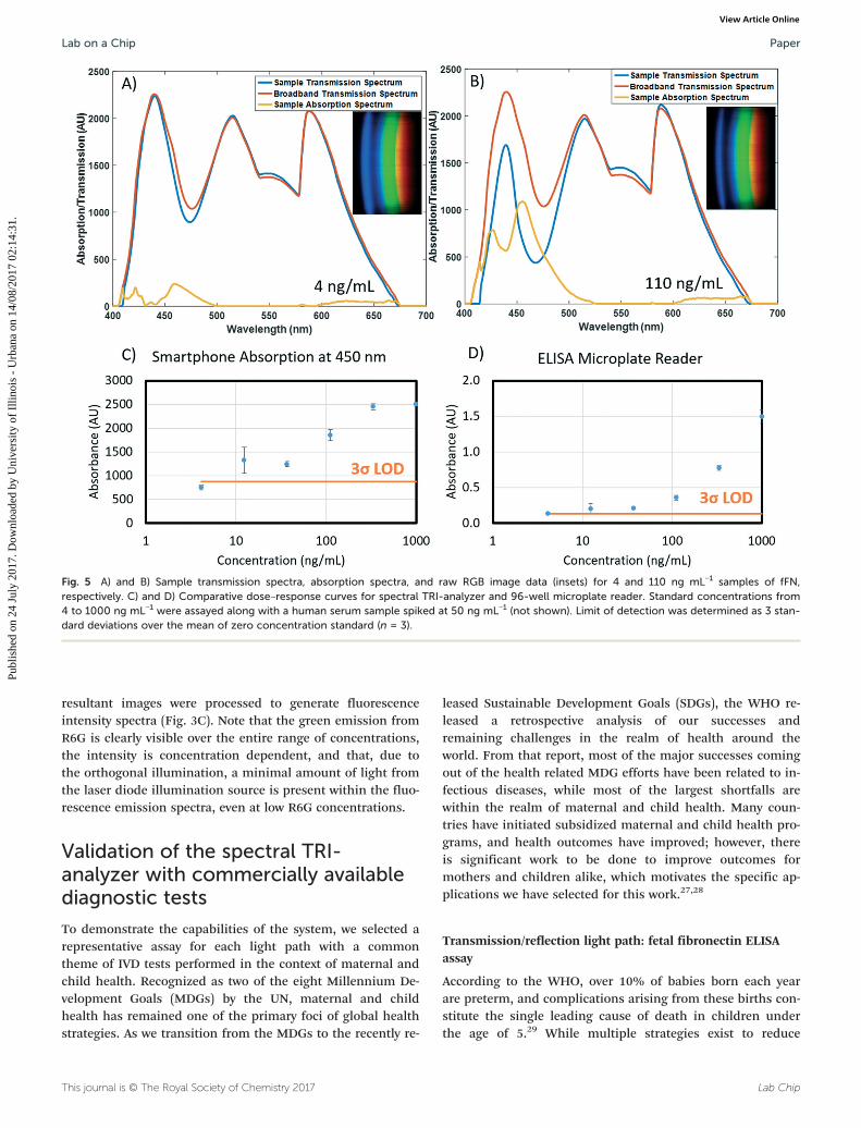
Fig. 5 A) and B) Sample transmission spectra, absorption spectra, and raw RGB image data (insets) for 4 and 110 ng mL−1 samples of fFN,respectively. C) and D) Comparative dose–response curves for spectral TRI-analyzer and 96-well microplate reader. Standard concentrations from4 to 1000 ng mL−1 were assayed along with a human serum sample spiked at 50 ng mL−1 (not shown). Limit of detection was determined as 3 stan-dard deviations over the mean of zero concentration standard (n = 3).
Lab on a Chip Paper
Publ
ishe
d on
24
July
201
7. D
ownl
oade
d by
Uni
vers
ity o
f Il
linoi
s -
Urb
ana
on 1
4/08
/201
7 02
:14:
31.
View Article Online

Lab Chip This journal is © The Royal Society of Chemistry 2017
complications associated with preterm births, diagnosis canoften be challenging.30 Fetal fibronectin is a glycoprotein thathas a high negative predictive value for spontaneous pretermbirth.31,32 Particularly when combined with a physical mea-surement of the cervical length, fetal fibronectin is an impor-tant diagnostic indicator to facilitate treatment, continuedmonitoring, or hospital referral of at-risk mothers. While
rapid lateral flow-based assays exist, they are not quantitative,and require specialized proprietary readers.33,34 Serologicaldetection of fFN is routine in a laboratory setting, and thetranslation of a fFN test to a point-of-care diagnostic wouldprovide useful information to health care providers, particu-larly with populations at-risk for pre-term births.
For fFN detection, a commercially available ELISA kit waspurchased (CUSABIO) and used to generate standard curves, aswell as measure spiked serum concentrations within physiolog-ically relevant ranges. Kit reagents were prepared followingmanufacturer instructions, and reactions were completed inthe included 96-well microplate. Standard concentrations wereserially diluted and run in triplicate at concentrations rangingfrom 4 to 1000 ng mL−1. Spiked plasma samples were createdat a concentration of 50 ng mL−1 from human plasma (n = 3).All samples were added to the microplate, and ELISA stepswere completed per manufacturer directions. After the additionof stop solution, the samples were immediately read in abenchtop 96-well plate reader (BioTek, Synergy HT), and thentransferred to a sample cartridge with a multichannel pipette.
Filled cartridges were measured using the developed Appto take five images of each sample as it was slid throughthe cartridge slot. The cylindrical lens that extends the non-spectral dimension of spectra onto the image sensor intro-duces spherical aberration, resulting in a curved appearanceof the resultant spectrum images, as shown inFig. 4A and B. As this is a function of internal optics andnot the alignment of the smartphone and the cradle, thecurvature observed is constant throughout multiple mea-surements and multiple phone removals/insertions (datanot shown). By fitting the pixel locations of where the redfilter drops off (∼580 nm) in each spectral row to a circulararc, spectra can be linearized and then summed in the non-
Fig. 6 Bland–Altman analysis of spectral TRI-analyzer compared withstandard microplate reader for readout of fFN assay. A variation on thestandard Bland–Altman method using the geometric mean and log dif-ference was applied to all non-zero values for each assay (Bland). Thelog differences showed a dependence on the magnitude of the mea-surement, so a regressive approach was used to model the relation, asdescribed by Bland (mean of regression shown with dashed line). ±1.96S.D. limits of agreement are shown as solid lines.
Fig. 7 Video processing and normalization. A) Average pixel intensityof a data-collection video showing clear demarcation of 8 samplewells. B and C) Example fluorescent spectrum showing regions used tonormalize the data.
Fig. 8 Bland–Altman analysis of spectral TRI-analyzer compared withstandard microplate reader for readout of phenylalanine assay. A varia-tion on the standard Bland–Altman method using the geometric meanand log difference was applied to all non-zero values for each assay(Bland). The log differences showed a dependence on the magnitudeof the measurement, so a regressive approach was used to model therelation, as described by Bland (mean of regression shown with dashedline). One point was found to be outside the ±1.96 S.D. limits of agree-ment (solid lines).
Lab on a ChipPaper
Publ
ishe
d on
24
July
201
7. D
ownl
oade
d by
Uni
vers
ity o
f Il
linoi
s -
Urb
ana
on 1
4/08
/201
7 02
:14:
31.
View Article Online

Lab ChipThis journal is © The Royal Society of Chemistry 2017
spectral dimension (Fig. 4C). The result is a spectrum wherepixel distance corresponds directly to wavelength, a relation-ship that can be interpolated from measuring two known la-sers with the smartphone system and interpolating thewavelengths in between (Fig. 4C). Finally, to adjust for over-all intensity variation resulting from imperfect cartridgealignment with the optical path, a linear scaling least-squares fit was performed on the spectrum above 500 nm(outside the region of interest of our chromogen absor-bance) when compared to a broadband spectrum (measuredwith ultrapure water). Resultant normalized transmissionspectra were subtracted from the same broadband spec-trum, resulting in absorption spectra (Fig. 5). Similar limitsof detection were observed between the 96-well microplatereader and spectral TRI-analyzer, both well under the mini-mum assay detection range (25 ng mL−1), with the lowestdiscernable assayed concentration being 37.0 and 12.3 ngmL−1, respectively. Bland–Altman analysis was performed onthe measurements from the 96-well plate reader and thespectral TRI-analyzer, and all data were shown to be wellwithin the 95% confidence intervals (Fig. 6).
Intensity light path: phenylalanine fluorescent assay
The prevention of phenylketonuria-associated mental deficitsbeen recognized at one of the largest public health successesof the past 50 years with over 4000 lives saved since screeningmethods began in the US 50 years ago.35 Global prevalence isregionally varied, ranging from 1/200000 in Finland to 1/2600in Turkey.36 Traditional newborn screening techniques arebased on a heel prick, originally used for bacterial metabolicassay, but now often run via mass spectroscopy.37–39 Benefitsof translating newborn birth screening tests to a smartphone-based point-of-care device including cloud-integration anddata storage, familiarity of user-interface, cost-effectivenessand portability, directly map to challenges identified by theUS Department of Health and Human Services, including thefeasibility of statewide surveillance, tracking, and reporting,the roles and responsibilities of a broad and varied user base,and the cost and number of tests needed.40
A commercially available assay kit was purchased (SigmaAldrich) for the quantification of human phenylalanine. Thephenylalanine standard was reconstituted in water, and
Fig. 9 Results of phenylketonuria assay. A) and B) Sample transmission spectra, absorption spectra, and raw RGB image data (insets) for 0.8 and0.2 nmol samples of phenylalanine, respectively. C) and D) Comparative dose–response curves for spectral TRI-analyzer and 96-well microplatereader. Standard concentrations from 0 to 8 nmol were assayed. Limit of detection was determined as 3 standard deviations over the mean of zeroconcentration standard (n = 3).
Lab on a Chip Paper
Publ
ishe
d on
24
July
201
7. D
ownl
oade
d by
Uni
vers
ity o
f Il
linoi
s -
Urb
ana
on 1
4/08
/201
7 02
:14:
31.
View Article Online

Lab Chip This journal is © The Royal Society of Chemistry 2017
serially diluted to concentrations of 0.8, 0.6, 0.4, 0.2, 0.1,0.05, and 0 nmol per well. Human serum samples weredeproteinized using a 10 kDa MWCO spin filter before beingdiluted with assay buffer and added to wells in triplicate (n =3). The samples were pre-treated for 10 minutes at room tem-perature with tyrosinase to control for background interfer-ence. A sample/standard reaction mixture of developer, en-zyme mix, and buffer was added to each of the wells permanufacturer directions. The solutions were allowed to incu-bate, covered and protected from light, for 20 minutes at 37C. Using a commercially available 96-well microplate reader(BioTek, Synergy HT), the completed assay was measured forfluorescence, exciting at λ = 535 nm and measuring at λ = 587nm. Samples were then transferred to cartridges, as de-scribed previously, and measured for fluorescence using thespectral TRI-analyzer.
For data acquisition of the fluorescence data, an improveddata collection procedure was developed. For absorption-based measurements, the stability of light output is time in-sensitive; however, with fluorescent dyes, both excitationintensity variation resulting from commercial laser diodes(even after circuit-based feedback control) and fluorophorephotobleaching result in a time-dependence of our measure-ments. To mitigate these variances, we developed a post-processing method that allows us to capture data as a videoas the user slides the cartridge through the cradle, therebyreconstituting the spatial dimension in which the multiplesample chambers are introduced into the light path. By ana-lyzing mean pixel value across each frame of the video, con-tiguous framesets of signal corresponding to each of the sam-ple chambers in the cartridge could be identified (Fig. 7).The center 80% of these frames were averaged, and used asan image to generate a spectrum, as done with the ELISAdata. These spectra were then normalized using the mea-sured integrated area of the excitation laser of the samplechambers before assay completion. This normalizationmethod was validated using Bland–Altman analysis (Fig. 8).The resultant fluorescence spectra resulted in a clear dose–re-sponse curve with a comparable limit of detection to that ofthe commercially available plate reader (Fig. 9). The limit ofdetection for the spectral TRI-analyzer was less than the low-est assayed concentration (0.5 nmol), while the limit of detec-tion for the 96-well plate reader was between 0.5 nmol and1.0 nmol. We observe saturation of our signal at higher ana-lyte concentrations, suggesting our system may have a lowerdynamic range than the microplate reader.
Conclusion
Our unique spectral TRI-analyzer system demonstrates boththe breadth of IVD assay possibilities and applications thatcan be achieved within a single instrument. Each of the threeoptical biosensing modalities offers up a wide array ofexisting assays that have been developed for the laboratorysetting that can be rapidly translated to be used with such aPOCT device.
The development of this device takes advantage of both therapid improvements to smartphone hardware and software aswell as the significant improvements in resolution and afford-ability of SLA-based 3D printing. This demonstration paves theway for a variety of prototyping applications in optical systemdesign where 3D-printed optical mounts could augment oreven replace conventional benchtop optical assembly andalignment. In particular, within the realm of optical biosensorsdesigned for the point-of-care, this presents significant poten-tial to create new optical systems that can proceed from com-puter simulation directly to handheld prototype.
Similarly, we have demonstrated a rapid-prototype-friendlymethod for micro- and milli-fluidic cartridges for use withmobile biosensing devices. By linearizing fluid chambers intomulti-sample cartridges, we can provide simple multiplexingwhich is useful both for assays requiring multiple standardsor controls, or situations where many samples might be runat once. The simplicity of our laser-cut acrylic sheet and DSAassembly opens the door for a myriad of other possible car-tridge configurations, including increasing the number ofsample chambers per cartridge for further multiplexing, orincorporating other microfluidic assay steps such as samplelysis, mixing, or other mainstays of microfluidic-chip basedtechnology. For example, the specific dimensions and layoutof our cartridges was designed to maximize convenience forinterfacing between the assay solutions run on our benchtopcomparison instruments that used a 96-well plate format byhaving sample ports spaced and sized to match the end of an8-well multichannel pipette.
In the demonstration of our system with existing commer-cial assays we sought to exemplify the sorts of laboratory-grade tests that could be directly transferred to the point-of-care. Thousands of these kits exist that rely on optical read-out of sandwich immunoassays with either absorbance orfluorescence-based reporting molecules (ELISA, FIA, CLIA),and many new assays are being developed based upon othernovel techniques, including those using bioluminescence,SPR, PCs or other selective-resonance phenomena.
Video-based capture of sample signals is a natural comple-ment to our developed linear cartridge, further simplifyingmulti-analyte multiplexing into a more user-friendly inter-face. This represents a novel data collection method that rep-resents yet another natural benefit of harnessing extantsmartphone imaging technology for spectral analysis and bio-sensing applications. To our knowledge, no such video-basedmultiplexing of spectral data has been described. The devel-oped image processing holds potential for further expansioninto future devices that can take advantage of such a ‘swipe’based interface. The swiping of a credit card is a ubiquitousmotion that readily simplifies multiplexed sample measure-ments compared to those in a typical cuvette-based measure-ment system.
The presented results of both proof-of-concept assaysdemonstrate that the spectral TRI-analyzer can replicate thesensitivity of conventional benchtop laboratory instruments.In both assays, our smartphone-based system could detect
Lab on a ChipPaper
Publ
ishe
d on
24
July
201
7. D
ownl
oade
d by
Uni
vers
ity o
f Il
linoi
s -
Urb
ana
on 1
4/08
/201
7 02
:14:
31.
View Article Online

Lab ChipThis journal is © The Royal Society of Chemistry 2017
concentrations of assayed analyte less than those detected ona conventional laboratory instrument. The single largest com-promise observed with our system was a decrease in dynamicrange for the assays. In the fFN experiment, near-completeabsorption of light at 450 nm at high concentrations resultedin signal saturation. For this particular assay, this wouldlikely be acceptable as threshold ranges from literature rangefrom 10–200 ng mL−1.41–43 Similarly, in the phenylalanine as-say, our instrument did not produce a linear dose–responsecurve, instead demonstrating a saturation–curve relationshipat higher concentrations. Likely using a 4-parameter logisticgrowth curve instead of a linear fit would allow for appropri-ate unknown concentrations to be determined using our sys-tem, but as shown by our Bland–Altman analysis for thisdata, even the unknown concentration in our serum samplewas shown to be measured with agreement between it andour benchtop analyzer. This level of agreement was demon-strated for both assays at all concentrations, suggesting thatour device can successfully reproduce the expected assaymeasurements from identical samples.
A key challenge in developing any successful IVD for use atthe point-of-care is achieving a device that is financially feasi-ble. To construct a single system, the cost of components ofour system was approximately $550. Many previous proof-of-concept demonstrations have focused on a single usage case,which significantly decreases the cost effectiveness of any diag-nostic device. The broad applicability of our device provides anecessary linkage between this previous work and the futurepossibility of truly portable IVD devices. Demonstrated here isjust one example set of related tests that individually might notresult in a commercially viable IVD device, but together, pro-vide a powerful opportunity to distribute maternal and childhealth monitoring from central laboratories to clinics andhealth outposts worldwide. The potential for this device tofunction as the backbone of a portable laboratory promises torevolutionize the future of mobile health diagnostics.
Acknowledgements
We would like to acknowledge the National Science Founda-tion for their support of this work via Grant no. CBET 12-64377 and the National Institutes of Health GrantF30AI122925. Additionally, we would like to express our grati-tude for the assistance of P. T. Clark for his assistance withthe initial optical layout.
Notes and references
1 Frost & Sullivan, Global Battery Market for Wearables, 2016.2 S. A. Soper, K. Brown, A. Ellington, B. Frazier, G. Garcia-
Manero, V. Gau, S. I. Gutman, D. F. Hayes, B. Korte, J. L.Landers, D. Larson, F. Ligler, A. Majumdar, M. Mascini, D.Nolte, Z. Rosenzweig, J. Wang and D. Wilson, Biosens.Bioelectron., 2006, 21, 1932–1942.
3 J. Kirkwood, The Next Step for Molecular Point-of-Care Test-ing, 2016.
4 L. Bissonnette and M. G. Bergeron, Clin. Microbiol. Infect.,2016, 16, 1044–1053.
5 C. D. Chin, V. Linder and S. K. Sia, Lab Chip, 2012, 12(12),2118–2134.
6 E. McLeod and A. Ozcan, Rep. Prog. Phys., 2016, 79, 076001.7 S. A. Lee and C. Yang, Lab Chip, 2014, 14(16), 3056–3063.8 H. Yu, Y. Tan and B. T. Cunningham, Anal. Chem.,
2014, 86(17), 8805–8813.9 M. A. Hossain, J. Canning, S. Ast, K. Cook, P. J. Rutledge and
A. Jamalipour, Opt. Lett., 2015, 40(8), 1737–1740.10 K. D. Long, H. Yu and B. T. Cunningham, Biomed. Opt.
Express, 2014, 5(11), 3792–3806.11 L. Jiang, M. Mancuso, Z. Lu, G. Akar, E. Cesarman and D.
Erickson, Sci. Rep., 2014, 4, 4137.12 O. Mudanyali, S. Dimitrov, U. Sikora, S. Padmanabhan, I.
Navruz and A. Ozcan, Lab Chip, 2012, 12(15), 2678–2686.13 A. St John and C. P. Price, Clin. Biochem. Rev., 2014, 35,
155–167.14 L. Kwon, K. D. Long, Y. Wan, H. Yu and B. T. Cunningham,
Biotechnol. Adv., 2016, 34, 291–304.15 D. Zhang and Q. Liu, Biosens. Bioelectron., 2016, 75, 273–284.16 H. Zhu, I. Sencan, J. Wong, S. Dimitrov, D. Tseng, K.
Nagashima and A. Ozcan, Lab Chip, 2013, 13, 1282–1288.17 D. Ji, H. Song, N. Zhang, Q. Gan, S. Pi, S. Jiang and X. Zeng,
Front. Opt., 2016, DOI: 10.1364/FIO.2016.JW4A.30.18 M. Seydack, Biosens. Bioelectron., 2005, 20, 2454–2469.19 K. A. Willets and R. P. Van Duyne, Annu. Rev. Phys. Chem.,
2007, 58, 267–297.20 J.-Y. Yoon, Biol. Eng., 2008, 1, 79–94.21 M. F. Pineda, L. L.-Y. Chan, T. Kuhlenschmidt, C. J. Choi, M.
Kuhlenschmidt and B. T. Cunningham, IEEE Sens. J.,2009, 9, 470–477.
22 D. Gallegos, K. D. Long, H. Yu, P. P. Clark, Y. Lin, S. George,P. Nath and B. T. Cunningham, Lab Chip, 2013, 13(11),2124–2132.
23 Frost & Sullivan, Advances in Label-Free Detection, ReportD12D, 2008.
24 R. Tiwari, Immunoassay Market Growth Driven by ELISA toHit 8.2% CAGR to 2021, 2016.
25 Frost & Sullivan, Overview of the In-vitro Diagnostics Marketin India, 2013.
26 Frost & Sullivan, Analysis of the US Immunochemistry Market,Report NE6F, 2014.
27 WHO, Health in 2015: from MDGs to SDGs, World HealthOrganization, 2015.
28 WHO, From MDGs to SDGs, WHO launches new report, WorldHealth Organization, 2016.
29 WHO, Preterm birth, World Health Organization, 2016.30 WHO, WHO recommendations on interventions to improve
preterm birth outcomes, World Health Organization, 2015.31 D. G. Kiefer and A. M. Vintzileos, Rev. Obstet. Gynecol.,
2008, 1, 106–112.32 F. Audibert, S. Fortin, E. Delvin, A. Djemli, S. Brunet, J. Dube
and W. D. Fraser, J. Obstet. Gynaecol. Can., 2010, 307–312.33 D. S. Abbott, S. K. Radford, P. T. Seed, R. M. Tribe and A. H.
Shennan, Am. J. Obstet. Gynecol., 2013, 208, 122.
Lab on a Chip Paper
Publ
ishe
d on
24
July
201
7. D
ownl
oade
d by
Uni
vers
ity o
f Il
linoi
s -
Urb
ana
on 1
4/08
/201
7 02
:14:
31.
View Article Online

Lab Chip This journal is © The Royal Society of Chemistry 2017
34 S. Deshpande, A. V. Asselt, F. Tomini, N. Armstrong, A.Allen, C. Noake, K. Khan, J. Severens, J. Kleijnen and M.Westwood, Health Technol. Assess., 2013, 17(40), 1–138.
35 CDC, Second National Report on Biochemical Indicators ofDiet and Nutrition in the US Population, Centers for DiseaseControl, 2012.
36 R. A. Williams, C. D. Mamotte and J. R. Burnett, in ClinBiochem Rev, 2008, vol. 29, pp. 31–41.
37 Medical Research Council Working Group, Br. Med. J.,1968, 4, 7–13.
38 K. Bhattacharya, T. Wotton and V. Wiley, Transl. Pediatr.,2014, 3, 63–70.
39 E. J. Schoen, J. C. Baker, C. J. Colby and T. T. To, Pediatrics,2002, 110, 781–786.
40 A. R. Kemper, C. A. Kus, R. J. Ostrander, A. M.Comeau, C. A. Boyle, D. Dougherty, M. Y. Mann, J. R.Botkin and N. S. Green, Implementing Point-of-Care New-born Screening, US Department of Health and HumanServices, 2012.
41 H. Leitich and A. Kaider, BJOG, 2003, 110(Suppl 20),66–70.
42 D. S. Abbott, N. L. Hezelgrave, P. T. Seed, J. E. Norman,A. L. David, P. R. Bennett, J. C. Girling, M.Chandirimani, S. J. Stock, J. Carter, R. Cate, J. Kurtzman,R. M. Tribe and A. H. Shennan, Obstet. Gynecol.,2015, 125, 1168–1176.
43 G. C. Lu, R. L. Goldenberg, S. P. Cliver, U. S. Kreaden andW. W. Andrews, Obstet. Gynecol., 2001, 97, 225–228.
Lab on a ChipPaper
Publ
ishe
d on
24
July
201
7. D
ownl
oade
d by
Uni
vers
ity o
f Il
linoi
s -
Urb
ana
on 1
4/08
/201
7 02
:14:
31.
View Article Online