Embed Size (px)
DESCRIPTION
mikroteknik tumbuhan
Citation preview

Welcome

Sum of steps used for preparation of a
microscopic slide ready to examination
Lab. 1 Microtechniques
“Histotechniques”

1. Sectioning method Done by Microtome
2. Non-Sectioning
method
Smear,squash,spreading
Methods of preparing microscopic slides:

1. Autopsy post-mortem specimens
2. Biopsy
specimens took from the living animals
or humans…
Types of specimen

Sectioning/mounting 4
Fixation 1
Dehydration 2
Embedding 3
Staining 5
Processing for slide preparation
Microscopic examination 6
To perform histological examination, must first prepare tissues:

1- Fixation
Fixation is a complex series of chemical events that comes by
reaction between the fixative and protein which form a gel like
subs. , so keeping every thing as their in vivo relation to each other.
The aim of fixation: 1- To prevent autolysis and bacterial attack.
2- fix the tissues so they will not change their volume and shape during
processing.
3- prepare tissue for clear staining of sections.
4- leave tissue as close as their living state , and no small molecules
should be lost.

• Formalin 10% (very common)
• Bouin fixative (expensive)
• Alcohols (70%)
• Gluteraldehyde ( E.M)
Types of fixatives

• The process to get rid of the excessive
fixative solution.
• Depend on the type of the fixative used.
(wether w/water base or alcoholic)…
Washing
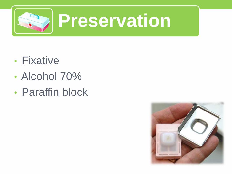
• Fixative
• Alcohol 70%
• Paraffin block
Preservation

2- Dehydration
• Remove water from fixed tissue
• graded alcohol series followed by “clearing agent” (xylenes,
histoclear, toluene) to remove alcohol (making tissue somewhat
transparent).
• why?
• most fixatives = water soluble, most embedding media = non-polar
(not water soluble) don’t get along
• must convert tissue from polar (water-based) medium to non-polar
medium that is miscible w/ embedding medium
• miscible = liquids will mix to form a homogenous solution

50% EtOH / 50% Phosphate-buffered Saline (PBS)
75% EtOH / 25% PBS
90% EtOH / 10% PBS
95% EtOH / 5% PBS
100% EtOH
clearing agent
paraffin wax (if doing paraffin embedding/sectioning)
Dehydration

• Replacing the dehydrating fluid with a fluid that is totally miscible with
both the dehydrating fluid and the embedding medium.
Choice of a clearing agent depends on:
1. type of tissues to be processed
2. Safety factors.
3. Cost and convenience.
4. Speedy removal of dehydrating agent .
5. easiness of removal by molten paraffin wax .
6. Minimal tissue damage .
3- Clearing

Xylene
Toluene
Chloroform
Benzene
Some clearing agents:

A process by which tissues are surrounded by a
medium such as agar, gelatin, or wax which when
solidified will provide sufficient external support during
sectioning.
• paraffin wax, plastic (glycol metacrylate, epoxy resins)
• Why so ?
structural support to tissue during sectioning
makes tissue easier to cut
tissue needs to withstand sectioning process
components must stay in natural positions
4- Embedding

Tissue embedded in “hard” Paraffin wax medium
Embedding

• Using microtome to cut transparent thin slices from embedded tissue specimen & mount on microscope slides
• generally 5-15 mm sections
• thickness depends upon tissue, subsequent procedures
• why?
• allows you to see internal structure of tissue
• allows stains or specific markers such as antibodies to more easily infiltrate tissue
• allows light to pass through tissue making structure visible
5- Sectioning

• A microtome is a mechanical instrument used to cut biological specimens into very thin segments for microscopic examination.
• Most microtomes use a steel blade and are used to prepare sections of animal or plant tissues.?
Types:
• Rotary microtome
• Freezing microtome
• Sliding microtome
• Ultra microtome(E.M)
Sectioning by microtome

• We use microtome to cut
transparent thin slices from
embedded tissue
specimens
• generally 5-15 mm sections
• thickness depends upon
tissue, subsequent
procedures treatment.
Sectioning by microtome

A. 40o C water bath
1. Flattens paraffin
section
2. Permits mounting
on slide
B. Gelatin & albumin
C. Glass slides
D. Oven / air dry
Mounting sections

• Use many techniques to stain tissue sections w/ dyes
• first must remove embedding medium, rehydrate(series alcohol)
tissues (dyes generally in aqueous solution)
• common stains - hemotoxylin & eosin (H&E), trichrome
• why?
• creates higher contrast that allows observation of structures that are not visible in unstained tissues.
• may reveal differences in chemical nature of certain regions of tissue (e.g. - some dyes, immunohistochemistry, immunofluorescence)
6- Staining

• Chemistry of Staining:
• Cationic dyes (+) - stain basophilic structures
e.g., nuclei, ribosomes, DNA (Basic dye ex
: Hematoxylin)
• Anionic dyes (-) - stain acidophilic structures
e.g., mitochondria, collagen (Acidophilic dye ex
: Eosin)
Staining

• Acid-Base
• hematoxylin - basic
dye, stains nucleous blue
• eosin - acidic dye stains,
cytoplasm & intercellular
components pink
Categories of Stains

• Trichrome (many methods) • 3 colors, allows
differentiation between cytoplasmic & intercellular components
• blue = collagen
• (eg, Mallory's stain,Masson's stain )
Collagen (blue)
Human skin
Keratin(pink)
nuclei (blue-black)
Categories of Stains

• Specific (special) stains • Periodic-Acid Schiff - stains glycogen, muco & glycoproteins,
glycosaminoglycans red
• Silver Impregnation - outlines reticular fibers
• Orcein, Resorcin-Fuchsin - elastic fibers
• Sudan Black B - stains fat
Categories of Stains

*A process in which we use a medium for adhesion of coverslip to the slide to protect the tissue from the dust, Mo. & dirt.
Coverslip & mounting medium (not miscible with water) Such as Kanada balsam ie .yellow natural resin dissolve in xylene.
Processs:
1. Remove the slides from the stainer dry it .
2. Coverslip using appropriate sized coverglass.
3. Label slides if necessary and arrange accordingly.
Coverslipping / Mounting mov

The slides are ready to be examined Microscopically
Thank you


![[ASM] Lab1](https://img.pdfslide.net/doc/110x75/588121881a28abb9388b706b/asm-lab1.jpg)